Embed Size (px)
Citation preview
Hair follicle dermalpapilla cells at a glance
Ryan R. Driskell1, Carlos Clavel2,Michael Rendl2,* and Fiona M.Watt1,3,*1Laboratory for Epidermal Stem Cell Biology,Wellcome Trust Centre for Stem Cell Research,University of Cambridge, Cambridge CB2 1QR, UK2Black Family Stem Cell Institute and Department ofDevelopmental and Regenerative Biology, MountSinai School of Medicine, New York, NY 10029, USA3CRUK Cambridge Research Institute, Li Ka ShingCentre, Robinson Way, Cambridge CB2 0RE, UK*Authors for correspondence([email protected]; [email protected])
Journal of Cell Science 124, 1179-1182 © 2011. Published by The Company of Biologists Ltddoi:10.1242/jcs.082446
IntroductionMammalian skin is a highly tractable tissue inwhich to explore epithelial–mesenchymal
(See poster insert)
Cell Science at a Glance 1179
interactions during development and inpostnatal life (Blanpain and Fuchs, 2009;Müller-Röver et al., 2001; Schmidt-Ullrich andPaus, 2005; Watt and Jensen, 2009). Onepopulation of mesenchymal cells in the skin,known as dermal papilla (DP) cells, is the focusof intense interest because the DP not onlyregulates hair follicle development and growth,but is also thought to be a reservoir of multi-potent stem cells. In this article and theaccompanying poster we review the origins ofthe DP during skin development, and discuss DPheterogeneity and the changes in the DP thatoccur during the hair growth cycle. We alsoconsider the different cell lineages along whichDP cells can differentiate as well as potentialclinical applications of DP cells.
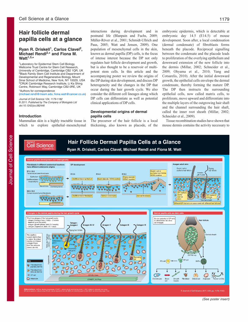
Developmental origins of dermalpapilla cellsThe precursor of the hair follicle is a localthickening, also known as placode, of the
embryonic epidermis, which is detectable atembryonic day 14.5 (E14.5) of mousedevelopment. Soon after, a local condensation(dermal condensate) of fibroblasts formsbeneath the placode. Reciprocal signallingbetween the condensate and the placode leadsto proliferation of the overlying epithelium anddownward extension of the new follicle intothe dermis (Millar, 2002; Schneider et al.,2009; Ohyama et al., 2010; Yang andCotsarelis, 2010). After the initial downwardgrowth, the epithelial cells envelope the dermalcondensate, thereby forming the mature DP.The DP then instructs the surroundingepithelial cells, now called matrix cells, toproliferate, move upward and differentiate intothe multiple layers of the outgrowing hair shaftand the channel surrounding the hair shaft,called the inner root sheath (Millar, 2002;Schneider et al., 2009).
Tissue recombination studies have shown thatmouse dermis contains the activity necessary to
Jour
nal o
f Cel
l Sci
ence

1180
induce hair follicle formation as early as E12.5,before the dermal condensate has developed(Dhouailly, 1973; Song and Sawyer, 1996).Dermis from hair-forming regions of skin caninduce follicles in both hair- and non-hair-forming epithelium, whereas dermis from non-hair-forming sites cannot support the formationof hair follicles. Several of the pathways that areinvolved in reciprocal signalling between theepithelial cells and DP of the developing folliclehave been identified, with reciprocal Wntsignalling emerging as one of the earliest andmost-important (Kishimoto et al., 2000; Millar,2002; Schneider et al., 2009; Ohyama et al.,2010; Yang and Cotsarelis, 2010). However,relatively little is known about how dermalcondensate and DP cells become hair-inducingspecialised fibroblasts (Schneider et al., 2009;Ohyama et al., 2009; Yang and Cotsarelis,2009).
Fibroblasts and, therefore, DP in differentbody sites have different embryonic origins(Fernandes et al., 2004; Rendl et al., 2005;Ohtola et al., 2008; Wong et al., 2006; Jinno etal., 2010). Head and facial fibroblasts arederived from the neural crest, whereas dorsaland ventral trunk fibroblasts come from thedermomyotome of somitic and lateral plateorigin, respectively. Cell autonomous, site-specific homeobox (Hox) gene expressionconfers positional memory on fibroblasts fromdifferent body sites and has a role in specifyingthe gene expression profile of the overlyingepidermis (Rinn et al., 2008).
Dermal papilla heterogeneityMouse skin contains several distinct hair follicletypes that differ in length, thickness and theshape of the hair shaft, i.e. straight or kinked. Inback skin, the most abundant follicles formzigzag hairs, which have two kinks in the shaft,whereas the hairs of other follicle types (guard,awl and auchene) have longer shafts that areeither straight or have a single kink (Schlake,2007). Guard hairs develop during the first waveof hair follicle morphogenesis around E14.5;awl and auchene hairs form in the second wavearound E16.5; and zigzag hairs form during thethird wave, starting at E18.5 (Schlake, 2007).The DP of zigzag hairs are smaller than those ofthe other follicle types (Elliott et al., 1999).Between E14.5 and E16.5, all developing DP(that is, those associated with guard, awl orauchene follicles) express the transcriptionfactor sex determining region Y-box 2 (Sox2),but SOX2 is undetectable in the DP of zigzaghairs, which develop from E18.5 onwards(Driskell et al., 2009).
Differential Sox2 expression can be used toisolate DP cells originating from different hairfollicle types in early postnatal (P2) mouse skin
(Driskell et al., 2009). At this stage, all DP cellsexpress CD133 (also known as PROM1) andalkaline phosphatase (Handjiski et al., 1994),but DP of zigzag hairs are SOX2-negative andguard, awl and auchene follicles are SOX2-positive. When different DP populations aresorted and used in skin reconstitution assays,SOX-positive DP cells are found to be necessaryfor the formation of guard, awl or auchenefollicles (Driskell et al., 2009). By contrast, DPexpression of SOX is required for the formationof zigzag hair follicles (James et al., 2003). Thisindicates different roles for two SRYtranscription factors in specifying hair follicletype during development.
Gene expression profiling of DP cells isolatedfrom developing mouse skin has resulted in thedefinition of a core DP ‘signature’ of 184 genes(Rendl et al., 2005), and in signatures that arespecific for SOX2-positive and -negative DPtypes (Driskell et al., 2009). These signaturesare beginning to provide information about thesignalling pathways that control DP and hairfollicle function, in particular the importance ofWnt, bone morphogenetic protein (BMP) andfibroblast growth factor (FGF) (Greco et al.,2009; Kishimoto et al., 2000; Rendl et al., 2008).In addition, comparison of the properties of hairfollicles in different body sites revealsdifferences in the properties of their DP. Oneexample, in human skin, is the observation thatandrogens stimulate hair follicle growth in theface but cause follicle miniaturisation inthe scalp. DP cells express androgen receptorsand 5a-OH-reductase – a key enzyme inandrogen metabolism – and DP from differentbody sites differ in their responsiveness toandrogen (Rutberg et al., 2006).
The role of the DP in the hair growthcycleIn postnatal life the hair follicles undergocyclical growth. The resting phase is known astelogen, the growth phase as anagen and theregression phase as catagen (Müller-Röver etal., 2001; Schmidt-Ullrich and Paus, 2005;Ohyama et al., 2010; Yang and Cotsarelis,2010). During catagen, the epithelial cells at thebase of the follicle undergo apoptosis, butthe DP remains intact and is pulled or migratesupwards, until it comes to rest next to the stemcells of the hair follicle bulge. This situationpersists during telogen. In anagen, cells at thebase of the follicle start to proliferate, whichresults in downward growth of the follicle andenvelopment of the DP. DP cells themselves arethought to not divide. However, the number ofcells in the DP increases during anagen, possiblyas a result of replenishment from neighbouringcells of the dermal sheath (Tobin et al., 2003;Chi et al., 2010).
At the onset of anagen the DP activates stemcells in the secondary hair germ leading to newdownward growth of follicles. In the hairlessmutant mouse, DP cells become stranded deepin the dermis during catagen and lose contactwith the bulge; the follicles in these mice cannotundergo anagen and eventually degenerate(Panteleyev et al., 1999). Interruption of -catenin signalling in the DP results in reducedproliferation of cells at the base of the follicle,which induces catagen and prevents anageninduction (Enshell-Seijffers et al., 2010). -catenin activity in the DP regulates a number ofother signalling pathways, including the FGFpathway, which mediate the inductive effects ofthe DP on the hair follicle epithelium (Enshell-Seijffers et al., 2010). Fgf7 and Fgf10 areexpressed in the DP and stimulate proliferationof the adjacent epithelial cells of the hair follicle(Greco et al., 2009).
Some DP markers, such as alkalinephosphatase and cellular retinoic-acid-bindingprotein 1 (CRABP1), are expressed throughoutthe hair growth cycle (Collins and Watt, 2008).Others, such as the serine protease Corin, areupregulated during anagen (Enshell-Seijffers etal., 2008). In adult mouse skin Sox2 expressionin the DP varies during the hair growth cycle andis only detectable during anagen (Biernaskie etal., 2009).
Potential therapeutic applications ofDP cells in restoring hair growthHair loss (alopecia) is a common and distressingproblem for men and women, and there istherefore considerable interest in treatments thatcan prevent or reverse it. Harnessing the abilityof the differentiated and highly specialisedfibroblasts of the DP to induce neighbouringepidermal cells to differentiate along the hairfollicle lineages is an attractive approach totreating alopecia.
The hair-inductive ability of DP cells is notrestricted to embryonic development, and DPcells from postnatal skin retain the ability todirect epithelial cells to form hair follicles(Jahoda et al., 1984). Furthermore, formation ofnew DP can be induced in adult skin byactivating the Wnt pathway in the epidermis(Silva-Vargas et al., 2005). These observationssuggest that it is possible to generate DP cells inorder to treat hair loss.
One obvious strategy is to expand DP cells inculture before transplantation. DP cells not onlyretain the ability to form DP following in vitroculture, but they can also contribute to dermalsheath cells and non-follicle-associatedfibroblasts during skin reconstitution andwound-healing (Biernaskie et al., 2009; Rendl etal., 2008). However, after a few passagescultured DP cells lose their trichogenic
Journal of Cell Science 124 (8)
Jour
nal o
f Cel
l Sci
ence
1181
properties (i.e. their ability to induce hairfollicles) (Ohyama et al., 2010; Yang andCotsarelis, 2010; Horne et al., 1986; Kishimotoet al., 2000; Lichti et al., 1993; Rendl et al.,2008). Culture media have been developed thatextend the time for which DP cells can becultured (Limat et al., 1993; Osada et al., 2007;Roh et al., 2004), and activation of Wnt andBmp signalling pathways in mouse DP cells candelay loss of trichogenicity (Kishimoto et al.,2000; Rendl et al., 2008). Other strategies topreserve the properties of DP cells are to growthem in three-dimensional aggregates (Osada etal., 2007; Higgins et al., 2010) or to culture themtogether with keratinocytes on extracellularmatrix substrates in order to mimic the in vivomicroenvironment (Havlickova et al., 2009).
DP cells as stem cells with multi-lineage differentiation potentialSurprisingly, the therapeutic potential of DPcells extends far beyond inducing new hairfollicles. To study, or eventually correct, a widevariety of degenerative disorders, inducedpluripotent stem (iPS) cells are being generatedfrom patient biopsies (Yamanaka and Blau,2010). Plucking hair from patients is a non-invasive way to obtain cells for reprogramming,and recent studies have shown that mouse DPcells can be more readily reprogrammed intoiPS cells than most other cell types (Tsai et al.,2010).
Another recent finding is that SOX2-positiveDP cells are the origin of skin-derivedprogenitor cells (SKPs). SKPs are cells that canbe cultured to form nestin-positive spheres withthe capacity to differentiate into neurons, glia,smooth muscle cells, adipocytes and other celltypes (Toma et al., 2001; Fernandes et al., 2004;Lavoie et al., 2009; Biernaskie et al., 2009).Since Sox2 is also expressed in dermal sheathcells close to the DP, it is possible thatsheath cells have the ability to form SKPs inculture. SKPs can be generated not only fromrodent skin, but also from human hair follicleDP (Hunt et al., 2008). The multi-lineagedifferentiation potential of cultured DP anddermal sheath cells is not dependent on priorculture as spheroids: they can also differentiateinto adipogenic, osteogenic and hematopoieticlineages under other culture conditions (Lako etal., 2002; Jahoda et al., 2003).
The observation that SKPs can be isolatedfrom back skin (Biernaskie et al., 2009) issurprising because multipotent dermal cellshave previously been identified to originatefrom neural crest cells (Fernandes et al., 2004;Wong et al., 2006) and dorsal skin DP arise fromthe dermomyotome. Recent lineage tracingstudies using the Wnt1 promoter to drive Crerecombinase expression in neural crest
derivatives and the myogenic regulatory factor 5(Myf5) promoter to express Cre in cells ofsomitic origin show that SKPs from bothlocations can differentiate into Schwann cells, acell type previously thought to be exclusivelyderived from the neural crest (Jinno et al., 2010).This suggests that the hair follicle environment,rather than the developmental origin of the cells,induces expression of neural-crest-related genesand generates cells with the characteristics ofneural crest derivatives.
ConclusionsThe cells of the DP are not only essential for hairfollicle development and function, but are also areservoir of cells with the potential todifferentiate into a range of cell types that are ofpotential therapeutic importance. Improvedmethods for culturing DP cells can be exploitedto treat hair loss, and the ability to direct DP cellsto differentiate into other lineages, in particularSchwann cells, could provide a source of cells torepair damaged nerves (Biernaskie et al., 2007).
Deposited in PMC for release after 6 months.
Individual poster panels are available as JPEG files athttp://jcs.biologists.org/cgi/content/full/124/8/1179/DC1
ReferencesBiernaskie, J., Sparling, J. S., Liu, J., Shannon, C. P.,Plemel, J. R., Xie, Y., Miller, F. D. and Tetzlaff, W. (2007).Skin-derived precursors generate myelinating Schwanncells that promote remyelination and functional recoveryafter contusion spinal cord injury. J. Neurosci. 27, 9545-9559.Biernaskie, J., Paris, M., Morozova, O., Fagan, B. M.,Marra, M., Pevny, L. and Miller, F. D. (2009). SKPsderive from hair follicle precursors and exhibit properties ofadult dermal stem cells. Cell Stem Cell 5, 610-623.Blanpain, C. and Fuchs, E. (2009). Epidermalhomeostasis: a balancing act of stem cells in the skin. Nat.Rev. Mol. Cell Biol. 10, 207-217.Chi, W. Y., Enshell-Seijffers, D. and Morgan, B. A.(2010). De novo production of dermal papilla cells duringthe anagen phase of the hair cycle. J. Invest. Dermatol. 130,2664-2666.Collins, C. A. and Watt, F. M. (2008). Dynamic regulationof retinoic acid-binding proteins in developing, adult andneoplastic skin reveals roles for beta-catenin and Notchsignalling. Dev. Biol. 324, 55-67.Dhouailly, D. (1973). Dermo-epidermal interactionsbetween birds and mammals: differentiation of cutaneousappendages. J. Embryol. Exp. Morphol. 30, 587-603.Driskell, R. R., Giangreco, A., Jensen, K. B., Mulder, K.W. and Watt, F. M. (2009). Sox2-positive dermal papillacells specify hair follicle type in mammalian epidermis.Development 136, 2815-2823.Elliott, K., Stephenson, T. J. and Messenger, A. G.(1999). Differences in hair follicle dermal papilla volumeare due to extracellular matrix volume and cell number:implications for the control of hair follicle size and androgenresponses. J. Invest. Dermatol. 113, 873-877.Enshell-Seijffers, D., Lindon, C. and Morgan, B. A.(2008). The serine protease Corin is a novel modifier of theAgouti pathway. Development 135, 217-225.Enshell-Seijffers, D., Lindon, C., Kashiwagi, M. andMorgan, B. A. (2010). -catenin activity in the dermalpapilla regulates morphogenesis and regeneration of hair.Dev. Cell 18, 633-642.Fernandes, K. J., McKenzie, I. A., Mill, P., Smith, K. M.,Akhavan, M., Barnabe-Heider, F., Biernaskie, J., Junek,A., Kobayashi, N. R., Toma, J. G. et al. (2004). A dermalniche for multipotent adult skin-derived precursor cells. Nat.Cell Biol. 6, 1082-1093.
Greco, V., Chen, T., Rendl, M., Schober, M., Pasolli, H.A., Stokes, N., Dela Cruz-Racelis, J. and Fuchs, E.(2009). A two-step mechanism for stem cell activationduring hair regeneration. Cell Stem Cell 4, 155-169.Handjiski, B. K., Eichmuller, S., Hofmann, U.,Czarnetzki, B. M. and Paus, R. (1994). Alkalinephosphatase activity and localization during the murine haircycle. Br. J. Dermatol. 131, 303-310.Havlickova, B., Biro, T., Mescalchin, A., Tschirschmann,M., Mollenkopf, H., Bettermann, A., Pertile, P., Lauster,R., Bodo, E. and Paus, R. (2009). A human folliculoidmicrosphere assay for exploring epithelial-mesenchymalinteractions in the human hair follicle. J. Invest. Dermatol.129, 972-983.Higgins, C. A., Richardson, G. D., Ferdinando, D.,Westgate, G. E. and Jahoda, C. A. (2010). Modelling thehair follicle dermal papilla using spheroid cell cultures. Exp.Dermatol. 19, 546-548.Horne, K. A., Jahoda, C. A. and Oliver, R. F. (1986).Whisker growth induced by implantation of culturedvibrissa dermal papilla cells in the adult rat. J. Embryol.Exp. Morphol. 97, 111-124.Hunt, D. P., Morris, P. N., Sterling, J., Anderson, J. A.,Joannides, A., Jahoda, C., Compston, A. and Chandran,S. (2008). A highly enriched niche of precursor cells withneuronal and glial potential within the hair follicle dermalpapilla of adult skin. Stem Cells 26, 163-172.Jahoda, C. A., Horne, K. A. and Oliver, R. F. (1984).Induction of hair growth by implantation of cultured dermalpapilla cells. Nature 311, 560-562.Jahoda, C. .A., Whitehouse, J., Reynolds, A. J., Hole, N.(2003) Hair follicle dermal cells differentiate intoadipogenic and osteogenic lineages. Exp. Dermatol. 12,849-859.James, K., Hosking, B., Gardner, J., Muscat, G. E. andKoopman, P. (2003). Sox18 mutations in the ragged mousealleles ragged-like and opossum. Genesis 36, 1-6.Jinno, H., Morozova, O., Jones, K. L., Biernaskie, J. A.,Paris, M., Hosokawa, R., Rudnicki, M. A., Chai, Y.,Rossi, F., Marra, M. A. et al. (2010). Convergent genesisof an adult neural crest-like dermal stem cell from distinctdevelopmental origins. Stem Cells 28, 2027-2040.Kishimoto, J., Burgeson, R. E. and Morgan, B. A. (2000).Wnt signaling maintains the hair-inducing activity of thedermal papilla. Genes Dev. 14, 1181-1185.Lako, M., Armstrong, L., Cairns, P. M., Harris, S., Hole,N. and Jahoda, C. A. (2002). Hair follicle dermal cellsrepopulate the mouse haematopoietic system. J. Cell Sci.115, 3967-3974.Lavoie, J. F., Biernaskie, J. A., Chen, Y., Bagli, D.,Alman, B., Kaplan, D. R. and Miller, F. D. (2009). Skin-derived precursors differentiate into skeletogenic cell typesand contribute to bone repair. Stem Cells Dev. 18, 893-906.Lichti, U., Weinberg, W. C., Goodman, L., Ledbetter, S.,Dooley, T., Morgan, D. and Yuspa, S. H. (1993). In vivoregulation of murine hair growth: insights from graftingdefined cell populations onto nude mice. J. Invest.Dermatol. 101, 124S-129S.Limat, A., Hunziker, T., Waelti, E. R., Inaebnit, S. P.,Wiesmann, U. and Braathen, L. R. (1993). Soluble factorsfrom human hair papilla cells and dermal fibroblastsdramatically increase the clonal growth of outer root sheathcells. Arch. Dermatol. Res. 285, 205-210.Millar, S. E. (2002). Molecular mechanisms regulating hairfollicle development. J. Invest. Dermatol. 118, 216-225.Müller-Röver, S., Handjiski, B., van der Veen, C.,Eichmuller, S., Foitzik, K., McKay, I. A., Stenn, K. S. andPaus, R. (2001). A comprehensive guide for the accurateclassification of murine hair follicles in distinct hair cyclestages. J. Invest. Dermatol. 117, 3-15.Ohtola, J., Myers, J., Akhtar-Zaidi, B., Zuzindlak, D.,Sandesara, P., Yeh, K., Mackem, S., Atit, R. (2008) -Catenin has sequential roles in the survival and specificationof ventral dermis. Development 135, 2321-2329.Ohyama, M., Zheng, Y., Paus, R. and Stenn, K. S. (2010).The mesenchymal component of hair follicle neogenesis:background, methods and molecular characterization. Exp.Dermatol. 19, 89-99.Osada, A., Iwabuchi, T., Kishimoto, J., Hamazaki, T. S.and Okochi, H. (2007). Long-term culture of mousevibrissal dermal papilla cells and de novo hair follicleinduction. Tissue Eng. 13, 975-982.Panteleyev, A. A., Botchkareva, N. V., Sundberg, J. P.,Christiano, A. M. and Paus, R. (1999). The role of the
Journal of Cell Science 124 (8)
Jour
nal o
f Cel
l Sci
ence

1182
hairless (hr) gene in the regulation of hair follicle catagentransformation. Am. J. Pathol. 155, 159-171.Pierard, G. E. and De la Brassinne, M. (1975). Cellularactivity in the dermis surrounding the hair bulb in alopeciaareata. J. Cutan. Pathol. 2, 240-245.Rendl, M., Lewis, L. and Fuchs, E. (2005). Moleculardissection of mesenchymal-epithelial interactions in the hairfollicle. PLoS Biol. 3, e331.Rendl, M., Polak, L. and Fuchs, E. (2008). BMP signalingin dermal papilla cells is required for their hair follicle-inductive properties. Genes Dev. 22, 543-557.Rinn, J. L., Wang, J. K., Allen, N., Brugmann, S. A.,Mikels, A. J., Liu, H., Ridky, T. W., Stadler, H. S., Nusse,R., Helms, J. A. et al. (2008). A dermal HOXtranscriptional program regulates site-specific epidermalfate. Genes Dev. 22, 303-307.Roh, C., Tao, Q. and Lyle, S. (2004). Dermal papilla-induced hair differentiation of adult epithelial stem cellsfrom human skin. Physiol. Genomics 19, 207-217.Rutberg, S. E., Kolpak, M. L., Gourley, J. A., Tan, G.,Henry, J. P. and Shander, D. (2006). Differences inexpression of specific biomarkers distinguish human beardfrom scalp dermal papilla cells. J. Invest. Dermatol. 126,2583-2595.Schlake, T. (2007). Determination of hair structure andshape. Semin. Cell Dev. Biol. 18, 267-273.
Schmidt-Ullrich, R. and Paus, R. (2005). Molecularprinciples of hair follicle induction and morphogenesis.BioEssays 27, 247-261.Schneider, M. R., Schmidt-Ullrich, R. and Paus, R.(2009). The hair follicle as a dynamic miniorgan. Curr. Biol.19, R132-R142.Silva-Vargas, V., Lo Celso, C., Giangreco, A., Ofstad, T.,Prowse, D. M., Braun, K. M. and Watt, F. M. (2005).Beta-catenin and Hedgehog signal strength can specifynumber and location of hair follicles in adult epidermiswithout recruitment of bulge stem cells. Dev. Cell 9, 121-131.Song, H. K. and Sawyer, R. H. (1996). Dorsal dermis ofthe scaleless (sc/sc) embryo directs normal feather patternformation until day 8 of development. Dev. Dyn. 205, 82-91.Tobin, D. J., Gunin, A., Magerl, M. and Paus, R. (2003).Plasticity and cytokinetic dynamics of the hair folliclemesenchyme during the hair growth cycle: implications forgrowth control and hair follicle transformations. J. Invest.Dermatol. Symp. Proc. 8, 80-86.Toma, J. G., Akhavan, M., Fernandes, K. J., Barnabe-Heider, F., Sadikot, A., Kaplan, D. R. and Miller, F. D.(2001). Isolation of multipotent adult stem cells from thedermis of mammalian skin. Nat. Cell Biol. 3, 778-784.Tsai, S. Y., Clavel, C., Kim, S., Ang, Y. S., Grisanti, L.,Lee, D. F., Kelley, K. and Rendl, M. (2010). Oct4 and klf4
reprogram dermal papilla cells into induced pluripotent stemcells. Stem Cells 28, 221-228.Watt, F. M. and Jensen, K. B. (2009). Epidermal stem celldiversity and quiescence. EMBO Mol. Med. 1, 260-267.Wong, C. E, Paratore, C., Dours-Zimmermann, M. T,Rochat, A., Pietri, T., Suter, U., Zimmermann, D. R.,Dufour, S., Thiery, J. P., Meijer et al. (2006) Neural crest-derived cells with stem cell features can be traced back tomultiple lineages in the adult skin. J. Cell Biol. 175, 1005-1015. Yamanaka, S. and Blau, H. M. (2010). Nuclearreprogramming to a pluripotent state by three approaches.Nature 465, 704-712.Yang, C. C. and Cotsarelis, G. (2010). Review of hairfollicle dermal cells. J. Dermatol. Sci. 57, 2-11.
Journal of Cell Science 124 (8)
Cell Science at a Glance on the WebElectronic copies of the poster insert areavailable in the online version of this articleat jcs.biologists.org. The JPEG images canbe downloaded for printing or used asslides.
Jour
nal o
f Cel
l Sci
ence