Embed Size (px)
Citation preview
Basics Hair has little remaining physiologic importance,
but great psychological importance. Five million total hair follicles in the adult human,
without significant racial or sexual differences Scalp has highest density, but declines with age from
1135/cm2 at birth to 300-500/cm2 in adults for a total of 100,000 follicles.
Basics Ethnic differences in hair shape are the result of
differences in follicle shape. The follicle keratinizes (hardens) 1st & molds the hair
to its shape Caucasian=round to oval Black=elliptical/flattened Asian=round, also largest diameter

Embryology and Development Early formation of hair follicles depends on a very
complicated series of mesenchymal epithelial interactions that are being actively investigated.
Why study these complex interactions? One day it may be possible to induce new follicle
development as a treatment for the scarring alopecias Timeline:
10 Weeks: Follicle formation begins on the head (particularly eyebrows, lower and upper lip)
16 weeks: Follicles develop cephalocaudally over rest of the body
22 weeks: New follicle formation complete
Embryology No hair follicles form after birth (under normal
circumstances) The nonrandom symmetric distribution is probably
determined by patterning genes such as hox, msx, enthat were originally discovered in Drosophila
The establishment of a dermal papilla (DP) is vital to the development of all hair follicles.
DP is a group of specialized dermal fibroblast cells. They are derived from the embryonic mesoderm They aggregate in the dermis just below the epidermis.
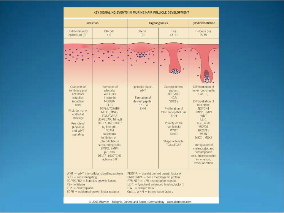
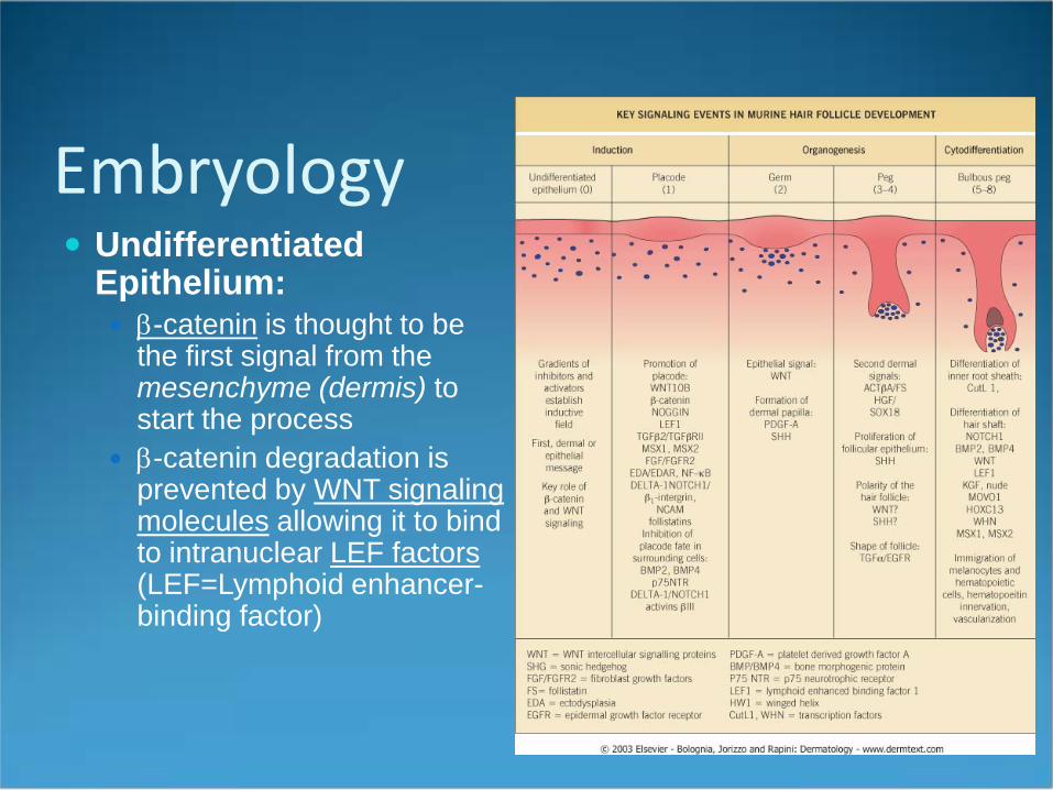
Embryology Undifferentiated
Epithelium: β-catenin is thought to be
the first signal from the mesenchyme (dermis) to start the process
β-catenin degradation is prevented by WNT signaling molecules allowing it to bind to intranuclear LEF factors(LEF=Lymphoid enhancer-binding factor)
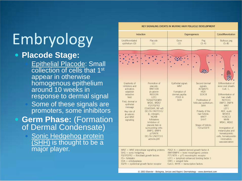
Embryology Placode Stage:
Epithelial Placode: Small collection of cells that 1st
appear in otherwise homogenous epithelium around 10 weeks in response to dermal signal
Some of these signals are promoters, some inhibitors
Germ Phase: (Formation of Dermal Condensate) Sonic Hedgehog protein
(SHH) is thought to be a major player.
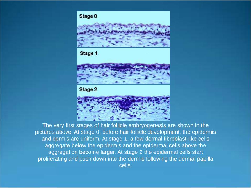
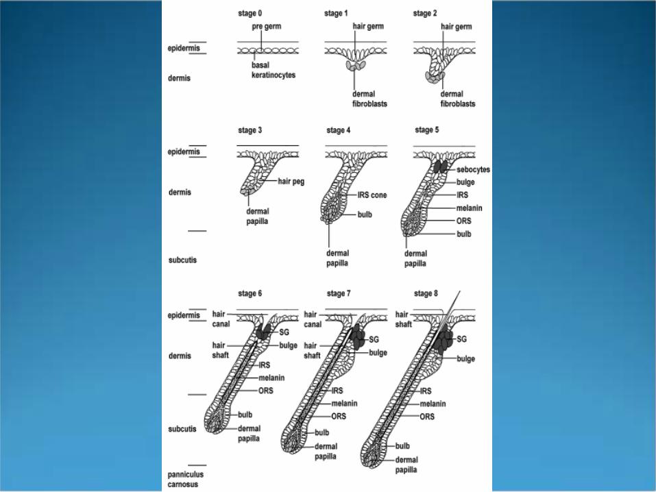
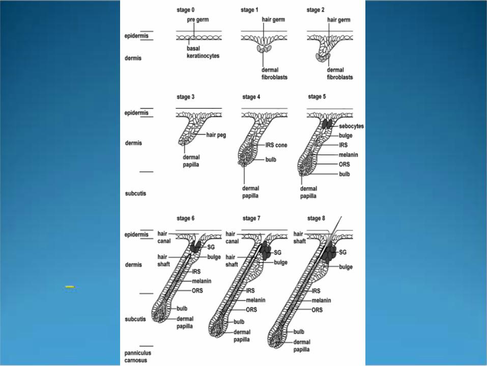
The very first stages of hair follicle embryogenesis are shown in the pictures above. At stage 0, before hair follicle development, the epidermis
and dermis are uniform. At stage 1, a few dermal fibroblast-like cells aggregate below the epidermis and the epidermal cells above the
aggregation become larger. At stage 2 the epidermal cells start proliferating and push down into the dermis following the dermal papilla
cells.
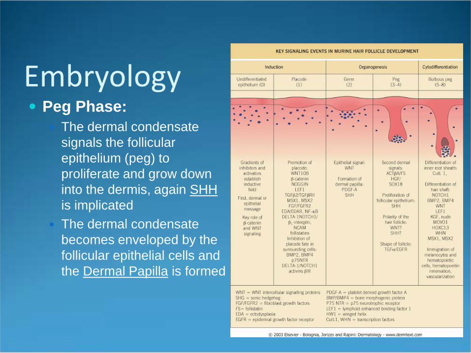
Embryology Peg Phase:
The dermal condensate signals the follicular epithelium (peg) to proliferate and grow down into the dermis, again SHHis implicated
The dermal condensate becomes enveloped by the follicular epithelial cells and the Dermal Papilla is formed
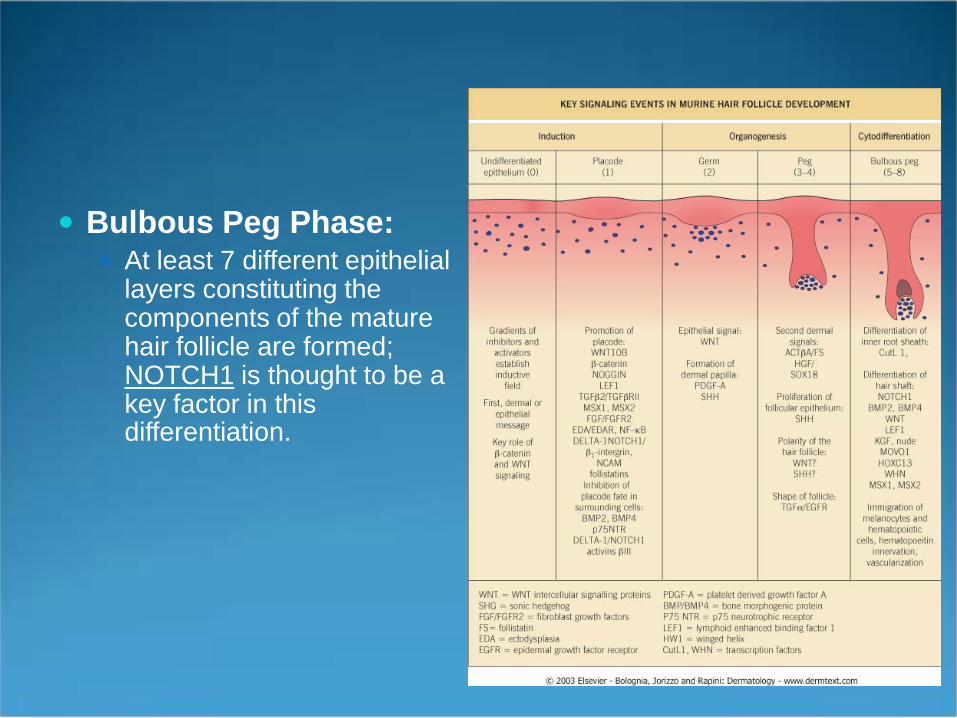
Bulbous Peg Phase: At least 7 different epithelial
layers constituting the components of the mature hair follicle are formed; NOTCH1 is thought to be a key factor in this differentiation.
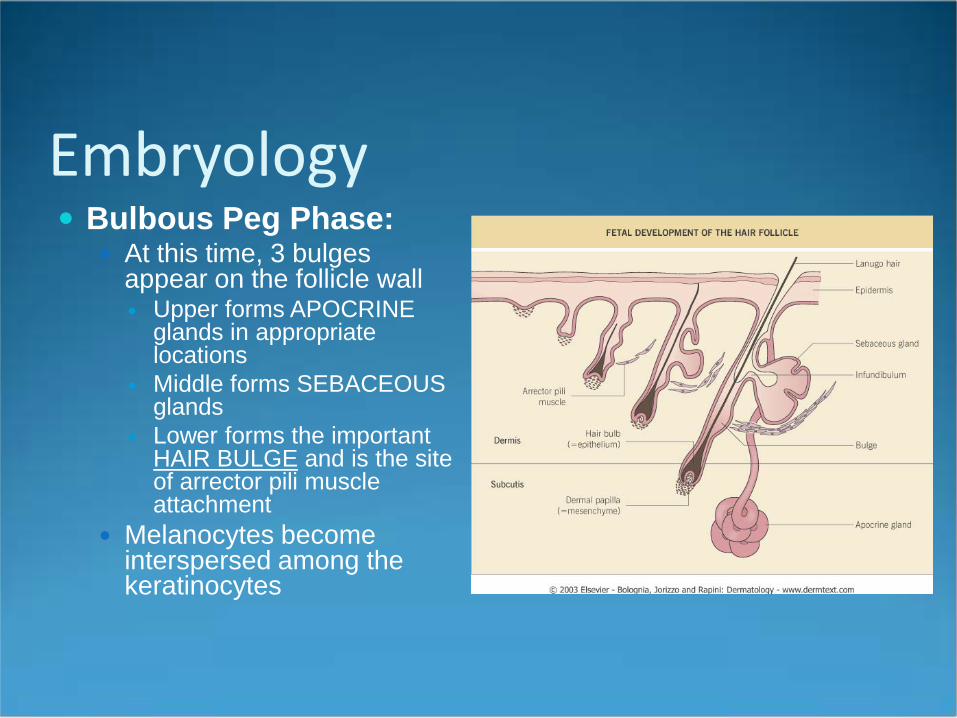
Embryology Bulbous Peg Phase:
At this time, 3 bulges appear on the follicle wall Upper forms APOCRINE
glands in appropriate locations
Middle forms SEBACEOUS glands
Lower forms the important HAIR BULGE and is the site of arrector pili muscle attachment
Melanocytes become interspersed among the keratinocytes
Embryology As the epidermal plug penetrates down into the
dermis mesodermal cells congregate around it. The mesodermal cells develop into a fibrous follicular
sheath or collagen capsule to encase the epidermal cells.
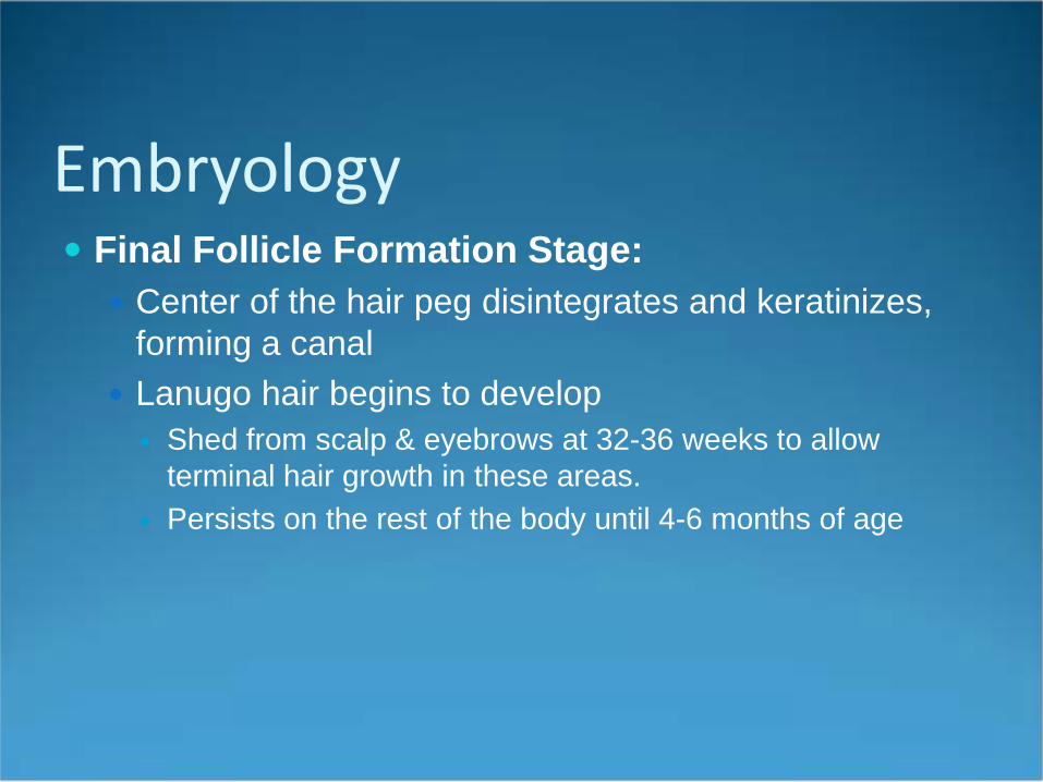
Embryology Final Follicle Formation Stage:
Center of the hair peg disintegrates and keratinizes, forming a canal
Lanugo hair begins to develop Shed from scalp & eyebrows at 32-36 weeks to allow
terminal hair growth in these areas. Persists on the rest of the body until 4-6 months of age
1. Ectoderm2. Mesoderm
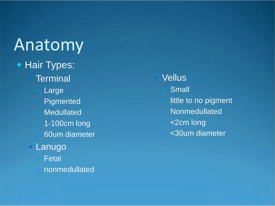
Anatomy Hair Types:
Terminal Large Pigmented Medullated 1-100cm long 60um diameter
Lanugo Fetal nonmedullated
Vellus Small little to no pigment Nonmedullated <2cm long <30um diameter
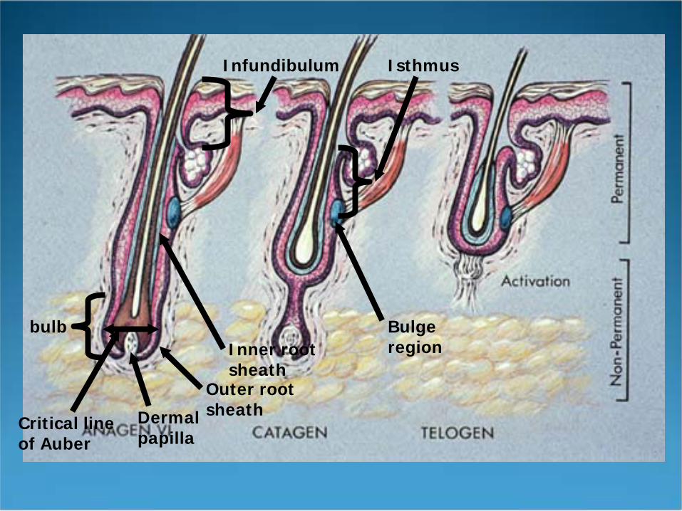
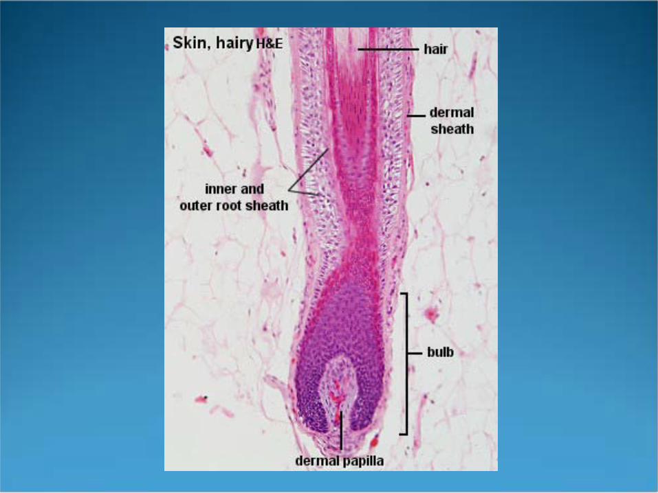
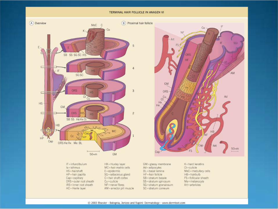
Infundibulum Isthmus
bulb
Dermal papilla
Outer root sheath
Inner root sheath
Bulge region
Critical line of Auber
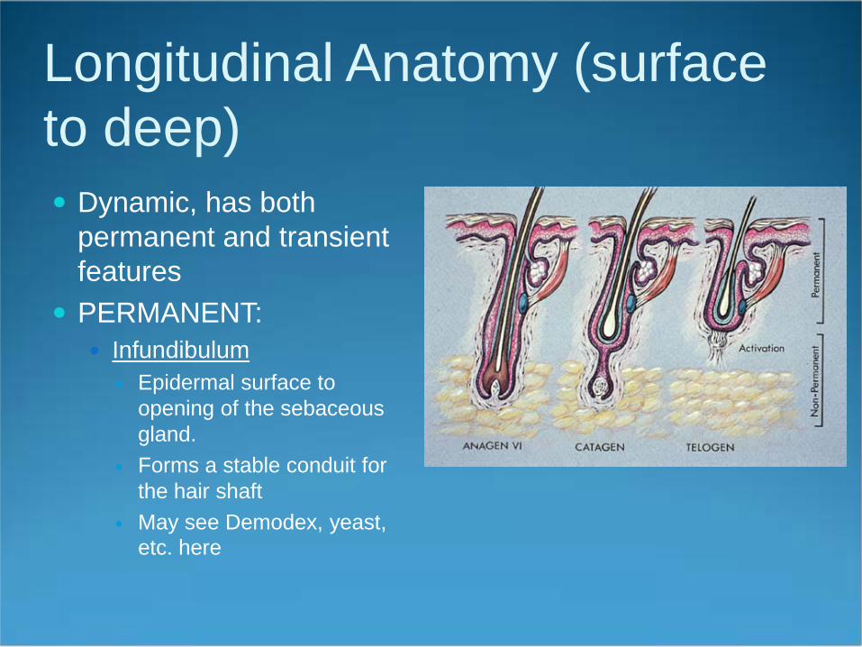
Longitudinal Anatomy (surface to deep) Dynamic, has both
permanent and transient features
PERMANENT: Infundibulum
Epidermal surface to opening of the sebaceous gland.
Forms a stable conduit for the hair shaft
May see Demodex, yeast, etc. here
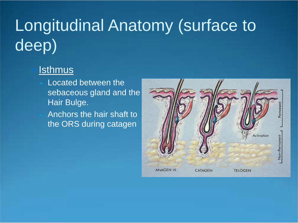
Longitudinal Anatomy (surface to deep)
Isthmus Located between the
sebaceous gland and the Hair Bulge.
Anchors the hair shaft to the ORS during catagen
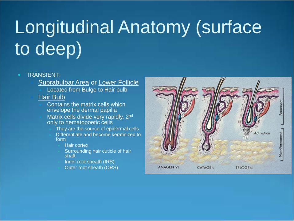
Longitudinal Anatomy (surface to deep) TRANSIENT:
Suprabulbar Area or Lower Follicle Located from Bulge to Hair bulb
Hair Bulb Contains the matrix cells which
envelope the dermal papilla Matrix cells divide very rapidly, 2nd
only to hematopoetic cells They are the source of epidermal cells Differentiate and become keratinized to
form Hair cortex Surrounding hair cuticle of hair
shaft Inner root sheath (IRS) Outer root sheath (ORS)
Longitudinal Anatomy (surface to deep) Transient
Hair Bulb Critical Line of Auber
widest diameter of bulb highest mitotic activity
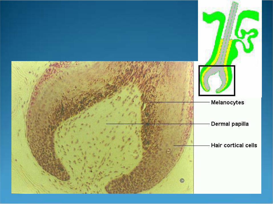
Dermal Papilla Dictates the embryonic generation of a hair follicle Modifies the extent of proliferation and differentiation of the epithelial hair
germ, and thus controls the size of the hair follicle Derived from the dermis mesenchyme Consists of small group of fibroblast cells derived from the mesoderm Basement membrane or glassy membrane separates the DP cells from the hair
fiber/sheath cells. The bigger the DP the thicker the hair fiber
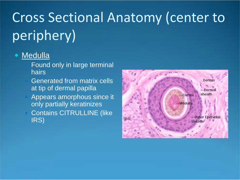
Cross Sectional Anatomy (center to periphery) Medulla
Found only in large terminal hairs
Generated from matrix cells at tip of dermal papilla
Appears amorphous since it only partially keratinizes
Contains CITRULLINE (like IRS)
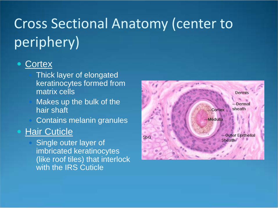
Cross Sectional Anatomy (center to periphery) Cortex
Thick layer of elongated keratinocytes formed from matrix cells
Makes up the bulk of the hair shaft
Contains melanin granules Hair Cuticle
Single outer layer of imbricated keratinocytes (like roof tiles) that interlock with the IRS Cuticle
Cross Sectional Anatomy (center to periphery) Inner Root Sheath (IRS)
Made up of 3 layers Does NOT contain melanin Formed from matrix cells (at periphery of follicle) in
pace with the hair shaft It dictates the shape of the hair since it hardens
(keratinizes) first IRS cells are shed into the infundibulum as the hair
shaft grows Products of the sebaceous gland help break down the
IRS Contains CITRULLINE (Like Medulla)
Henle’s
Huxley’s and cuticle

Cross Sectional Anatomy (center to periphery) Layers of IRS
IRS Cuticle (Inner) Huxley’s Layer (Middle) 3-4 cells thick Henle’s Layer (Outer) One cell layer thick. FIRST TO
BE KERATINIZED! Contain Keratins 1/10 (like upper layers of epidermis)
Keratinization order (1) Henle’s (2) Cuticle of IRS/Hair (3)Huxley’s
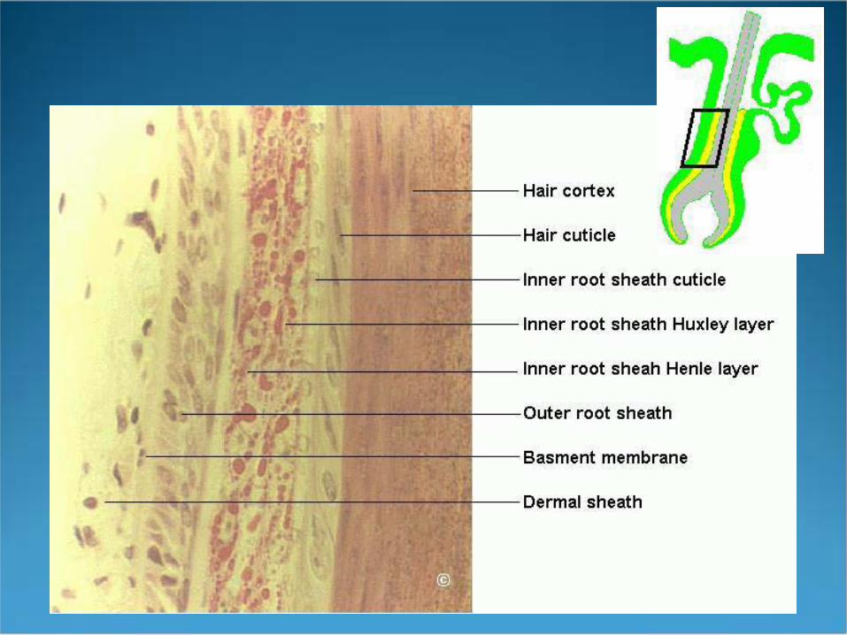
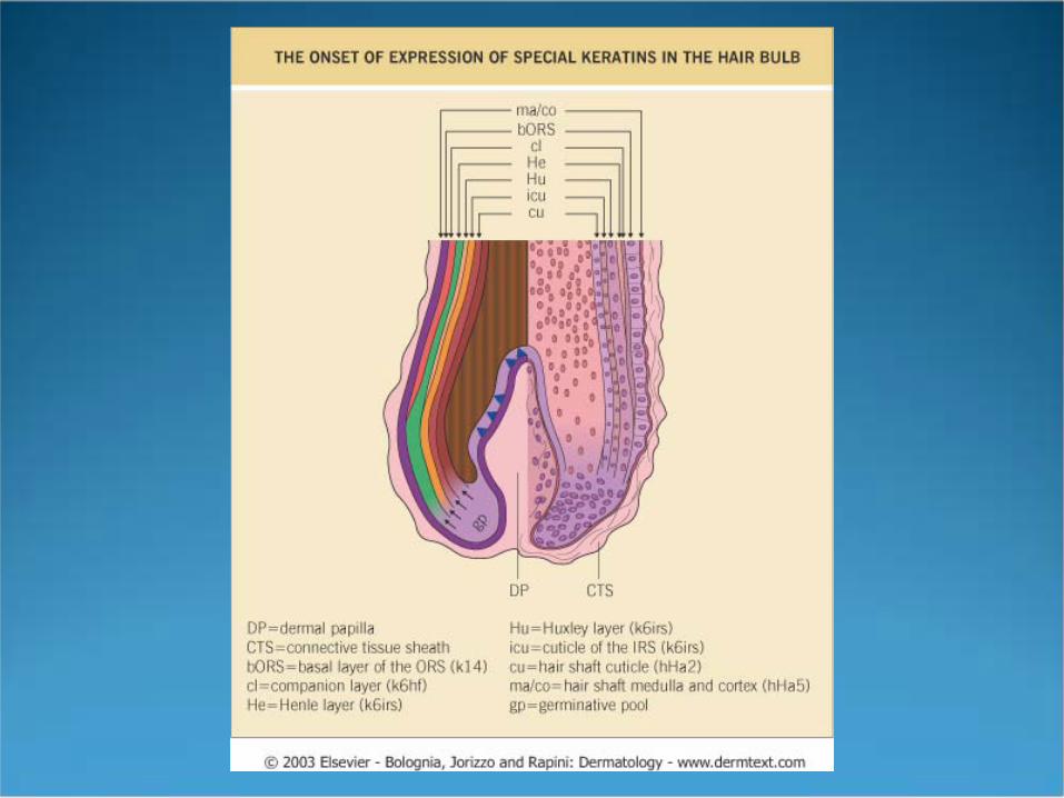
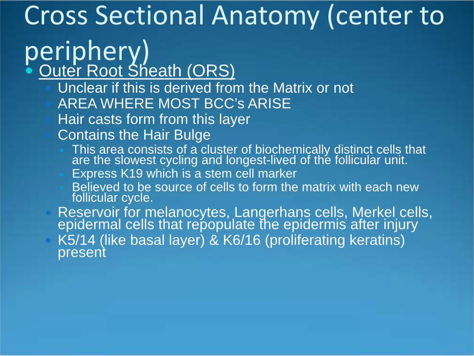
Cross Sectional Anatomy (center to periphery) Outer Root Sheath (ORS)
Unclear if this is derived from the Matrix or not AREA WHERE MOST BCC’s ARISE Hair casts form from this layer Contains the Hair Bulge
This area consists of a cluster of biochemically distinct cells that are the slowest cycling and longest-lived of the follicular unit.
Express K19 which is a stem cell marker Believed to be source of cells to form the matrix with each new
follicular cycle. Reservoir for melanocytes, Langerhans cells, Merkel cells,
epidermal cells that repopulate the epidermis after injury K5/14 (like basal layer) & K6/16 (proliferating keratins)
present
Cross Sectional Anatomy (center to periphery) Glassy or Vitreous Membrane:
A-cellular basement membrane bounding the entire follicle
Becomes corrugated in catagen, therefore, can be used as marker of this phase
Fibrous Root Sheath Outermost layer continuous with the dermis

Hair Keratinization Cortical keratins
classified as “hard” keratins as opposed to epidermal “soft” keratins
Have a higher Cysteine content which allows for more disulfide bonds and greater durability
These bonds are broken and reestablished by perms and straighteners (alkali substances)
Cortical Keratins are divided into 2 types: Ha = Acidic 1-8 (Chromosome 17) Hb = Basic 1-6 (Chromosome 12): Monilethrix #6 Occur in pairs with a highly conserved alpha helical
domain like epidermal keratins
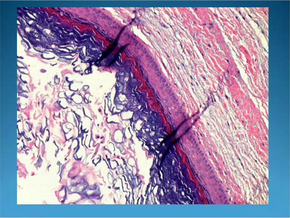
Hair Keratinization ORS and Hair Shaft undergo Tricholemmal
keratinization without the appearance of a granular layer
IRS keratinizes in a manner similar to the epidermis via the formation of Trichohyalin granules which are categorized as soft keratins
Cysts Epidermal inclusion cyst
Derived from the infundibulum Will see trichohyalin granules Soft keratin like epidermis
Tricholemmal cyst Derived from the isthmus Will not see trichohyalin granules Hard keratin like hair and ORS
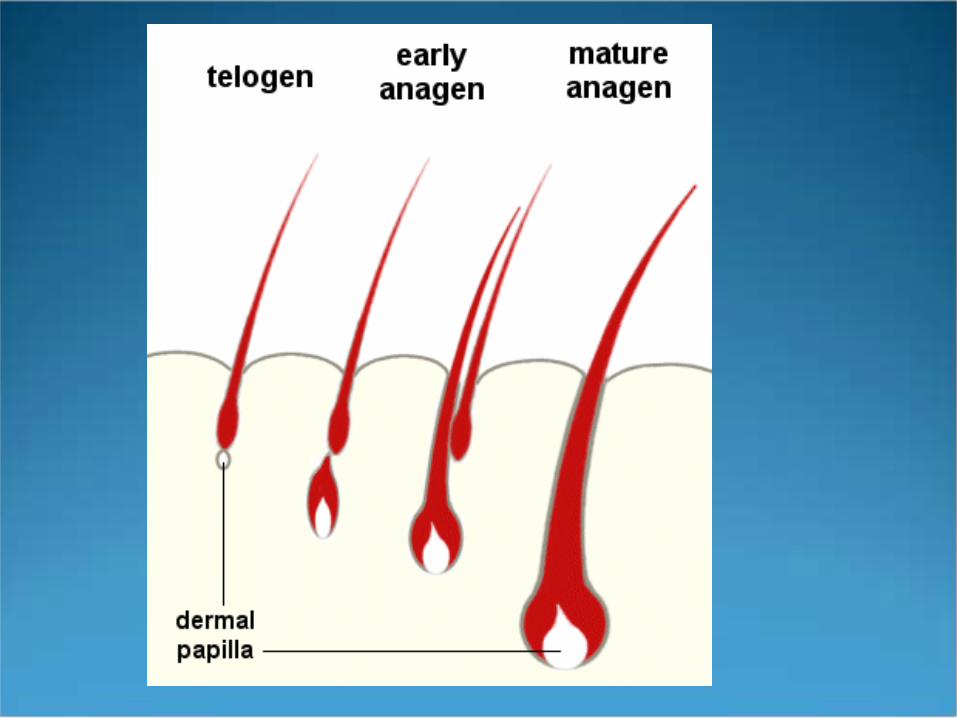
Hair Growth Cycle All animals (except Merino sheep & poodles)
have hair that cycles between active (Anagen) & resting (Telogen) states
Human hair exhibits mosaic growth except for lanugo hair in early life
Hair length is dependent on length of Anagen Scalp hair has the longest anagen phase (3-7 years) eyebrows have the shortest Anagen phase
1. Anagen2. Catagen3. Telogen
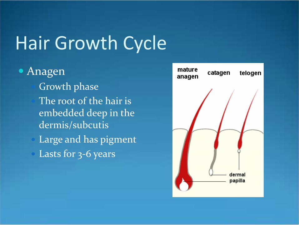
Hair Growth Cycle Anagen
Growth phase The root of the hair is
embedded deep in the dermis/subcutis
Large and has pigment Lasts for 3-6 years
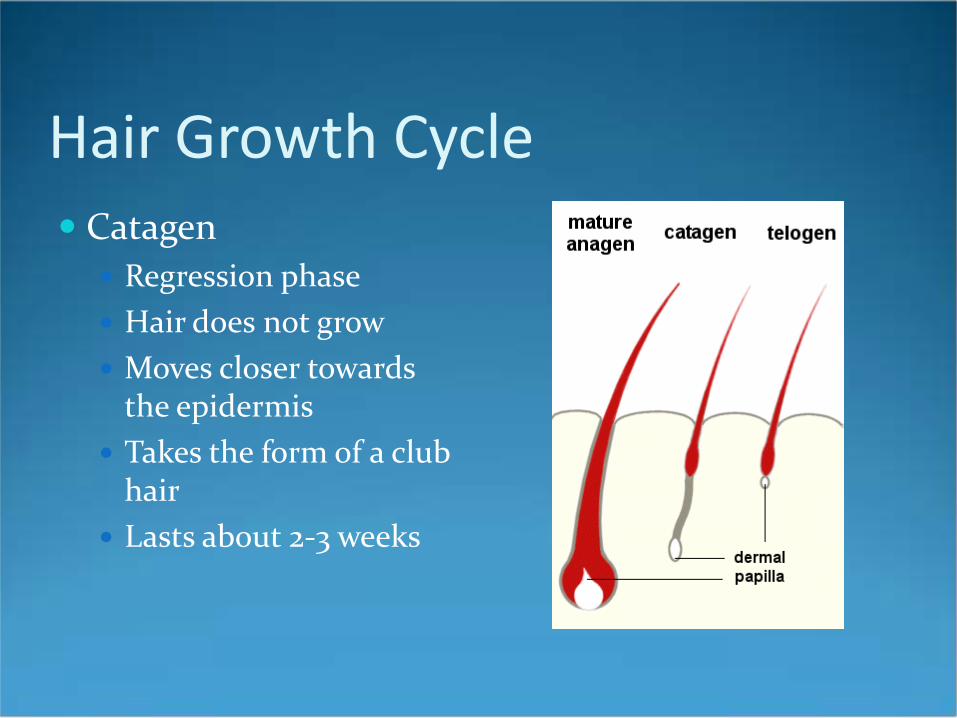
Hair Growth Cycle Catagen
Regression phase Hair does not grow Moves closer towards
the epidermis Takes the form of a club
hair Lasts about 2-3 weeks
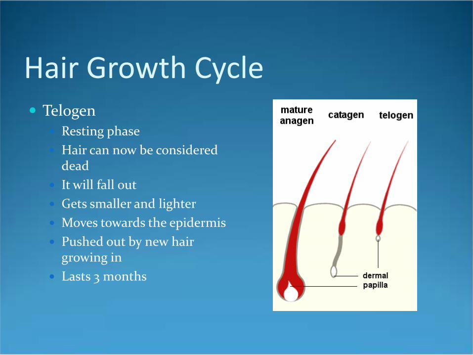
Hair Growth Cycle Telogen
Resting phase Hair can now be considered
dead It will fall out Gets smaller and lighter Moves towards the epidermis Pushed out by new hair
growing in Lasts 3 months

Hair Growth Cycle ANAGEN:
80-90% of scalp hair in growing phase Unclear what physiologic events stimulate growth assumed that factors from the dermal papilla regulate
anagen onset Keratinocyte Growth Factor (KGF or FGF-7) is
implicated complicated process involving redundant signals
Cyclosporin, Minoxidil, PTH-related peptide & estrogen receptor antagonists induce anagen in mice
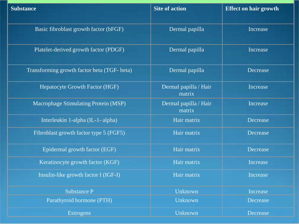
Substance Site of action Effect on hair growth
Basic fibroblast growth factor (bFGF) Dermal papilla Increase
Platelet-derived growth factor (PDGF) Dermal papilla Increase
Transforming growth factor beta (TGF- beta) Dermal papilla Decrease
Hepatocyte Growth Factor (HGF) Dermal papilla / Hair matrix
Increase
Macrophage Stimulating Protein (MSP) Dermal papilla / Hair matrix
Increase
Interleukin 1-alpha (IL-1- alpha) Hair matrix Decrease
Fibroblast growth factor type 5 (FGF5) Hair matrix Decrease
Epidermal growth factor (EGF) Hair matrix Decrease
Keratinocyte growth factor (KGF) Hair matrix Increase
Insulin-like growth factor I (IGF-I) Hair matrix Increase
Substance P Unknown IncreaseParathyroid hormone (PTH) Unknown Decrease
Estrogens Unknown Decrease
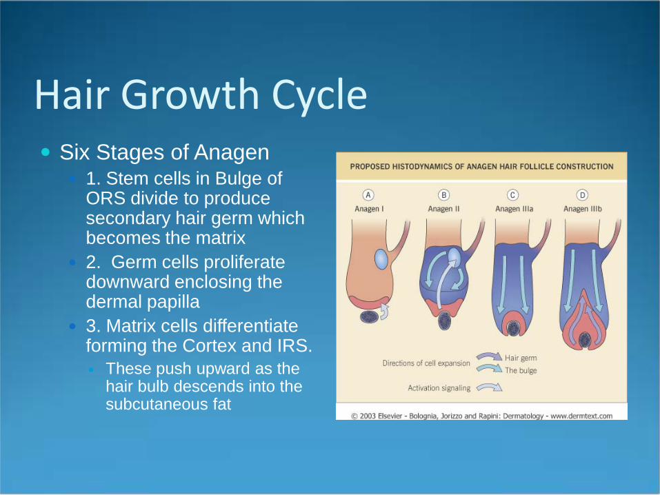
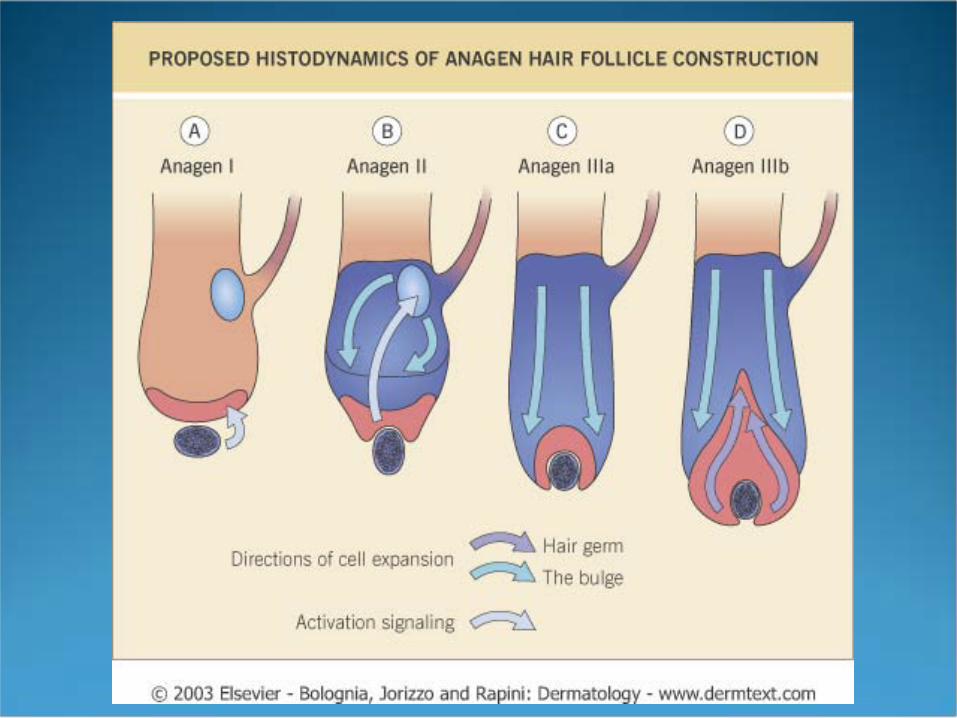
Hair Growth Cycle Six Stages of Anagen
1. Stem cells in Bulge of ORS divide to produce secondary hair germ which becomes the matrix
2. Germ cells proliferate downward enclosing the dermal papilla
3. Matrix cells differentiate forming the Cortex and IRS. These push upward as the
hair bulb descends into the subcutaneous fat
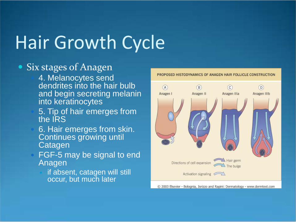
Hair Growth Cycle Six stages of Anagen
4. Melanocytes send dendrites into the hair bulb and begin secreting melanin into keratinocytes
5. Tip of hair emerges from the IRS
6. Hair emerges from skin. Continues growing until Catagen
FGF-5 may be signal to end Anagen if absent, catagen will still
occur, but much later
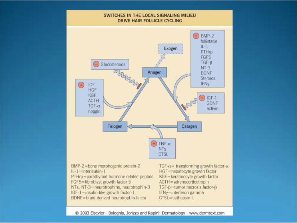
Hair Growth Cycle CATAGEN
Lasts only 2-3 weeks Only 1% of follicles in this stage Mitosis ceases in the matrix and the cells keratinize
forming a club hair Apoptosis occurs Melanocytes stop producing pigment Melanocytes withdraw their dendrites so the club end
of the hair is NOT pigmented The Suprabulbar area collapses like an accordion and
involutes while the vitreous membrane becomes thick and corrugated
Hair Growth Cycle CATGEN
IRS disintegrates & the ORS forms a sac from which the next generation hair germ will form
The dermal papilla follows the collapsing bulb and comes to rest just below the Bulge.
If this fails to happen no more hairs will be formed. This is seen in the mouse hairless gene and in a Pakistani family (Atrichia with Papular Lesions)
Hair Growth Cycle TELOGEN
Period of complete inactivity lasting about 100 days (3 months) in the scalp
5-10% of follicles Scalp loses 100-150 telogen hairs a day, which is only
a small percentage of the total # of hairs in this phase. Club Hair either falls out during telogen or is pushed
out by new hair growth during the next anagen phase.

Hair Growth Cycle EXOGEN
There is speculation that hair shedding might be an active process
For example, if 10% of scalp hairs (~10,000) are in telogen, then why does one only lose 100 hairs a day
Hair shedding might involve release by Desmoglein 3 which is believed to anchor the club hair during Telogen
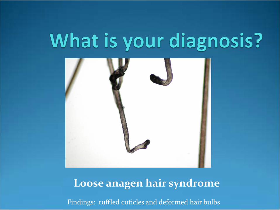
Loose anagen hair syndrome
Findings: ruffled cuticles and deformed hair bulbs
Control of Hair Growth Scalp hair grows at 1cm/month or 0.34mm/day Plucking telogen hairs seems to advance the
onset of anagen Shaving has no effect on hair growth or diameter Differences in maximal achievable scalp hair
length are the result of different anagen lengths Follicles are inherently programmed to cycle There are other control mechanisms, especially
via the endocrine system

Control of Hair GrowthHormones Androgens
THE MOST IMPORTANT hormonal regulators Act through receptors in the dermal papilla Increases the length of anagen, diameter, and growth
rate in susceptible follicles Paradoxically cause shorter anagen time,
miniaturization, and slower growth in areas such as the scalp.
Axillary & Pubic hair respond to testosterone, rest of body hair only responds to DHT made by 5-alpha reductase.
Control of Hair GrowthHormones Androgens
5-alpha reductase deficiency have axillary & pubic, but no body hair, balding or prostatic hypertrophy)
5αR Type I: Sebaceous glands Type II: Prostate and dermal papilla Puberty
Pubic hair responds 1st
Axillary & beard hair growth ~2 years later. Adult pattern not fully complete until 4th decade
Control of Hair GrowthHormones Estrogens
Prolongs anagen but decreases the growth rate. Responsible for the post-partum telogen effluvium
Thyroxine Advances onset of anagen increases growth rate. Excesses can be stressful and lead to telogen
effluvium. Deficiency can do the same in addition to slowing
growth rate.