Embed Size (px)
Citation preview
Copyright © 2017 Korean Stroke SocietyThis is an Open Access article distributed under the terms of the Creative Commons Attribution Non-Commercial License (http://creativecommons.org/licenses/by-nc/4.0/) which permits unrestricted non-commercial use, distribution, and reproduction in any medium, provided the original work is properly cited.
pISSN: 2287-6391 • eISSN: 2287-6405 http://j-stroke.org 67
Haemorrhagic Transformation after Ischaemic Stroke in Patients Taking Non-vitamin K Antagonist Oral AnticoagulantsJan C. Purrucker,a Kirsten Haas,b Marcel Wolf,c Timolaos Rizos,a Shujah Khan,a Peter Kraft,d Sven Poli,e Rainer Dziewas,f Johannes Meyne,g Frederick Palm,h Sebastian Jander,i Markus Möhlenbruch,c Peter U. Heuschmann,b,j and Roland Veltkamp,a,k on behalf of the RASUNOA investigatorsaDepartment of Neurology, Heidelberg University Hospital, Heidelberg, GermanybInstitute of Clinical Epidemiology and Biometry, University Würzburg, GermanycDepartment of Neuroradiology, Heidelberg University Hospital, Heidelberg, GermanydDepartment of Neurology, University Hospital Würzburg, Würzburg, GermanyeDepartment of Neurology, Tübingen University, Tübingen, GermanyfDepartment of Neurology, University Hospital Münster, GermanygDepartment of Neurology, University Hospital Schleswig-Holstein, Kiel, GermanyhDepartment of Neurology, Klinikum Ludwigshafen, Ludwigshafen, GermanyiDepartment of Neurology, Heinrich-Heine-University, Medical Faculty, Düsseldorf, GermanyjComprehensive Heart Failure Center, and Clinical Trial Center, University Hospital Würzburg, Würzburg, Germany kDepartment of Stroke Medicine, Imperial College London, London, United Kingdom
Correspondence: Roland VeltkampDepartment of Stroke Medicine, Imperial College London, Charing Cross Campus, 3 East 6, Fulham Palace Road, London, W6 8RF, United Kingdom Phone: +44-20-33130133, Fax: +44-20-83833309, E-mail: [email protected]
Received: June 16, 2016Revised: October 11, 2016Accepted: November 28, 2016
The authors have no financial conflicts of interest.
Background and Purpose To evaluate the frequency and outcome of haemorrhagic transforma-tion (HT) after ischaemic stroke in patients treated with non-vitamin K antagonist oral anticoagulants (NOACs).Methods Patients with stroke on treatment with a NOAC were prospectively enrolled in this multi-centre observational study between February 2012 and 2015. Brain imaging at admission and follow-up imaging until day 7 were reviewed for HT. Functional outcome was assessed by the modified Rankin scale (mRS) before the index event, at discharge, and at 3-months.Results 231 patients without recanalisation therapy (no-RT), and 32 patients with RT were eligible for analysis. Any HT was present at admission in 9/231 no-RT patients (3.9%, 95% CI 2.0 to 7.3) and in none of the patients with RT. In patients with follow-up imaging (no-RT, n= 129, and RT, n= 32), HT was present in 14.0% (no-RT; 95% CI, 8.9 to 21.1), and 40.6% (RT, 95% CI, 25.5 to 57.8), respectively. After adjustment for stroke severity, this difference between the no-RT and RT groups became non-significant. Symptomatic ICH was observed in 1 patient per group. HT was not associated with unfa-vourable outcome (mRS 3-6) at 3-months in multivariable analysis. Resumption of OAC after stroke was delayed in patients with HT compared to those without (15 d [IQR, 5–26] vs. 1 d [0–4], P < 0.001).Conclusions The frequency and severity of HT after stroke on NOAC appears similar to previous re-ports for vitamin K antagonists and no anticoagulation. Whether asymptomatic HT should delay re-sumption of preventive anticoagulation requires further investigation.
Keywords Stroke, Anticoagulation, Haemorrhagic Transformation
Original Article
Journal of Stroke 2017;19(1):67-76https://doi.org/10.5853/jos.2016.00542
Purrucker, et al. Haemorrhagic Transformation and NOAC Use
https://doi.org/10.5853/jos.2016.00542 68 http://j-stroke.org
Introduction
Any haemorrhagic transformation (HT) is found in approxi-mately 8.5% (95% CI 7-10%), of non-thrombolysed acute isch-aemic stroke (AIS) patients.1 Symptomatic HT associated with clinical worsening or death occurs in 1.5% (95% CI 0.8-2.2%).1,2 In the randomised European Cooperative Acute Stroke Study III, the rate of any HT was notably higher in both the IV thromboly-sis and placebo-arm (27.0% vs. 17.6%).3 However, differences in assessment criteria and HT definitions hamper direct compari-sons.4 While oral anticoagulation (OAC) with vitamin K antago-nists (VKA) does not appear to affect the occurrence of HT,2,5 data regarding the frequency and severity of HT in patients treated with non-Vitamin K antagonist oral anticoagulants (NO-ACS) is not available. The potential effects of HT on neurological outcome and on the interval until re-initiation of anticoagula-tion are unknown.
We evaluated the frequency and severity of HT at admission and during the first 7 days in acute stroke patients taking NO-ACs, and examined effects on the management and functional outcome of AIS under NOAC treatment using data from the multicentre Registry of Acute Stroke Under New Oral Anticoagu-lants (RASUNOA-pilot).
Methods
Study design, setting and variablesPatients presenting between February 2012 and February 2015
with an acute ischaemic stroke and taking NOACs at the time of the event were prospectively enrolled into the RASUNOA-pilot registry, an investigator-initiated, multicentre, observational co-hort study (ClinicalTrials.gov, NCT01850797). Overall, 38 neurol-ogy departments with certified stroke units across Germany par-ticipated in the registry. Inclusion criteria were age ≥18 years, acute ischaemic stroke, and current therapy with a NOAC (i.e., Apixaban, Dabigatran or Rivaroxaban) at the time of stroke on-set. For the present analysis, we excluded (i) patients without any available brain imaging, and (ii) patients with transient isch-aemic attack (as defined by presentation without neurological deficit according to the National Institute of Health Stroke Scale (NIHSS) at the time of admission and absence of an acute isch-aemic lesion on brain imaging6). Due to the observational char-acter of the registry, all diagnostic and treatment decisions were left to the discretion of the attending physicians. Patient charac-teristics including demographic information, clinical data and laboratory parameters were prospectively collected using a stan-dardized case report file. The modified Rankin scale score (mRS) was used for functional assessment prior to the stroke (pmRS),
at admission, and at hospital discharge. Moreover, 3-month out-come was assessed by a structured telephone follow-up. The in-dividual stroke risk in patients with atrial fibrillation was calcu-lated using the CHA2DS2VASc-score (excluding the index event), and the individual bleeding risk using the HAS BLED-score, re-spectively, with the item “labile INR” set to zero.7,8
The analysis was done stratified for patients not undergoing thrombolysis and/or thrombectomy (no-RT) and patients who received recanalisation therapy (RT), respectively (a focused analysis of the RT group without IV-only treated patients has been previously published.9
Neuroimaging analysisCranial CT and MRI examinations were assessed for signs of
ischaemic infarction, as well as intracranial haemorrhage by an experienced reader (JP). Assessment was performed on scans ob-tained at admission, and on follow-up scans carried out until day 7 if available. Regarding diagnosis and classification of haemor-rhage, findings were reviewed by a board-certified neuroradiolo-gist (MW). Disagreement between the two readers was resolved by consensus. Readers were blinded for patient details. HT was defined as any level of hyperdensity within the area of low at-tenuation (CT), or hypointensities on T2* or susceptibility weight-ed (SWI) sequences10 in areas corresponding to DWI/FLAIR hy-perintense regions on MRI. If rapid attenuation of initial hyper-densities on CT scans was observed, this was categorized as con-trast staining rather than hemorrhage.9
Haemorrhagic transformation was classified in accordance with the ECASS-I-criteria as either haemorrhagic infarction (HI1 and 2), or parenchymal haemorrhage (PH1 and 2).11 Accordingly, HI1 was defined as small petechiae along the margin of the in-farct, HI2 as more confluent petechiae within the infarct but without space-occupying effect, PH1 as a blood clot not exceed-ing 30% of the infarct area with some mild space-occupying ef-fect, and PH2 as blood clots exceeding 30% of the infarct area with significant space-occupying effect (Figure 1B).11 Moreover, we recorded intracerebral haemorrhage remote from the infarct, which was defined as small or medium-sized blood clots (PHr1) or large confluent dense blood clots (PHr2). Evidence of intra-ventricular or subarachnoidal haemorrhage was also captured. Symptomatic intracranial haemorrhage (sICH) was defined fol-lowing the ECASS-II definition.12 The extent of infarction was quantified using the Alberta Stroke Program Early Computed To-mography Score (ASPECTS; range 0 to 10 with 1 point subtract-ed for any evidence of ischaemic change in each defined region on axial cuts). In case of posterior circulation stroke, the pc-AS-PECTS score was calculated.13,14 For quantification of the site and size of ischaemic infarction the classification by Paciaroni and
Vol. 19 / No. 1 / January 2017
https://doi.org/10.5853/jos.2016.00542 http://j-stroke.org 69
colleagues was used.2 Accordingly, infarct size was classified as (1) small (lesion in the anterior or posterior circulation <1.5 cm), (2) medium (lesion in a cortical superficial branch of middle ce-rebral artery [MCA], or lesion involving the MCA deep branch, or lesion in internal borderzone territories, or lesion in a cortical su-perficial branch of posterior cerebral artery [PCA], or lesion in-volving the PCA branch or lesion in a cortical superficial branch of anterior cerebral artery [ACA]), (3) large anterior (lesion in-volving complete territory of MCA, PCA or ACA or lesion involv-ing 2 cortical superficial branches of MCA or lesion involving a cortical superficial branch of MCA associated to the MCA deep branch, or lesion involving more than 1 artery territory [e.g., MCA associated to ACA territories]) or (4) large posterior (lesion in-volving brain stem or cerebellum >1.5 cm).2 Cases without de-finitive evidence of acute cerebral infarction on available scans were added to the Paciaroni category 1.
Statistical analysisAll analyses were descriptive and exploratory. Continuous vari-
ables were described by mean and standard deviation (SD) or median and interquartile-range (IQR) for categorical variables, absolute and relative frequencies were reported. The Shapiro-Wilk test was used to ascertain distribution of data. Post-hoc, groups of patients with haemorrhagic transformation (HT) and without (non-HT) were defined. Fishers-exact test was used to compare proportions in baseline and radiological characteristics, dosing categories (standard [apixaban 5 mg BID, dabigatran 150 mg BID, rivaroxaban 20 mg QD] vs. low drug dose[apixaban 2.5 mg BID, dabigatran 110 mg BID, rivaroxaban 15 mg QD], and considering the actual dose, current renal function and age, ade-quate or overdosing) and outcomes, as appropriate. To compare continuous variables, t-test or non-parametric Mann-Whitney U test was used according to the skewness of the data. To calculate 95% confidence intervals (CI) of proportions, the adjusted Wald
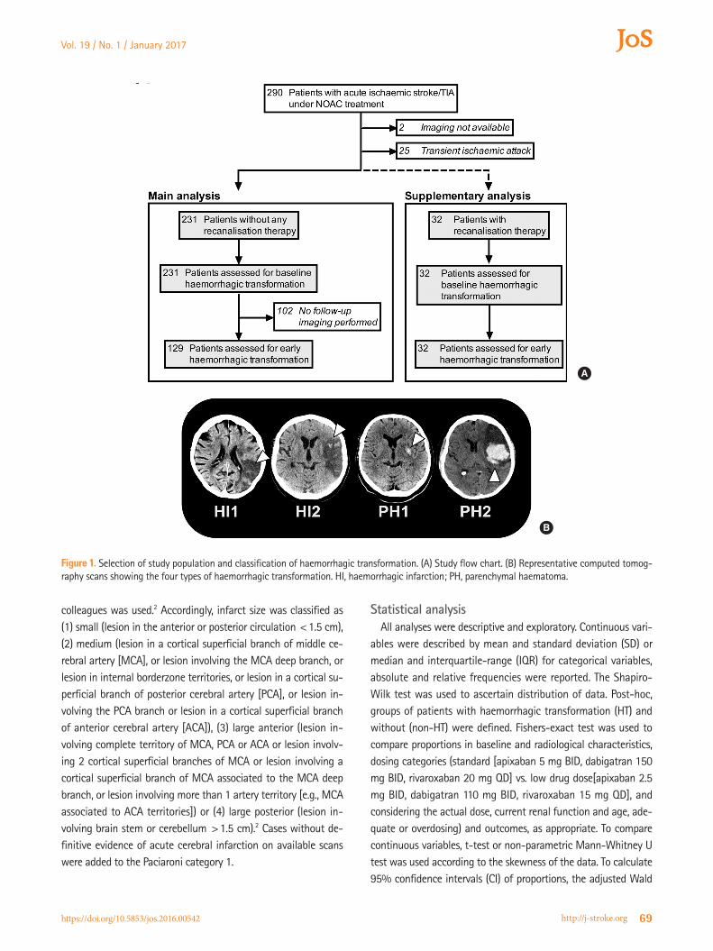
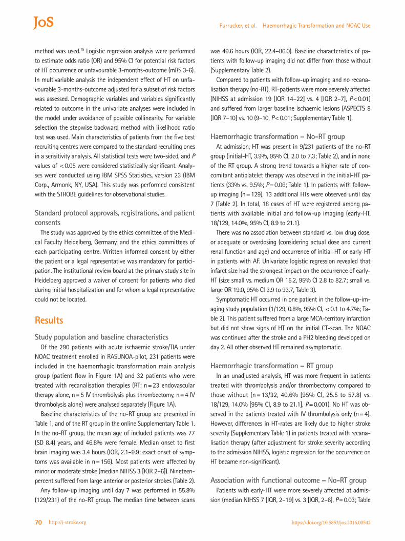
Figure 1. Selection of study population and classification of haemorrhagic transformation. (A) Study flow chart. (B) Representative computed tomog-raphy scans showing the four types of haemorrhagic transformation. HI, haemorrhagic infarction; PH, parenchymal haematoma.
A
B
Purrucker, et al. Haemorrhagic Transformation and NOAC Use
https://doi.org/10.5853/jos.2016.00542 70 http://j-stroke.org
method was used.15 Logistic regression analysis were performed to estimate odds ratio (OR) and 95% CI for potential risk factors of HT occurrence or unfavourable 3-months-outcome (mRS 3-6). In multivariable analysis the independent effect of HT on unfa-vourable 3-months-outcome adjusted for a subset of risk factors was assessed. Demographic variables and variables significantly related to outcome in the univariate analyses were included in the model under avoidance of possible collinearity. For variable selection the stepwise backward method with likelihood ratio test was used. Main characteristics of patients from the five best recruiting centres were compared to the standard recruiting ones in a sensitivity analysis. All statistical tests were two-sided, and P values of <0.05 were considered statistically significant. Analy-ses were conducted using IBM SPSS Statistics, version 23 (IBM Corp., Armonk, NY, USA). This study was performed consistent with the STROBE guidelines for observational studies.
Standard protocol approvals, registrations, and patient consents
The study was approved by the ethics committee of the Medi-cal Faculty Heidelberg, Germany, and the ethics committees of each participating centre. Written informed consent by either the patient or a legal representative was mandatory for partici-pation. The institutional review board at the primary study site in Heidelberg approved a waiver of consent for patients who died during initial hospitalization and for whom a legal representative could not be located.
Results
Study population and baseline characteristicsOf the 290 patients with acute ischaemic stroke/TIA under
NOAC treatment enrolled in RASUNOA-pilot, 231 patients were included in the haemorrhagic transformation main analysis group (patient flow in Figure 1A) and 32 patients who were treated with recanalisation therapies (RT; n=23 endovascular therapy alone, n=5 IV thrombolysis plus thrombectomy, n=4 IV thrombolysis alone) were analysed separately (Figure 1A).
Baseline characteristics of the no-RT group are presented in Table 1, and of the RT group in the online Supplementary Table 1. In the no-RT group, the mean age of included patients was 77 (SD 8.4) years, and 46.8% were female. Median onset to first brain imaging was 3.4 hours (IQR, 2.1–9.9; exact onset of symp-toms was available in n=156). Most patients were affected by minor or moderate stroke (median NIHSS 3 [IQR 2–6]). Nineteen-percent suffered from large anterior or posterior strokes (Table 2).
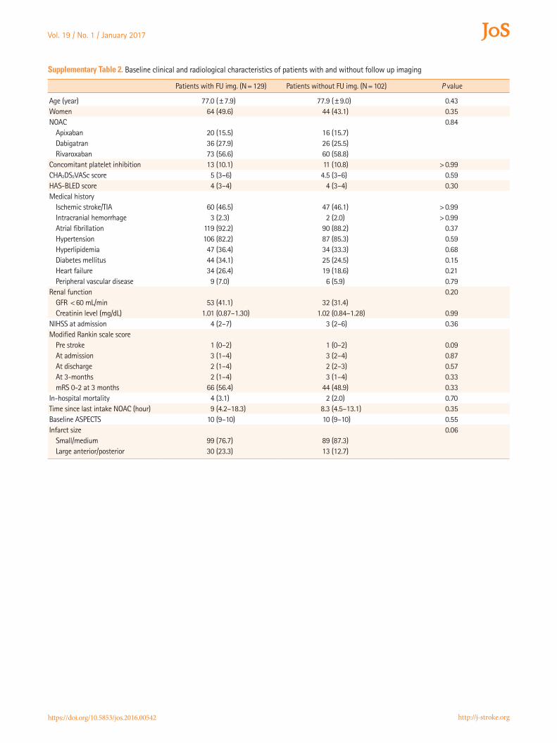
Any follow-up imaging until day 7 was performed in 55.8% (129/231) of the no-RT group. The median time between scans
was 49.6 hours (IQR, 22.4–86.0). Baseline characteristics of pa-tients with follow-up imaging did not differ from those without (Supplementary Table 2).
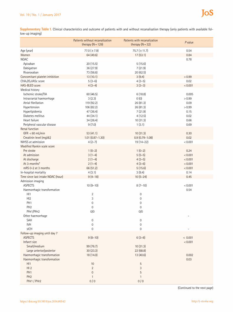
Compared to patients with follow-up imaging and no recana-lisation therapy (no-RT), RT-patients were more severely affected (NIHSS at admission 19 [IQR 14–22] vs. 4 [IQR 2–7], P<0.01) and suffered from larger baseline ischaemic lesions (ASPECTS 8 [IQR 7–10] vs. 10 (9–10, P<0.01; Supplementary Table 1).
Haemorrhagic transformation – No-RT groupAt admission, HT was present in 9/231 patients of the no-RT
group (initial-HT, 3.9%, 95% CI, 2.0 to 7.3; Table 2), and in none of the RT group. A strong trend towards a higher rate of con-comitant antiplatelet therapy was observed in the initial-HT pa-tients (33% vs. 9.5%; P=0.06; Table 1). In patients with follow-up imaging (n=129), 13 additional HTs were observed until day 7 (Table 2). In total, 18 cases of HT were registered among pa-tients with available initial and follow-up imaging (early-HT, 18/129, 14.0%, 95% CI, 8.9 to 21.1).
There was no association between standard vs. low drug dose, or adequate or overdosing (considering actual dose and current renal function and age) and occurrence of initial-HT or early-HT in patients with AF. Univariate logistic regression revealed that infarct size had the strongest impact on the occurrence of early-HT (size small vs. medium OR 15.2, 95% CI 2.8 to 82.7; small vs. large OR 19.0, 95% CI 3.9 to 93.7, Table 3).
Symptomatic HT occurred in one patient in the follow-up-im-aging study population (1/129, 0.8%, 95% CI, <0.1 to 4.7%; Ta-ble 2). This patient suffered from a large MCA-territory infarction but did not show signs of HT on the initial CT-scan. The NOAC was continued after the stroke and a PH2 bleeding developed on day 2. All other observed HT remained asymptomatic.
Haemorrhagic transformation – RT groupIn an unadjusted analysis, HT was more frequent in patients
treated with thrombolysis and/or thrombectomy compared to those without (n=13/32, 40.6% [95% CI, 25.5 to 57.8) vs. 18/129, 14.0% [95% CI, 8.9 to 21.1], P=0.001). No HT was ob-served in the patients treated with IV thrombolysis only (n=4). However, differences in HT-rates are likely due to higher stroke severity (Supplementary Table 1) in patients treated with recana-lisation therapy (after adjustment for stroke severity according to the admission NIHSS, logistic regression for the occurrence on HT became non-significant).
Association with functional outcome – No-RT groupPatients with early-HT were more severely affected at admis-
sion (median NIHSS 7 [IQR, 2–19] vs. 3 [IQR, 2–6], P=0.03; Table

Vol. 19 / No. 1 / January 2017
https://doi.org/10.5853/jos.2016.00542 http://j-stroke.org 71
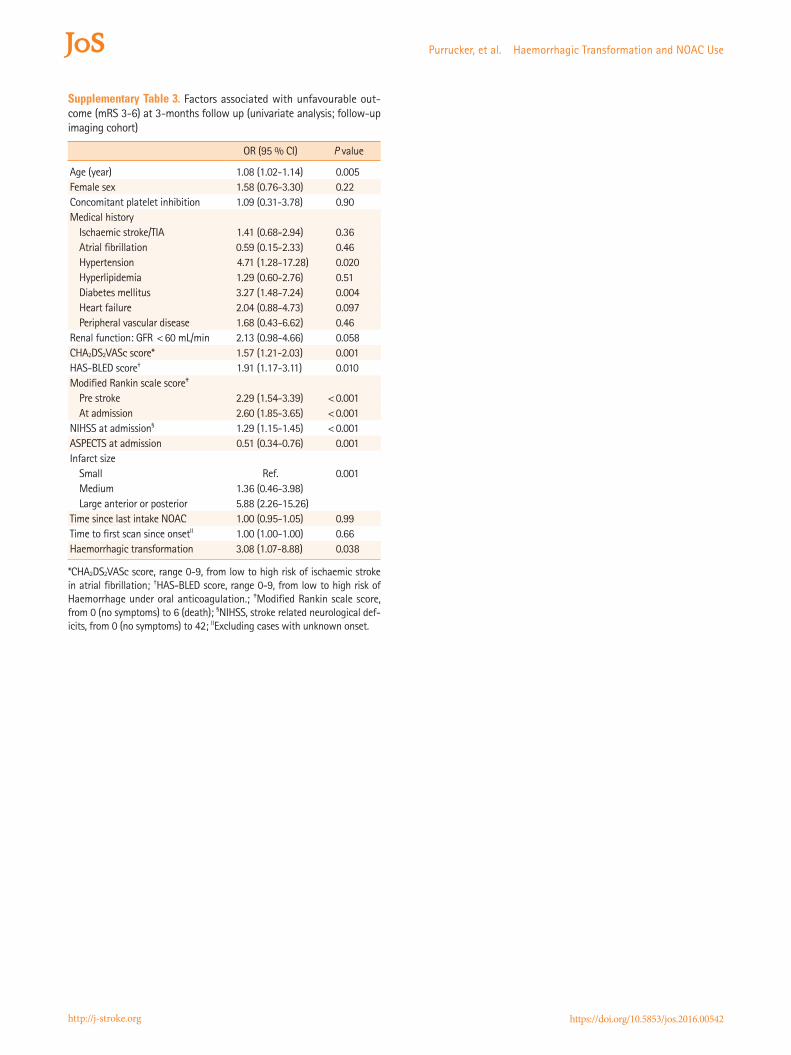
1), and suffered more frequently from larger anterior and poste-rior infarctions than patients without early-HT (P<0.001; Table 2). Patients with early-HT had a significantly worse outcome compared to those without (median mRS 4 [IQR, 1–6] vs. mRS 2 [IQR 1–4], P=0.001; Table 1). However, in multivariate analysis, HT was not independently associated with unfavourable out-come (mRS 3-6) at 3-months (Table 4; for univariate analysis, see Supplementary Table 3). In-hospital mortality was not signif-icantly increased among patients with HT.
Resumption of OAC – No-RT groupIn stroke survivors, OAC was resumed during the acute inpa-
tient stay in the majority of patients (n=184/230, 80%; thereof 97% NOAC, 3% phenprocoumon). In patients with early-HT, OAC was resumed after occurrence of HT in all but one case. Median time to recommended resumption was significantly lon-ger in patients with early-HT (15 d [IQR, 5–26]) compared to pa-tients without (1 d [0–4]; P<0.001). Notably, the delay was at least partly mediated through larger infarct-sizes in the early-HT group (see above), as a generally longer time until resumption of OAC was observed in patients without HT but large infarct com-pared to patients without HT and a small infarct (10 d [IQR, 0–15] and 1 d [IQR, 0–2]; P=0.034; data not shown).
Table 1. Baseline characteristics and functional outcome variables of patients with and without haemorrhagic transformation (no-RT group)
All PatientsAdmission imaging cohort Follow-up imaging cohort
Without initial-HT
Initial-HT P valueWithout early-HT
Early-HT P value
N 231 222 9 111 18Age (year) 77.4 (±8.4) 77.5 (±8.4) 75.4 (±8.3) 0.45 77.2 (±8.2) 76.1 (±5.6) 0.47Women 108 (46.8) 106 (47.7) 2 (22.2) 0.18 57 (51.4) 7 (38.9) 0.45NOAC 0.86 0.29
Apixaban 36 (15.6) 35 (15.8) 1 (11.1) 19 (17.1) 1 (5.6)Dabigatran 62 (26.8) 59 (26.6) 3 (33.3) 32 (28.8) 4 (22.2)Rivaroxaban 133 (57.6) 128 (57.7) 5 (55.6) 60 (54.1) 13 (72.2)
Time since last intake NOAC (hour) 8.7 (4.3–15.2) 8.8 (4.3–15.4) 5.9 (4.4–18.2) 0.87 9.0 (4.2–18.1) 12.8 (4.5–20.8) 0.48Time until resumption of OAC (day) 1 (0-3) 1 (0-3) 2 (1-15) 0.017 1 (0-3) 15 (5-26) < .001Concomitant platelet inhibition 24 (10.4) 21 (9.5) 3 (33.3) 0.06 10 (9.0) 3 (16.7) 0.39CHA2DS2VASc score* 5 (3–6) 5 (3–6) 4 (3–8) 0.63 5 (4–6) 4 (3–6) 0.24HAS-BLED† 4 (3–4) 4 (3–4) 3 (3–4) 0.52 4 (3–4) 3 (3–4) 0.09Medical history‡
Ischaemic stroke/TIA 107 (46.3) 102 (45.9) 5 (55.6) 0.74 53 (47.7) 7 (38.9) 0.61Intracranial Haemorrhage 5 (2.2) 5 (2.3) 0 (0) >0.99 3 (2.7) 0 (0) >0.99Atrial fibrillation 209 (90.5) 200 (90.1) 9 (100) >0.99 101 (91.0) 18 (100) 0.36Hypertension 193 (83.5) 186 (83.8) 7 (77.8) 0.65 93 (83.8) 13 (72.2) 0.32Hyperlipidemia 81 (35.1) 80 (36.0) 1 (11.1) 0.17 42 (37.8) 5 (27.8) 0.60Diabetes mellitus 69 (29.9) 63 (28.4) 6 (66.7) 0.02 38 (34.2) 6 (33.3) >0.99Heart failure 53 (22.9) 51 (23.0) 2 (22.2) >0.99 28 (25.2) 6 (33.3) 0.57Peripheral vascular disease 15 (6.5) 14 (6.3) 1 (11.1) 0.46 8 (7.2) 1 (5.6) >0.99
Renal functionGFR <60 mL/min 85/ 206 (41.3) 82/197 (36.9) 3 (33.3) 0.74 45/99 (45.5) 8/18 (44.4) >0.99Creatinin level (mg/dL) 1.0 (0.9–1.3) 1.0 (0.9–1.3) 1.0 (0.7–1.2) 0.37 1.0 (0.8–1.3) 1.0 (0.9–1.2) 0.95
NIHSS at admission 3 (2–6) 3 (2–6) 4 (1–7) 0.87 3 (2–6) 7 (2–19) 0.03Modified Rankin scale score
Pre stroke 1 (0–2) 1 (0–2) 2 (1–3) 0.14 1 (0–2) 1 (1–2) 0.13At admission 3 (1–4) 3 (1–4) 4 (1–5) 0.21 2 (1–4) 5 (3–5) 0.001At discharge 2 (1–3) 2 (1–3) 3 (2–5) 0.15 2 (1–3) 4 (2–5) 0.001At 3-months§ 2 (1–4) 2 (1–4) 2 (1–6) 0.78 1 (1–4) 4 (1–6) 0.001mRS 0-2 at 3-months 110 (53.1) 105 (47.3) 5 (55.6) >0.99 60 (60.6) 6 (33.3) 0.04
In-hospital mortality 6/229 (2.6) 6/220 (2.7) 0 (0) >0.99 2 (1.8) 2 (11.1) 0.09
Data are mean (±SD), median (IQR) or n (%). Missing data for creatinin level (n=5), GFR (n=25), mRS at discharge (n=2) and at 3-months (n=24), time un-til resumption of anticoagulation, (n=29).NOAC, non-vitamin K antagonist oral anticoagulant; TIA, transient ischaemic attack; GFR, glomerular filtration rate (electronic GFR as reported by the centres); NIHSS, National Institutes of Health Stroke Scale; mRS, modified Rankin score [from 0 (no symptoms) to 6 (death)]; no-RT, no recanalisation therapy group.*CHA2DS2VASc score, range 0-9, from low to high risk of ischemic stroke in atrial fibrillation; †HAS-BLED score, range 0-9, from low to high risk of haemor-rhage under oral anticoagulation; ‡Medical history, excluding the index-event; §3-months modified Rankin data available for 207/231 patients (89.6%).

Purrucker, et al. Haemorrhagic Transformation and NOAC Use
https://doi.org/10.5853/jos.2016.00542 72 http://j-stroke.org
Discussion
The new findings of our study are that (1) spontaneous HT oc-curs at a similar rate in acute stroke patients treated with NOAC as previously reported for non-anticoagulated and VKA-antico-agulated stroke patients,1,2,5 (2) recanalisation therapy did not in-crease the proportion of patients with HT after adjustment for stroke severity, (3) presence of HT on imaging appeared to delay the resumption of OAC, and (4) the presence of HT in NOAC treated patients was not independently associated to an unfa-vourable outcome at 3 months.
Ischaemic stroke occurred under NOAC therapy in 1 to 2% of NOAC treated patients with AF per year in large randomised controlled trials.16-19 The incidence of strokes in patients taking NOACs is expected to increase in the future due to an increase of the prevalence of atrial fibrillation,20 higher utilisation of
NOAC in the population,21 and a potential extension of indica-tions.22 A major advantage of NOAC compared to VKA treatment is a significant reduction of intracranial haemorrhage (ICH).23 Nevertheless, NOAC-related ICH has a similar poor prognosis as VKA-related ICH.24 Three preclinical studies in mice undergoing transient middle cerebral occlusion that were anticoagulated with either warfarin or dabigatran supported the assumption that NOAC are also associated with a lower risk of HT in isch-aemic stroke compared to VKA.25-27 Moreover, Bohmann and col-leagues suggested that early continuation of dabigatran might be safe in experimental ischaemic stroke.26 In contrast to these experimental data, current expert guidance regarding the re-sumption of anticoagulation recommends starting oral antico-agulation in TIA and stroke patients with short or longer delay depending on the severity of the stroke.28
According to the present analysis, 14.0% (95% CI, 8.9 to 21.1)
Table 2. Radiological characteristics and outcomes (no-RT group)
Admission imaging cohort Follow-up imaging cohort
Without initial-HT Initial-HT P value Without early-HT Early-HT P value
N 222 9 – 111 18 –MRI available 118 (53.2) 3 (33.3) 0.32 76 (68.5) 9 (50.0) 0.18Proximal vessel occlusion* 21/121 (17.4) 0/2 (0) >0.99 11/69 (15.9) 4/9 (44.4) 0.06Infarct size 0.001 <0.001
Small 148 (66.7) 0 (0) – 76 (68.5) 2 (11.1) –Medium 36 (16.2) 4 (44.4) – 15 (13.5) 6 (33.3) –Large anterior 29 (13.1) 4 (44.4) – 16 (14.4) 8 (44.4) –Large posterior 9 (4.1) 1 (11.1) – 4 (3.6) 2 (11.1) –
Admission imagingASPECTS 10 (9–10) 9 (8–9) 0.003 10 (9–10) 9 (5–9) <0.001HI1 0 (0) 6 (66.7) – 0 (0) 2 (11.1) –HI2 0 (0) 3 (33.3) – 0 (0) 3 (16.7) –PH1 0 (0) 0 (0) – 0 (0) 0 (0) –PH2 0 (0) 0 (0) – 0 (0) 0 (0) –PHr1/PHr2 0 (0)/0 (0) 0 (0)/0 (0) – 0 (0)/0 (0) 0 (0)/0 (0) –SAH 0 (0) 0 (0) – 0 (0) 0 (0) –IVH 0 (0) 0 (0) – 0 (0) 0 (0) –sICH 0 (0) 0 (0) – 0 (0) 0 (0) –
Follow-up imaging until day 7ASPECTS – – – 9 (8–10) 5 (4–9) 0.001HI1 – – – 0 (0)/0 (0) 10 (55.6) –HI 2 – – – 0 (0) 2 (11.1) –PH1 – – – 0 (0) 0 (0) –PH2 – – – 0 (0) 1 (5.6) –PHr1/PHr2 – – – 0 (0) 0 (0)/0 (0) –SAH – – – 0 (0) 0 (0) –IVH – – – 0 (0) 0 (0) –sICH – – – 0 (0) 1 (5.6) –
Data are median (IQR) or n (%). Note: In the follow-up imaging group, a total of 5 haemorrhages were present at admission, and 13 additional haemorrhages became present during follow-up imaging, resulting in a total of 18 hemorrhagic transformations in this group.MRI, magnetic resonance imaging; ASPECTS, Alberta Stroke Program Early Computed Tomography Score; HI1/2, haemorrhagic infarction type 1/2; PH1/2, pa-renchymal haemorrhage type 1/2; SAH, subarachnoid haemorrhage; IVH, intraventricular haemorrhage; sICH, symptomatic intracerebral haemorrhage (ac-cording to the ECASS-II criteria); no-RT, no recanalisation therapy group.*CT, MR or digital subtraction angiography available in 123/231 patients (53.2%).
Vol. 19 / No. 1 / January 2017
https://doi.org/10.5853/jos.2016.00542 http://j-stroke.org 73
of the NOAC anticoagulated patients with initial- and follow-up imaging developed HT, which is slightly higher than reported for VKA treated patients (10% in patients with INR <2, but no data on patients with INR >2 available),5 and for patients without antithrombotic or thrombolysis treatment (8.5%, 95% CI 7% to 10%).1 In contrast, substantially higher rates of HT have been re-ported in the placebo arm of intravenous thrombolysis trials (21.6 to 29.6%).29
Although there is an ongoing debate about the relevance of both initial clinically symptomatic and asymptomatic forms of HT in terms of functional outcome,30-33 the present study high-lights a more direct clinical consequence: Delayed resumption of OAC after occurrence of HT. Primary or secondary prevention of ischaemic stroke in (non-valvular) atrial fibrillation was the indi-cation of oral anticoagulation in the majority of our patients. These patients are at a high risk of recurrent stroke, indicated by a median CHA2DS2VASc score of 5 at admission (excluding the
index event). Once a patient has suffered from HT, resumption of OAC is usually postponed until HT is resolved or at least consid-ered radiologically stable. In our study, OAC was indeed resumed significantly later in patients in whom early-HT occurred. How-ever, larger infarct-sizes in the HT group might have contributed to this finding.
Interestingly, the median admission NIHSS of 4 in our popula-tion was relatively low compared to cardioembolic stroke.34 A potential explanation might be that orally anticoagulated pa-tients have less severe stroke compared to non-anticoagulated patients with AF in observational studies.35,36 Alternatively, stroke may have been caused by another etiology of cardioembolism.
Larger scale prospective studies are needed to determine whether early restart or continued anticoagulation in non-dis-abling small ischaemic stroke with less severe blood-brain barrier damage is safe and beneficial. The proportion of 9% of patients who developed HT despite small-size infarction in our cohort however still warrants caution. A recent single-centre open-label non-randomised study found no increased risk of HT after initia-tion of dabigatran therapy within 24 hours of TIA or minor stroke (NIHSS ≤3).37 Another single-centre open-label study reports a progression of asymptomatic HT after administration of rivaroxa-ban on a median of 3 days after TIA and mild-to-moderate stroke (NIHSS ≤9) comparing baseline to follow-up MRI at day 7.38
Our study has some limitations. Recruitment was imbalanced among centres with six centres recruiting 57% of patients. How-ever, a sensitivity analysis revealed no difference in terms of age, sex, and stroke severity in patients from top-recruiting versus other centres (Supplementary Table 4). As the RASUNOA pilot study did not have a simultaneously recruited control group, only indirect comparisons with other anticoagulation or no anticoag-ulation are possible. We do not have data regarding intercurrent antiplatelet therapy, which might have influenced the risk of HT.
Although the present study comprises the largest prospectively
Table 3. Factors associated with early haemorrhagic transformation (univariate analysis) (follow-up imaging cohort)
OR (95 % CI) P value
Age (year) 0.98 (0.92-1.05) 0.57Female sex 0.60 (0.22-1.67) 0.33Concomitant platelet inhibition 2.02 (0.50-8.19) 0.33Medical history
Ischaemic stroke/TIA 0.70 (0.25-1.93) 0.49Hypertension 0.50 (0.16-1.59) 0.24Hyperlipidemia 0.63 (0.21-1.90) 0.41Diabetes mellitus 0.96 (0.33-2.76) 0.94Heart failure 1.48 (0.51-4.32) 0.47Peripheral vascular disease 0.76 (0.09-6.45) 0.80
Renal function: GFR <60 mL/min 0.96 (0.35-2.64) 0.94CHA2DS2VASc score* 0.89 (0.68-1.17) 0.41HAS-BLED score† 0.68 (0.39-1.19) 0.17Modified Rankin scale score‡
Pre stroke 1.25 (0.84-1,86) 0.27At admission 1.95 (1.29-2.94) 0.002
NIHSS at admission§ 1.09 (1.03-1.16) 0.006ASPECTS at admission 0.61 (0.46-0.80) <0.001Infarct size 0.001
Small Ref.Medium 15.20 (2.80-82.66)Large anterior or posterior 19.00 (3.85-93.74)
Time since last intake NOAC 1.01 (0.95-1.08) 0.68Time to first scan since onsetII 1.02 (0.99-1.05) 0.24
TIA, transient ischaemic attack; GFR, glomerular filtration rate (electronic GFR as reported by the centres); NIHSS, National Institutes of Health Stroke Scale; ASPECTS, Alberta Stroke Program Early Computed Tomography Score; NOAC, non-vitamin K antagonist oral anticoagulant.*CHA2DS2VASc score, range 0-9, from low to high risk of ischaemic stroke in atrial fibrillation; †HAS-BLED score, range 0-9, from low to high risk of Haemorrhage under oral anticoagulation; ‡Modified Rankin scale score, from 0 (no symptoms) to 6 (death); §NIHSS, stroke related neurological def-icits, from 0 (no symptoms) to 42; IIExcluding cases with unknown onset.
Table 4. Factors associated with unfavourable outcome (mRS 3-6) at 3-months follow up (multivariate analysis) (follow-up imaging cohort)
OR (95% CI) P value
Age (year) 1.07 (0.97-1.13) 0.20Gender: female 1.46 (0.52-4.14) 0.47Medical history: Hypertension 9.81 (1.92-50.04) 0.006NIHSS admission (0-3 vs. ≥4)* 3.33 (1.18-9.39) 0.023Modified Rankin scale score pre stroke† 1.97 (1.23-3.17) 0.005Infarct size 0.007
Small Ref.Medium 2.40 (0.61-9.35)Large anterior or posterior 6.46 (2.02-20.73)
Haemorrhagic transformation 3.26 (0.66-16.04) 0.14
*NIHSS grouped into (0-3 vs. ≥4) based on Baird et al., 2001.39; †Modified Rankin scale score, pre stroke from 0 (no symptoms) to 5 (severe disability).
Purrucker, et al. Haemorrhagic Transformation and NOAC Use
https://doi.org/10.5853/jos.2016.00542 74 http://j-stroke.org
collected observational cohort of IS patients under NOAC treat-ment to our knowledge, its sample size is still limited and larger scale prospective studies are needed in order to confirm the re-sults. Nevertheless, due to its observational character with only few exclusion criteria, we expect generalisability of the findings. Finally, neither the study design nor the sample-size allowed de-tection of potential differences in rate and severity of HT related to specific NOAC agents or specific doses.
Conclusions
In conclusion, we found a similar risk of HT after ischemic stroke in patients on NOAC as previously reported for VKA-anti-coagulated, and non-anticoagulated patients. However, replica-tion of the results in future prospective studies using matched control groups is therefore necessary, and current indirect com-parisons should be interpreted with caution. While clinicians should carefully avoid continuation of NOAC therapy in patients with large infarcts, more evidence is needed to avoid potentially unnecessary delays of restarting anticoagulation in patients with HT of small or moderate ischaemic strokes, who are at risk of re-current thromboembolism.
Acknowledgments
We thank all principal investigators of the RASUNOA study and participating hospitals who enrolled at least one ischaemic stroke patient (A-Z). A Binder (Kiel), M Dichgans (München), R Dziewas (Münster), K Gröschel (Mainz), M Eicke (Idar-Oberstein), M Ertl (Regensburg), MG Hennerici (Mannheim), C Hobohm (Leipzig), T Höhle (Herne), S Jander (Düsseldorf), E Jüttler (Ulm), A Khaw (Greifswald), C Kleinschnitz (Würzburg), A Kraft (Halle), M Köhrmann (Erlangen), F Meisel (Karlsruhe), T Neumann-Haefelin (Fulda), C Opherk (Heilbronn), F Palm (Ludwigshafen), S Poli (Tübingen), J Röther (Hamburg), E Schmid (Stuttgart), G Seidel (Hamburg), H Sodan (Bad Neustadt), C Tanislav (Gießen), G Thomalla (Hamburg), R Veltkamp (Heidelberg), K Wartenberg (Halle-Wittenberg), C Weimar (Essen).
JCP received travel and congress participation support from Pfizer, and personal fees from Boehringer Ingelheim, outside the submitted work. TR received consulting honoraria, speakers’ honoraria and travel support from BMS Pfizer, Boehringer Ingel-heim, Bayer HealthCare and Daichii Sankyo, outside the submit-ted work. PK received consulting honoraria, speakers’ honoraria, travel support or research support from Boehringer Ingelheim, Bayer HealthCare, BMS Pfizer and Daiichi Sankyo. SP received honoraria from C.R. Bard, Bayer Healthcare, BeneChill, BMS-Pfizer, Boehringer-Ingelheim, Daiichi Sankyo, EMCOOLS, and
ZOLL; institutional research support from BeneChill, BMS-Pfizer, Covidien, EMCOOLS, Helena Laboratories, HVM Medical, Rau-medic, and ZOLL; congress/meeting traveling and accommoda-tion costs: C.R. Bard, Bayer Healthcare, BeneChill, Boehringer-In-gelheim, EMCOOLS, and ZOLL. RD has received speaker fees and consultation honoraria from Bayer, Boehringer Ingelheim, BMS, Pfizer and Daiichi Sankyo. FP received consulting honoraria, speakers’ honoraria or travel support from Boehringer Ingelheim, BMS, Pfizer and Daiichy-Sankyo. SJ received honoraria for speak-ing and consultancy from Boehringer Ingelheim, Bayer Health-care, BMS, Pfizer, and Daiichi Sankyo.PUH reports grants from BMBF, EU, Charité, Berlin Chamber of Physicians, German Par-kinson Society, University Hospital Würzburg, Robert-Koch-Insti-tute, Charité–Universitätsmedizin Berlin (within MonDAFIS; MonDAFIS is supported by an unrestricted research grant to the Charité from Bayer), University Göttingen (within FIND-AFran-domised, supported by an unrestricted research grant to the University Göttingen from Boehringer-Ingelheim), and University Hospital Heidelberg (within RASUNOA-prime; supported by an unrestricted research grant to the University Hospital Heidelberg from Bayer, BMS, Boehringer-Ingelheim, Daiichi Sankyo), outside the submitted work. RV has received speaker fees, consulting honoraria and research support from Bayer, Boehringer Ingel-heim, BMS, Pfizer, Daiichi Sankyo, CSL Behring. The other authors report no competing interests.
References1. Lindley RI, Wardlaw JM, Sandercock PA, Rimdusid P, Lewis SC,
Signorini DF, et al. Frequency and risk factors for spontaneous
hemorrhagic transformation of cerebral infarction. J Stroke Cerebrovasc Dis 2004;13:235-246.
2. Paciaroni M, Agnelli G, Corea F, Ageno W, Alberti A, Lanari A,
et al. Early hemorrhagic transformation of brain infarction:
rate, predictive factors, and influence on clinical outcome: re-
sults of a prospective multicenter study. Stroke 2008;39:2249-
2256.
3. Hacke W, Kaste M, Bluhmki E, Brozman M, Davalos A, Guidetti
D, et al. Thrombolysis with alteplase 3 to 4.5 hours after acute
ischemic stroke. N Engl J Med 2008;359:1317-1329.
4. Alvarez-Sabin J, Maisterra O, Santamarina E, Kase CS. Factors
influencing haemorrhagic transformation in ischaemic stroke.
Lancet Neurol 2013;12:689-705.
5. O’Donnell M, Oczkowski W, Fang J, Kearon C, Silva J, Bradley C,
et al. Preadmission antithrombotic treatment and stroke se-
verity in patients with atrial fibrillation and acute ischaemic
stroke: an observational study. Lancet Neurol 2006;5:749-754.
6. Easton JD, Saver JL, Albers GW, Alberts MJ, Chaturvedi S, Feld-
Vol. 19 / No. 1 / January 2017
https://doi.org/10.5853/jos.2016.00542 http://j-stroke.org 75
mann E, et al. Definition and evaluation of transient ischemic
attack. Stroke 2009;40:2276-2293.
7. Lip GY, Lane DA, Buller H, Apostolakis S. Development of a
novel composite stroke and bleeding risk score in patients with
atrial fibrillation: the AMADEUS Study. Chest 2013;144:1839-
1847.
8. Lip GY, Frison L, Halperin JL, Lane DA. Comparative validation
of a novel risk score for predicting bleeding risk in anticoagu-
lated patients with atrial fibrillation: the HAS-BLED (Hyper-
tension, Abnormal Renal/Liver Function, Stroke, Bleeding His-
tory or Predisposition, Labile INR, Elderly, Drugs/Alcohol Con-
comitantly) score. J Am Coll Cardiol 2011;57:173-180.
9. Purrucker JC, Wolf M, Haas K, Rizos T, Khan S, Dziewas R, et al.
Safety of Endovascular Thrombectomy in Patients Receiving
Non-Vitamin K Antagonist Oral Anticoagulants. Stroke 2016;
47:1127-1130.
10. Haacke EM, Mittal S, Wu Z, Neelavalli J, Cheng YC. Suscepti-
bility-weighted imaging: technical aspects and clinical appli-
cations, part 1. Am J Neuroradiol 2009;30:19-30.
11. Hacke W, Kaste M, Fieschi C, Toni D, Lesaffre E, von Kummer R,
et al. Intravenous thrombolysis with recombinant tissue plas-
minogen activator for acute hemispheric stroke. The European
Cooperative Acute Stroke Study (ECASS). JAMA 1995;274:1017-
1025.
12. Hacke W, Kaste M, Fieschi C, von Kummer R, Davalos A, Meier
D, et al. Randomised double-blind placebo-controlled trial of
thrombolytic therapy with intravenous alteplase in acute isch-
aemic stroke (ECASS II). Second European-Australasian Acute
Stroke Study Investigators. Lancet 1998;352:1245-1251.
13. Barber PA, Demchuk AM, Zhang J, Buchan AM. Validity and
reliability of a quantitative computed tomography score in
predicting outcome of hyperacute stroke before thrombolytic
therapy. ASPECTS Study Group. Alberta Stroke Programme
Early CT Score. Lancet 2000;355:1670-1674.
14. Puetz V, Sylaja PN, Coutts SB, Hill MD, Dzialowski I, Mueller P,
et al. Extent of hypoattenuation on CT angiography source im-
ages predicts functional outcome in patients with basilar ar-
tery occlusion. Stroke 2008;39:2485-2490.
15. Agresti A, Coull BA. Approximate is better than “exact” for in-
terval estimation of binomial proportions. The American Statis-tician 1998;52:119-126.
16. Connolly SJ, Ezekowitz MD, Yusuf S, Eikelboom J, Oldgren J,
Parekh A, et al. Dabigatran versus warfarin in patients with
atrial fibrillation. N Engl J Med 2009;361:1139-1151.
17. Patel MR, Mahaffey KW, Garg J, Pan G, Singer DE, Hacke W, et
al. Rivaroxaban versus warfarin in nonvalvular atrial fibrilla-
tion. N Engl J Med 2011;365:883-891.
18. Granger CB, Alexander JH, McMurray JJ, Lopes RD, Hylek EM,
Hanna M, et al. Apixaban versus warfarin in patients with
atrial fibrillation. N Engl J Med 2011;365:981-992.
19. Giugliano RP, Ruff CT, Braunwald E, Murphy SA, Wiviott SD,
Halperin JL, et al. Edoxaban versus warfarin in patients with
atrial fibrillation. N Engl J Med 2013;369:2093-2104.
20. Kirchhof P, Breithardt G, Bax J, Benninger G, Blomstrom-Lun-
dqvist C, Boriani G, et al. A roadmap to improve the quality of
atrial fibrillation management: proceedings from the fifth
Atrial Fibrillation Network/European Heart Rhythm Associa-
tion consensus conference. Europace 2016;18:37-50.
21. Huisman MV, Rothman KJ, Paquette M, Teutsch C, Diener HC,
Dubner SJ, et al. Antithrombotic Treatment Patterns in Patients
with Newly Diagnosed Nonvalvular Atrial Fibrillation: The
GLORIA-AF Registry, Phase II. Am J Med 2015;128:1306-1313.
22. Hart RG, Diener HC, Coutts SB, Easton JD, Granger CB, O’Donnell
MJ, et al. Embolic strokes of undetermined source: the case for
a new clinical construct. Lancet Neurol 2014;13:429-438.
23. Ruff CT, Giugliano RP, Braunwald E, Hoffman EB, Deenadayalu
N, Ezekowitz MD, et al. Comparison of the efficacy and safety
of new oral anticoagulants with warfarin in patients with
atrial fibrillation: a meta-analysis of randomised trials. Lancet 2014;383:955-962.
24. Purrucker JC, Haas K, Rizos T, Khan S, Wolf M, Hennerici MG,
et al. Early Clinical and Radiological Course, Management, and
Outcome of Intracerebral Hemorrhage Related to New Oral
Anticoagulants. JAMA Neurol 2016;73:169-177.
25. Pfeilschifter W, Spitzer D, Czech-Zechmeister B, Steinmetz H,
Foerch C. Increased risk of hemorrhagic transformation in
ischemic stroke occurring during warfarin anticoagulation: an
experimental study in mice. Stroke 2011;42:1116-1121.
26. Bohmann F, Mirceska A, Pfeilschifter J, Lindhoff-Last E, Stein-
metz H, Foerch C, et al. No influence of dabigatran anticoagu-
lation on hemorrhagic transformation in an experimental
model of ischemic stroke. PLoS ONE 2012;7:e40804.
27. Gliem M, Hermsen D, van Rooijen N, Hartung HP, Jander S.
Secondary intracerebral hemorrhage due to early initiation of
oral anticoagulation after ischemic stroke: an experimental
study in mice. Stroke 2012;43:3352-3357.
28. Heidbuchel H, Verhamme P, Alings M, Antz M, Hacke W, Old-
gren J, et al. European Heart Rhythm Association Practical
Guide on the use of new oral anticoagulants in patients with
non-valvular atrial fibrillation. Europace 2013;15:625-651.
29. Lees KR, Bluhmki E, von Kummer R, Brott TG, Toni D, Grotta JC,
et al. Time to treatment with intravenous alteplase and out-
come in stroke: an updated pooled analysis of ECASS, ATLAN-
TIS, NINDS, and EPITHET trials. Lancet 2010;375:1695-1703.
30. Park JH, Ko Y, Kim WJ, Jang MS, Yang MH, Han MK, et al. Is
asymptomatic hemorrhagic transformation really innocuous?
Purrucker, et al. Haemorrhagic Transformation and NOAC Use
https://doi.org/10.5853/jos.2016.00542 76 http://j-stroke.org
Neurology 2012;78:421-426.
31. Lei C, Wu B, Liu M, Chen Y. Asymptomatic hemorrhagic trans-
formation after acute ischemic stroke: is it clinically innocu-
ous? J Stroke Cerebrovasc Dis 2014;23:2767-2772.
32. Dzialowski I, Pexman JH, Barber PA, Demchuk AM, Buchan AM,
Hill MD, et al. Asymptomatic hemorrhage after thrombolysis
may not be benign: prognosis by hemorrhage type in the Ca-
nadian alteplase for stroke effectiveness study registry. Stroke
2007;38:75-79.
33. Jickling GC, Liu D, Stamova B, Ander BP, Zhan X, Lu A, et al.
Hemorrhagic transformation after ischemic stroke in animals
and humans. J Cereb Blood Flow Metab 2014;34:185-199.
34. Kimura K, Minematsu K, Yamaguchi T, Japan Multicenter
Stroke Investigators C. Atrial fibrillation as a predictive factor
for severe stroke and early death in 15,831 patients with acute
ischaemic stroke. J Neurol Neurosurg Psychiatry 2005;76:679-
683.
35. Hylek EM, Go AS, Chang Y, Jensvold NG, Henault LE, Selby JV,
et al. Effect of intensity of oral anticoagulation on stroke se-
verity and mortality in atrial fibrillation. N Engl J Med 2003;
349:1019-1026.
36. Audebert HJ, Schenk B, Schenkel J, Heuschmann PU. Impact of
prestroke oral anticoagulation on severity and outcome of
ischemic and hemorrhagic stroke in patients with atrial fibril-
lation. Cerebrovasc Dis 2010;29:476-483.
37. Kate M, Gioia L, Buck B, Sivakumar L, Jeerakathil T, Shuaib A,
et al. Dabigatran Therapy in Acute Ischemic Stroke Patients
Without Atrial Fibrillation. Stroke 2015;46:2685-2687.
38. Gioia LC, Kate M, Sivakumar L, Hussain D, Kalashyan H, Buck
B, et al. Early Rivaroxaban Use After Cardioembolic Stroke
May Not Result in Hemorrhagic Transformation: A Prospective
Magnetic Resonance Imaging Study. Stroke 2016;47:1917-
1919.
39. Baird AE, Dambrosia J, Janket S, Eichbaum Q, Chaves C, Silver
B, et al. A three-item scale for the early prediction of stroke
recovery. Lancet 2001;357:2095-2099.
Vol. 19 / No. 1 / January 2017
https://doi.org/10.5853/jos.2016.00542 http://j-stroke.org
Supplementary Table 1. Clinical characteristics and outcome of patients with and without recanalisation therapy (only patients with available fol-low-up imaging)
Patients without recanalization therapy (N=129)
Patients with recanalization therapy (N=32)
P value
Age (year) 77.0 (±7.9) 75.7 (±11.7) 0.54Women 64 (49.6) 17 (53.1) 0.84NOAC 0.78
Apixaban 20 (15.5) 5 (15.6)Dabigatran 36 (27.9) 7 (21.9)Rivaroxaban 73 (56.6) 20 (62.5)
Concomitant platelet inhibition 13 (10.1) 3 (9.4) >0.99CHA2DS2VASc score 5 (3–6) 4 (3–5) 0.02HAS-BLED score 4 (3–4) 3 (3–3) <0.001Medical history
Ischemic stroke/TIA 60 (46.5) 6 (18.8) 0.005Intracranial haemorrhage 3 (2.3) 0 (0) >0.99Atrial fibrillation 119 (92.2) 26 (81.3) 0.09Hypertension 106 (82.2) 26 (81.3) >0.99Hyperlipidemia 47 (36.4) 7 (21.9) 0.15Diabetes mellitus 44 (34.1) 4 (12.5) 0.02Heart failure 34 (26.4) 10 (31.3) 0.66Peripheral vascular disease 9 (7.0) 1 (3.1) 0.69
Renal functionGFR <60 mL/min 53 (41.1) 10 (31.3) 0.30Creatinin level (mg/dL) 1.01 (0.87–1.30) 0.9 (0.79–1.08) 0.02
NIHSS at admission 4 (2–7) 19 (14–22) <0.001Modified Rankin scale score
Pre stroke 1 (0–2) 1 (0–2) 0.24At admission 3 (1–4) 5 (5–5) <0.001At discharge 2 (1–4) 4 (3–5) <0.001At 3-months* 2 (1–4) 4 (3–6) <0.001mRS 0-2 at 3 months 66 (51.2) 5 (15.6) <0.001
In-hospital mortality 4 (3.1) 3 (9.4) 0.14Time since last intake NOAC (hour) 9 (4–18) 10 (5–24) 0.45Admission imaging
ASPECTS 10 (9–10) 8 (7–10) <0.001Haemorrhagic transformation 0.54
HI1 2 0HI2 3 0PH1 0 0PH2 0 0PHr1/PHr2 0/0 0/0
Other haemorrhage -SAH 0 0IVH 0 0sICH 0 0 -
Follow-up imaging until day 7ASPECTS 9 (8–10) 6 (3–8) < 0.001Infarct size <0.001
Small/medium 99 (76.7) 10 (31.3)Large anterior/posterior 30 (23.3) 22 (68.8)
Haemorrhagic transformation 18 (14.0) 13 (40.6) 0.002Haemorrhagic transformation 0.03
HI1 10 5HI 2 2 3PH1 0 5PH2 1 1PHr1 / PHr2 0 / 0 0 / 0
(Continued to the next page)

Purrucker, et al. Haemorrhagic Transformation and NOAC Use
https://doi.org/10.5853/jos.2016.00542 http://j-stroke.org
Patients without recanalization therapy (N=129)
Patients with recanalization therapy (N=32)
P value
Other haemorrhageSAH 0 2 0.04IVH 0 1 -sICH 1 1 -
Data are mean (±SD), median (IQR) or n (%).*3-months modified Rankin data available for n=117/129 (90.7%, patients without recanalization therapy), and n=31/32 patients (96.9%, patients with re-canalization therapy).
Supplementary Table 1. Continued from the previous page
Vol. 19 / No. 1 / January 2017
https://doi.org/10.5853/jos.2016.00542 http://j-stroke.org
Supplementary Table 2. Baseline clinical and radiological characteristics of patients with and without follow up imaging
Patients with FU img. (N=129) Patients without FU img. (N=102) P value
Age (year) 77.0 (±7.9) 77.9 (±9.0) 0.43Women 64 (49.6) 44 (43.1) 0.35NOAC 0.84
Apixaban 20 (15.5) 16 (15.7)Dabigatran 36 (27.9) 26 (25.5)Rivaroxaban 73 (56.6) 60 (58.8)
Concomitant platelet inhibition 13 (10.1) 11 (10.8) >0.99CHA2DS2VASc score 5 (3–6) 4.5 (3–6) 0.59HAS-BLED score 4 (3–4) 4 (3–4) 0.30Medical history
Ischemic stroke/TIA 60 (46.5) 47 (46.1) >0.99Intracranial hemorrhage 3 (2.3) 2 (2.0) >0.99Atrial fibrillation 119 (92.2) 90 (88.2) 0.37Hypertension 106 (82.2) 87 (85.3) 0.59Hyperlipidemia 47 (36.4) 34 (33.3) 0.68Diabetes mellitus 44 (34.1) 25 (24.5) 0.15Heart failure 34 (26.4) 19 (18.6) 0.21Peripheral vascular disease 9 (7.0) 6 (5.9) 0.79
Renal function 0.20GFR <60 mL/min 53 (41.1) 32 (31.4)Creatinin level (mg/dL) 1.01 (0.87–1.30) 1.02 (0.84–1.28) 0.99
NIHSS at admission 4 (2–7) 3 (2–6) 0.36Modified Rankin scale score
Pre stroke 1 (0–2) 1 (0–2) 0.09At admission 3 (1–4) 3 (2–4) 0.87At discharge 2 (1–4) 2 (2–3) 0.57At 3-months 2 (1–4) 3 (1–4) 0.33mRS 0-2 at 3 months 66 (56.4) 44 (48.9) 0.33
In-hospital mortality 4 (3.1) 2 (2.0) 0.70Time since last intake NOAC (hour) 9 (4.2–18.3) 8.3 (4.5–13.1) 0.35Baseline ASPECTS 10 (9–10) 10 (9–10) 0.55Infarct size 0.06
Small/medium 99 (76.7) 89 (87.3)Large anterior/posterior 30 (23.3) 13 (12.7)
Purrucker, et al. Haemorrhagic Transformation and NOAC Use
https://doi.org/10.5853/jos.2016.00542 http://j-stroke.org
Supplementary Table 3. Factors associated with unfavourable out-come (mRS 3-6) at 3-months follow up (univariate analysis; follow-up imaging cohort)
OR (95 % CI) P value
Age (year) 1.08 (1.02-1.14) 0.005Female sex 1.58 (0.76-3.30) 0.22Concomitant platelet inhibition 1.09 (0.31-3.78) 0.90Medical history
Ischaemic stroke/TIA 1.41 (0.68-2.94) 0.36Atrial fibrillation 0.59 (0.15-2.33) 0.46Hypertension 4.71 (1.28-17.28) 0.020Hyperlipidemia 1.29 (0.60-2.76) 0.51Diabetes mellitus 3.27 (1.48-7.24) 0.004Heart failure 2.04 (0.88-4.73) 0.097Peripheral vascular disease 1.68 (0.43-6.62) 0.46
Renal function: GFR <60 mL/min 2.13 (0.98-4.66) 0.058CHA2DS2VASc score* 1.57 (1.21-2.03) 0.001HAS-BLED score† 1.91 (1.17-3.11) 0.010Modified Rankin scale score‡
Pre stroke 2.29 (1.54-3.39) <0.001At admission 2.60 (1.85-3.65) <0.001
NIHSS at admission§ 1.29 (1.15-1.45) <0.001ASPECTS at admission 0.51 (0.34-0.76) 0.001Infarct size
Small Ref. 0.001Medium 1.36 (0.46-3.98)Large anterior or posterior 5.88 (2.26-15.26)
Time since last intake NOAC 1.00 (0.95-1.05) 0.99Time to first scan since onsetII 1.00 (1.00-1.00) 0.66Haemorrhagic transformation 3.08 (1.07-8.88) 0.038
*CHA2DS2VASc score, range 0-9, from low to high risk of ischaemic stroke in atrial fibrillation; †HAS-BLED score, range 0-9, from low to high risk of Haemorrhage under oral anticoagulation.; ‡Modified Rankin scale score, from 0 (no symptoms) to 6 (death); §NIHSS, stroke related neurological def-icits, from 0 (no symptoms) to 42; IIExcluding cases with unknown onset.

Vol. 19 / No. 1 / January 2017
https://doi.org/10.5853/jos.2016.00542 http://j-stroke.org
Supplementary Table 4. Characteristics of Patients from top-recruiting centres versus standard recruiting centres
Patients from top-recruiting
centres
Patients from standard recruiting centres
P value
Age (year) 76.9 (±8.9) 77.7 (±8.7) 0.43Women 73 (48.3) 52 (46.4) 0.80Pre-stroke mRS 1 (0–2) 1 (0–2) 0.20Stroke severity
NIHSS at admission 4 (2–11) 4 (2–7) 0.15mRS at admission 3 (2–5) 3 (1–4) 0.10ASPECTS at admission 9 (8–10) 10 (9–10) 0.34
OutcomeEarly HT 13 (8.6) 10 (8.9) .93mRS 0-2 at 3-months 61 (45.9) 54 (51.4) .39
Data are mean (±SD), median (IQR) or n (%).N=263, main analysis group (no-RT group) plus patients treated with re-canalisation therapies.