Embed Size (px)
Citation preview
Haematological disorders during pregnancy
Catherine LAMBERT, MD
Haemostasis and Thrombosis Unit Division of Haematology
Cliniques Universitaires Saint-Luc
Contents
• Haematological physiology in normal pregnancy
• Thrombocytopenia during pregnancy : diagnosis and management
• Neonatal allo-immune thrombopenia
• Haemostasis in normal pregnancy
• Pregnancy in women with inherited bleeding disorder
• Gestational venous thrombo-embolism
• Thrombophilia and obstetrical complications
Haematological physiologic changes during pregnancy
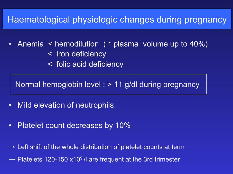
Haematological physiologic changes during pregnancy
• Anemia < hemodilution (;�plasma volume up to 40%) < iron deficiency < folic acid deficiency Normal hemoglobin level : > 11 g/dl during pregnancy • Mild elevation of neutrophils
• Platelet count decreases by 10% → Left shift of the whole distribution of platelet counts at term
→ Platelets 120-150 x109 /l are frequent at the 3rd trimester

Thrombocytopenia during pregnancy
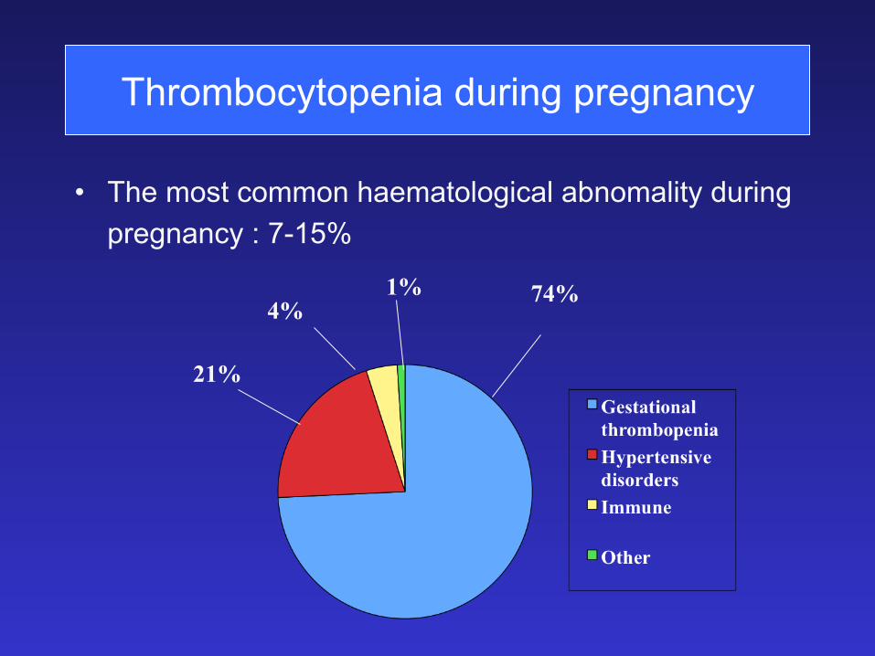
Thrombocytopenia during pregnancy
• The most common haematological abnomality during
pregnancy : 7-15%
74%
21%
4% 1%
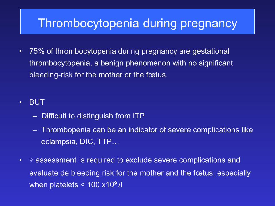
Thrombocytopenia during pregnancy
• 75% of thrombocytopenia during pregnancy are gestational thrombocytopenia, a benign phenomenon with no significant bleeding-risk for the mother or the fœtus.
• BUT
– Difficult to distinguish from ITP
– Thrombopenia can be an indicator of severe complications like eclampsia, DIC, TTP…
• � assessment is required to exclude severe complications and
evaluate de bleeding risk for the mother and the fœtus, especially when platelets < 100 x109 /l
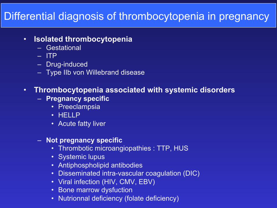
Differential diagnosis of thrombocytopenia in pregnancy
• Isolated thrombocytopenia – Gestational – ITP – Drug-induced – Type IIb von Willebrand disease
• Thrombocytopenia associated with systemic disorders – Pregnancy specific
• Preeclampsia • HELLP • Acute fatty liver
– Not pregnancy specific • Thrombotic microangiopathies : TTP, HUS • Systemic lupus • Antiphospholipid antibodies • Disseminated intra-vascular coagulation (DIC) • Viral infection (HIV, CMV, EBV) • Bone marrow dysfuction • Nutrionnal deficiency (folate deficiency)
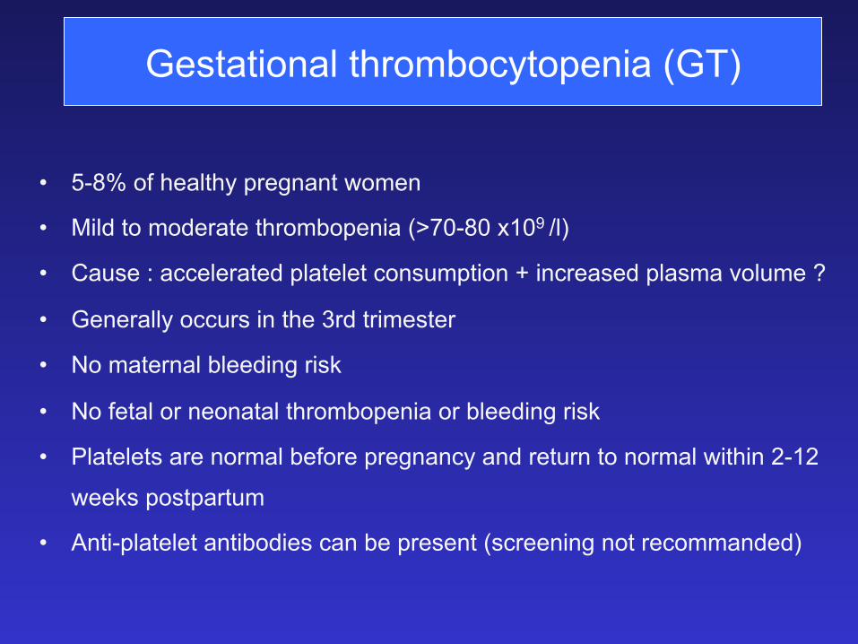
Gestational thrombocytopenia (GT)
• 5-8% of healthy pregnant women
• Mild to moderate thrombopenia (>70-80 x109 /l)
• Cause : accelerated platelet consumption + increased plasma volume ?
• Generally occurs in the 3rd trimester
• No maternal bleeding risk
• No fetal or neonatal thrombopenia or bleeding risk
• Platelets are normal before pregnancy and return to normal within 2-12
weeks postpartum
• Anti-platelet antibodies can be present (screening not recommanded)
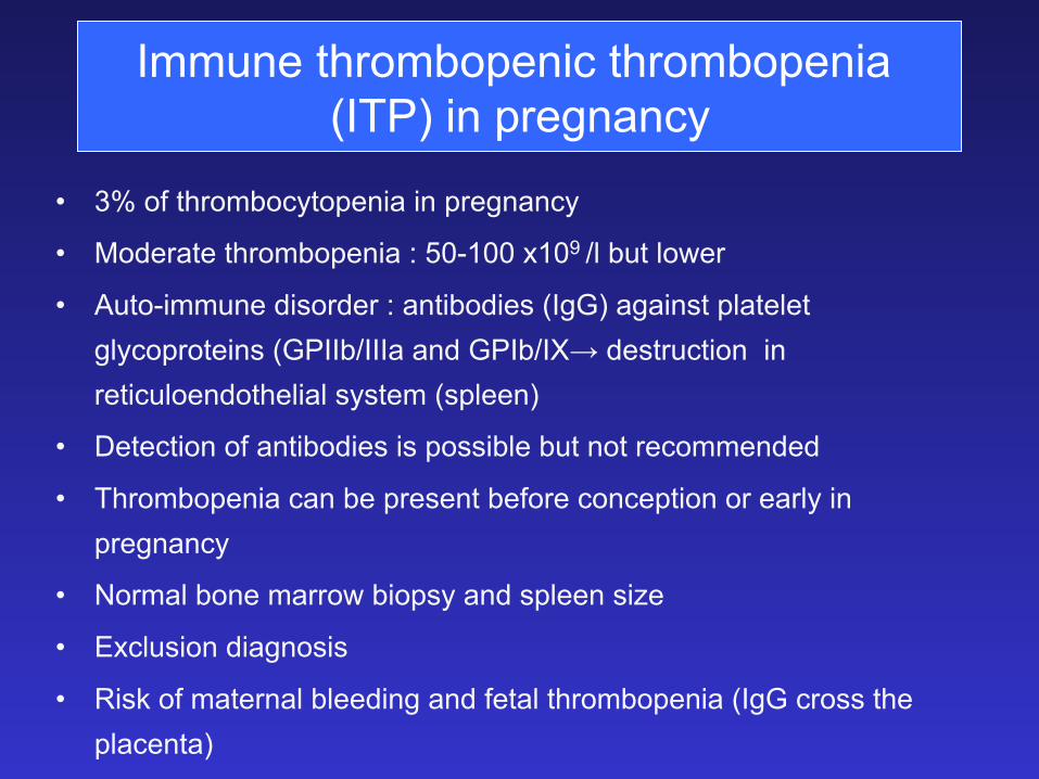
Immune thrombopenic thrombopenia (ITP) in pregnancy
• 3% of thrombocytopenia in pregnancy
• Moderate thrombopenia : 50-100 x109 /l but lower
• Auto-immune disorder : antibodies (IgG) against platelet glycoproteins (GPIIb/IIIa and GPIb/IX→ destruction in reticuloendothelial system (spleen)
• Detection of antibodies is possible but not recommended
• Thrombopenia can be present before conception or early in pregnancy
• Normal bone marrow biopsy and spleen size
• Exclusion diagnosis
• Risk of maternal bleeding and fetal thrombopenia (IgG cross the placenta)
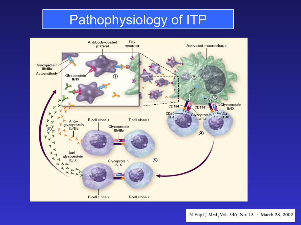
Pathophysiology of ITP
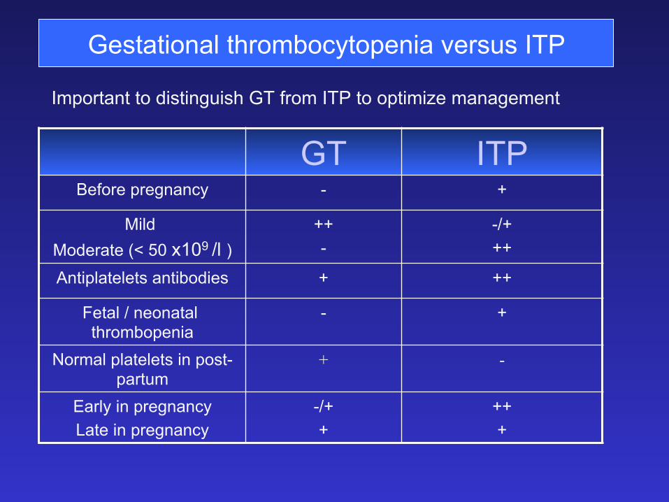
Gestational thrombocytopenia versus ITP
Important to distinguish GT from ITP to optimize management
GT ITP Before pregnancy - +
Mild Moderate (< 50 x109 /l )
++ -
-/+ ++
Antiplatelets antibodies + ++
Fetal / neonatal thrombopenia
- +
Normal platelets in post-partum
+ -
Early in pregnancy Late in pregnancy
-/+ +
++ +
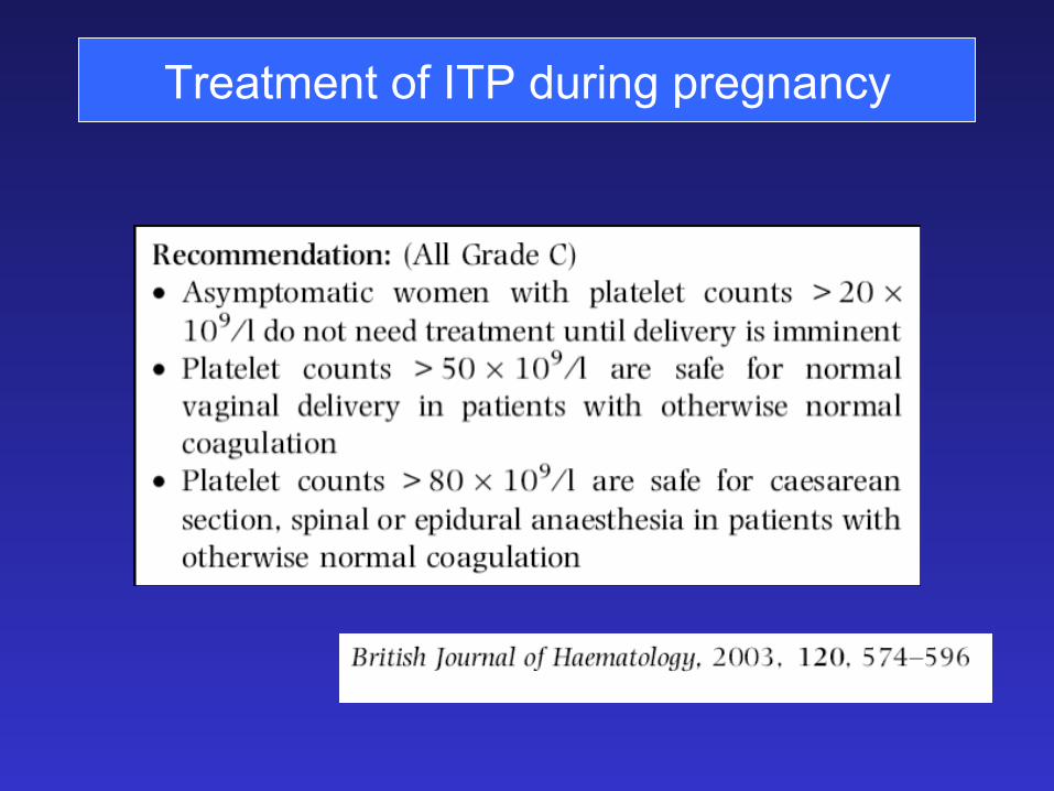
Treatment of ITP during pregnancy
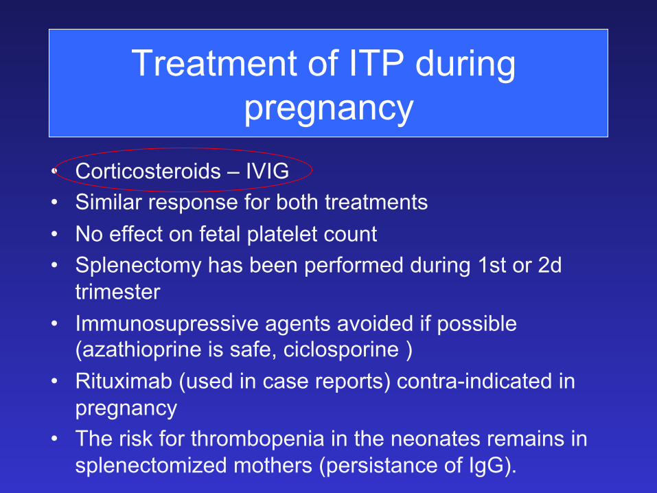
• Corticosteroids – IVIG • Similar response for both treatments • No effect on fetal platelet count • Splenectomy has been performed during 1st or 2d
trimester • Immunosupressive agents avoided if possible
(azathioprine is safe, ciclosporine ) • Rituximab (used in case reports) contra-indicated in
pregnancy • The risk for thrombopenia in the neonates remains in
splenectomized mothers (persistance of IgG).
Treatment of ITP during pregnancy
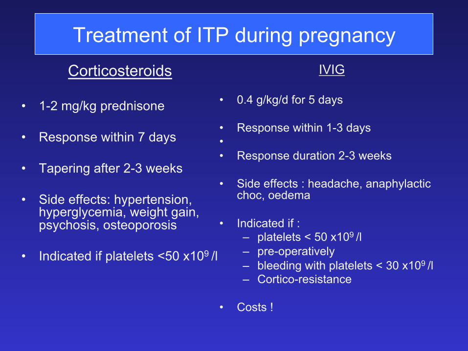
Treatment of ITP during pregnancy Corticosteroids
• 1-2 mg/kg prednisone • Response within 7 days
• Tapering after 2-3 weeks
• Side effects: hypertension, hyperglycemia, weight gain, psychosis, osteoporosis
• Indicated if platelets <50 x109 /l
IVIG
• 0.4 g/kg/d for 5 days
• Response within 1-3 days • • Response duration 2-3 weeks • Side effects : headache, anaphylactic
choc, oedema
• Indicated if : – platelets < 50 x109 /l – pre-operatively – bleeding with platelets < 30 x109 /l – Cortico-resistance
• Costs !
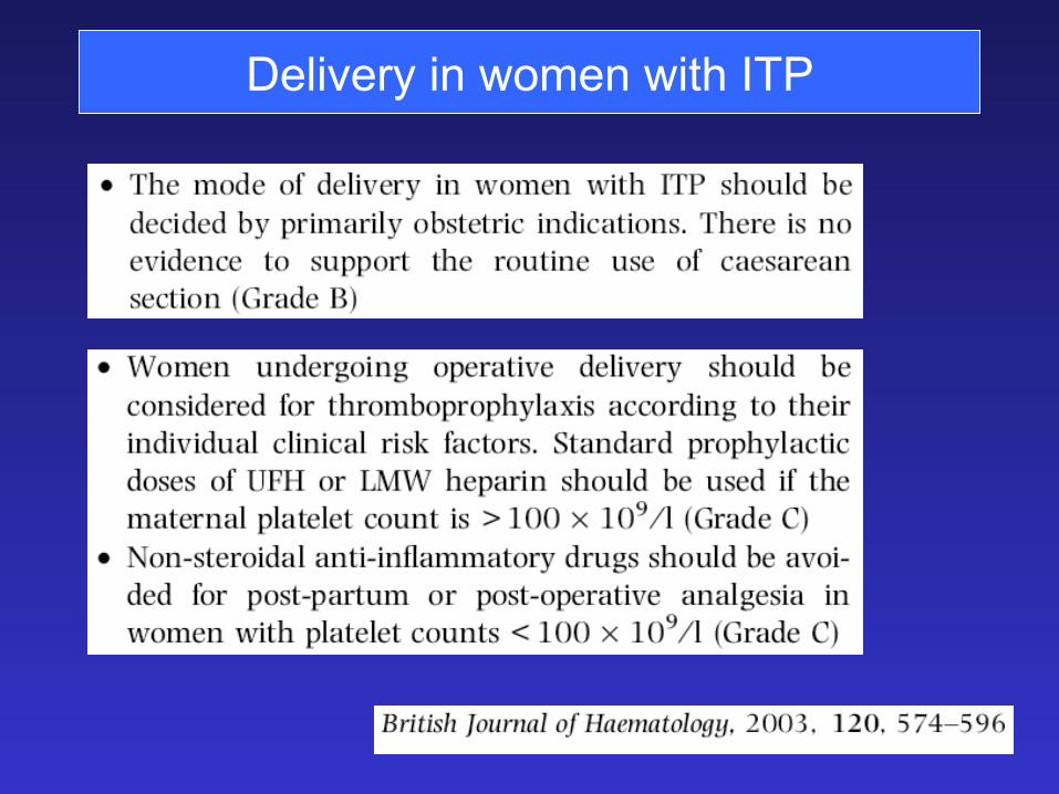
Delivery in women with ITP
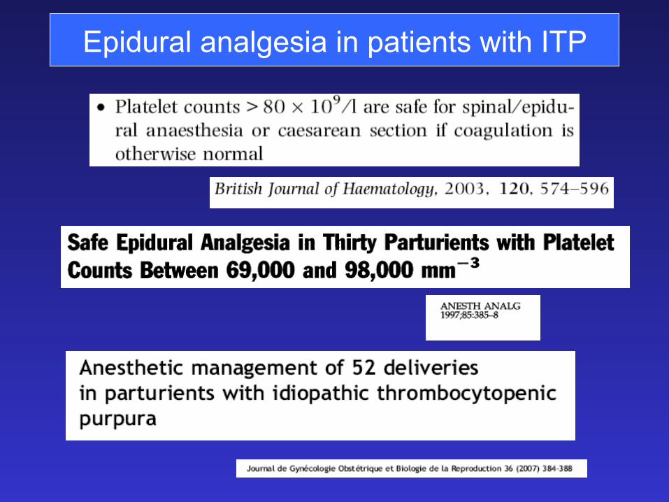
Epidural analgesia in patients with ITP
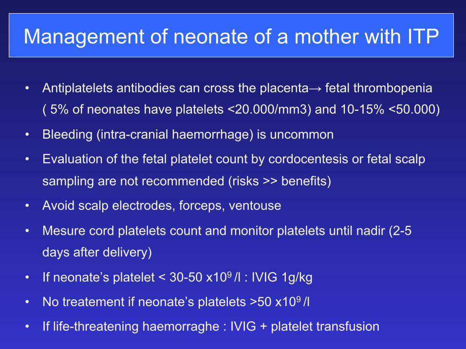
Management of neonate of a mother with ITP
• Antiplatelets antibodies can cross the placenta→ fetal thrombopenia ( 5% of neonates have platelets <20.000/mm3) and 10-15% <50.000)
• Bleeding (intra-cranial haemorrhage) is uncommon
• Evaluation of the fetal platelet count by cordocentesis or fetal scalp
sampling are not recommended (risks >> benefits)
• Avoid scalp electrodes, forceps, ventouse
• Mesure cord platelets count and monitor platelets until nadir (2-5 days after delivery)
• If neonate’s platelet < 30-50 x109 /l : IVIG 1g/kg
• No treatement if neonate’s platelets >50 x109 /l
• If life-threatening haemorraghe : IVIG + platelet transfusion
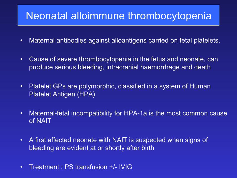
Neonatal alloimmune thrombocytopenia
• Maternal antibodies against alloantigens carried on fetal platelets. • Cause of severe thrombocytopenia in the fetus and neonate, can
produce serious bleeding, intracranial haemorrhage and death
• Platelet GPs are polymorphic, classified in a system of Human Platelet Antigen (HPA)
• Maternal-fetal incompatibility for HPA-1a is the most common cause of NAIT
• A first affected neonate with NAIT is suspected when signs of bleeding are evident at or shortly after birth
• Treatment : PS transfusion +/- IVIG
• Rhesus alloimmunization
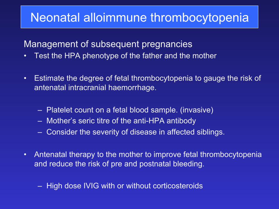
Management of subsequent pregnancies • Test the HPA phenotype of the father and the mother
• Estimate the degree of fetal thrombocytopenia to gauge the risk of antenatal intracranial haemorrhage.
– Platelet count on a fetal blood sample. (invasive) – Mother’s seric titre of the anti-HPA antibody – Consider the severity of disease in affected siblings.
• Antenatal therapy to the mother to improve fetal thrombocytopenia and reduce the risk of pre and postnatal bleeding.
– High dose IVIG with or without corticosteroids
Neonatal alloimmune thrombocytopenia
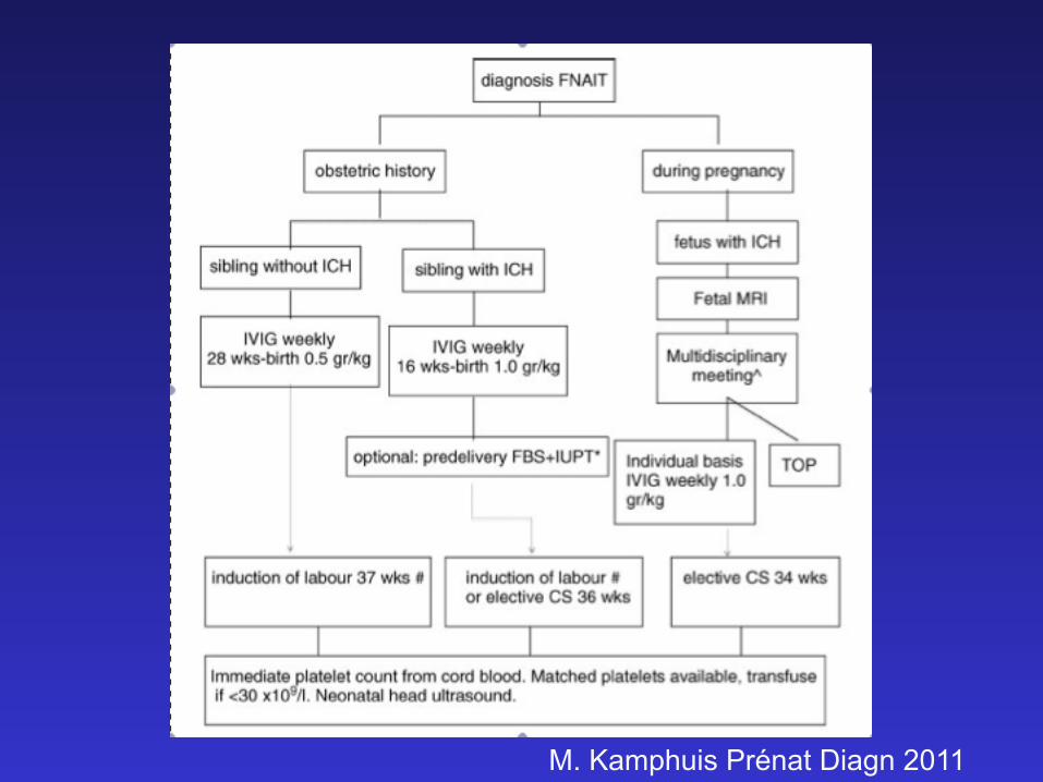
M. Kamphuis Prénat Diagn 2011
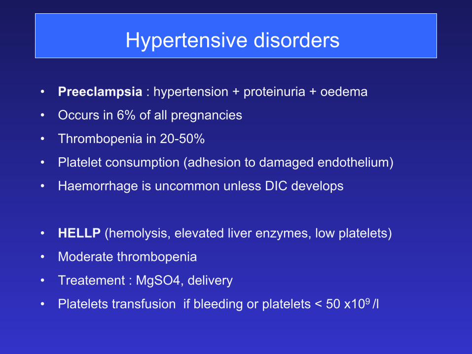
Hypertensive disorders
• Preeclampsia : hypertension + proteinuria + oedema
• Occurs in 6% of all pregnancies
• Thrombopenia in 20-50%
• Platelet consumption (adhesion to damaged endothelium)
• Haemorrhage is uncommon unless DIC develops
• HELLP (hemolysis, elevated liver enzymes, low platelets)
• Moderate thrombopenia
• Treatement : MgSO4, delivery
• Platelets transfusion if bleeding or platelets < 50 x109 /l
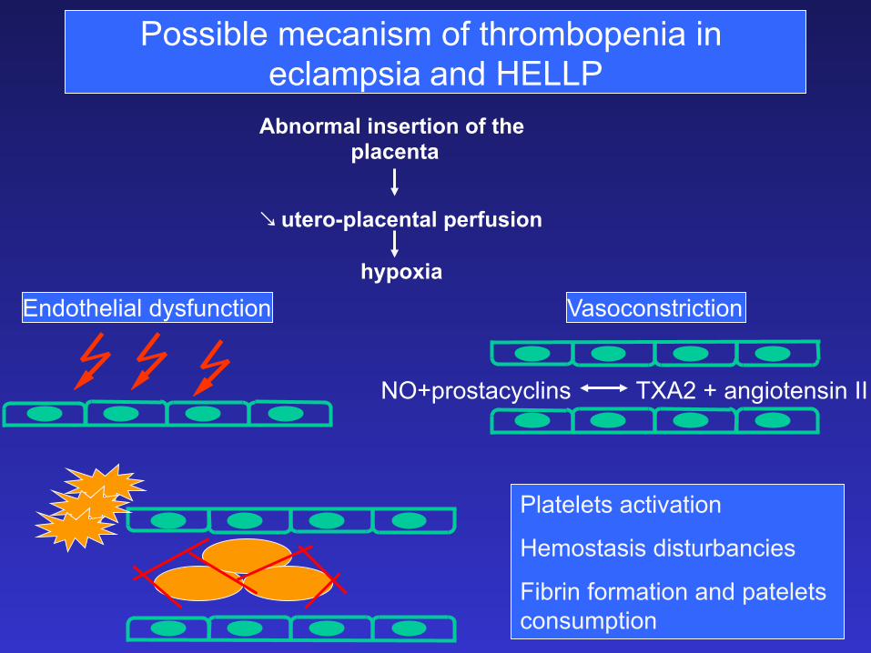
� utero-placental perfusion
hypoxia Endothelial dysfunction
Abnormal insertion of the placenta
Vasoconstriction
NO+prostacyclins TXA2 + angiotensin II
Platelets activation
Hemostasis disturbancies
Fibrin formation and patelets consumption
Possible mecanism of thrombopenia in eclampsia and HELLP
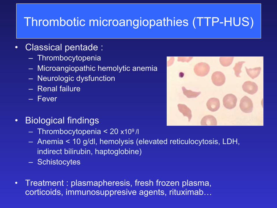
Thrombotic microangiopathies (TTP-HUS)
• Classical pentade : – Thrombocytopenia – Microangiopathic hemolytic anemia – Neurologic dysfunction – Renal failure – Fever
• Biological findings – Thrombocytopenia < 20 x109 /l – Anemia < 10 g/dl, hemolysis (elevated reticulocytosis, LDH, indirect bilirubin, haptoglobine) – Schistocytes
• Treatment : plasmapheresis, fresh frozen plasma, corticoids, immunosuppresive agents, rituximab…
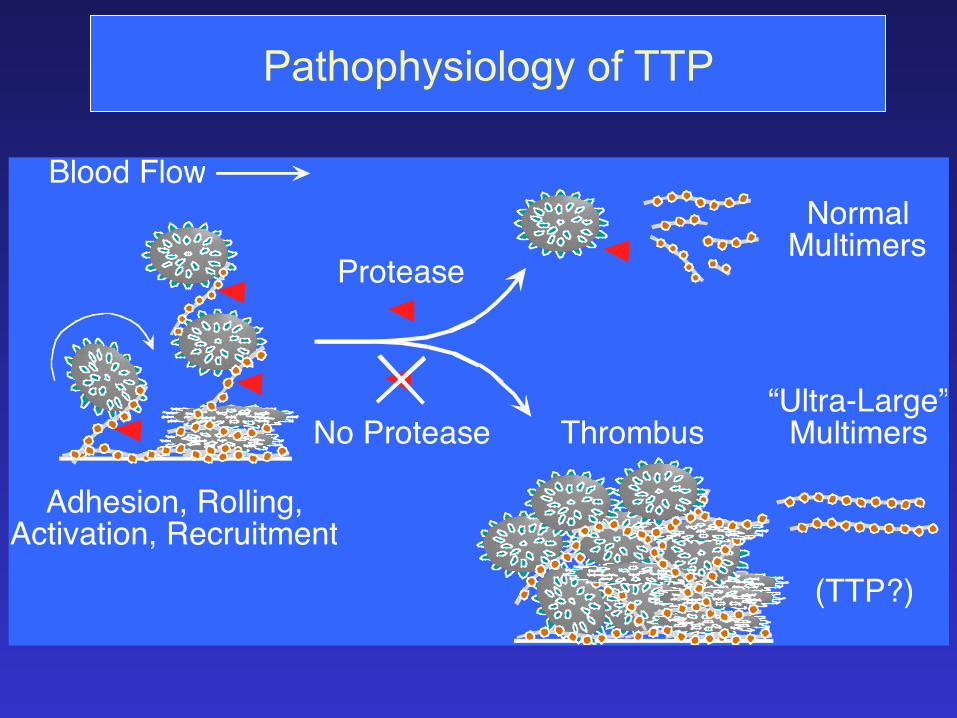
Pathophysiology of TTP
Blood Flow
Adhesion, Rolling,Activation, Recruitment
NormalMultimers
Protease
No Protease Thrombus“Ultra-Large”
Multimers
(TTP?)
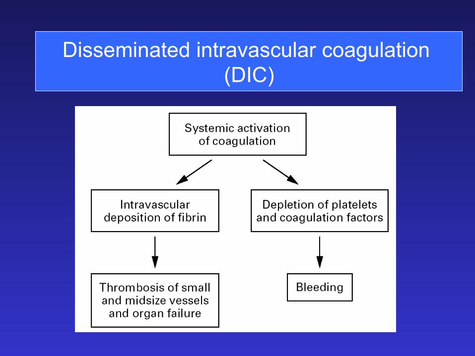
Disseminated intravascular coagulation (DIC)
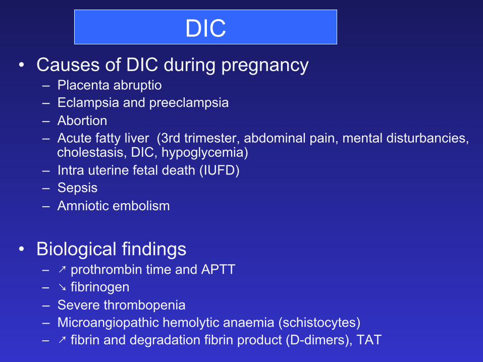
DIC • Causes of DIC during pregnancy
– Placenta abruptio – Eclampsia and preeclampsia – Abortion – Acute fatty liver (3rd trimester, abdominal pain, mental disturbancies,
cholestasis, DIC, hypoglycemia) – Intra uterine fetal death (IUFD) – Sepsis – Amniotic embolism
• Biological findings 9 ; prothrombin time and APTT 9 < fibrinogen – Severe thrombopenia – Microangiopathic hemolytic anaemia (schistocytes) 9 ; fibrin and degradation fibrin product (D-dimers), TAT
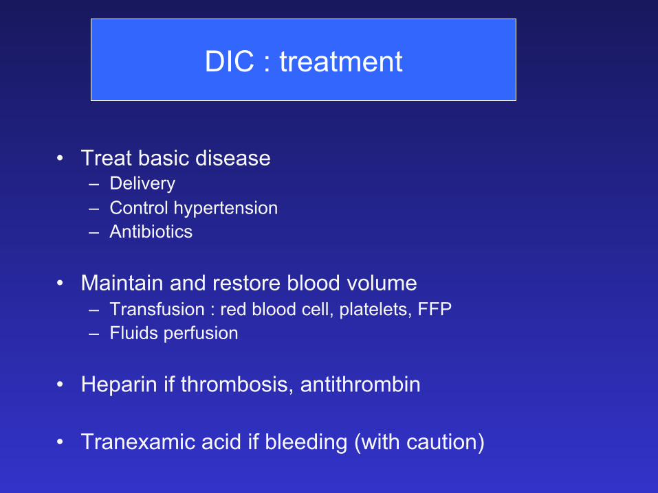
• Treat basic disease – Delivery – Control hypertension – Antibiotics
• Maintain and restore blood volume – Transfusion : red blood cell, platelets, FFP – Fluids perfusion
• Heparin if thrombosis, antithrombin
• Tranexamic acid if bleeding (with caution)
DIC : treatment
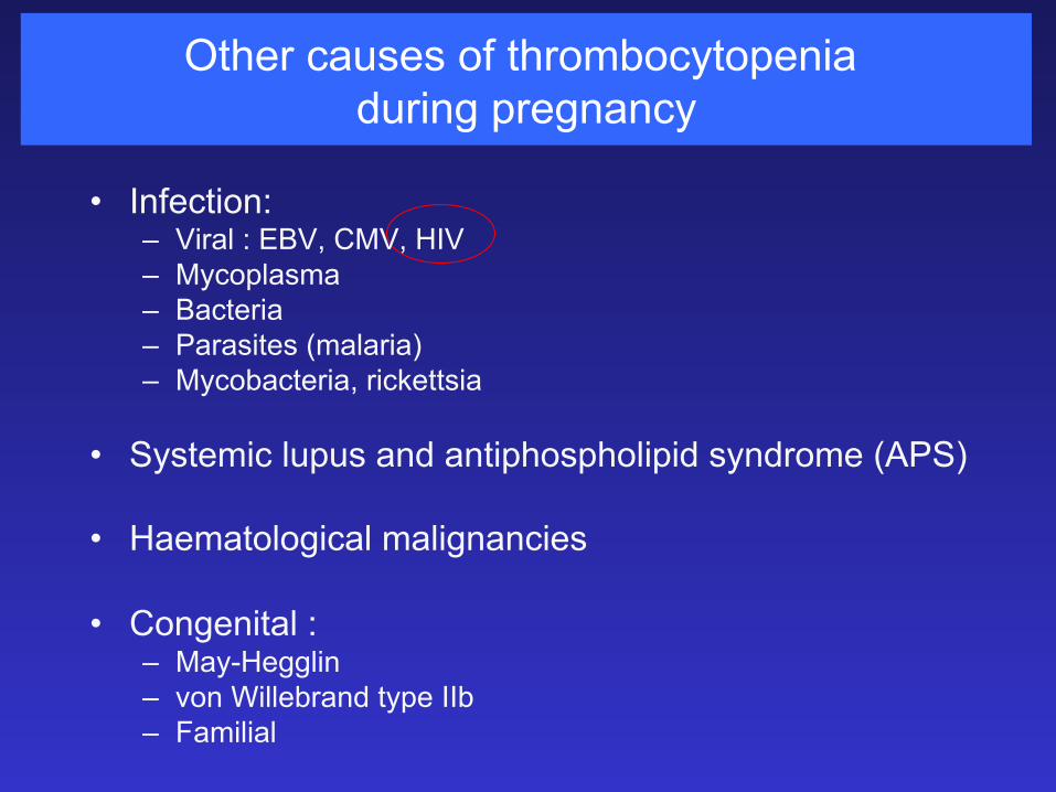
Other causes of thrombocytopenia during pregnancy
• Infection: – Viral : EBV, CMV, HIV – Mycoplasma – Bacteria – Parasites (malaria) – Mycobacteria, rickettsia
• Systemic lupus and antiphospholipid syndrome (APS) • Haematological malignancies • Congenital :
– May-Hegglin – von Willebrand type IIb – Familial
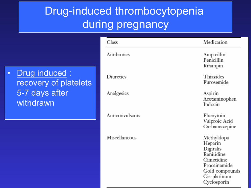
Drug-induced thrombocytopenia during pregnancy
• Drug induced : recovery of platelets 5-7 days after withdrawn
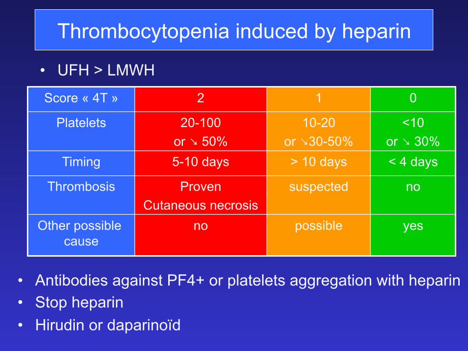
Thrombocytopenia induced by heparin
• UFH > LMWH
Score « 4T » 2 1 0
Platelets 20-100 .1�< 50%
10-20 .1�<�����
<10 .1�<����
Timing 5-10 days > 10 days < 4 days
Thrombosis Proven Cutaneous necrosis
suspected no
Other possible cause
no possible yes
• Antibodies against PF4+ or platelets aggregation with heparin • Stop heparin • Hirudin or daparinoïd
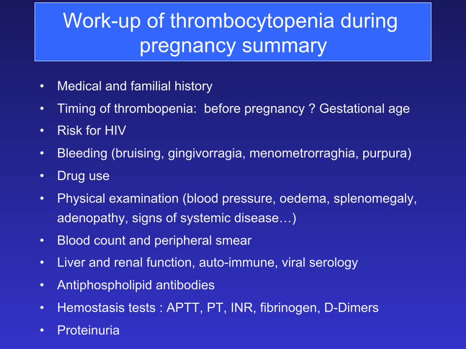
Work-up of thrombocytopenia during pregnancy summary
• Medical and familial history
• Timing of thrombopenia: before pregnancy ? Gestational age
• Risk for HIV
• Bleeding (bruising, gingivorragia, menometrorraghia, purpura)
• Drug use
• Physical examination (blood pressure, oedema, splenomegaly, adenopathy, signs of systemic disease…)
• Blood count and peripheral smear
• Liver and renal function, auto-immune, viral serology
• Antiphospholipid antibodies
• Hemostasis tests : APTT, PT, INR, fibrinogen, D-Dimers
• Proteinuria
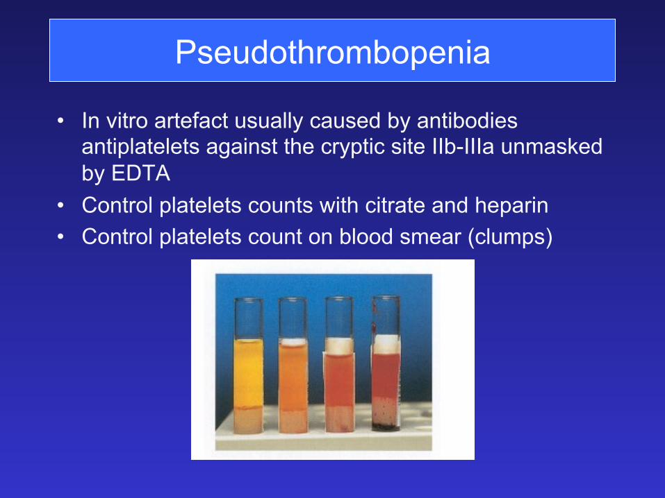
Pseudothrombopenia
• In vitro artefact usually caused by antibodies antiplatelets against the cryptic site IIb-IIIa unmasked by EDTA
• Control platelets counts with citrate and heparin • Control platelets count on blood smear (clumps)
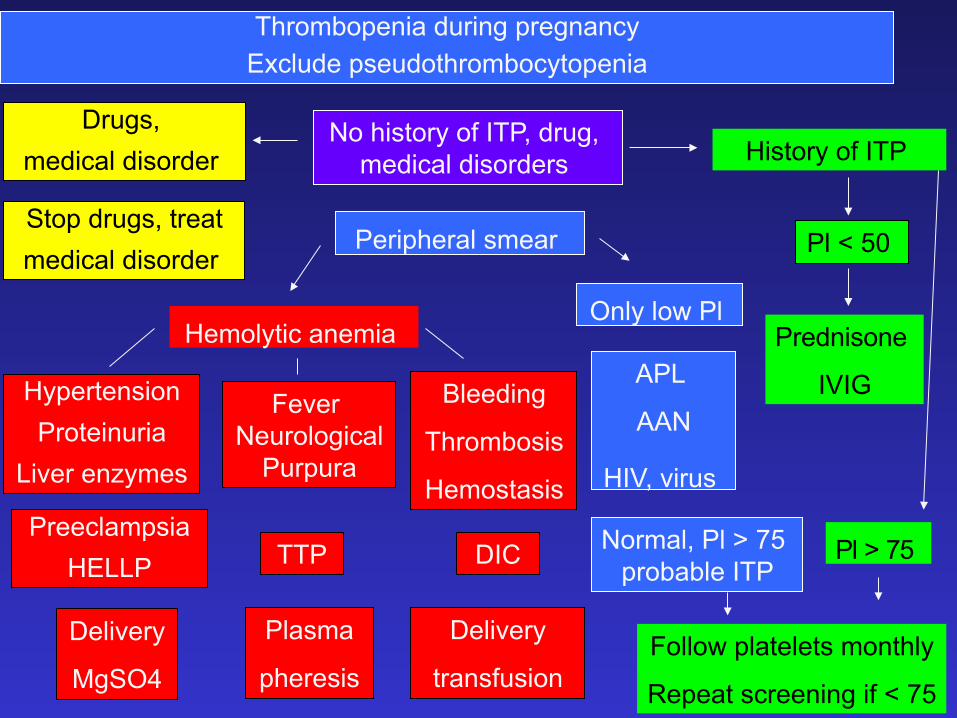
Thrombopenia during pregnancy Exclude pseudothrombocytopenia
No history of ITP, drug, medical disorders
Peripheral smear
Follow platelets monthly
Repeat screening if < 75
History of ITP
Pl < 50
Prednisone
IVIG
Pl > 75
Only low Pl
APL
AAN
HIV, virus
Normal, Pl > 75 probable ITP
Drugs, medical disorder
Stop drugs, treat medical disorder
Hemolytic anemia
Hypertension Proteinuria
Liver enzymes
Fever Neurological
Purpura
Bleeding
Thrombosis
Hemostasis Preeclampsia
HELLP TTP DIC
Delivery
MgSO4
Delivery
transfusion
Plasma
pheresis
Haemostasis in normal pregnancy
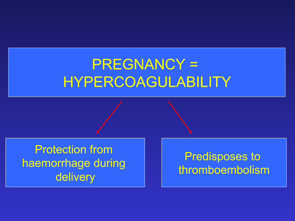
PREGNANCY = HYPERCOAGULABILITY
Protection from haemorrhage during
delivery
Predisposes to thromboembolism
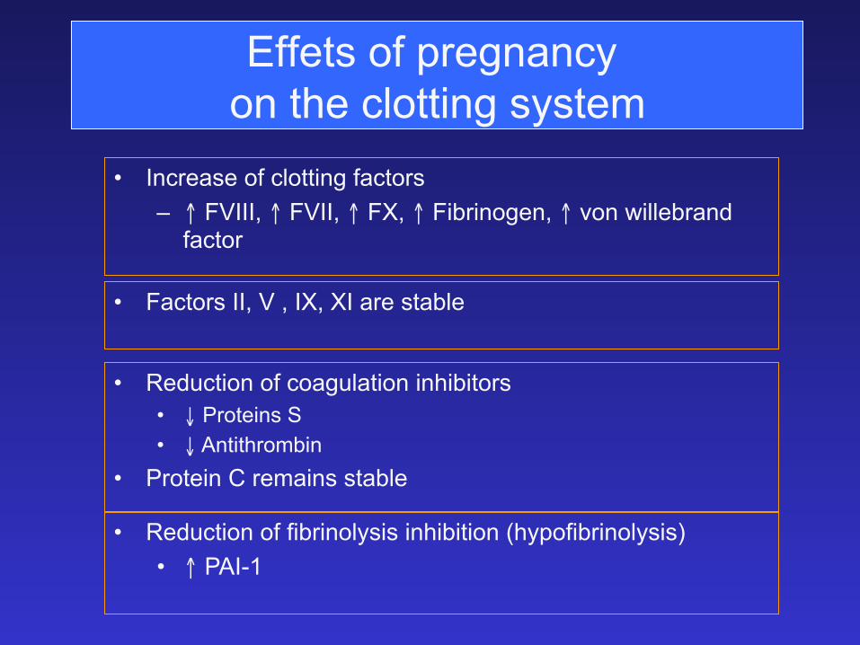
Effets of pregnancy on the clotting system
• Increase of clotting factors – ↑ FVIII, ↑ FVII, ↑ FX, ↑ Fibrinogen, ↑ von willebrand
factor
• Reduction of coagulation inhibitors • ↓ Proteins S • ↓ Antithrombin
• Protein C remains stable
• Reduction of fibrinolysis inhibition (hypofibrinolysis) • ↑ PAI-1
• Factors II, V , IX, XI are stable

Pregnancy in women with inherited bleeding disorder
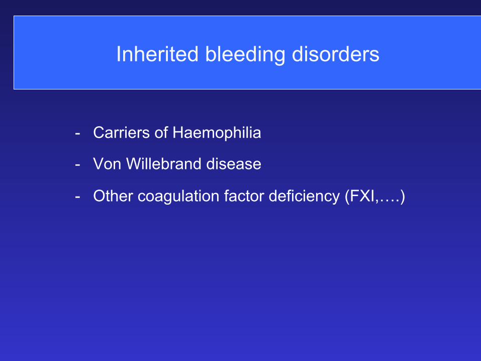
Inherited bleeding disorders
- Carriers of Haemophilia
- Von Willebrand disease
- Other coagulation factor deficiency (FXI,….)
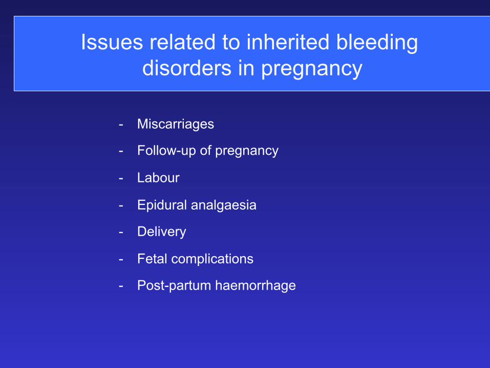
Issues related to inherited bleeding disorders in pregnancy
- Miscarriages
- Follow-up of pregnancy
- Labour
- Epidural analgaesia
- Delivery
- Fetal complications
- Post-partum haemorrhage
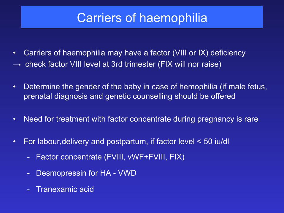
Carriers of haemophilia
• Carriers of haemophilia may have a factor (VIII or IX) deficiency → check factor VIII level at 3rd trimester (FIX will nor raise) • Determine the gender of the baby in case of hemophilia (if male fetus,
prenatal diagnosis and genetic counselling should be offered • Need for treatment with factor concentrate during pregnancy is rare • For labour,delivery and postpartum, if factor level < 50 iu/dl
- Factor concentrate (FVIII, vWF+FVIII, FIX)
- Desmopressin for HA - VWD
- Tranexamic acid
VWD during pregnancy • Variable and unpredictable increase of :
- FVIII - von Willebrand factor : > 12 weeks, not in vWD type 3 → factors levels should be checked during the last trimester
• Miscarriages
– Similar incidence in women with vWD (10-20 %) and without vWD (12-15%)
– Miscarriages or abortion early in pregnancy carry an increased risk of maternal haemorrhage
• Vaginal bleeding – During first trimester : 33% in VWD 33% > mothers without vWD
(16%)
– Antepartum : frequency not higher than normal
Epidural analgaesia
• Limited reported experience
• Many anesthaetists are reluctant to preform the procedure
• Can be safely performed if FVIII and von Willebrand factor (VWF)
antigen and activity levels are above 50 % at time of delivery
• Von Willebrand factor concentrates or DDAVP should be
administered if FVIII and VWF levels below 50 %
• Any other clotting abnormality should be excluded (low platelets)
• Individualised assessment is required
vWD and delivery
• Primary post-partum haemorrhage
– Blood loss > 500 ml during 24 hrs after birth of the infant
– 3 to 5 % in normal vaginal delivery
– 15 to 18.5 % in women with vWD
• Secondary post-partum haemorrhage – 1.3 % in normal individuals
– 20-25 % in patients with vWD (rapid fall of FVIII and VWF)
Always check VWF and FVIII before delivery and factors + haemoglobin on discharge
Management of peri- and post-partum haemorrhage
- Minimise genital and peritoneal trauma at delivery
- Factor levels should be kept > 50 % for 3-4 days after vaginal delivery
and 4-5 days after cesarean section
- At time of delivery, a sample of cord-blood should be collected and
carried promptly to the lab to exclude severe deficiency
- Good communication between anesthaesiologist, obstetrician and
bleeding expert is mandatory
Management of the fetus born to a mother with a bleeding disorder
• Determine the gender of the baby in case of hemophilia
• Reduce the risk of bleeding complications at delivery by avoiding :
• Instrumental deliveries (vacuum extractions, forceps)
• Prolonged deliveries
• Invasive monitoring techniques
• Desmopressin do not contra-indicate breast feeding
• After birth, avoid IM injection of Vit K, IM immunisations and post-
pone surgery when possible (circumcision)
Ohter rare bleeding disorders in pregnancy
Global management like hemophilia and vWD • Afibrinogenaemia
– Infusion of fibrinogen or FFP → Fg level > 100 mg/dl
• Factor XI deficiency – No raise during pregancy – Correlation between factor level and bleeding is difficult (<
15 iu/dl) – Fresh frozen plasma if required
• Factor VII deficiency – Tranexamic acid, Novoseven®
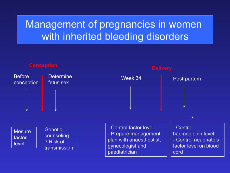
Management of pregnancies in women with inherited bleeding disorders
- Control factor level - Prepare management plan with anaesthestist, gynecologist and paediatrician
Genetic counseling ? Risk of transmission
Determine fetus sex
Conception
Before conception
Mesure factor level
Delivery
Week 34 Post-partum
- Control haemoglobin level - Control neaonate’s factor level on blood cord
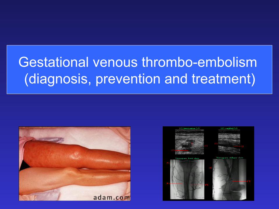
Gestational venous thrombo-embolism (diagnosis, prevention and treatment)
Embolism
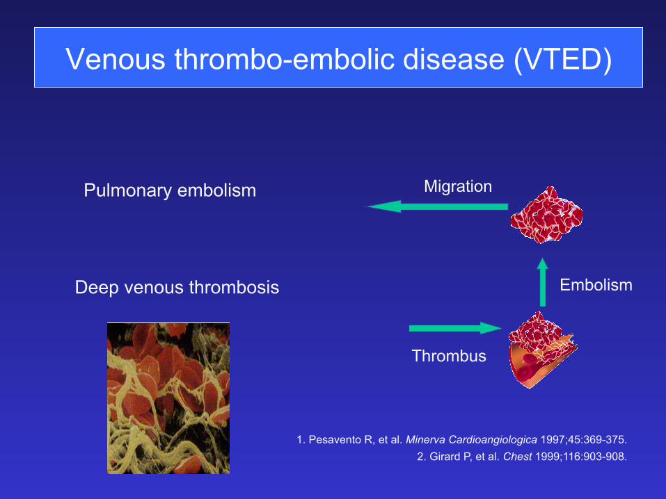
Venous thrombo-embolic disease (VTED)
Pulmonary embolism
Deep venous thrombosis
Migration
Thrombus
1. Pesavento R, et al. Minerva Cardioangiologica 1997;45:369-375. 2. Girard P, et al. Chest 1999;116:903-908.
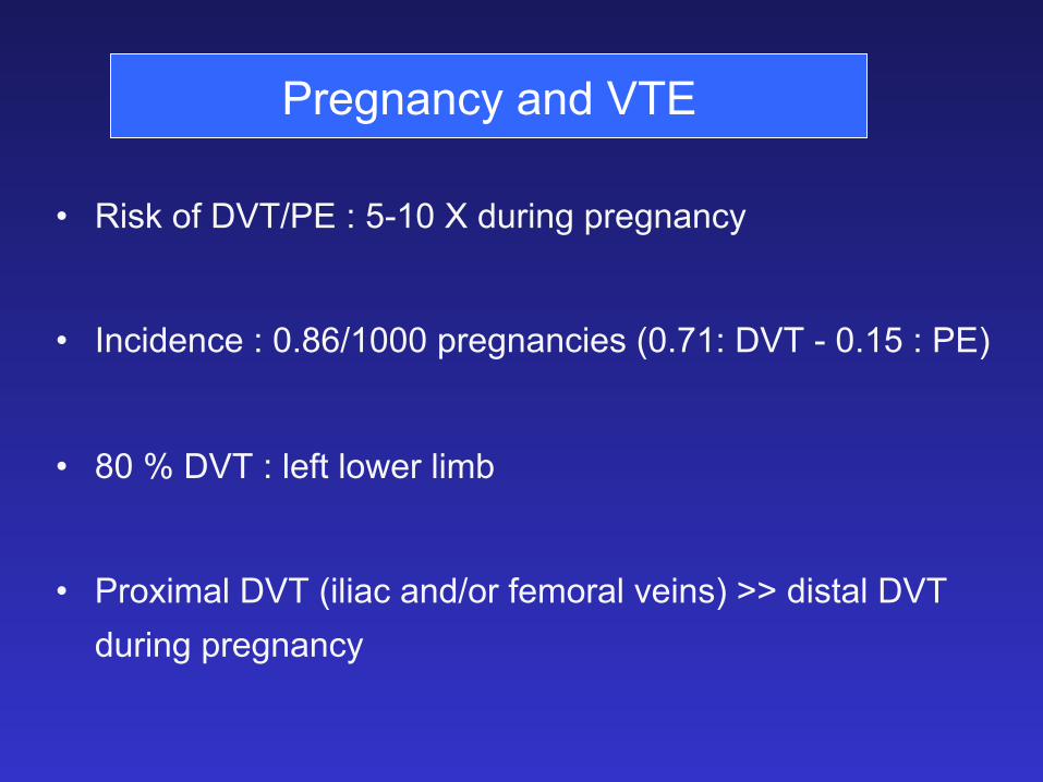
Pregnancy and VTE
• Risk of DVT/PE : 5-10 X during pregnancy
• Incidence : 0.86/1000 pregnancies (0.71: DVT - 0.15 : PE)
• 80 % DVT : left lower limb
• Proximal DVT (iliac and/or femoral veins) >> distal DVT during pregnancy
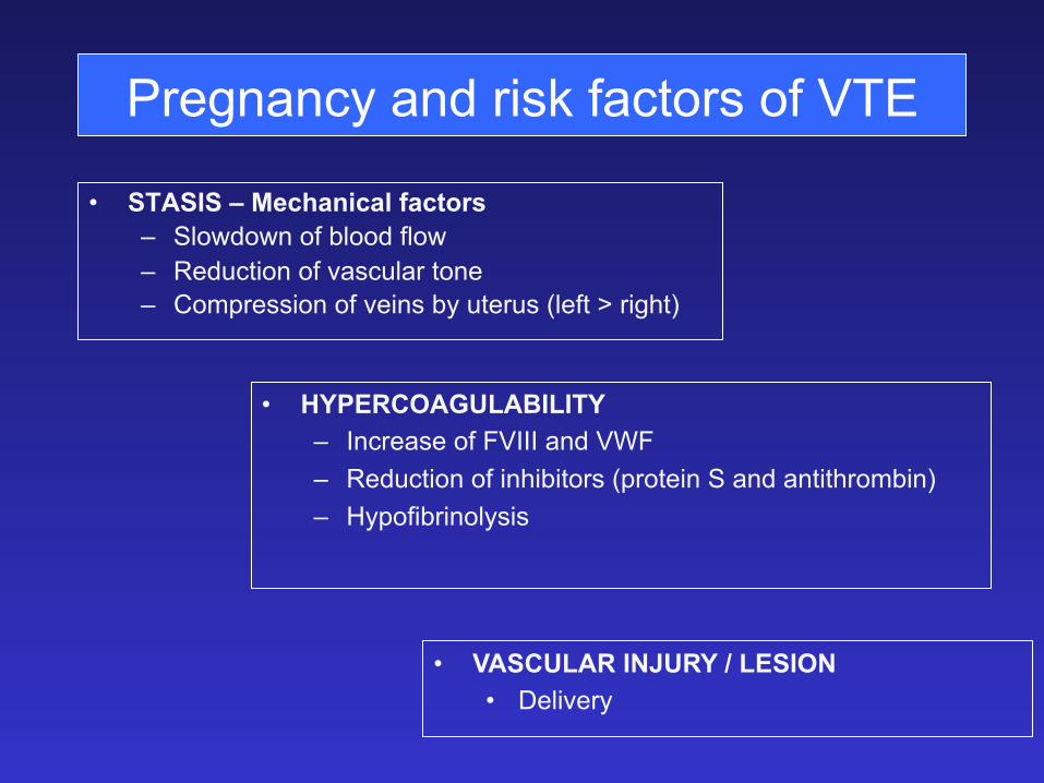
Pregnancy and risk factors of VTE
• STASIS – Mechanical factors – Slowdown of blood flow – Reduction of vascular tone – Compression of veins by uterus (left > right)
• HYPERCOAGULABILITY – Increase of FVIII and VWF – Reduction of inhibitors (protein S and antithrombin) – Hypofibrinolysis
• VASCULAR INJURY / LESION • Delivery
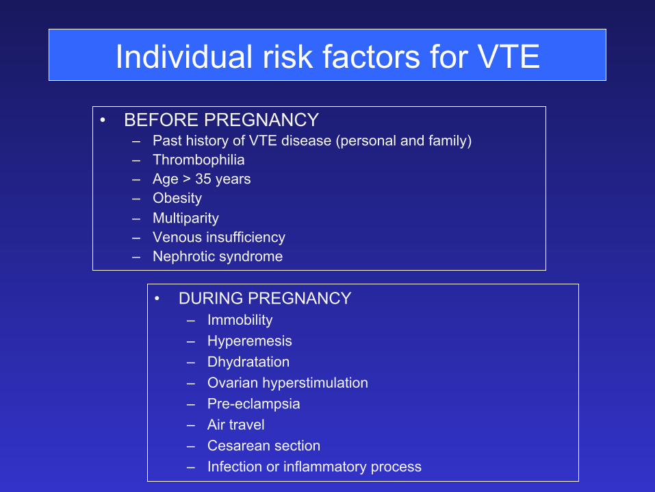
Individual risk factors for VTE
• BEFORE PREGNANCY – Past history of VTE disease (personal and family) – Thrombophilia – Age > 35 years – Obesity – Multiparity – Venous insufficiency – Nephrotic syndrome
• DURING PREGNANCY – Immobility – Hyperemesis – Dhydratation – Ovarian hyperstimulation – Pre-eclampsia – Air travel – Cesarean section – Infection or inflammatory process
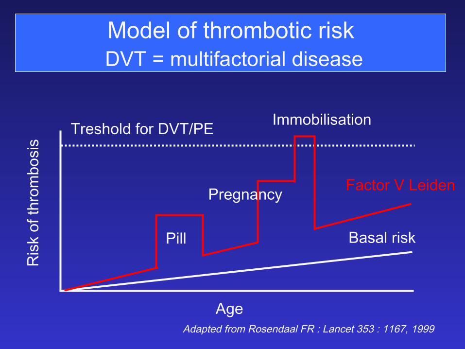
Model of thrombotic risk DVT = multifactorial disease
�'%�
�)2*�.&�3(1.,".2)2�
�1%2(.+$�&.1��������
�!2!+�1)2*�
�!#3.1����%)$%-�
�)++�
�1%'-!-#8�
�,,.")+)2!3).-�
������������ ���������� �������������������������
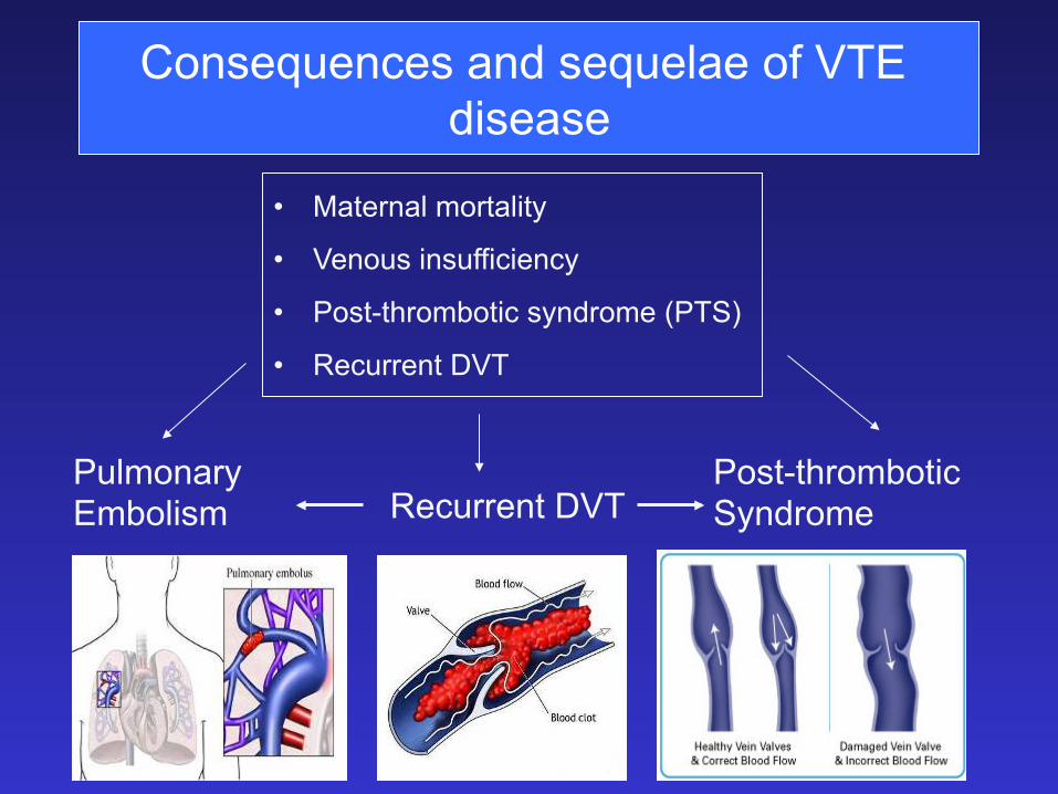
Pulmonary Embolism Recurrent DVT
Post-thrombotic Syndrome
Consequences and sequelae of VTE disease
• Maternal mortality
• Venous insufficiency
• Post-thrombotic syndrome (PTS)
• Recurrent DVT
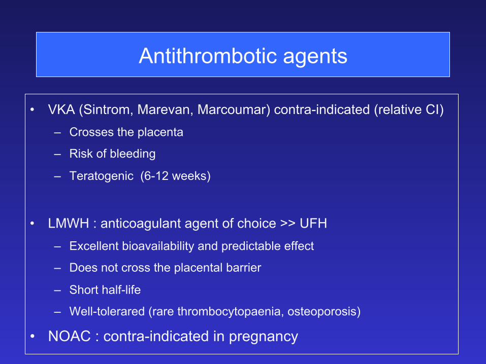
Antithrombotic agents
• VKA (Sintrom, Marevan, Marcoumar) contra-indicated (relative CI) – Crosses the placenta
– Risk of bleeding
– Teratogenic (6-12 weeks)
• LMWH : anticoagulant agent of choice >> UFH – Excellent bioavailability and predictable effect
– Does not cross the placental barrier
– Short half-life
– Well-tolerared (rare thrombocytopaenia, osteoporosis)
• NOAC : contra-indicated in pregnancy
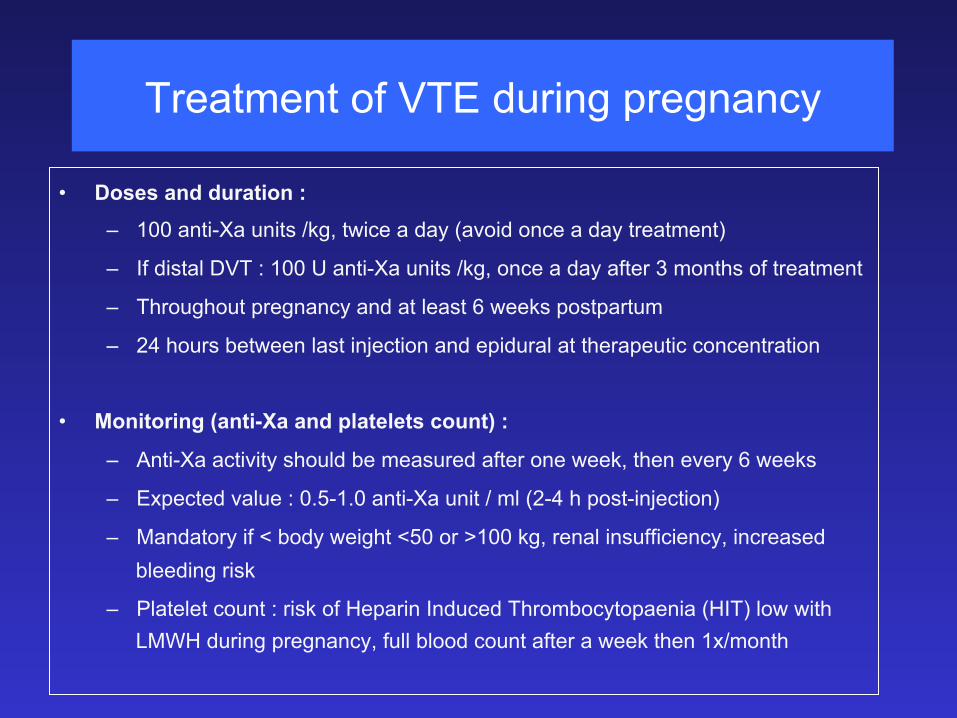
Treatment of VTE during pregnancy
• Doses and duration :
– 100 anti-Xa units /kg, twice a day (avoid once a day treatment)
– If distal DVT : 100 U anti-Xa units /kg, once a day after 3 months of treatment
– Throughout pregnancy and at least 6 weeks postpartum
– 24 hours between last injection and epidural at therapeutic concentration
• Monitoring (anti-Xa and platelets count) :
– Anti-Xa activity should be measured after one week, then every 6 weeks
– Expected value : 0.5-1.0 anti-Xa unit / ml (2-4 h post-injection)
– Mandatory if < body weight <50 or >100 kg, renal insufficiency, increased bleeding risk
– Platelet count : risk of Heparin Induced Thrombocytopaenia (HIT) low with LMWH during pregnancy, full blood count after a week then 1x/month

Pregnant Women With Past DVT/PE • Prophylaxis for 6 weeks after delivery (Grade 2B) for all
• Moderate/high recurrence risk should consider taking prophylactic or intermediate-dose LMWH during pregnancy (Grade 2C). This includes women with a prior DVT or PE that was unprovoked, or related to pregnancy or estrogen use.
• Low risk for recurrence (because their VTE was provoked by a transient risk factor unrelated to pregnancy or estrogen use) and their doctors should watch carefully for signs or symptoms of DVT or PE during pregnancy, but ACCP suggests they not use LMWH as prophylaxis (Grade 2C).
VTE prophylaxis during pregnancy ACCP 2012
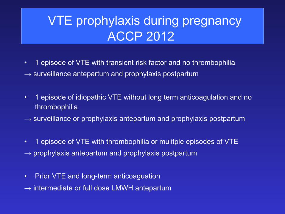
VTE prophylaxis during pregnancy ACCP 2012
• 1 episode of VTE with transient risk factor and no thrombophilia → surveillance antepartum and prophylaxis postpartum
• 1 episode of idiopathic VTE without long term anticoagulation and no
thrombophilia → surveillance or prophylaxis antepartum and prophylaxis postpartum • 1 episode of VTE with thrombophilia or mulitple episodes of VTE → prophylaxis antepartum and prophylaxis postpartum • Prior VTE and long-term anticoaguation
→ intermediate or full dose LMWH antepartum
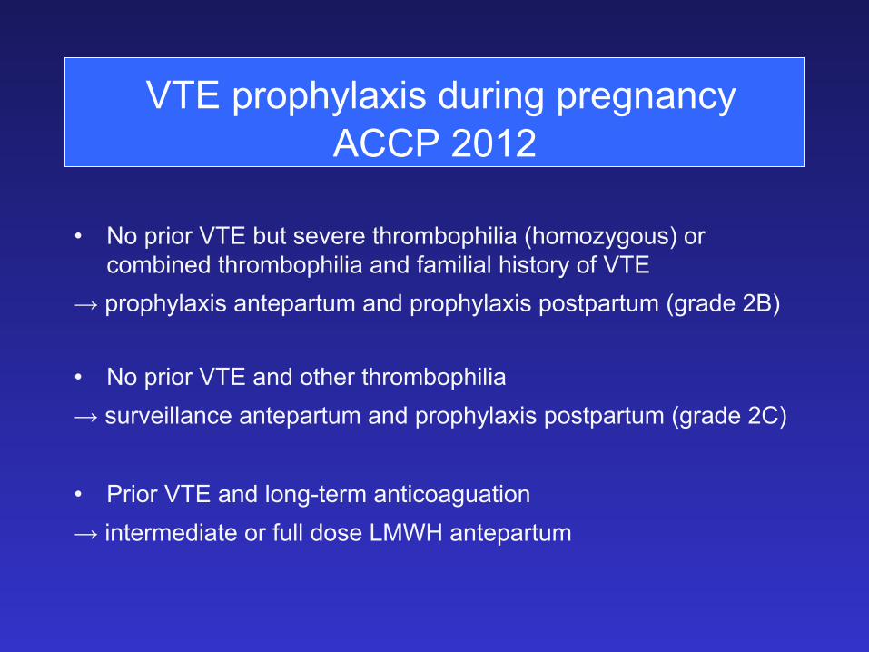
• No prior VTE but severe thrombophilia (homozygous) or combined thrombophilia and familial history of VTE
→ prophylaxis antepartum and prophylaxis postpartum (grade 2B)
• No prior VTE and other thrombophilia → surveillance antepartum and prophylaxis postpartum (grade 2C) • Prior VTE and long-term anticoaguation → intermediate or full dose LMWH antepartum
VTE prophylaxis during pregnancy ACCP 2012
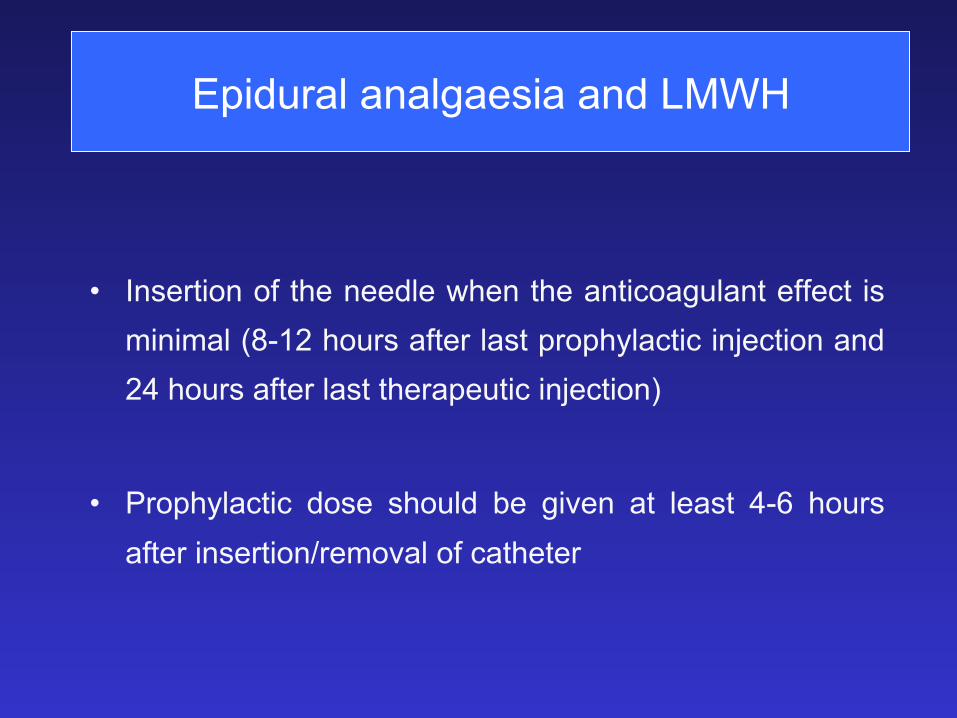
Epidural analgaesia and LMWH
• Insertion of the needle when the anticoagulant effect is minimal (8-12 hours after last prophylactic injection and 24 hours after last therapeutic injection)
• Prophylactic dose should be given at least 4-6 hours after insertion/removal of catheter
�(1.,"./()+)!�
: �-(%1)3%$�.1�!#04)1%$�/1%$)2/.2)3).-�3.6!1$2�3(1.,".2)2�
: �1),!1)+8�!22.#)!3%$�6)3(�!-�)-#1%!2%$�1)2*�.&�5%-.42�3(1.,".%,".+)#�$)2%!2%�
: �!-�"%�!22.#)!3%$�6)3(�!$5%12%�/1%'-!-#8�.43#.,%2�������
�
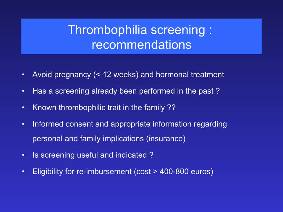
Thrombophilia screening : recommendations
• Avoid pregnancy (< 12 weeks) and hormonal treatment
• Has a screening already been performed in the past ?
• Known thrombophilic trait in the family ??
• Informed consent and appropriate information regarding
personal and family implications (insurance)
• Is screening useful and indicated ?
• Eligibility for re-imbursement (cost > 400-800 euros)
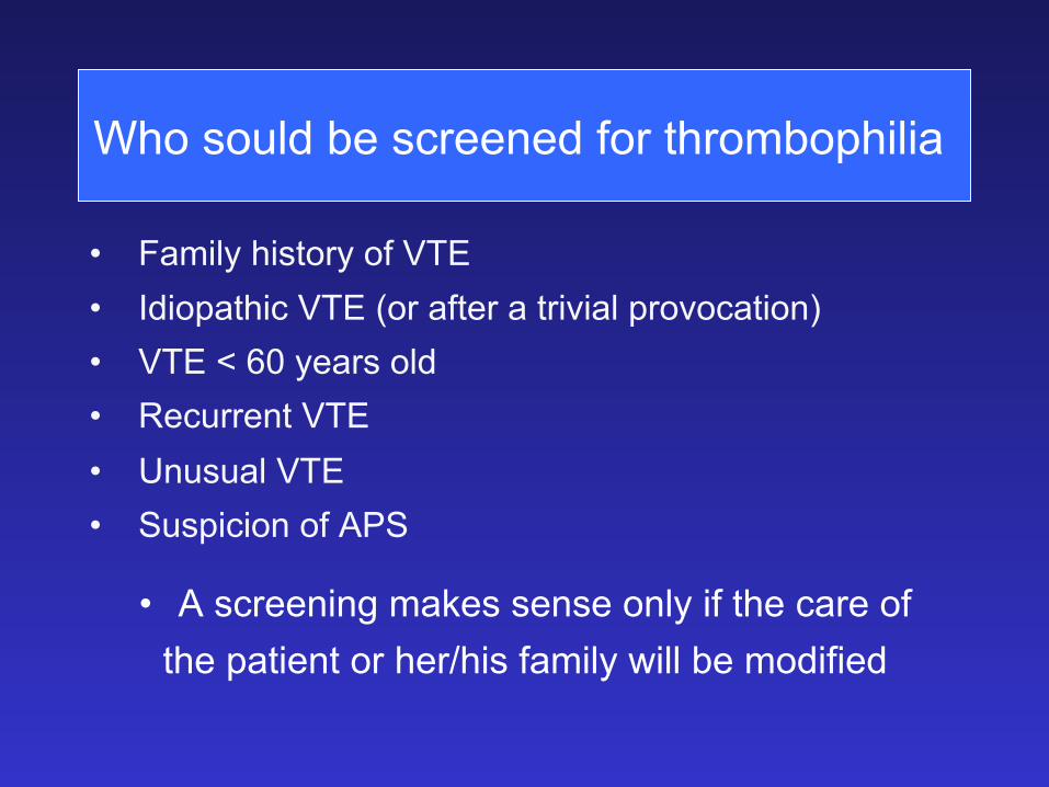
• Family history of VTE • Idiopathic VTE (or after a trivial provocation) • VTE < 60 years old • Recurrent VTE • Unusual VTE • Suspicion of APS
• A screening makes sense only if the care of the patient or her/his family will be modified
Thrombophilia screening : indications
Who sould be screened for thrombophilia
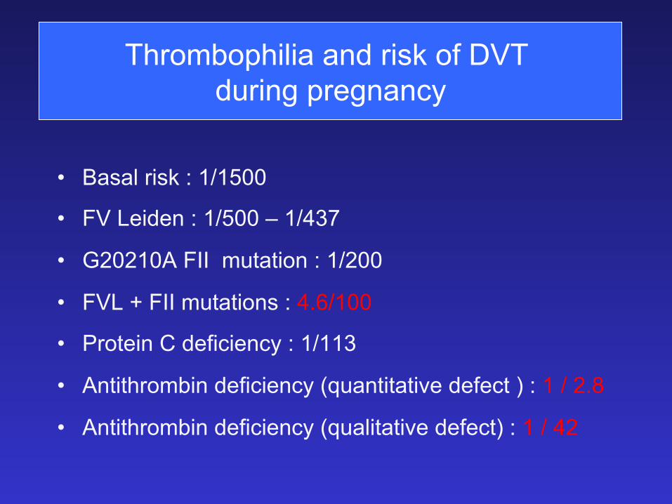
Thrombophilia and risk of DVT during pregnancy
• Basal risk : 1/1500
• FV Leiden : 1/500 – 1/437
• G20210A FII mutation : 1/200
• FVL + FII mutations : 4.6/100
• Protein C deficiency : 1/113
• Antithrombin deficiency (quantitative defect ) : 1 / 2.8
• Antithrombin deficiency (qualitative defect) : 1 / 42

Ovarian stimulation
• Thromboprophylaxis with LMWH (3800 à 5000 anti/Xa units /day) :
– Past history of DVT/PE
– Risk factors for DVT-PE
– Known thrombophilia
– Ovarian hyperstimulation
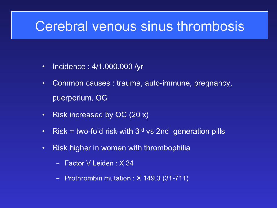
Cerebral venous sinus thrombosis
: �-#)$%-#%��� �����81�
: �.,,.-�#!42%2���31!4,!��!43.�),,4-%��/1%'-!-#8��
/4%1/%1)4,�����
: �)2*�)-#1%!2%$�"8�������7��
: �)2*���36.�&.+$�1)2*�6)3(��1$�52��-$��'%-%1!3).-�/)++2�
: �)2*�()'(%1�)-�6.,%-�6)3(�3(1.,"./()+)!��
9 �!#3.1����%)$%-��� �� �
9 �1.3(1.,")-�,43!3).-��� � ����������
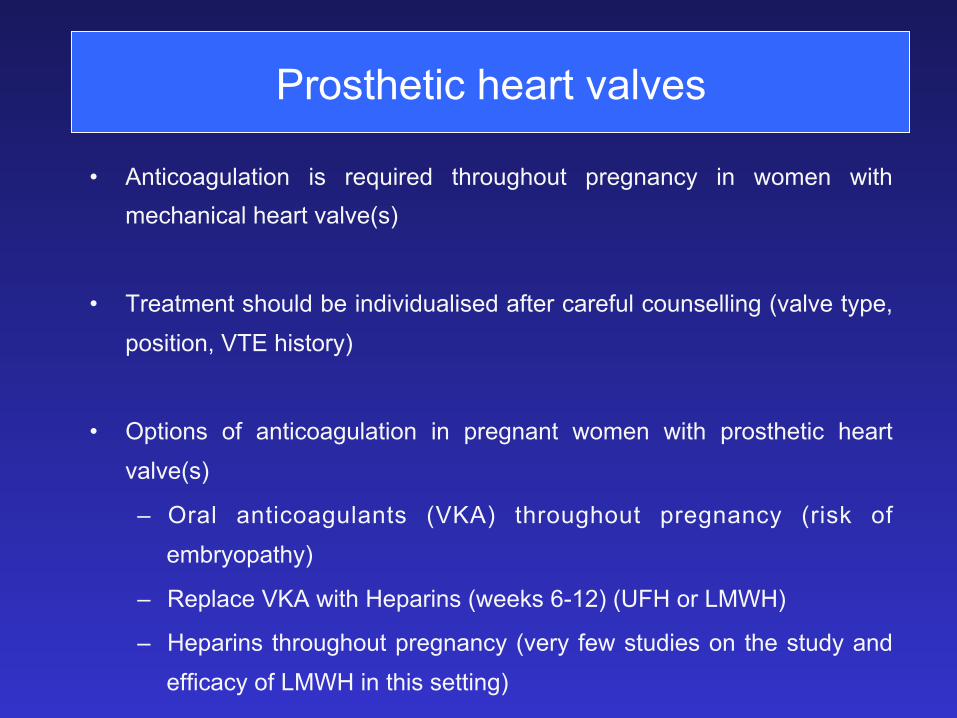
Prosthetic heart valves
• Anticoagulation is required throughout pregnancy in women with mechanical heart valve(s)
• Treatment should be individualised after careful counselling (valve type, position, VTE history)
• Options of anticoagulation in pregnant women with prosthetic heart valve(s)
– Oral anticoagulants (VKA) throughout pregnancy (risk of embryopathy)
– Replace VKA with Heparins (weeks 6-12) (UFH or LMWH)
– Heparins throughout pregnancy (very few studies on the study and efficacy of LMWH in this setting)
Arterial thrombo-embolism in pregnancy
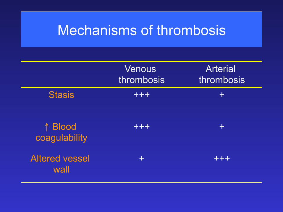
Mechanisms of thrombosis
Venous thrombosis
Arterial thrombosis
Stasis +++ +
↑ Blood coagulability
+++ +
Altered vessel wall
+ +++
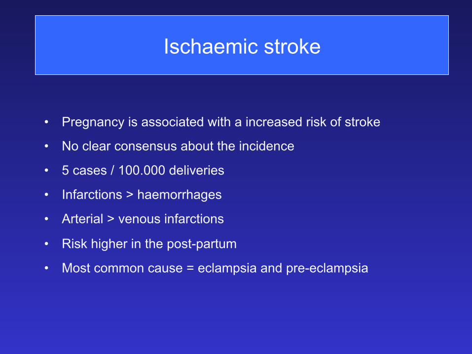
Ischaemic stroke
• Pregnancy is associated with a increased risk of stroke
• No clear consensus about the incidence
• 5 cases / 100.000 deliveries
• Infarctions > haemorrhages
• Arterial > venous infarctions
• Risk higher in the post-partum
• Most common cause = eclampsia and pre-eclampsia
Thrombophilia and fetal loss / gestational vascular complications
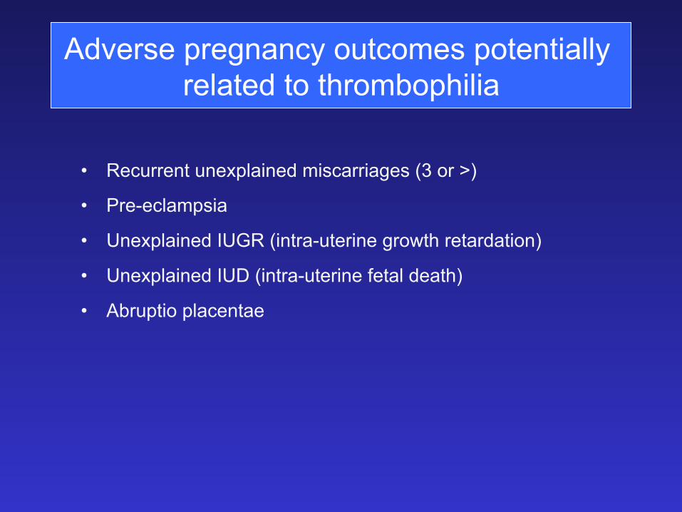
Adverse pregnancy outcomes potentially related to thrombophilia
• Recurrent unexplained miscarriages (3 or >)
• Pre-eclampsia
• Unexplained IUGR (intra-uterine growth retardation)
• Unexplained IUD (intra-uterine fetal death)
• Abruptio placentae
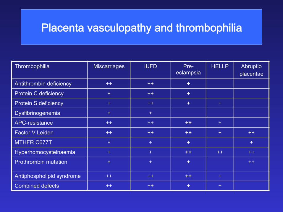
�+!#%-3!�5!2#4+./!3(8�!-$�3(1.,"./()+)!�
Thrombophilia Miscarriages IUFD Pre-eclampsia
HELLP Abruptio placentae
Antithrombin deficiency ++ ++ +
Protein C deficiency + ++ +
Protein S deficiency + ++ + +
Dysfibrinogenemia + +
APC-resistance ++ ++ ++ +
Factor V Leiden ++ ++ ++ + ++
MTHFR C677T + + + +
Hyperhomocysteinaemia + + ++ ++ ++
Prothrombin mutation + + + ++
Antiphospholipid syndrome ++ ++ ++ +
Combined defects ++ ++ + +
Thrombophilia and complicated pregnancy : evidence and controversies
• With the exception of Anti-Phospholipid antibodies, there is little evidence to support systematic screening for thrombophilia in women with complicated pregnancy
• In our experience, combined treatment with LMWH and aspirin with close monitoring although not validated is safe and efficient in most cases to prevent recurrent complications
• For women with past history of eclampsia and/or pre-eclampsia
and no thrombophilia, low dose aspirin is recommended
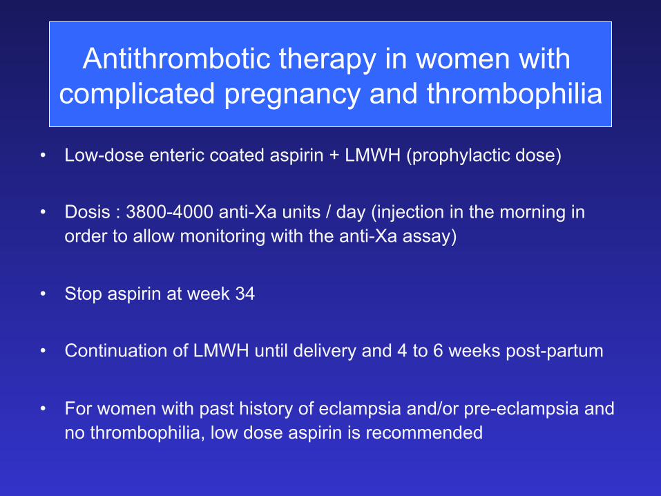
Antithrombotic therapy in women with complicated pregnancy and thrombophilia
• Low-dose enteric coated aspirin + LMWH (prophylactic dose)
• Dosis : 3800-4000 anti-Xa units / day (injection in the morning in order to allow monitoring with the anti-Xa assay)
• Stop aspirin at week 34
• Continuation of LMWH until delivery and 4 to 6 weeks post-partum
• For women with past history of eclampsia and/or pre-eclampsia and no thrombophilia, low dose aspirin is recommended

Thank you for your attention


![E-ISSN: Pharmacognostical and chromatographic evaluation ... · disorders), Vibandha (Constipation), Gulma (abdominal lump), Raktavikara (haematological disorders) etc [7] . Rohitak](https://img.pdfslide.us/doc/110x75/5d5e687e88c993f7068b6270/e-issn-pharmacognostical-and-chromatographic-evaluation-disorders-vibandha.jpg)