Embed Size (px)
Citation preview

Salmonella Suppress Innate Immunity by Targeting Mast Cells
by
Hae Woong Choi
Department of Pathology Duke University
Date: _________________________ Approved:
___________________________ Soman Abraham, Supervisor
___________________________ Herman Staats
___________________________ Laura Hale
___________________________ Michael Dee Gunn
___________________________ Meta Kuehn
Dissertation submitted in partial fulfillment of the requirements for the degree of Doctor of Philosophy in the
Department of Pathology in the Graduate School of Duke University
2014

ABSTRACT
Salmonella Suppress Innate Immunity by Targeting Mast Cells
by
Hae Woong Choi
Department of Pathology Duke University
Date: _________________________ Approved:
___________________________ Soman Abraham, Supervisor
___________________________ Herman Staats
___________________________ Laura Hale
___________________________ Michael Dee Gunn
___________________________ Meta Kuehn
An abstract of a dissertation submitted in partial fulfillment of the requirements for the degree of Doctor of Philosophy in the
Department of Pathology in the Graduate School of Duke University
2014

Copyright by
Hae Woong Choi
2014

iv
Abstract
Mast cells (MCs) are increasingly recognized as powerful sentinel cells responsible for
modulating the early immune responses to a wide range of infectious agents. This protective
role is attributable in part to their preponderance at the host-environment interface and their
innate capacity to rapidly release modulators of immune cell trafficking, which promote the
early recruitment of pathogen-clearing immune cells from the blood. However, host-adapted
pathogens have been a critical threat to humans for a long time because they have evolved
mechanisms directed at overcoming protective immunity.
In this work, we outline how Salmonella enterica serovar Typhimurium has evolved a
novel mechanism to inactivate peripheral MCs, resulting in limited neutrophil responses at
infection sites during early stages of infection. Due to the delay in bacterial clearance at the
point of entry, Salmonella are able to multiply and rapidly disseminate to distal sites.
Suppression of local MC degranulation restricts the outflow of vascular contents into sites of
infection, thus facilitating bacterial spread.
We discovered that MC suppression is mediated by the Salmonella Protein Tyrosine
Phosphatase (SptP), which shares structural homology with YopH, an effector protein expressed
by plague-causing Yersinia pestis. Interestingly, SptP also shares homology with phosphatases
found in MCs, and these phosphatases are also homologous to YopH. We show that YopH
possesses the ability to suppress MCs like SptP, suggesting that this activity is common among
some of the more virulent bacterial pathogens. The functionally relevant domain in SptP is its
enzymatic site, which works by dephosphorylating the vesicle fusion protein N-ethylmalemide-
sensitive factor (NSF) and by blocking phosphorylation of Syk, which are located downstream
and upstream of tyrosine phosphorylation signaling pathway in MCs, respectively.

v
Without SptP, oral challenge with S. Typhimurium fails to suppress MC degranulation
and exhibits limited colonization of the mesenteric lymph nodes. Administration of SptP to sites
of Escherichia coli infection markedly enhances its virulence. Thus, SptP-mediated inactivation of
local MCs is a powerful mechanism utilized by S. Typhimurium to impede early innate immunity.
This finding provides a logical explanation for why previous attempts by others to demonstrate a
protective role for MCs against Salmonella infection have given equivocal results.
Taken together, this work highlights an overlooked virulence mechanism possessed by
certain host-adapted pathogens to avoid the host innate immune system. Additionally, this
innate immune-quelling property of SptP may hold future promise for tempering harmful
inflammatory disorders in immune competent hosts.

vi
Contents
Abstract ....................................................................................................................................................................... iv
List of Tables ............................................................................................................................................................. ix
List of Figures ............................................................................................................................................................. x
List of Abbreviations ............................................................................................................................................ xii
Acknowledgments ................................................................................................................................................ xiv
1. Introduction to Mast Cells and Salmonella ............................................................................................... 1
1.1 Mast Cell Regulation of Innate Immunity. ......................................................................................... 1
1.1.1 Mast cells as Pivotal Immune Surveillance Cells .................................................................... 1
1.1.2 Mast Cells Respond Rapidly to Invading Pathogens Through Degranulation. .......... 4
1.1.3 Mast Cells Express a Wide Array of Receptors that Recognize a Broad Range of
Pathogens. ......................................................................................................................................................... 5
1.1.4 Signaling Events in Mast Cells that Regulate Mast Cell Exocytosis. ............................... 6
1.1.5 Physiological Consequences of MC Degranulation. ............................................................... 9
1.2 Salmonella Typhimurium ....................................................................................................................... 11
1.2.1 Salmonella Typhimurium: a Pathogenic Bacteria Involved in Food Poisoning. ..... 11
1.2.2 Salmonella Typhimurium can Impede the Development of Adaptive Immune
Responses. ....................................................................................................................................................... 14
1.2.3 Salmonella Typhimurium Evades Recognition by Immune Surveillance Cells. ...... 14
1.2.4 Salmonella Typhimurium May Also Modulate the Innate Immune Response. ....... 14
1.2.5 S. Typhimurium Survives Within Phagosomes to Establish Persistent Infections.
.............................................................................................................................................................................. 15
2. Salmonella Typhimurium Impedes Innate Immunity with a Mast Cell-Suppressing Tyrosine Phosphatase, SptP .............................................................................................................................. 17
2.1 Introduction ................................................................................................................................................. 17
2.2 Results ............................................................................................................................................................ 18
2.2.1 Failure of local MCs to degranulate and rapidly recruit neutrophils following S.
Typhimurium infection .............................................................................................................................. 18
2.2.2 S. Typhimurium Inhibited Degranulation of Murine MCs ................................................ 22
2.2.3 S. Typhimurium Actively Suppresses Degranulation of Murine MCs .......................... 23
2.2.4 S. Typhimurium Actively Suppresses Degranulation of Peritoneal MCs in vivo. ... 24
2.2.5 S. Typhimurium Actively Suppresses the Degranulation of Human MCs .................. 26
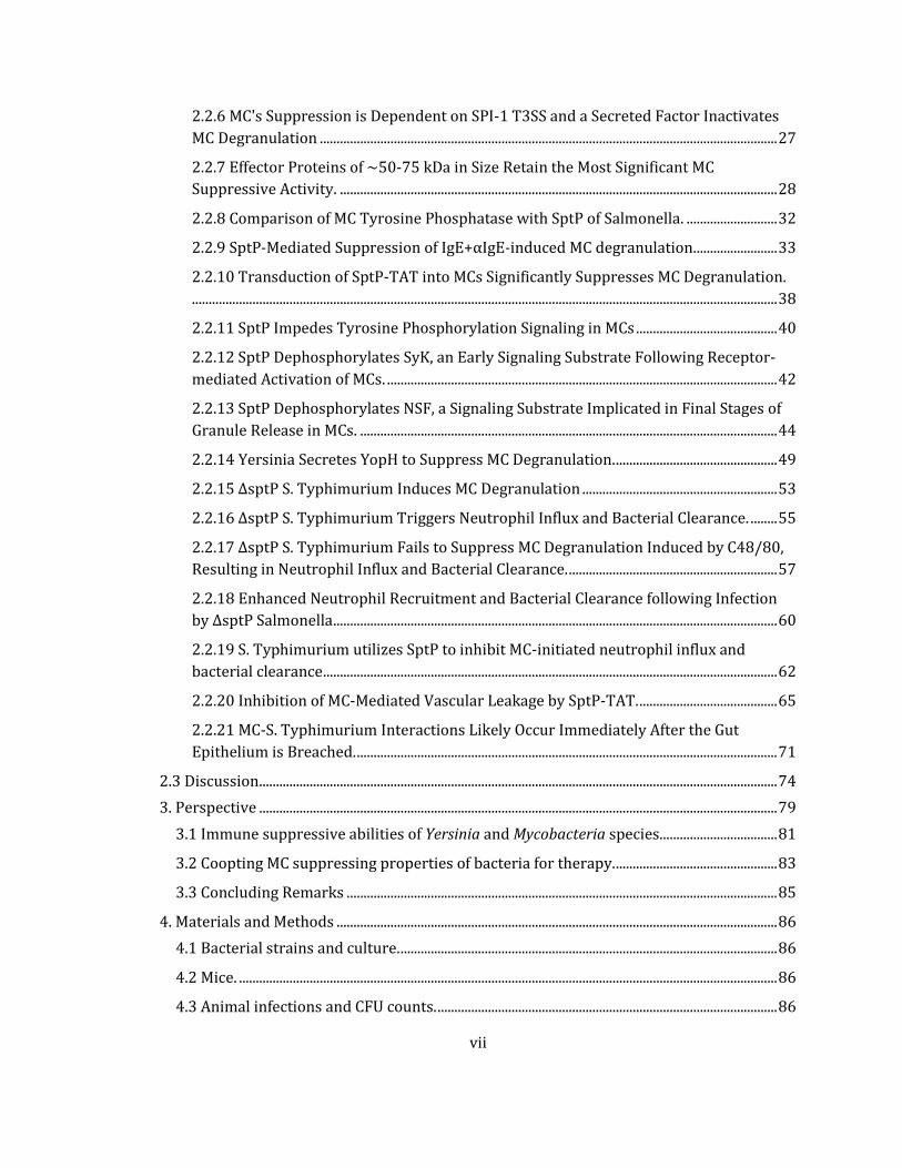
vii
2.2.6 MC's Suppression is Dependent on SPI-1 T3SS and a Secreted Factor Inactivates
MC Degranulation ........................................................................................................................................ 27
2.2.7 Effector Proteins of ~50-75 kDa in Size Retain the Most Significant MC
Suppressive Activity. .................................................................................................................................. 28
2.2.8 Comparison of MC Tyrosine Phosphatase with SptP of Salmonella. ........................... 32
2.2.9 SptP-Mediated Suppression of IgE+αIgE-induced MC degranulation. ........................ 33
2.2.10 Transduction of SptP-TAT into MCs Significantly Suppresses MC Degranulation.
.............................................................................................................................................................................. 38
2.2.11 SptP Impedes Tyrosine Phosphorylation Signaling in MCs .......................................... 40
2.2.12 SptP Dephosphorylates SyK, an Early Signaling Substrate Following Receptor-
mediated Activation of MCs. .................................................................................................................... 42
2.2.13 SptP Dephosphorylates NSF, a Signaling Substrate Implicated in Final Stages of
Granule Release in MCs. ............................................................................................................................ 44
2.2.14 Yersinia Secretes YopH to Suppress MC Degranulation. ................................................ 49
2.2.15 ΔsptP S. Typhimurium Induces MC Degranulation .......................................................... 53
2.2.16 ΔsptP S. Typhimurium Triggers Neutrophil Influx and Bacterial Clearance. ........ 55
2.2.17 ΔsptP S. Typhimurium Fails to Suppress MC Degranulation Induced by C48/80,
Resulting in Neutrophil Influx and Bacterial Clearance. .............................................................. 57
2.2.18 Enhanced Neutrophil Recruitment and Bacterial Clearance following Infection
by ΔsptP Salmonella .................................................................................................................................... 60
2.2.19 S. Typhimurium utilizes SptP to inhibit MC-initiated neutrophil influx and
bacterial clearance ....................................................................................................................................... 62
2.2.20 Inhibition of MC-Mediated Vascular Leakage by SptP-TAT. ......................................... 65
2.2.21 MC-S. Typhimurium Interactions Likely Occur Immediately After the Gut
Epithelium is Breached. ............................................................................................................................. 71
2.3 Discussion .......................................................................................................................................................... 74
3. Perspective .......................................................................................................................................................... 79
3.1 Immune suppressive abilities of Yersinia and Mycobacteria species. .................................. 81
3.2 Coopting MC suppressing properties of bacteria for therapy. ................................................ 83
3.3 Concluding Remarks ................................................................................................................................ 85
4. Materials and Methods ................................................................................................................................... 86
4.1 Bacterial strains and culture. ................................................................................................................ 86
4.2 Mice. ................................................................................................................................................................ 86
4.3 Animal infections and CFU counts. ..................................................................................................... 86
viii
4.4 Cell culture. .................................................................................................................................................. 87
4.5 β-hexosaminidase assay. ........................................................................................................................ 87
4.6 Microscopy. .................................................................................................................................................. 88
4.7 Myeloperoxidase activity assay. .......................................................................................................... 88
4.8 Construction of SptP plasmids and transfected cell lines. ........................................................ 89
4.9 SptP-TAT purification and Column Chromatography ................................................................ 90
4.10 Statistical analysis. ................................................................................................................................. 91
References ................................................................................................................................................................ 92
Biography ............................................................................................................................................................... 102

ix
List of Tables
Table 1: Diverse physiological reactions of MCs to various pathogenic bacteria………….……10
Table 2: Sequence alignments of tyrosine phosphatase in Yersinia pestis, Salmonella Typhimurium, and MCs………………………………………………………………………………………….………49
Table 3: PCR Primer Sequence………………………………………………………………………………………..90

x
List of Figures
Figure 1. Strategic location of mast cells at the host-environment Interface ................................. 2
Figure 2. MC proximity to blood and lymphatic vessels. ......................................................................... 3
Figure 3. Schematic of the signaling pathway associated with the Fcε receptor on mast cells. ......................................................................................................................................................................................... 8
Figure 4. Salmonella spp. utilizes the type III secretion system to achieve successful invasion and intracellular survival ................................................................................................................................... 13
Figure 5. S. Typhimurium fails to elicit neutrophil recruitment and bacterial clearance in vivo. .............................................................................................................................................................................. 19
Figure 6. S. Typhimurium fails to elicit MC activation in vivo.............................................................. 21
Figure 7. S. Typhimurium inhibited MC degranulation, but E. coli activated it. .......................... 22
Figure 8. S. Typhimurium pretreatment actively suppresses MC degranulation in response to MC secretagogues. ............................................................................................................................................ 24
Figure 9. S. Typhimurium suppresses peritoneal MCs in vivo. ............................................................ 25
Figure 10. S. Typhimurium pretreatment actively suppresses human MC degranulation to MC secretagogues. ................................................................................................................................................. 26
Figure 11. MC suppression is dependent on SPI-1 TTSS and its secreted factor. ....................... 28
Figure 12. Fractions 24-44 harboring ~50-75 kDa proteins demonstrate potent MC suppressive activity. ............................................................................................................................................. 29
Figure 13. Tyrosine phosphatase activity can inhibit MC degranulation. ...................................... 31
Figure 14. Sequence alignments of tyrosine phosphatase in S. Typhimurium and MCs. ......... 32
Figure 15. The protein tyrosine phosphatase domain in SptP inhibits MC degranulation. .... 34
Figure 16. SptP-eGFP stable expression in MCs suppresses MC degranulation. ......................... 35
Figure 17. Introduction of recombinant SptP into MC culture media suppresses MC degranulation. ......................................................................................................................................................... 37
Figure 18. Treatment with SptP-TAT suppresses IgE-anti-IgE mediated MC degranulation.39
Figure 19. SptP-TAT inhibited tyrosine phosphorylation in MC granule chambers to block MC degranulation. ................................................................................................................................................. 41
Figure 20. Syk, one of tyrosine phosphorylation cascade, is the target of SptP........................... 43

xi
Figure 21. Intergranular fusion induced by WT Salmonella through dephosphorylation of NSF. .............................................................................................................................................................................. 47
Figure 22. Morphology of RBLs after exposure to WT or ΔsptP mutants with ionomycin. .... 48
Figure 23. Yersinia pestis suppresses MC degranulation. ...................................................................... 50
Figure 24. Yersinia pestis YopH suppresses MC activation. .................................................................. 52
Figure 25. ΔsptP Salmonella initiated phosphotyrosine signaling and degranulation of MCs. ....................................................................................................................................................................................... 54
Figure 26. Enhanced neutrophil recruitment and bacterial clearance with ΔsptP Salmonella infection, and decreased effects with ΔsptP(psptPWT) Salmonella. .................................................... 56
Figure 27. ΔsptP S. Typhimurium infected peritoneal MCs evoke a degranulation response to C48/80. ...................................................................................................................................................................... 58
Figure 28. ΔsptP S. Typhimurium infected peritoneal MCs evoke enhanced neutrophil recruitment and bacterial clearance following exposure to C48/80. .............................................. 59
Figure 29. Enhanced neutrophil recruitment and bacterial clearance following infection by ΔsptP S. Typhimurium. ........................................................................................................................................ 61
Figure 30. Administration of SptP-TAT at sites of E. coli infection inhibits MC-mediated neutrophil recruitment resulting in impaired bacterial clearance ................................................... 64
Figure 31. Vascular leakage resulting from MC degranulation following E. coli infection is suppressed by the administration of SptP-TAT. ....................................................................................... 66
Figure 32. SptP-TAT-suppressed MC degranulation and accompanying vascular leakage during E. coli infection ......................................................................................................................................... 67
Figure 33. SptP-TAT inhibited tyrosine phosphorylation signaling and degranulation in MCs. ....................................................................................................................................................................................... 68
Figure 34. SptPC481S-TAT and SptP-Hisx6 failed to suppress MCs in vivo. ................................... 70
Figure 35. MC degranulation in the cecum of mice infected with ΔsptP but not in WT Salmonella infected mice. ................................................................................................................................... 71
Figure 36. Intestinal MCs reduce the bacterial burden in the mesenteric lymph nodes following oral infection with ΔsptP S. Typhimurium. ............................................................................. 73
Figure 37. Model depicting mechanism of MC Suppression by SptP derived from S. Typhimurium. ......................................................................................................................................................... 78
Figure 38. Pathogenic infection in human body. ...................................................................................... 80
xii
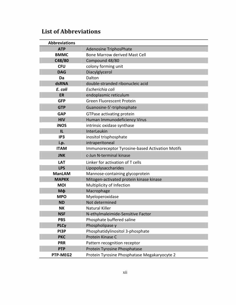
List of Abbreviations
Abbreviations
ATP Adenosine TriphosPhate BMMC Bone Marrow derived Mast Cell C48/80 Compound 48/80
CFU colony forming unit DAG Diacylglycerol Da Dalton
dsRNA double-stranded ribonucleic acid E. coli Escherichia coli
ER endoplasmic reticulum
GFP Green Fluorescent Protein
GTP Guanosine-5'-triphosphate
GAP GTPase activating protein HIV Human Immunodeficiency Virus
iNOS intrinsic oxidase synthase IL InterLeukin
IP3 inositol trisphosphate i.p. intraperitoneal
ITAM Immunoreceptor Tyrosine-based Activation Motifs
JNK c-Jun N-terminal kinase
LAT Linker for activation of T cells LPS Lipopolysaccharides
ManLAM Mannose-containing glycoprotein MAPKK Mitogen-activated protein kinase kinase
MOI Multiplicity of Infection Mφ Macrophage
MPO Myeloperoxidase ND Not determined NK Natural Killer NSF N-ethylmaleimide-Sensitive Factor PBS Phosphate buffered saline PLCγ Phospholipase γ PI3P Phosphatidylinositol 3-phosphate PKC Protein Kinase C
PRR Pattern recognition receptor
PTP Protein Tyrosine Phosphatase
PTP-MEG2 Protein Tyrosine Phosphatase Megakaryocyte 2
xiii
Abbreviations
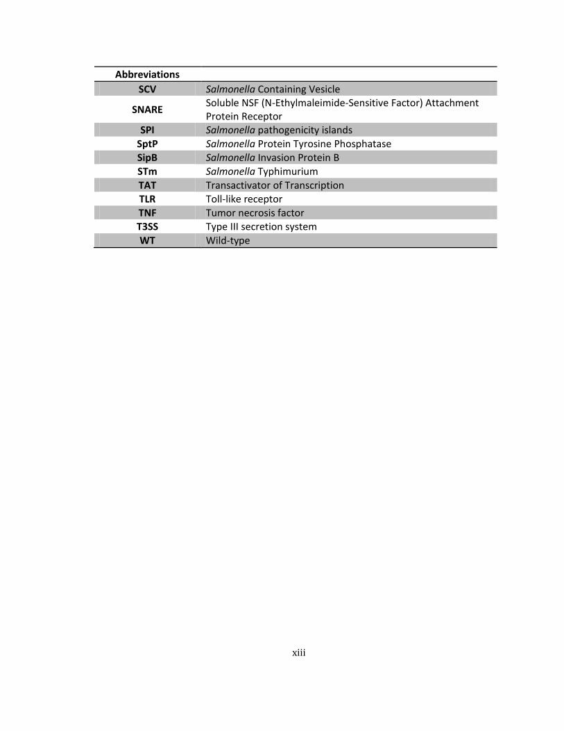
SCV Salmonella Containing Vesicle
SNARE Soluble NSF (N-Ethylmaleimide-Sensitive Factor) Attachment Protein Receptor
SPI Salmonella pathogenicity islands SptP Salmonella Protein Tyrosine Phosphatase SipB Salmonella Invasion Protein B STm Salmonella Typhimurium TAT Transactivator of Transcription TLR Toll-like receptor TNF Tumor necrosis factor T3SS Type III secretion system
WT Wild-type
xiv
Acknowledgments
I would like to express my sincere thanks to my advisor, Dr. Soman Abraham, for
supporting and guiding me throughout my graduate school life. He taught me how to think
scientifically, and how to connect my different findings into a logical and coherent whole, which
was difficult for me. His scientific expertise and advice were of great help in solving difficult
questions, and allowed me to complete several projects. Moreover, he was always open to talk
anytime on any issue, not only on current projects but also about life as scientist. I am also very
thankful to him for guiding my development into an independent scientist.
I am also very grateful to my PhD committee members; Professors Herman Staats, Meta
Kuehn, Laura Hale, and Michael Dee Gunn. Their insightful comments and critiques on my
projects were greatly helpful for their completion. I am also thankful to Professor Edward Miao
from UNC Chapel Hill, whose advice has been valuable to the development and completion of
the Salmonella project.
I thank Dr. Chris Shelburne for training me in handling mice. Special thanks are also
owed to Dr. Samantha Bowen and Gladys Ang for proofreading of my manuscripts. I also must
thank to all of the past and current lab members in Dr. Abraham lab: Dr. Jorn Karhausen, Dr.
Ashley St. John, Yuxuan Miao, Viraj Parekh, Mohammad Arifuzzaman, and Laura Mitrescu for
their help over the past 6 years.
I deeply want to thank my family, who have offered their support over the past six years.
My father and mother supported me with their unconditional love and encouraged me
throughout my life. My younger brother also stood behind me and encouraged me. I also thank
my parents-in-law for their advice and support.

xv
In particular, I thank my wife, Nah Hyung Kim. These few sentences can't express the
magnitude of my gratitude for her support and encouragement during my entire graduate
school life. Her encouragement always helped me to stand up when I was struggling with
difficult times. The extensive support that I have received from her enabled me to finish
graduate school at Duke.
Finally, I cannot finish without acknowledging God who gave me the strength and
perseverance to continue when I wanted to give up.
1
1. Introduction to Mast Cells and Salmonella
1.1 Mast Cell Regulation of Innate Immunity.
1.1.1 Mast cells as Pivotal Immune Surveillance Cells
After heart diseases, microbial infections are the second most common cause of human
death in the world [2]. MCs have several innate properties that make them uniquely able to
contribute to immune surveillance against infectious agents. These properties include their
selective location in relatively large numbers immediately underneath the epithelial surface of
the skin and the mucosa of the genitourinary tract and respiratory tract (Figure 1). Because
these sites are typically where infections begin, MCs appear strategically located to be among
the first immune cells that pathogens encounter after they have breached the epithelial barrier.
MCs are also found in close proximity around peripheral blood and lymphatic vessels (Figure 2),
and thus they are also ideally positioned to recruit immune cells from blood vessels into infected
tissue or coordinate the movement of immune cells from infected sites into lymphatic vessels
leading to the draining lymph nodes [3,4]. Recently, it was reported that MCs proximal to blood
vessels constantly sample blood contents by employing cytoplasmic protrusions [5] suggesting
that these cells are also capable of detecting and interacting with blood borne pathogens.

2
Figure 1. Strategic location of mast cells at the host-environment Interface
(A) Nasal passage from mouse was dissected and cryosectioned longitudinally. (Upper panel) MCs, B cells, and lymphatics were stained with avidin, anti-B220, and anti-LYVE-1 antibodies, respectively. (Bottom left panel) Macrophages were stained with anti- F4/80 antibody. Respiratory epithelial cells were visualized with autofluorescence and covered with fluorescent ovalbumin by nasal challenge. (Bottom right panel) DCs and B cells were stained with anti-CD11c and anti-B220 antibodies. Respiratory epithelial cells were also visualized with autofluorescence. L indicates airway lumen. (B) Bladder tissue was dissected and whole-mount-stained with wheat germ agglutinin, avidin, and anti-CD31 antibody for superficial epithelial cells, MCs, and blood vessels, respectively. The image is a 3D reconstruction view from Z-stacked images taken by confocal microscope. Scale bar: 300 µm.

3
Figure 2. MC proximity to blood and lymphatic vessels.
Abundant MCs are found near blood vessels in the mouse ear (left). Red fluorescence identified cells are MCs stained with avidin. Blue represents blood vessels stained with anti-CD31 antibody. Green depicts fluorescent dextran (150 kDa). When these MCs undergo degranulation, they induce blood vessel leakage that can be detected by the exudation of fluorescent dextran from blood vessels, indicated by green particles located in interstitial spaces, arrow. MCs are also typically found near lymphatics (right). Blue depicts lymphatic vessels stained for LYVE-1. Green depicts B cells stained with anti-B220 antibody. Red depicts MCs stained with avidin. Scale bar: 10 μm
4
1.1.2 Mast Cells Respond Rapidly to Invading Pathogens Through Degranulation.
MCs are highly specialized for the synthesis and secretion of a myriad of
pharmacologically-active products. MC mediators have traditionally been divided into two major
groups: those that are preformed, which include histamine, heparin, serine proteases, some
select cytokines and chemokines such as tumor necrosis factors (TNFs) [6] and CXCL1/CXCL2 [7],
and those that are synthesized de novo when the cells are activated, which include a wide range
of cytokines and eicosanoids such as leukotrienes, prostaglandins and the thromboxanes [8].
The release of pre-packaged mediators into the surrounding tissue typically occurs beginning
within seconds to minutes following stimulation, a strategy that gives MC-derived products a
temporal advantage over those produced by other immune surveillance cells, which take
markedly longer. The prepackaged MC products can be released in an explosive fashion or
piecemeal.
Piecemeal degranulation consists of a slow emptying of granule chambers in the
absence of large scale intergranular fusion. Because of their longevity, MCs at sites of
inflammation can undergo multiple cycles of degranulation followed by re-granulation; in this
way MCs are able to sustain inflammatory responses to pathogens. Unlike the degranulation
response, which is initiated within seconds of MC activation, de novo synthesis of eicosinoids,
cytokines and chemokines occurs significantly later, with a time frame that is comparable to
when other immune cells secrete their mediators following activation. Many studies have
shown that de novo production of these mediators by MCs in response to pathogens can vary
greatly depending on the stimulus and the experimental conditions.

5
1.1.3 Mast Cells Express a Wide Array of Receptors that Recognize a Broad Range of Pathogens.
Like other immune cells involved in immune surveillance, MCs express a wide range of
cell surface receptors that can directly bind pathogens, secreted toxins, or other microbial
products. These receptors include most pattern recognition receptors (PRRs) as well as other
receptors that specifically recognize microbial cell surface components. One of the best studied
bacterial receptors on MCs is CD48, a GPI-anchored protein that binds type I fimbriae on various
enterobacteria and a cell surface component on Mycobacterum tuberculosis [9,10]. Cross-linking
of CD48 not only causes MC degranulation but also uptake of adherent bacteria. Additionally,
MCs also express receptors on their cell surface for host proteins that coat pathogens, such as
complement fragments and antibodies [11]. Depending on the receptor that is engaged, their
cross-linking can trigger extensive or piece-meal MC degranulation. For example, cross-linking of
CD48 by type I fimbriated E. coli can result in extensive MC degranulation and release of a wide
range of mediators, whereas activation of Toll-like receptor (TLR) 4 by lipopolysaccharide (LPS)
results in the significant secretion of soluble mediators but little or no MC degranulation.
Similarly, binding of cholera toxin to the ganglioside GM1 results in the significant secretion of
interleukin-6 (IL-6) but no accompanying degranulation of MCs [12].
MC activation during infection is not always the result of contact with an infectious
agent, its products or with opsonized microbes; many infected or otherwise stressed cells
release danger signals or alarmins that serve as potent activators of MCs [13,14]. Therefore,
MCs do not have to be in immediate proximity to a site of infection to contribute to the immune
response. The signaling agents released by stressed host cells range from antimicrobial peptides
and adenosine triphosphate (ATP) to IL-33, which is typically released when cells degrade [15].
Binding of any of these cellular products to MCs can trigger extensive MC degranulation.

6
1.1.4 Signaling Events in Mast Cells that Regulate Mast Cell Exocytosis.
Despite the large body of information regarding immune recognition receptors for
pathogens or their products found on MCs, the question of how MCs signal intracellularly to
induce a secretory response remains largely unknown. However, several key signaling events are
likely shared with the well-characterized IgE signaling pathway, which is triggered when
allergens bind and crosslink IgE bound to the high-affinity IgE receptor (FcεRI) on MCs [16]. As
Figure 3 demonstrates, when FcεRI receptors aggregate, they initiate a cascade of tyrosine
phosphorylation events via multiple signaling molecules in the MC cytoplasm. Early on, activated
Lyn kinase phosphorylates tyrosine residues at immunoreceptor tyrosine-based activation
motifs (ITAMs) on the receptor subunits [16,17]. Subsequent amplification of tyrosine
phosphorylation occurs through activation of Syk kinase, which phosphorylates another
substrate, linker for activation of T cells (LAT) [17,18]. LAT phosphorylation activates
Phospholipase γ1 (PLCγ1), leading to the cleavage of phosphatidylinositol into inositol
trisphosphate (IP3) and diacylglycerol (DAG) [20,21]. DAG activates Protein Kinase C (PKC) and
inositol triphosphate (IP3), which in turn releases calcium from the endoplasmic reticulum (ER)
into the cytoplasm. This free calcium is utilized in the final stages of degranulation for the direct
fusion of granules into plasma membrane. For degranulation and extracellular release of MC
granules, fusion between granule membrane and plasma membrane must occur. V-soluble NSF
(N-ethylmaleimide-sensitive factor) attachment protein receptor (v-SNARE/VAMP8) on the
granule membrane and t-SNARE (SNAP-23) on the plasma membrane form stable trans-SNARE
complexes to guide secretory vesicles to the plasma membrane [22,23]. Additionally, multiple
accessory proteins are required to regulate cognate fusion processes and to assist in the
formation of various protein complexes. As in the early signaling events that occur around the
7
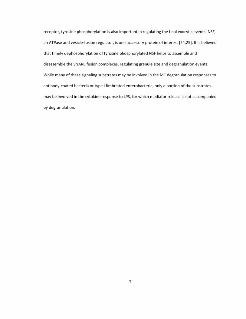
receptor, tyrosine phosphorylation is also important in regulating the final exocytic events. NSF,
an ATPase and vesicle-fusion regulator, is one accessory protein of interest [24,25]. It is believed
that timely dephosphorylation of tyrosine phosphorylated NSF helps to assemble and
disassemble the SNARE fusion complexes, regulating granule size and degranulation events.
While many of these signaling substrates may be involved in the MC degranulation responses to
antibody-coated bacteria or type I fimbriated enterobacteria, only a portion of the substrates
may be involved in the cytokine response to LPS, for which mediator release is not accompanied
by degranulation.
8
Figure 3. Schematic of the signaling pathway associated with the Fcε receptor on mast cells.
Top panel of image shows that antigen binding to the Fcε receptor induces the phosphorylation of Syk, amplifying signaling to induce degranulation and cytokine and lipid mediator release. Middle panel of image shows the brief signaling events during IgE receptor-mediated MC degranulation. Bottom panel of image shows the signaling events invovled in secretory vesicle fusion during degranulation. PTP-MEG2 (protein tyrosine phosphatase megakaryocyte 2) dephosphorylates the vesicle-fusion regulator NSF (N-ethylmaleimide-sensitive factor), which is a prerequisite to induce granule fusion to the plasma membrane.

9
1.1.5 Physiological Consequences of MC Degranulation.
Although MCs produce antimicrobial agents that directly reduce the microbial burden in
the body, for the most part the antimicrobial role of MCs appears to involve the mobilization of
other immune cells to combat infection. The MC response to microbial challenge is typically
biphasic. First, rapid degranulation facilitates the release of pre-formed inflammatory mediators,
including TNF-α, proteases, and histamine, that initiate the early recruitment of immune cells to
sites of infection [26]. This initial response is followed by de novo synthesis and secretion of
various immune mediators several hours later. This biphasic response permits MCs not only to
initiate but to sustain critical immune responses for prolonged periods of time. Because MCs are
found in close proximity to the vasculature, many MC-derived mediators readily traffic into the
bloodstream, initiate blood vessel dilation, and promote the extravasation of various immune
cells [27,28]. The functional importance of MCs has been best studied following bacterial
infections. The protective role of MCs and MC-derived TNF was first demonstrated nearly two
decades ago against Klebsiella pneumoniae infection [29] and against polymicrobial intra-
abdominal sepsis [30]. Upon contact with bacteria, MCs release TNF-α, which initiates the early
recruitment of neutrophils to clear the pathogen.
MCs have been shown to play similar protective roles during Pseudomonas aeruginosa
infection of the mouse peritoneum [31], E. coli infection of the peritoneum and urinary tract
[32], Citrobacter rodentium [33] and Helicobacter felis [34] infections of the gastrointestinal
tract, and Haemophilus influenzae infection of the ear [35]. MCs are equally effective against
several Gram-positive bacteria, including Streptococcus pyogenes, Mycoplasma pulmonis [36],
Mycoplasma pneumonia and Listeria monocytogenes [37]. Table 1 shows various pathogenic

10
bacteria that infect humans and summarizes the protective effects of MCs exerted against these
pathogens.
Table1. Diverse physiological reactions of MCs to various pathogenic bacteria
Pathogenic bacteria are grouped by Gram-stain reaction. Each bacterium is categorized by corresponding MC mediators and their effects on the immune system. ND: not determined.
Gram Stain Reaction
Pathogenic Bacteria MC
mediators Physiological Consequences References
Gram- Negative
Klebsiella pneumoniae TNF-α, IL-6 Recruitment of neutrophils [29], [38]
Escherichia coli TNF-α Recruitment of neutrophils / DCs
/ T cells
[32], [39] [40], [30]
Escherichia coli Leukotriene Recruitment of neutrophils [41]
Citrobacter rodentium ND Antibacterial activity
by directly killing bacteria in vitro [33]
Francisella tularensis IL-4 Alternative activation of Mφ for
host resistance [42], [43]
Pseudomonas aeruginosa
IL-1 α/β Recruitment of neutrophils [44], [45]
Gram-Positive
Clostridium difficile IL-8 Recruitment of neutrophils [46], [47]
Group A streptococcus Cathelicidin Secreting antimicrobial peptides [48]
Mycoplasma pneumonia ND Enhanced bacterial clearance [36]
Listeria monocytogenes TNF-α Recruitment of neutrophils [49], [50]
11
MCs in peripheral tissue are activated upon contact with Dengue virus, and in response
they release diverse cytokines and chemokines to recruit natural killer (NK) and NK T cells to
clear the virus [51-54]. It is not necessary for virus to infect MCs in order to activate them. Viral
components such as the protein Fv and synthetic viral double-stranded ribonucleic acid (dsRNA)
effectively activate MCs [55,56] and as a result can elicit the recruitment of CD8+ T cells [57].
Although it is unclear whether MCs contribute to the clearance of human immunodeficiency
virus (HIV), it appears that these cells become infected and serve as reservoirs, contributing to
persistent HIV infection [58-60].
Upon contact with cell surface components of various fungi, MC de novo synthesize and
secrete selected mediators without extensive degranulation [61]. During Aspergillus [62] and C.
neoformans infections [63], MC products promote significant anti-fungal innate immunity, which
mostly involves the recruitment of phagocytic cells and other immune cells. A powerful MC-
mediated mechanism to reduce the burden of gut parasites such as N. brasiliensis [64],
Trichinella spiralis and Strongyloides which is independent of immune cell recruitment, occurs
via through the release of MC protease, which triggers smooth muscle contraction.
1.2 Salmonella Typhimurium
1.2.1 Salmonella Typhimurium: a Pathogenic Bacteria Involved in Food Poisoning.
Salmonella Typhimurium is a highly invasive pathogen and a leading cause of foodborne
illness [65]. The global burden of nontyphoidal Salmonella in terms of human health is as high as
93.8 million cases, but because many mild cases are not diagnosed, the number is likely much
higher [66]. S. Typhimurium virulence has been associated with the ability to evade and

12
suppress the host immune system. Some of the earliest studies investigating the pathogenesis
of S. Typhimurium highlighted its remarkable capacity to invade and persist intracellularly within
gut epithelial cells and neighboring macrophages (Mφs) [67], where it can replicate while
avoiding immune cells and antimicrobial agents. Invasion and intracellular persistence are
mediated by a variety of effector proteins encoded in S. Typhimurium pathogenicity islands 1
and 2 (SPI-1 and SPI-2); the majority of these are exported out of the bacterial cell by the well-
characterized type III secretion system (T3SS) [68] (Figure 4).
13
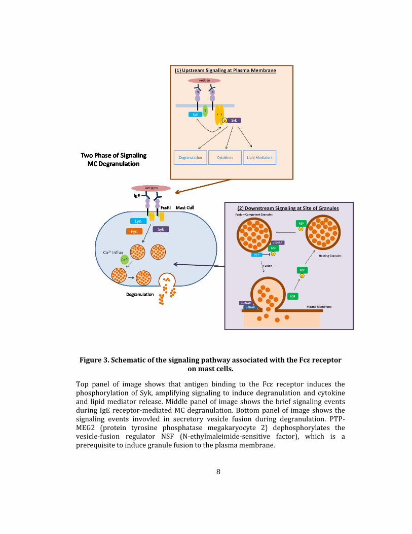
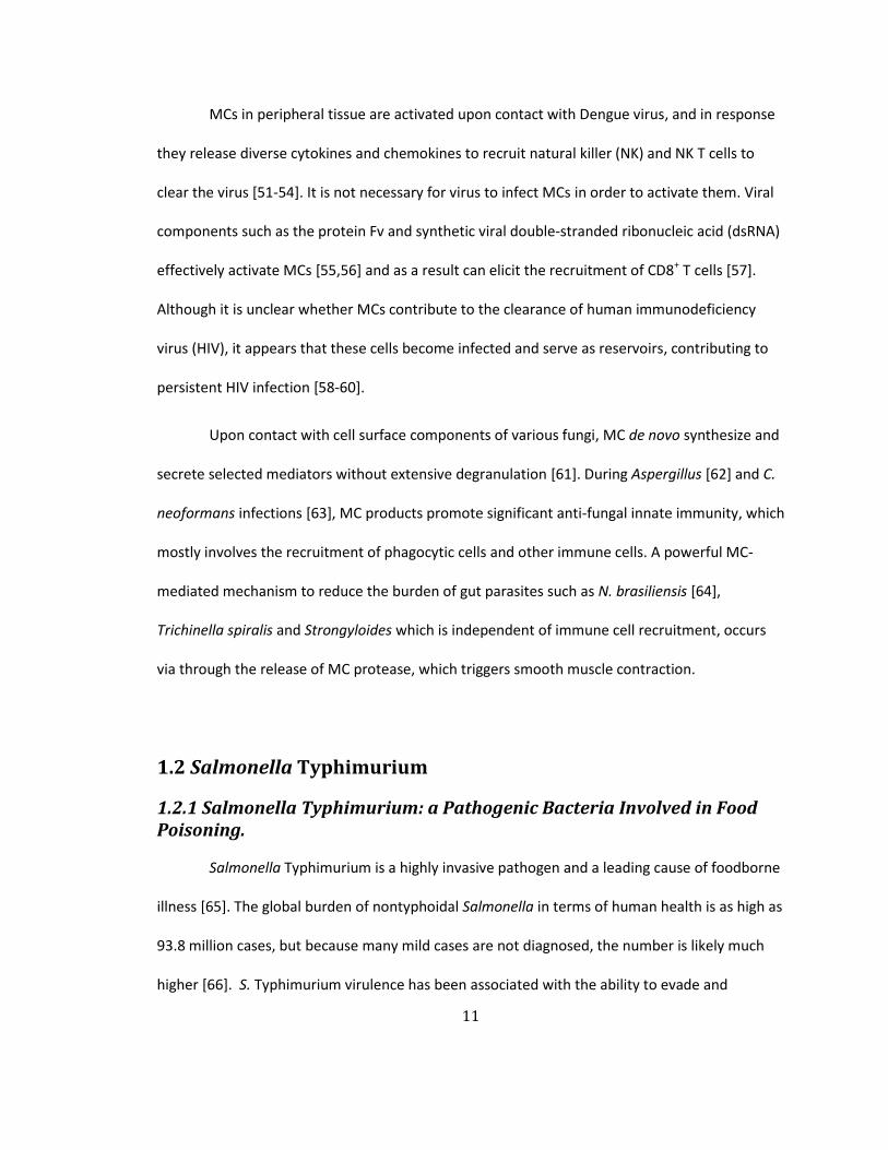
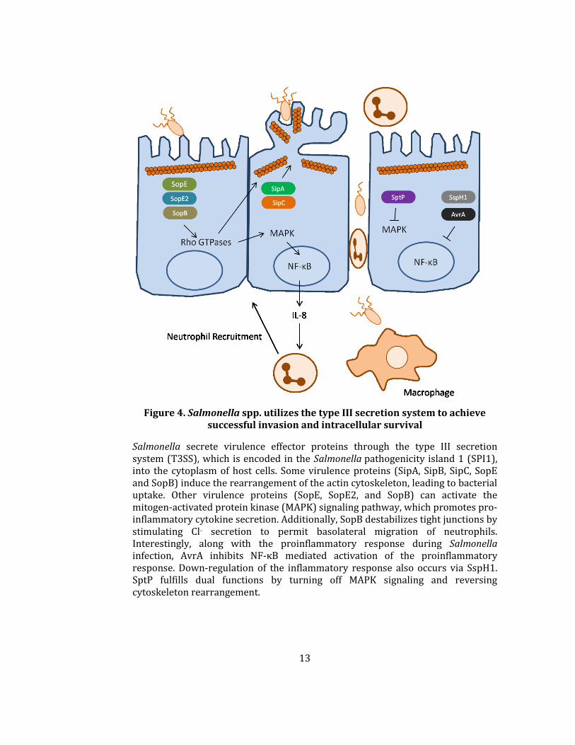
Figure 4. Salmonella spp. utilizes the type III secretion system to achieve successful invasion and intracellular survival
Salmonella secrete virulence effector proteins through the type III secretion system (T3SS), which is encoded in the Salmonella pathogenicity island 1 (SPI1), into the cytoplasm of host cells. Some virulence proteins (SipA, SipB, SipC, SopE and SopB) induce the rearrangement of the actin cytoskeleton, leading to bacterial uptake. Other virulence proteins (SopE, SopE2, and SopB) can activate the mitogen-activated protein kinase (MAPK) signaling pathway, which promotes pro-inflammatory cytokine secretion. Additionally, SopB destabilizes tight junctions by stimulating Cl- secretion to permit basolateral migration of neutrophils. Interestingly, along with the proinflammatory response during Salmonella infection, AvrA inhibits NF-κB mediated activation of the proinflammatory response. Down-regulation of the inflammatory response also occurs via SspH1. SptP fulfills dual functions by turning off MAPK signaling and reversing cytoskeleton rearrangement.

14
1.2.2 Salmonella Typhimurium can Impede the Development of Adaptive Immune Responses.
More recently, S. Typhimurium was found to directly suppress host adaptive immune
responses by impeding the actions of specific immune cells; for example, S. Typhimurium
induces antigen-presenting cells to adopt distinct migratory paths [69-71] and restricts T cell
proliferation and activation to limited regions of the body following infection [72,73]. Other
studies have pointed to a more global mechanism for the suppression of adaptive immune
responses that involves targeting the draining lymph node, which is the epicenter of the
adaptive immune response [74]. S. Typhimurium has been shown to target and disrupt the
architecture of lymph nodes by altering homeostatic chemokine gradients, resulting in aberrant
immune cell trafficking and an ineffective memory response to the pathogen.
1.2.3 Salmonella Typhimurium Evades Recognition by Immune Surveillance Cells.
The surface modification of pathogenic bacteria is a popular way to evade recognition
by host surveillance cells, such as alteration of the carbohydrate capsule by Haemophilus
influenza or Neisseria meningitides. Similarly, S. Typhimurium avoids the more immediate and
non-specific host innate immune response. S. Typhimurium is capable of modifying lipid A
expression on its surface; this is controlled by the PhoP/Q regulon. Regarding TLR4-mediated
activation of the proinflammatory response, this altered surface lipid A expression is 100-fold
less active than with the non-modified one [75,76].
1.2.4 Salmonella Typhimurium May Also Modulate the Innate Immune Response.
An increasing number of studies have shown that S. Typhimurium evasion of recognition
by host immune cells cannot explain the ability of S. Typhimurium to rapidly proliferate, as other

15
bacterial components such as flagella are readily recognized by the host PRR repertoire. The lack
of an adequate innate immune response to control S. Typhimurium growth and spread suggests
a more profound bacteria-mediated mechanism to delay or completely suppress non-specific
host responses.
To effectively achieve successful infection, S. Typhimurium has evolved virulence
effector proteins to regulate host innate immune response. In particular, anti-inflammatory
activity is beneficial to permit invading S. Typhimurium to achieve persistent infection. Although
S. Typhimurium is known for inducing pro-inflammatory reactions in the host, recent reports
suggest a role in suppressing pro-inflammatory responses in host as well. Several independent
studies have reported that AvrA, acetyltransferase, inhibits the pro-inflammatory response and
pro-apoptotic activity by modifying mitogen-activated protein kinase kinase, which leads to the
inactivation of the c-Jun N-terminal kinase (JNK) signaling pathway [77,78]. These observations
were made in intestinal epithelial cells and Mφs. In Mφs, AvrA was found to efficiently block
the JNK signaling pathway and delay the death of Mφ, permitting the bacteria to establsh an
intracellular niche [79].
1.2.5 S. Typhimurium Survives Within Phagosomes to Establish Persistent Infections.
After S. Typhimurium invades epithelial or phagocytic cells, it survives and replicates
successfully in endocytic vesicles within the host cell. Infected Salmonella is detected inside
Salmonella-containing vesicles (SCVs). Inside these SCVs, S. Typhimurium avoid the fusion of
degradative vesicles by redirecting the vesicles and thus establish a Salmonella-favorable
environment near ER without further maturation of phagosome. This establishment of
Salmonella-favorable niche is accomplished via virulence effector proteins located in the SPI2
16
locus. These effector proteins prevent trafficking of NADPH oxidase- and intrinsic oxidase
synthase (iNOS)-containing vesicles to SCVs [80-83]. Therefore, the failures of killing S.
Typhimurium by resident Mφs and DCs at the submucosa lead to drain into mesenteric lymph
nodes which connect bloodstream. Phagocytes harboring S. Typhimurium enter the blood
stream and transport the bacteria into liver and spleen, eventually causing systemic infections
[84]. The inability of phagocytes to eradicate Salmonella in phagosomes permits Salmonella to
survive and disseminate systemically.

17
2. Salmonella Typhimurium Impedes Innate Immunity with a Mast Cell-Suppressing Tyrosine Phosphatase, SptP
2.1 Introduction
An important component of the innate immune response to bacterial pathogens is the
MC, a morphologically distinct type of immune cell with specialized secretory functions that is
preferentially located in close proximity to the epithelium of the gastrointestinal tract and other
mucosal surfaces. Given their strategic location at potential sites of pathogen entry, MCs are
among the first immune cells to perceive and react to microbial penetration of the epithelial
barrier [3,4]. There is now a broad consensus that MCs are pivotal in initiating early innate
immune responses to invading pathogens. Studies investigating Gram-positive and Gram-
negative bacteria as well as viruses and fungi [26,85] have revealed that MCs promote the early
clearance of pathogens.
However, to date, S. Typhimurium has proven to be an exception to this paradigm.
Although some studies have indicated that MCs appear to contribute to S. Typhimurium
clearance, the adoptive transfer of cultured MCs into MC-deficient mice does not significantly
alter S. Typhimurium infection [86]. Furthermore, the presence of MCs during severe S.
Typhimurium infection even appears harmful [87].
Given the inconclusive role of MCs during S. Typhimurium infection, here we sought to
more closely examine interactions between MCs and S. Typhimurium in vitro and in vivo. We
discovered that S. Typhimurium possessed a remarkable capacity to inhibit the ability of MCs to
mount a degranulation response to powerful stimuli such as ionomycin and IgE-mediated
antigen recognition and that this inhibition was attributable to SptP, a Salmonella protein
tyrosine phosphatase secreted via the S. Typhimurium T3SS. Interestingly, SptP is structurally

18
analogous to several tyrosine phosphatases present in MCs and mediators found in other
pathogens, such as YopH of Yersinia pestis. We determined that SptP appeared to
dephosphorylate at least two proteins that are critical for MC degranulation. Our studies reveal
the distinct ability of S. Typhimurium to inactivate a key modulator of host innate immunity and
thereby facilitate stealthy infection.
2.2 Results
2.2.1 Failure of local MCs to degranulate and rapidly recruit neutrophils following S. Typhimurium infection
MCs possess an apparent inability to evoke protective responses in mice following S.
Typhimurium infection. To address this failure, we hypothesized that S. Typhimurium is able to
inactivate MCs. In order to test this notion, we injected late log phase S. Typhimurium SL1433
into the peritoneal cavity of WT and MC-deficient KitW-sh/KitW-sh (MC-deficient) mice. We chose
the peritoneum for the following reasons: (i) it is a self-contained body site where bacterial
numbers and neutrophil influx can be simultaneously and conveniently assessed, (ii) bacteria
can directly interact with MCs, and (iii) MCs can be readily visualized in the peritoneal fluid in
order to determine activation status by evaluating cell morphology. Interestingly, we observed
no significant difference in the bacterial burdens of peritoneal lavage from WT and MC-deficient
mice upon infection with S. Typhimurium (Figure 5A, top). Consistent with this finding,
myeloperoxidase (MPO) assays with lavage fluid revealed no significant difference in neutrophil
influx between the two groups of mice (Figure 5A, bottom). In contrast, when E. coli J96 was
intraperitoneally injected into the two mouse strains, a significant difference in bacterial
clearance was observed between WT and MC-deficient mice (Figure 5B, top). This observation
correlated with an increased neutrophil influx in WT mice compared to MC-deficient mice

19
(Figure 5B, bottom). The lack of an appreciable difference in bacterial load between WT and MC-
deficient mice is consistent with previous reports that found no clear protective function for
MCs during S. Typhimurium infection [87].
Figure 5. S. Typhimurium fails to elicit neutrophil recruitment and bacterial clearance in vivo.
(A, B, top) Residual bacterial counts (CFUs) in the peritoneal cavity of wild-type (WT) or MC-deficient mice 24 h following intraperitoneal (i.p.) infection with 5×10⁵ CFU S. Typhimurium or 1×10⁷ CFU E. coli. (A, B, bottom) Myeloperoxidase assay with peritoneal lavage fluid from WT or MC-deficient mice 5 h post-infection with 1×10⁷ CFU S. Typhimurium or E. coli J96. Mean ± SEM, *p<0.05, N.S., not significant.
20
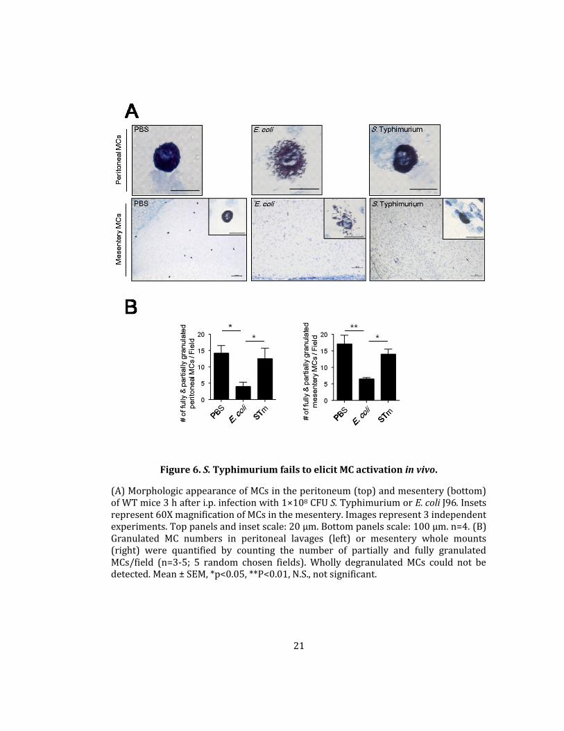
To determine whether our observations could be related to differential MC activation
upon contact with different pathogens, we investigated the morphology of MCs in the
peritoneal fluid (Figure 6A, top) and mesentery (bottom) of both groups of mice. For
comparative purposes, we also examined MCs from PBS-injected mice. MCs from mice
challenged with E. coli J96 were not readily visible with toluidine blue staining, as they were
extensively degranulated with an observable spray of isolated granules around each MC (Figure
6A, middle). In contrast, MCs from S. Typhimurium-challenged mice were readily detectable and
fully granulated (Figure 6A, right) and morphologically resembled MCs from controls (Figure 6A,
left). Quantification of fully and partially granulated MCs at both of these sites is provided in
Figure 6B. These observations collectively suggest that, unlike their vigorous response to E. coli
J96, MCs appear incapable of evoking a degranulation response to S. Typhimurium.
Consequently, limited neutrophil responses and corresponding bacterial clearance were
observed in S. Typhimurium-infected mice.
21
Figure 6. S. Typhimurium fails to elicit MC activation in vivo.
(A) Morphologic appearance of MCs in the peritoneum (top) and mesentery (bottom) of WT mice 3 h after i.p. infection with 1×10⁸ CFU S. Typhimurium or E. coli J96. Insets represent 60X magnification of MCs in the mesentery. Images represent 3 independent experiments. Top panels and inset scale: 20 µm. Bottom panels scale: 100 µm. n=4. (B) Granulated MC numbers in peritoneal lavages (left) or mesentery whole mounts (right) were quantified by counting the number of partially and fully granulated MCs/field (n=3-5; 5 random chosen fields). Wholly degranulated MCs could not be detected. Mean ± SEM, *p<0.05, **P<0.01, N.S., not significant.

22
2.2.2 S. Typhimurium Inhibited Degranulation of Murine MCs
To more closely investigate the limited MC degranulation response to S. Typhimurium,
we utilized the MC model cell line RBL-2H3 in standard in vitro β-hexosaminidase release assays
to assess MC degranulation activity following exposure to S. Typhimurium, E. coli J96, or
ionomycin, a potent MC secretagogue that works by triggering intracellular calcium flux [16].
Whereas ionomycin and E. coli J96 evoked significant responses, no degranulation was observed
in response to S. Typhimurium or to PBS (Figure 7A). Compared to the dose-dependent MC
degranulation observed with E. coli J96 (Figure 7B, left), the degranulation response to S.
Typhimurium was minimal (Figure 7B, right).
Figure 7. S. Typhimurium inhibited MC degranulation, but E. coli activated it.
(A, B) In vitro β-hexosaminidase release assays with RBL cells after 1 h exposure to S. Typhimurium (STm) or E. coli J96. (B) Effect of bacterial multiplicity of infection (MOIs) (10:1, 100:1, 1000:1) on MC degranulation. Mean ± SEM, *p<0.001, **p<0.01, ***p<0.05, N.S., not significant.
23
2.2.3 S. Typhimurium Actively Suppresses Degranulation of Murine MCs
The limited MC degranulation to S. Typhimurium could potentially be attributable to the
ability of S. Typhimurium to actively block MC degranulation or to the inability of MCs to evoke a
response to S. Typhimurium. To test the latter possibility, we exposed MCs to live and heat-
killed S. Typhimurium and observed that MCs failed to degranulate in response to either
stimulus, indicating that the MCs were inherently unresponsive to S. Typhimurium (Figure 8A,
left). Next, to investigate whether S. Typhimurium also possesses the ability to inhibit MC
degranulation, we pre-treated MCs with live or heat-killed S. Typhimurium and then exposed
the cells to the potent secretagogue ionomycin. We found that in contrast to killed S.
Typhimurium, live S. Typhimurium was able to block subsequent MC responses to ionomycin
(Figure 8A, right), indicating that S. Typhimurium has the innate capacity to actively block MC
degranulation. To assess the specificity of this activity, we pre-treated MCs with live E. coli strain
CI5 or the Gram-positive pathogen Staphylococcus aureus and found that neither of these
bacteria were able to inhibit MC degranulation in response to ionomycin (Figure 8B).

24
2.2.4 S. Typhimurium Actively Suppresses Degranulation of Peritoneal MCs in vivo.
In addition to in vitro experiments, we tested whether S. Typhimurium could also block
the MC degranulation response to MC activators in vivo. Before we started the experiment with
S. Typhimurium, we tested whether MC degranulation could be measured in vivo. To induce MC
degranulation, we injected intraperitoneal (i.p.) Compound 48/80 (C48/80), a MC-specific
activator, and performed double staining with an anti-c-kit antibody and avidin. As Figure 9A
shows, a population of c-Kit+Avidin+ stained MCs were clearly visible in negative control mice,
but this double-positive population dramatically decreased when C48/80 was injected. Based on
Figure 8. S. Typhimurium pretreatment actively suppresses MC degranulation in response to MC secretagogues.
(A, left) β-hexosaminidase release from RBLs following 1 h exposure to live or heat-treated (60˚C, 1 h) Salmonella Typhimurium (STm). (A, right and B) RBL degranulation to ionomycin after 30 min pretreatment with (A, right) live or heat-
killed (HK)-STm, and to (B) various bacteria. Mean ± SEM, *p<0.001.

25
this in vivo model system, we first injected mice i.p. with S. Typhimurium or E. coli J96, and then
injected the peritoneum with the MC-specific activator C48/80. The populations of c-Kit+Avidin+
stained MCs from mice administered C48/80 alone or C48/80 + E. coli J96 were depleted, while
a significant portion of the MC population in mice administered C48/80 + S. Typhimurium was
still present in the peritoneum (Figure 9B).
Figure 9. S. Typhimurium suppresses peritoneal MCs in vivo.
(A) Dose-dependent i.p. injection was performed with C48/80 (50 ug or 100 ug). (B) 5x10⁸ CFU of S. Typhimurium was injected i.p.. After 30 min, C48/80 (60 ug) was injected i.p. to activate MCs. (A and B) After 30 min, the peritoneal fluid was stained with an anti-c-kit antibody and avidin for flow cytometry. Bar graph indicated the
percent population of avidin+c-kit+ cells. Mean ± SD, *p<0.01. n=2-5 mice. N.S., not
significant.

26
2.2.5 S. Typhimurium Actively Suppresses the Degranulation of Human MCs
As our previous observations were made in a rodent cell line and mice in vivo, we
sought to confirm them in the LAD2 human MC cell line. Pre-treatment of human MCs with S.
Typhimurium not only blocked subsequent responses to ionomycin but also to other known
secretagogues, C48/80 and complement fragment C5a (Figure 10). This suggests that S.
Typhimurium possesses both the ability to circumvent MC activation and also the capacity to
suppress the MC degranulation response to activators utilizing distinct signaling pathways,
suggesting multipronged inhibitory activity.
Figure 10. S. Typhimurium pretreatment suppresses human MC degranulation to MC secretagogues.
Human mast cell (LAD2) degranulation in vitro was analyzed with a β-hexosaminidase release assay in response to ionomycin (left), C48/80 (middle), or
C5a (right) after 30 min pretreatment with live STm or PBS (control). Mean ± SEM,
*p<0.001.

27
2.2.6 MC's Suppression is Dependent on SPI-1 T3SS and a Secreted Factor Inactivates MC Degranulation
Virulence factors associated with S. Typhimurium are typically encoded by genes located
in one or more pathogenicity islands. Therefore, we examined S. Typhimurium null mutants for
the two best characterized pathogenicity islands, SPI1 and SPI2, as well as a double mutant
(SPI1&SP2) for their ability to block MC degranulation. In contrast to the SPI2 mutant (ΔSPI2),
we found that the SPI1 mutant (ΔSPI1) as well as the double SPI1&SPI2 mutant (ΔSPI1&2) failed
to block ionomycin-induced degranulation (Figure 11A). The SPI1 T3SS translocates multiple
effectors directly into host cells, many of which activate or impede specific cellular functions
[67]. In addition to whole bacterial infection, we tested whether Salmonella culture supernatant
retained the ability to suppress MCs (Figure 11B). The results indicated partial inhibition by the
WT Salmonella culture supernatant compared to the ΔSPI1 mutant supernatant, suggesting that
SPI1-dependent effector molecules from S. Typhimurium are secreted into culture media and
are responsible for MC suppression.

28
2.2.7 Effector Proteins of ~50-75 kDa in Size Retain the Most Significant MC Suppressive Activity.
So far, we observed that heat-killed S. Typhimurium and ΔSPI1 S. Typhimurium fail to
suppress MCs. S. Typhimurium translocates bacterial virulence effector proteins through SPI-1
T3SS and at least 20 effector proteins had been identified. At this point, we hypothesized that
effector molecules having capacity to suppress MC are Salmonella secreted proteins. As we
observed that MC suppressive effector proteins are secreted into Salmonella culture
supernatant, we utilized column chromatography (Figure 12A), which separates these effector
proteins based on protein size, to further identify these factors. If a collected fraction from the
Figure 11. MC suppression is dependent on SPI-1 T3SS and its secreted factor.
(A) MC degranulation to ionomycin after 30 min pretreatment with WT, ΔSPI1 mutant, ΔSPI2 or ΔSPI1&ΔSPI2 STm strains and (B) MC degranulation to ionomycin after 2 h pretreatment with culture supernatant from WT or ΔSPI1 mutant strain,
were measured by performing a β-hexosaminidase release assay. Mean ± SEM,
*p<0.001.
29
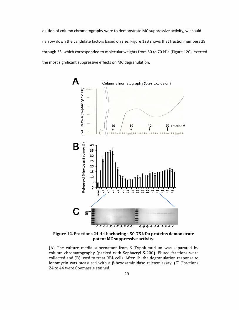
elution of column chromatography were to demonstrate MC suppressive activity, we could
narrow down the candidate factors based on size. Figure 12B shows that fraction numbers 29
through 33, which corresponded to molecular weights from 50 to 70 kDa (Figure 12C), exerted
the most significant suppressive effects on MC degranulation.
Figure 12. Fractions 24-44 harboring ~50-75 kDa proteins demonstrate potent MC suppressive activity.
(A) The culture media supernatant from S. Typhiumurium was separated by column chromatography (packed with Sephacryl S-200). Eluted fractions were collected and (B) used to treat RBL cells. After 1h, the degranulation response to ionomycin was measured with a β-hexosaminidase release assay. (C) Fractions 24 to 44 were Coomassie stained.

30
A previous study done by the Galán group showed that S. Typhimurium secretes SPI1
effector proteins into culture media, and five effector proteins have been identified [1] (Figure
13A). Among them, two proteins are identified as ~50-70 kDa in size. When we connected these
two results, the possible candidates for MC suppressive activity were Salmonella invasion
protein B (SipB) and Salmonella protein tyrosine phosphatase (SptP). SipB is one of the
components of the translocon complex which is assembled on the host membrane and is
required for proper secretion from T3SS [88,89]. SptP retains protein tyrosine phosphatase
activity to mediate host-cell recovery after bacterial invasion [90,91].
As we noticed in Figure 3, MC degranulation requires signaling cascades mediated by a
tyrosine phosphorylation at each signaling molecules [16]. To inhibit overwhelming activation
signals, MCs express endogenous tyrosine phosphatases near the plasma membrane to stop the
tyrosine phosphorylation signaling pathway [92]. Intuitively, we hypothesized that protein
tyrosine phosphatase activity from SptP is a potential candidate for MC suppression. Therefore,
we tested whether Salmonella culture supernatant exhibits tyrosine phosphatase activity, which
can interfere with tyrosine phosphorylation signaling in MCs. To inhibit tyrosine phosphatase
activity in Salmonella culture supernatant, sodium orthovanadate was pre-incubated with MCs
before treatment with ionomycin. The MC suppressive activity of Salmonella culture
supernatant was reduced significantly by pre-incubation with the tyrosine phosphatase inhibitor
(Figure 13B). Therefore, this data is consistent with the MC suppressor being SptP (Figure 13C), a
known SPI1 effector with distinct phosphatase and GTPase activities [90].

31
Figure 13. Tyrosine phosphatase activity can inhibit MC degranulation.
(A) Culture supernatant effector proteins of WT S. Typhimurium were separated by SDS-PAGE and stained with Coomassie blue. This data is adopted and modified from Hardt WD et al.[1]. (B) Culture supernatant from WT Salmonella was pretreated with sodium orthovanadate (100 μM or 10 μM) for 15 min. MC degranulation in response to ionomycin was measured by performing a β-hexosaminidase release assay after 2 h incubation with pretreated culture supernatant. (C) Diagram of SptP with two functional domains: GTPase activating protein (GAP) and protein tyrosine phosphatase. Mean ± SEM, *p<0.001, **p<0.05.

32
2.2.8 Comparison of MC Tyrosine Phosphatase with SptP of Salmonella.
We have noted that SptP shares significant homology with phosphatases naturally found
in MCs [90,92,93]. To compare SptP in Salmonella to protein tyrosine phosphatase 6 (PTP6) and
PTP9 in human cells including MCs, a sequence alignment and structural comparison were
performed. Figure 14 shows that catalytic active site and functional motifs in SptP are
structurally homologus to PTPs in human. Therefore, SptP can potentially target signaling
molecules in MCs and interfere with tyrosine phosphorylation signaling in MCs.
Figure 14. Sequence alignments of tyrosine phosphatase in S. Typhimurium and MCs.
(Top) Protein tyrosine phosphatase of S. Typhimurium is sequentially conserved with PTP in MCs. PTPN6 and PTPN9 in MCs share the following functional and characteristic protein tyrosine phosphatase motifs with SptP in S. Typhimurium. Cysteine residue (red) located in the active site is highly conserved in the three proteins. This cysteine residue is a phosphotyrosine binding site in the phosphatase. The PTP signature motif (yellow), which has the “HCXAGXGR(S/T)” amino acid sequence in the catalytic domain, is also highly conserved in the three proteins. The WPD loop (blue) which phosphatases utilize to dephosphorylate the target is also conserved in the three proteins.

33
2.2.9 SptP-Mediated Suppression of IgE+αIgE-induced MC degranulation.
To investigate the role of SptP in suppressing MC activity, we first compared the ability
of WT S. Typhimurium and an isogenic ΔsptP mutant to prevent degranulation. Here, we used
IgE+antigen as the MC stimulant because this well-characterized signaling pathway has several
potential targets for phosphatase activity. We found that in the case of MCs pretreated with the
ΔsptP mutant, the degranulation response to IgE stimulation was no longer inhibited and was
even higher than that seen with IgE stimulation (Figure 15). One possibility for the enhanced
degranulation response of ΔsptP treated MCs is that the GTPase-activating protein (GAP)
domain of SptP also has a known ability to promote recovery of the cellular cytoskeleton after
Salmonella-mediated invasion [91]. In the absence of a reorganized cytoskeleton, as is the case
with the ΔsptP mutant, MCs degranulate excessively following stimulation [94].
Next, we undertook a series of complementation studies to identify the relevant region
in the functionally distinct domains located at either end of SptP. To distinguish between the
effects of the GTPase-activating function of the amino-terminal region and the protein tyrosine
phosphatase function associated with the carboxy-terminal region [90,91], we complemented
ΔsptP S. Typhimurium with the plasmid psptPWT encoding WT sptP, psptPC481S encoding sptP with
an inactive phosphatase domain, or psptPR209A encoding sptP with an inactive GTPase domain
and compared the ability of the complemented strains to suppress MC degranulation (Figure 15).
Complementation with psptPWT or with psptPR209A but not with psptPC481S inhibited MC
degranulation in comparison to the ΔsptP mutant. These results suggest that SptP, and
specifically the catalytic activity of its tyrosine phosphatase domain, mediates suppression of
MC degranulation.

34
Figure 15. The protein tyrosine phosphatase domain in SptP inhibits MC degranulation.
Degranulation of IgE sensitized MCs in response to antigen (Ag, TNP-OVA) after 45 min pretreatment with WT, ΔsptP or complemented ΔsptP strains (ΔsptP(psptPWT), ΔsptP(psptPC481S), or ΔsptP(psptPR209A)). Mean ± SEM, *p<0.001.

35
To investigate whether SptP has the inherent ability to suppress MC degranulation, we
cloned and expressed SptP intracellularly in the RBL-2H3 MC cell by stably transfecting with
SptP-eGFP, resulting in SptP expression in approximately 60 % of cells based on fluorescent
activity (Figure 16A). We next examined the effects of SptP expression on the MC degranulation
response to IgE+anti-IgE in RBL-2H3 cells. We observed that RBL-2H3 MC cells transfected with
SptP-eGFP exhibited a significant (~60 %) reduction in degranulation compared to the control
(Figure 16B).
Figure 16. SptP-eGFP stable expression in MCs suppresses MC degranulation.
(A) Expression of transduced eGFP or SptP-eGFP was assessed by green fluorescence in transfected cells (top). Corresponding bright field images are shown at the bottom. (B) IgE sensitized MCs expressing retrovirally transduced eGFP or SptP-eGFP were cross-linked by anti-IgE and analyzed with a β-hexosaminidase release assay.

36
Next we investigated whether applying exogenous recombinant SptP into the culture
supernatant of MCs would block their subsequent degranulation. RBL-2H3 cells were treated
with increasing concentrations of recombinant SptP purified from E.coli BL21 (Figure 17A) and
then examined for their degranulation in response to different amounts of ionomycin (Figure
17B) or to IgE+anti IgE (Figure 17C). In both cases, a modest level of inhibition of the
degranulation response was observed. To confirm that tyrosine phosphatase activity from SptP
mediates suppression of MC degranulation, we pretreated recombinant SptP with
orthovanadate and examined its ability to block MC degranulation responses to IgE+anti IgE
(Figure 17D). However, we noticed that suppression with recombinant SptP was much less than
with S. Typhimurium by itself. We reasoned that the modest ability of recombinant SptP to block
MC degranulation may be attributable to its relative inability to permeate MC membranes as
this effector protein is typically introduced by S. Typhimurium directly into the MC cytosol via its
T3SS [68].

37
Figure 17. Introduction of recombinant SptP into MC culture media suppresses MC degranulation.
(A) SptP-Hisx6 was overexpressed in BL21 E. coli and purified with a Ni column. (B and C) Purified recombinant SptP-Hisx6 and (D) SptP-Hisx6 with sodium orthovanadate (1mM) were incubated with RBL-2H3 cells. After overnight incubation, the degranulation response to (B) ionomycin (1µg/ml) or (C and D) anti-IgE antibody was measured with β-hexosaminidase release assays. Mean ± SEM., *P<0.001, **p<0.05.
38
2.2.10 Transduction of SptP-TAT into MCs Significantly Suppresses MC Degranulation.
To facilitate trafficking of SptP into the MC cytosol via non-endocytic pathways after
exogenous treatment with SptP, we constructed membrane-permeant SptP by conjugating it to
the short peptide RKKRRQRRR derived from the HIV TAT (transactivator of transcription) protein,
which can translocate itself into the cytosol of various host cells [95]. To observe SptP-TAT
penetrance into bone marrow-derived mast cells (BMMCs), we employed confocal microscopy
to visualize the protein within BMMCs. We opted to employ BMMCs here to make the point that
observations made using the RBL-2H3 cell line are also applicable to primary MCs. We observed
that SptP-TAT readily entered BMMCs and could even be detected in the nuclei (Figure 18A,
bottom). Due to its superior ability to penetrate MC membranes, we predicted that SptP-TAT
would effectively block the MC degranulation response. We observed that in contrast to
controls, SptP-TAT significantly inhibited IgE+anti-IgE-mediated degranulation in a dose-
dependent manner (Figure 18B). We also generated a catalytically inactive form of SptP-TAT,
SptPC481S-TAT, to specifically ablate tyrosine phosphatase activity, that was observed in the
cytosol and nuclei (Figure 18A). Unlike SptP-TAT, recombinant SptPC481S-TAT was unable to
inhibit IgE+anti-IgE-mediated BMMC degranulation (Figure 18C), confirming that SptP inhibits
degranulation via its tyrosine phosphatase activity.

39
Figure 18. Treatment with SptP-TAT suppresses IgE-anti-IgE mediated MC degranulation.
(A) Confocal microscopy of BMMCs exposed to SptP-TAT or SptPC481S-TAT for 4 h. MC granules were probed with avidin (green) and intracellular SptP-TAT was probed with anti-His6 (red). (B and C) Degranulation of IgE-sensitized BMMCs in response to anti-IgE (B) after 4 h pretreatment with increasing amounts of SptP-TAT (10, 1, and 0.1 µg) or (C) after 2 h pretreatment with SptP-TAT (5 µg) or SptPC481S-TAT (5 µg). Scale bar
is 20 µm. Mean ± SEM, *p<0.001, **p<0.01, ***p<0.05

40
2.2.11 SptP Impedes Tyrosine Phosphorylation Signaling in MCs
Next, we sought to localize sites of tyrosine phosphorylation in MCs during
degranulation and determine their activation following exposure to SptP-TAT. Confocal
microscopy of BMMCs employing probes for MC granules and phosphotyrosine revealed that
tyrosine phosphorylation appeared to be localized primarily to empty compartments where
granules were housed prior to activation with IgE+anti-IgE (Figure 19, middle, arrowheads). This
is consistent with the notion that not only is tyrosine kinase activity important in the signaling
events leading to degranulation, but it is also important in the final stages of granule release
[24]. We thus hypothesized that intracellular SptP could interfere with the regulation of the
granule secretory pathway to block MC degranulation. In SptP-TAT-treated BMMCs (Figure 19,
right), which did not degranulate following exposure to IgE+anti-IgE, no tyrosine
phosphorylation was observed in the granule chambers, which is consistent with a lack of
degranulation. Notably, detectable amounts of SptP were localized to the granule
compartments. Thus, the absence of tyrosine phosphorylation proximal to sites of granule
discharge contributes to abrogated MC degranulation in SptP-TAT-treated MCs.

41
Figure 19. SptP-TAT inhibited tyrosine phosphorylation in MC granule chambers to block MC degranulation.
Morphological appearance of IgE sensitized BMMCs in response to anti-IgE after 2 h pretreatment with vehicle or SptP-TAT (10 µg). MC granules: avidin (red), SptP-TAT: anti-His6 (green), and intracellular tyrosine phosphorylation sites: anti-phosphotyrosine (p-Tyr) (blue) antibodies. Scale bars: 20 µm.
42
2.2.12 SptP Dephosphorylates SyK, an Early Signaling Substrate Following Receptor-mediated Activation of MCs.
The SptP-TAT-associated absence of tyrosine phosphorylation in MCs led us to seek
putative SptP targets in the IgE signaling pathway. Syk, a tyrosine kinase, is an important
substrate in IgE-mediated MC degranulation [16,96], and thus we examined the effects of SptP
on Syk activation following IgE mediated activation. SptP overexpression in transfected MCs
markedly suppressed Syk phosphorylation following stimulation with IgE+anti-IgE (Figure 20,
top). To further confirm the role of Syk, we also compared IgE-mediated Syk phosphorylation in
MCs pre-treated with WT or ΔsptP S. Typhimurium. Consistent with SptP overexpression, WT S.
Typhimurium markedly suppressed Syk phosphorylation, whereas ΔsptP S. Typhimurium failed
to do so. Indeed, ΔsptP S. Typhimurium appeared to enhance Syk phosphorylation, indicating
that in the absence of SptP, Salmonella might directly activate MCs and that the signaling events
involve Syk (Figure 20, bottom).

43
Figure 20. Syk, one of tyrosine phosphorylation cascade, is the target of SptP.
Western blots of cell preparations from IgE-sensitized RBLs that either stably expressed SptP-eGFP or control eGFP (top) or were pretreated with WT STm or ΔsptP STm for 1 h (bottom) before activation with anti-IgE. The cell preparations from both experiments were obtained at the indicated time points after activation with anti-IgE. Western blots were probed with α-p-Syk, α-Syk, and α-β-actin antibodies.

44
2.2.13 SptP Dephosphorylates NSF, a Signaling Substrate Implicated in Final Stages of Granule Release in MCs.
We also noticed that although granules of RBL-2H3 cells were not released following 1 h
exposure WT S. Typhimurium, they appeared to have fused with each other, resulting in
abnormally large intracellular granules (Figure 21A, middle). However, this phenotype was not
observed in MCs exposed to ΔsptP S. Typhimurium (Figure 21A, right). A critical determinant of
MC degranulation is NSF, a vesicle fusion protein localized to the cytoplasmic side of the granule
membrane [24,25]. In the steady state, NSF is typically phosphorylated which is important to
prevent MC granules from spontaneous intracellular fusion [97]. Upon MC activation, NSF
became dephosphorylated, promoting the fusion of granule membranes to plasma membranes
and resulting in extracellular release of MC granules. We hypothesized that in WT S.
Typhimurium-infected MCs, NSF is prematurely dephosphorylated, resulting in granule fusion
without extracellular release. To validate this hypothesis, control-treated, WT, and ΔsptP S.
Typhimurium-infected MCs were stained for NSF. In all cases, NSF colocalized to the outer
surface of MC granules (Figure 21B). Furthermore, the granules of WT S. Typhimurium-infected
MCs appeared to be 2-3 times the size of regular granules of control MCs and ΔsptP S.
Typhimurium-infected MCs, suggesting intergranular fusion in the former (Figure 21B). To
determine if intergranular fusion was associated with the dephosphorylation of NSF, we
examined phosphorylation of NSF around MC granules and found that whereas NSF appeared
phosphorylated around MC granules of PBS or ΔsptP S. Typhimurium-treated MCs, this was not
the case in WT S. Typhimurium treated MCs (Figure 21B, middle). Furthermore, when we
immunoprecipitated NSF from the three groups of MCs and probed with anti-phosphotyrosine
antibodies on a western blot, we observed that in contrast to the other two conditions, NSF in
WT S. Typhimurium-infected MCs was markedly dephosphorylated (Figure 21C) providing

45
support for our hypothesis of premature intergranular fusion induced by S. Typhimurium
phosphatases. That SptP acts at sites downstream of Syk can also be inferred by the fact that WT
S. Typhimurium, but not ΔsptP and ΔsptP(psptPC481S) blocked ionomycin induced degranulation
(Figure 21D), which occurs downstream of receptor-mediated MC signaling events [16].

46

47
Figure 21. Intergranular fusion induced by WT Salmonella through dephosphorylation of NSF.
(A, B) Morphology of RBLs after exposure to WT or ΔsptP STm. After 1 h treatment, (A) cells were stained with antibodies for fluorescence microscopy. MC granules: anti-serotonin (green), NSF: anti-NSF (red) antibodies and actin: phalloidin (blue). (B, C) After 1 h treatment, cells were treated with vanadate (100 µM) for 5 min to preserve tyrosine phosphorylation and (B) were stained with antibodies for fluorescence microscopy. NSF: anti-NSF (red) and intracellular tyrosine phosphorylation: anti-pTyr (green) and actin: phalloidin (blue). (C) Lysates from RBLs were analyzed by immunoprecipitation and immunoblotting for phosphotyrosine and NSF. Scale bar is 20 µm. (D) Degranulation responses of MCs to ionomycin after 30 min pretreatment with wild-type, ΔsptP, or complemented ΔsptP strains (ΔsptP(psptPWT), or ΔsptP(psptPC481S). Mean ± SEM, *p<0.001

48
Consistent with the β-hexosaminidase assay results (Figure 21D), the fluorescent
microscopy images showed that WT and ΔsptP(psptPWT) S. Typhimurium retained significant
amount of granules in the cytoplasm and were larger in size than those in control MCs when
infected MCs were treated with ionomycin (Figure 22, top right and bottom middle). On the
other hand, ΔsptP and ΔsptP(psptPC481S) failed to inhibit degranulation such that very few
granules were detected in cytoplasm (Figure 22, top middle, bottom left, and bottom right),
which is similar to the ionomycin alone-treated control.
Figure 22. Morphology of RBLs after exposure to WT or ΔsptP mutants with ionomycin.
Morphology of RBLs after exposure to WT or ΔsptP or ΔsptP(psptPWT) or ΔsptP(psptPC481S) S. Typhimurium. Salmonella: anti-Salmonella (red) antibody and granule: avidin (green) and actin: phalloidin (blue). Scale bar: 10 µm.
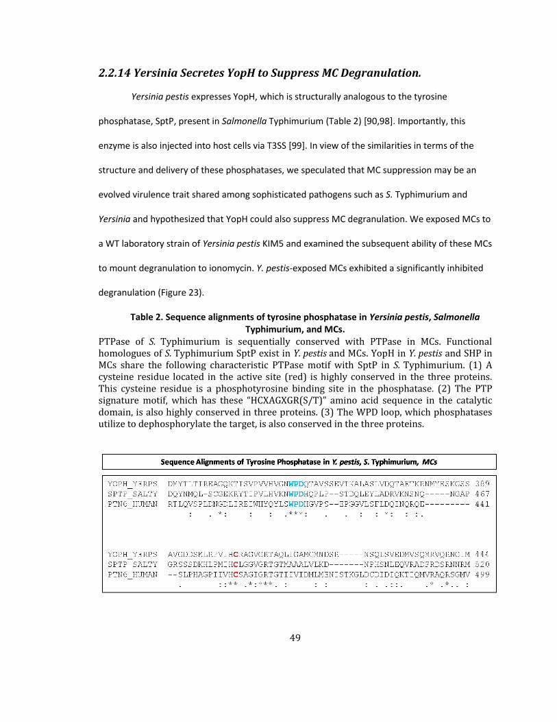
49
2.2.14 Yersinia Secretes YopH to Suppress MC Degranulation.
Yersinia pestis expresses YopH, which is structurally analogous to the tyrosine
phosphatase, SptP, present in Salmonella Typhimurium (Table 2) [90,98]. Importantly, this
enzyme is also injected into host cells via T3SS [99]. In view of the similarities in terms of the
structure and delivery of these phosphatases, we speculated that MC suppression may be an
evolved virulence trait shared among sophisticated pathogens such as S. Typhimurium and
Yersinia and hypothesized that YopH could also suppress MC degranulation. We exposed MCs to
a WT laboratory strain of Yersinia pestis KIM5 and examined the subsequent ability of these MCs
to mount degranulation to ionomycin. Y. pestis-exposed MCs exhibited a significantly inhibited
degranulation (Figure 23).
Table 2. Sequence alignments of tyrosine phosphatase in Yersinia pestis, Salmonella Typhimurium, and MCs.
PTPase of S. Typhimurium is sequentially conserved with PTPase in MCs. Functional homologues of S. Typhimurium SptP exist in Y. pestis and MCs. YopH in Y. pestis and SHP in MCs share the following characteristic PTPase motif with SptP in S. Typhimurium. (1) A cysteine residue located in the active site (red) is highly conserved in the three proteins. This cysteine residue is a phosphotyrosine binding site in the phosphatase. (2) The PTP signature motif, which has these “HCXAGXGR(S/T)” amino acid sequence in the catalytic domain, is also highly conserved in three proteins. (3) The WPD loop, which phosphatases utilize to dephosphorylate the target, is also conserved in the three proteins.

50
To determine whether YopH was the specific Yersinia factor involved in MC suppression,
we cloned and expressed this tyrosine phosphatase in RBL-2H3 cells. In contrast to control
eGFP-transfected cells, MCs transfected with YopH-eGFP exhibited limited degranulation in
response to IgE+antigen (Figure 24A). Figure 24B demonstrates the intracellular expression of
YopH, the granulation status of treated MCs, and the tyrosine phophorylation of eGFP- and
YopH-eGFP-transfected RBL-2H3 cells following exposure to IgE+antigen. In particular, MC
degranulation appeared to be directly correlated with tyrosine phosphorylation in empty
Figure 23. Yersinia pestis suppresses MC degranulation.
Degranulation of RBLs pretreated for 30 min with PBS or Y. pestis KIM5 in response to
ionomycin. Mean ± SEM, *p<0.001.

51
granule chambers (Figure 24B, middle, arrow heads) of IgE+antigen activated control MCs but
not in YopH-expressing MCs (Figure 24B).
Next, we cloned and expressed YopH as a fusion protein with TAT. To determine
whether exogenous YopH-TAT would inhibit MC degranulation, we exposed IgE-sensitized MCs
to increasing concentrations of YopH-TAT and examined their responses to antigen-induced
activation. We observed a significant and dose-dependent suppression of the MC degranulation
response to antigen (Figure 24C). Taken together, these observations indicate that YopH shares
the ability of SptP to suppress degranulation and suggest that MC suppression may be an
important virulence trait among host-adapted pathogens.
In this data, we observed that YopH targeted MCs to suppress innate immunity.
Previous studies show that YopH can suppress adaptive immunity. The tyrosine phosphatase
activity of YopH has been known to suppress the activity of lymphocytes [100]. Specifically,
YopH dephosphorylates adaptor proteins to interfere with the T cell receptor signaling pathway
and prevent the development of protective immunity against Yersinia infection [101,102].
Altogether, Yersinia utilizes a potent tyrosine phosphatase to inactivate diverse immune cells
and down-regulate innate and adaptive immunity.

52
Figure 24. Yersinia pestis YopH suppresses MC activation.
(A and B) RBLs were retrovirally transduced with control eGFP or YopH-eGFP before IgE+antigen stimulation (Ag, TNP-OVA) and were assayed for degranulation (A) or subjected to confocal microscopy (B). Granules were stained with serotonin (red) and tyrosine phosphorylation was stained with an anti-p-Tyr antibody (blue). (C) Degranulation in response to IgE+Ag (TNP-OVA) RBLs following exposure to
increasing concentrations of purified YopH-TAT. Scale bar: 10 µm, mean ± SEM,
*p<0.001, **p<0.01.

53
2.2.15 ΔsptP S. Typhimurium Induces MC Degranulation
To investigate the in vivo contribution of SptP to MC suppression during S. Typhimurium
infection, we challenged the peritoneal cavities of mice with WT or ΔsptP S. Typhimurium
(Figure 25A) and determined that MCs isolated from the peritoneal cavities exhibited widely
different phenotypes in terms of their granulation status. MCs exposed to WT S. Typhimurium
remained fully granulated (Figure 25A, third column), but MCs exposed to ΔsptP S. Typhimurium
underwent extensive degranulation (Figure 25A, fourth column). This degranulation in response
to the ΔsptP mutant when quantitated was comparable to the degranulation observed with E.
coli J96 (Figure 25B), which was included for comparative purposes. The vacant granule
chambers in extensively degranulated MCs stained strongly for tyrosine phosphorylation (Figure
25A, fourth column, arrowhead), whereas granule chambers in MCs exposed to WT S.
Typhimurium or PBS failed to exhibit tyrosine phosphorylation staining. We observed that the
overall phosphotyrosine fluorescence in ΔsptP-infected MCs was markedly higher (Figure 25A,
fourth column) than E. coli-infected MCs. This enhanced tyrosine phosphorylation was
previously observed in ΔsptP-exposed MCs in vitro (data not shown), which also consistently
exhibited higher Syk phosphorylation than IgE+anti-IgE positive control cells (Figure 20, bottom).
This is potentially due to the fact that significantly higher degranulation was observed with
ΔsptP S. Typhimurium than with the positive degranulation control (Figure 15).

54
Figure 25. ΔsptP Salmonella initiated phosphotyrosine signaling and degranulation of MCs.
(A) 1×10⁸ CFU WT STm, ΔsptP STm, or E. coli J96 were injected i.p. MCs were harvested 4 h later by peritoneal lavages, cytospun, and stained with toluidine blue for bright field microscopy (top) or with α-serotonin (red) for granules, and α-phosphotyrosine (blue) for immunofluorescence microscopy. (B) Granulated MC numbers in peritoneal lavages of (A) were quantified by counting partially and fully granulated MCs/field (n=3-6;
5 random chosen fields). Wholly degranulated MCs could not be detected. n=4 mice, mean ±
SEM, *p<0.05, **p<0.01. Scale bar: 20 µm.
55
2.2.16 ΔsptP S. Typhimurium Triggers Neutrophil Influx and Bacterial Clearance.
As MC degranulation has been closely associated with neutrophil recruitment and
subsequent bacterial clearance, we compared neutrophil responses in the peritoneum of mice
following 4 h challenge with WT or ΔsptP S. Typhimurium. Predictably, we observed significantly
higher neutrophil responses with ΔsptP than with WT S. Typhimurium (Figure 26A), and this
correlated with greater clearance of ΔsptP compared to WT S. Typhimurium 24 h after bacterial
challenge (Figure 26B), which was recovered with the complemented mutant strain
(ΔsptP(psptPWT)) (Figure 26C, D).

56
Figure 26. Enhanced neutrophil recruitment and bacterial clearance with ΔsptP Salmonella infection, and decreased effects with ΔsptP(psptPWT)
Salmonella.
(A) Assays for neutrophil recruitment (MPO) and (B) bacterial clearance (CFUs) were performed on lavages collected 4 h and 24 h, respectively, after i.p injection of 1×10⁶ CFU WT or ΔsptP STm; n=4 mice. (C) Assays for neutrophil recruitment (MPO) and (D) bacterial clearance (CFUs) were performed on peritoneal fluids collected 4 h and 24 h, respectively, after i.p. injection of 1×10⁶ CFU ΔsptP or
ΔsptP(psptPWT) complemented mutant strain STm. n=4-5 mice, Mean ± SEM,
*p<0.05.
57
2.2.17 ΔsptP S. Typhimurium Fails to Suppress MC Degranulation Induced by C48/80, Resulting in Neutrophil Influx and Bacterial Clearance.
Thus far, we observed that WT S. Typhimurium suppresses MC degranulation and
following neutrophil recruitment and bacterial clearance was affected, but ΔsptP S.
Typhimurium fails to do so. Next, we investigated whether S. Typhimurium-mediated MC
suppression continues even when a potent MC activator is administered. To test this hypothesis,
mice with WT S. Typhimurium or ΔsptP S. Typhimurium i.p. infection (1h) were challenged with
C48/80 i.p. (30min). As Figure 27 shows, both C48/80 alone and ΔsptP S. Typhimurium + C48/80
induced heavy degranulation from peritoneal MCs (second and fourth rows, respectively). In
contrast, peritoneal MCs from mice with WT S. Typhimurium infection failed to undergo
degranulation even when they were exposed to C48/80 (third row). This suggests SptP mediated
dephosphorylation is suppressing MC activation.

58
Figure 27. ΔsptP S. Typhimurium infected peritoneal MCs evoke a degranulation response to C48/80.
Mice were I.P. infected with WT or ΔsptP S. Typhimurium. After 1h, the mice were I.P. infected with C48/80 (20 µg/mouse). After 30min, isolated peritoneal MCs were stained with anti-serotonin (red) and anti-pTyr antibodies (blue). Scale bars are 10 µm.

59
As mentioned earlier, MC degranulation leads to neutrophil recruitment and bacterial
clearance. Therefore, we investigated whether massive suppression of MCs leads to inhibition of
neutrophil recruitment and bacterial clearance. We compared neutrophil responses in the
peritoneum of mice following 1 h infection with WT or ΔsptP S. Typhimurium and 4 h challenge
with C48/80. Similar to the results from Figure 26, significantly higher neutrophil responses
were observed with ΔsptP than with WT S. Typhimurium (Figure 28A), and this correlated with
greater clearance of ΔsptP compared to WT S. Typhimurium 6 h after bacterial challenge (Figure
28B). Clearance of ΔsptP S. Typhimurium was similar to that of E. coli that was used as a positive
control for neutrophil recruitment and bacterial clearance.
Figure 28. ΔsptP S. Typhimurium-infected peritoneal MCs evoke enhanced neutrophil recruitment and bacterial clearance following exposure to C48/80.
Mice were i.p. infected with WT or ΔsptP S. Typhimurium. After 1h, the mice were i.p. infected with C48/80 (20 µg/mouse). Isolated peritoneal fluid was assayed for
measuring MPO after 4h or bacterial counts after 6 h. Mean ± SEM, *p<0.01,
**p<0.05.
60
2.2.18 Enhanced Neutrophil Recruitment and Bacterial Clearance following Infection by ΔsptP Salmonella
Given the intrinsic ability of SptP to block MC degranulation, previous studies have not
been able to demonstrate a specific role for MCs in combating S. Typhimurium infections in WT
and MC-deficient mice. Indeed, when we initially compared neutrophil responses and bacterial
clearance in WT and MC-deficient mice following intraperitoneal challenge with WT S.
Typhimurium, we failed to notice any appreciable difference between the two strains of mice
(Figure 5A and Figure 6). However, based on our subsequent findings, we sought to
demonstrate a potential MC response to S. Typhimurium by employing our ΔsptP mutant strain.
We compared neutrophil responses and bacterial clearance of ΔsptP S. Typhimurium in WT and
MC-deficient mice following peritoneal challenge. Neutrophil responses to ΔsptP S.
Typhimurium were significantly more elevated in WT mice compared to MC-deficient mice
(Figure 29, left), and this was accompanied by better clearance of ΔsptP S. Typhimurium in WT
mice 24 h after bacterial challenge (Figure 29, right).

61
Figure 29. Enhanced neutrophil recruitment and bacterial clearance following infection by ΔsptP S. Typhimurium.
Neutrophil influx (left) and bacterial counts (right) in WT and MC-deficient mice,
following i.p. injection of 5×10⁵ CFU ΔsptP S. Typhimurium. Mean ± SEM, *p<0.05.

62
2.2.19 S. Typhimurium utilizes SptP to inhibit MC-initiated neutrophil influx and bacterial clearance
To demonstrate the potency of the MC suppressing activity of SptP-TAT, we co-
administered recombinant SptP-TAT i.p. with E. coli J96, which normally activates MCs, and
examined neutrophil recruitment and bacterial clearance. Compared to E. coli J96 alone, E. coli
J96 co-instilled with SptP-TAT evoked lower neutrophil responses and bacterial clearance from
the peritoneum (Figure 30A). To verify that this weak neutrophil response was linked to MCs, we
examined isolated peritoneal MCs and found that those from mice challenged with E. coli J96
with SptP-TAT were non-degranulated (Figure 30B, right column) in contrast to mice challenged
with E. coli J96 alone (Figure 30B, middle column, quantitated in Figure 30C). Furthermore,
these non-degranulated MCs exhibited a limited degree of tyrosine phosphorylation and
appeared to have accrued detectable amounts of SptP-TAT in their granule compartments
(Figure 30B, right column).

63
64
Figure 30. Administration of SptP-TAT at sites of E. coli infection inhibits MC-mediated neutrophil recruitment resulting in impaired bacterial clearance.
(A) Neutrophil influx (top) and bacterial counts (bottom) in WT mice were measured, after SptP-TAT (100 µg/mouse) or vehicle followed 30 min later with 1×10⁷ CFU E. coli. MPO assays were performed 5 h post-infection and CFUs 24 h post-infection. (B) Morphologies of MCs in the peritoneal cavities of control and SptP-TAT treated mice 3 h following E. coli infection. MC granules: avidin (red), SptP-TAT: anti-His6 (green) and sites of tyrosine phosphorylation: anti-p-Tyr antibodies (blue). Scale bars: 20 µm. (C) Granulated MC numbers in peritoneal lavages of (B) were quantified by counting the number of partially and fully granulated MCs/field (n=5; 5 random chosen fields). Fully degranulated MCs could not be detected.
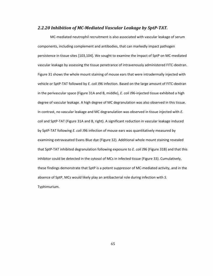
65
2.2.20 Inhibition of MC-Mediated Vascular Leakage by SptP-TAT.
MC-mediated neutrophil recruitment is also associated with vascular leakage of serum
components, including complement and antibodies, that can markedly impact pathogen
persistence in tissue sites [103,104]. We sought to examine the impact of SptP on MC-mediated
vascular leakage by assessing the tissue penetrance of intravenously administered FITC-dextran.
Figure 31 shows the whole mount staining of mouse ears that were intradermally injected with
vehicle or SptP-TAT followed by E. coli J96 infection. Based on the large amount of FITC-dextran
in the perivascular space (Figure 31A and B, middle), E. coli J96-injected tissue exhibited a high
degree of vascular leakage. A high degree of MC degranulation was also observed in this tissue.
In contrast, no vascular leakage and MC degranulation was observed in tissue injected with E.
coli and SptP-TAT (Figure 31A and B, right). A significant reduction in vascular leakage induced
by SptP-TAT following E. coli J96 infection of mouse ears was quantitatively measured by
examining extravasated Evans Blue dye (Figure 32). Additional whole mount staining revealed
that SptP-TAT inhibited degranulation following exposure to E. coli J96 (Figure 31B) and that this
inhibitor could be detected in the cytosol of MCs in infected tissue (Figure 33). Cumulatively,
these findings demonstrate that SptP is a potent suppressor of MC-mediated activity, and in the
absence of SptP, MCs would likely play an antibacterial role during infection with S.
Typhimurium.

66
F
Figure 31. Vascular leakage resulting from MC degranulation following E. coli infection is suppressed by the administration of SptP-TAT.
(A and B) Mouse ears were injected intradermally with vehicle or SptP-TAT (20 µg) and injected 1 h later with 1×10⁶ CFU E. coli J96 at the same site and FITC-dextran (green) i.v. At 1 h post-infection, the ears were dissected, (A) stained with avidin (red) and anti-CD31 (blue), (B) stained with avidin (red) and anti-E. coli (blue), and whole-mounted for immunofluorescence microscopy. Scale bars: 50 μm.

67
Figure 32. SptP-TAT-suppressed MC degranulation and accompanying vascular leakage during E. coli infection
(A) Vehicle or SptP-TAT or SptP-His6 or SptPC481S-TAT was intradermally injected into mouse ears. At 1 h post-infection, 1×10⁶ CFU E. coli were injected at the same site, and Evans Blue dye (0.5%) was simultaneously injected intravenously. At 1 h post-infection, (A) the extravasation of Evans Blue was visualized in infected ears. (B) Vascular leakage was quantified by measuring extracted dye from dissected ears at 620 nm absorbance. Y axis values are relative to negative control. Mean ± SD. *p<0.01

68
Figure 33. SptP-TAT inhibited tyrosine phosphorylation signaling and degranulation in MCs.
The same procedure described in Figure 31 was performed. Avidin (red), an anti-His antibody (green), and anti-pTyr antibody (blue) were used to stain whole ear tissue. The tissue was visualized by confocal microscopy. Scale bar: 50 µm.
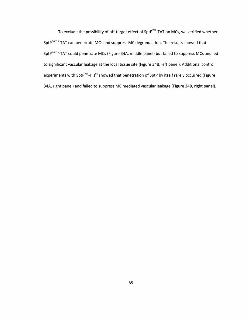
69
To exclude the possibility of off-target effect of SptPWT-TAT on MCs, we verified whether
SptPC481S-TAT can penetrate MCs and suppress MC degranulation. The results showed that
SptPC481S-TAT could penetrate MCs (Figure 34A, middle panel) but failed to suppress MCs and led
to significant vascular leakage at the local tissue site (Figure 34B, left panel). Additional control
experiments with SptPWT-Hisx6 showed that penetration of SptP by itself rarely occurred (Figure
34A, right panel) and failed to suppress MC mediated vascular leakage (Figure 34B, right panel).

70
Figure 34. SptPC481S-TAT and SptP-Hisx6 failed to suppress MCs in vivo.
(A) BMMCs exposed to SptPC481S-TAT or SptP-Hisx6 for 4 h. Confocal microscopy of MCs revealed granules probed with avidin (green) and intracellular SptPC481S-TAT probed with anti-Hisx6 (red). (B) Mouse ears were injected intradermally with SptPC481S-TAT (20 µg) or SptP-Hisx6 (20 µg) and 1 h later, 1×10⁶ CFU E. coli were injected into the same area of each ear. At the same time a marker of vascular contents, FITC-dextran (green), was intravenously administered. At 1 h post-infection, the ears were dissected and stained to visualize MC granules: avidin (red) and blood vessels: anti-CD31 (blue) antibody. Tissues were whole mounted for confocal microscopy.

71
2.2.21 MC-S. Typhimurium Interactions Likely Occur Immediately After the Gut Epithelium is Breached.
During natural infection, S. Typhimurium that successfully breach the gut epithelial
barrier typically target the mesenteric lymph nodes, where they proliferate with limited
interference from the host immune system [84]. However, before S. Typhimurium can reach the
draining lymph nodes, the bacteria must circumvent an array of MCs present in the lamina
propria of the gut epithelium. Shown in Figure 35 are sections of the cecal epithelium of mice
challenged with PBS, WT S. Typhimurium or ΔsptP S. Typhimurium. Numerous fully granulated
MCs were seen proximal to the epithelium in PBS or WT S. Typhimurium-challenged mice but
not in mice challenged with ΔsptP S. Typhimurium which are incapable of suppressing MCs.
Indeed, the apparent absence of fully granulated MCs in the cecum of ΔsptP S. Typhimurium
challenged mice was attributable to extensive MCs degranulation induced by the many mutant S.
Figure 35. MC degranulation in the cecum of mice infected with ΔsptP but not in WT Salmonella infected mice.
Wild-type mice were pre-treated with streptomycin (20 mg/mouse) for 24 h, followed by oral infection with 1x10⁸ CFU of WT or ΔsptP S. Typhimurium. After 12 h, the ceca were dissected, cryo-sectioned and immunostained for MCs: avidin (red), S. Typhimurium: α-Salmonella (blue), and epithelial cells: α-E-cadherin antibodies (green). Shown in the first panel is the cecum of PBS treated mice (control). Shown in the second and third panels are ceca of mice infected with WT or ΔsptP S. Typhimurium, respectively. Scale bar: 50 µm.

72
Typhimurium found at this site (Figure 35).
We investigated whether these MCs in the lamina propria played a role in controlling
bacterial numbers, thus suggesting a plausible role for S. Typhimurium-mediated MC
suppression during natural infection. To address this question, we assessed the number of
bacteria that reached the mesenteric lymph nodes in WT and MC-deficient mice shortly after
oral challenge with ΔsptP S. Typhimurium, which does not suppress MCs. If gut-resident MCs
contribute to bacterial clearance, we would expect fewer bacteria to reach the mesenteric
nodes in MC-sufficient mice compared to their MC-deficient counterparts. We detected
markedly greater numbers of bacteria in the immediate draining lymph nodes of MC-deficient
mice 24 h after oral infection with ΔsptP S. Typhimurium compared to WT mice (Figure 36A).
Because some recent studies have questioned the use of Kit-dependent MC-deficient mice
[25,26,85], we have also included here a recently described Kit-independent inducible model of
MC deficiency involving Cma1-cre+iDTR+ mice [25]. Using this model, we obtained very similar
findings regarding the contribution of MCs in the clearance of ΔsptP S. Typhimurium (Figure
36B). Additionally, we confirmed the contribution of the phosphatase domain of SptP by
challenging WT and MC-deficient mice with ΔsptP(psptPC481S) S. Typhimurium (Figure 36C). To
confirm that the SptP deletion was responsible for MC-dependent bacterial clearance, we also
infected WT and MC-deficient mice with ΔsptP(psptPWT) S. Typhimurium (Figure 36D).
Predictably, we observed that the bacterial burden in MC-sufficient mice was now comparable
to that of MC-deficient mice. This result is also consistent with the data presented in Figure 5A
demonstrating that the WT S. Typhimurium burden was comparable in MC-sufficient and MC-
deficient mice. Thus, MCs have the potential to regulate the colonization of the mesenteric

73
lymph nodes, which are among the first sites targeted by pathogens that successfully breach the
gut barrier.
Figure 36. Intestinal MCs reduce the bacterial burden in the mesenteric lymph nodes following oral infection with ΔsptP S. Typhimurium.
CFU of mesenteric lymph nodes in streptomycin-treated (A, C, D) WT or MC-deficient mice, (B) Cma1-cre+iDTR+ mice or WT controls after DT injections, following oral challenge with 5×10⁷ CFU (A, B) ΔsptP S. Typhimurium, (C) ΔsptP(psptPC481S) and (D) ΔsptP(psptPWT) 24 h post-infection. *p<0.01, **p<0.05, N.S., not significant.

74
2.3 Discussion
In this study, we have revealed a highly effective mechanism involving the inactivation
of MCs employed by S. Typhimurium to elude host innate immunity, which are immune
surveillance cells strategically located at host-environment interfaces. We demonstrated that
unlike E. coli J96, which provoked rapid and extensive MC degranulation, S. Typhimurium not
only failed to elicit a MC degranulation response but also prevented MC responses to a wide
array of potent secretagogues such as IgE+antigen, a calcium ionophore and complement
revealing a broad and powerful inhibitory activity (Figure 10, 15 and 27). This capacity to block
MC responses to other signals is relevant during infection because even though S. Typhimurium
do not activate MCs per se, various endogenous danger signals or alarmins are released at
infection sites and can potentially activate MCs [105]. In vivo studies in mice revealed that this
innate capacity of S. Typhimurium to block MC degranulation markedly reduced neutrophil
responses and limited bacterial clearance at the infection site. This is in sharp contrast to E. coli,
which elicited extensive MC degranulation, a strong neutrophil response, and marked bacterial
clearance from the site of infection. This capacity to elude early innate immune responses by
inactivating peripheral MCs may explain, at least in part, the well-recognized ability of S.
Typhimurium to avoid clearance by the innate immune system and to disseminate rapidly from
the site of infection to distal sites such as the draining lymph nodes and liver [84]. Our
observation that S. Typhimurium inhibited early neutrophil recruitment is not inconsistent with
earlier reports of strong neutrophil responses following S. Typhimurium infection [106,107]. In
this study, we focused on a narrow window of time during the early stages of the infection
process but observed the development of a vigorous MC-independent neutrophil response at
sites of infection with WT S. Typhimurium after the initial lag period. Our findings imply that the
75
initial immune response to a pathogen is an important determinant of infection outcome. A
delayed neutrophil response at the site of S. Typhimurium infection, regardless of its intensity,
may have limited protective value because many of the bacteria are either intracellular or have
become systemic at that point.
The ability of S. Typhimurium to block MC activity could, at least in part, explain why
others have not been able to demonstrate a protective role for MCs [86]. Much of our
conclusions were based on traditional Kit mutant MC-deficient mice; however, since this model
of MC deficiency has been recently challenged [25,26,85], we sought to confirm our findings by
employing Kit-independent Cma1-cre+iDTR+ mice in which MC deficiency was inducible. Our
findings utilizing this inducible model of MC deficiency corroborated our findings with Kit
mutant mice providing further validation of our conclusions.
S. Typhimurium-mediated suppression of MC degranulation was achieved through the
introduction of SptP into the MC cytosol. The suppressive actions of SptP are restricted to the
exocytic function of MCs because MCs readily internalize S. Typhimurium upon contact. SptP is a
SPI1 effector protein with two functional domains. The N-terminal domain functions during
pathogen entry into host cells by deactivating Rho GTPases and reversing pathogen-induced
cytoskeletal changes following S. Typhimurium uptake [91]. The second domain, a C-terminal
domain, comprises a protein tyrosine phosphatase reported to function late in the pathogen
entry process when S. Typhimurium is harbored within late endosomes [108]. In these
compartments, it is believed that SptP dephosphorylates a protein that facilitates cellular
membrane fusion and protein degradation, enhancing intracellular S. Typhimurium replication.
The tyrosine phosphatase domain of SptP is also thought to inactivate MAP kinase [109]. Our
finding that the tyrosine phosphatase region of SptP mediates MC inactivation represents a

76
distinct function for this domain, while our work with GTPase-inactive SptP as well as YopH,
which lacks a GTPase domain, suggests that GTPase activity is for the most part dispensable for
MC suppression. A comparison of the primary structure of SptP with multiple tyrosine
phosphatases found in the cytosol of MCs reveals high degree of homology [90], implying
multiple targets for this phosphatase. Morphological studies of MCs employing pTyr-specific
antibody probes revealed that degranulation was closely associated with phosphorylation
events at the cell surface as well as at intracellular sites proximal to cytosolic granules.
Phosphorylation of these proteins at both sites did not occur in the presence of SptP, suggesting
that SptP acted at multiple sites and on multiple targets. By examining S. Typhimurium-infected
MCs before or after activation with various secretagogues, we have been able to identify at least
two distinct substrates for SptP. Immunoblot analyses of S. Typhimurium-infected MCs and
control MCs following IgE mediated activation revealed one of the intracellular targets of SptP to
be Syk, which is a member of the Syk/Zap-70 family and is a substrate found early in the
signaling cascade. Microscopic and immunoprecipation studies of MCs infected with WT S.
Typhimurium also revealed that shortly after infection MC granules spontaneously coalesced
forming oversized granules that failed to be expelled upon subsequent exposure to potent
secretegogues. This phenomenon was not observed with ΔsptP S. Typhimurium, indicating that
it was an SptP-mediated activity. We have linked this intergranular fusion to SptP-mediated
dephophorylation of NSF, a fusion protein whose functions include the prevention of homotypic
intergranular fusion [24,93].
Although a number MC-inactivating compounds derived from various microbes have
been identified, including the toxic fungal metabolite gliotoxin [110,111], ES-62, a glycoprotein
secreted by filarial nematodes [112], FK506 from Streptomyces tsukubaensis [113], and

77
cyclosporine A from Tolypocladium inflatum [114], these do not, for the most part, appear to be
relevant to the pathogenesis of infection. Here, Our studies reveal that two highly successful
host-adapted pathogens, S. Typhimurium and Y. pestis, share a powerful mechanism to suppress
MC degranulation, suggesting that MC inhibition is an important virulence determinant. MCs are
among the first immune cells encountered by these pathogens after they traverse the epithelial
barrier. They are relatively abundant in dermal tissue, where Y. pestis is localized following
injection by fleas, and are also common in the gut epithelium below Peyer’s patches as well as in
the lamina propria, where they can encounter S. Typhimurium invading through M cells or gut
epithelial cells [115]. Orally-administered S. Typhimurium that successfully breach the epithelial
barrier traffic to the mesenteric lymph nodes, a process likely to be influenced by MC
interactions. Indeed, significantly higher numbers of the ΔsptP mutant were present in the
mesenteric nodes of MC-deficient mice following oral infection, implicating mucosal MCs as
primary responders to S. Typhimurium breaching the gut epithelium (Figure 37).
MCs and MC-like cells are found in a wide range of animal species including less evolved
species that are devoid of an adaptive immune system. In recent years, it has become clear that
MCs play a key role in modulating innate immune responses to various bacteria, parasites,
viruses and fungi, suggesting that their raison d'être is the modulation of immediate and
nonspecific immunity to microbial challenge. In view of this, it is not surprising that certain
virulent pathogens would have evolved special mechanisms to inactivate these cells to prevent
or delay host immune responses and promote their pathogenesis.
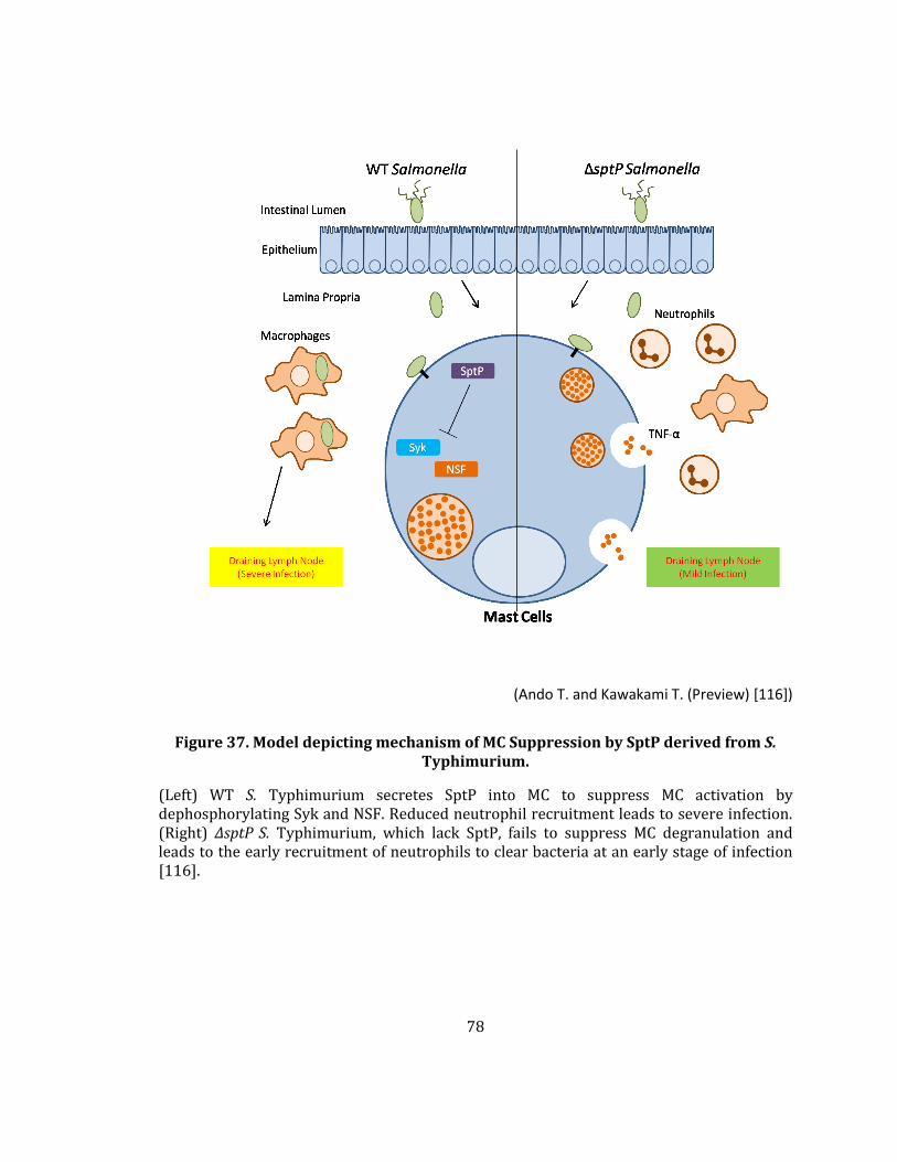
78
(Ando T. and Kawakami T. (Preview) [116])
Figure 37. Model depicting mechanism of MC Suppression by SptP derived from S. Typhimurium.
(Left) WT S. Typhimurium secretes SptP into MC to suppress MC activation by dephosphorylating Syk and NSF. Reduced neutrophil recruitment leads to severe infection. (Right) ΔsptP S. Typhimurium, which lack SptP, fails to suppress MC degranulation and leads to the early recruitment of neutrophils to clear bacteria at an early stage of infection [116].

79
3. Perspective
In this thesis I have described how pathogenic Salmonella have evolved powerful
mechanisms to inactivate MCs which are key immune surveillance cells located in the gut as well
as other mucosal sites. I have demonstrated that Salmonella achieve this feat by secreting a
tyrosine phosphatase, SptP, directly into the cytosol of MCs. The translocation of SptP into MCs
occurs via the T3SS which injects various Salmonella effector proteins via a syringe-like structure
that appears to traverse the plasma membranes of MCs and other target cells. Once introduced
into the MC cytosol, SptP was found to target multiple substrates involved in MC degranulation.
One of the substrates was Syk a key conduit of early signaling from receptors located in the MC
membrane and the other was NSF a critical mediator of the final steps of MC degranulation.
Interestingly, SptP was found to dephosphorylate NSF resulting in premature intergranular
fusion in MCs while it prevented phosphorylation of Syk following MC exposure to Salmonella.
Both of these activities completely blocked the MC degranulation response to Salmonella but
remarkably had no effect on the endocytic activities of the cell. Consequently infecting
Salmonella were able to readily infect MCs and seek intracellular refuge in the same immune
cells whose function it is to warn the host of the infection. Microscopic examination of the
cecum of mice following oral challenge with Salmonella revealed that the bacteria encountered
MCs as soon as they had circumvented intestinal epithelium in cecum (Figure 35). The
importance of SptP in modulating the outcome of the Salmonella-MC encounter could be
gauged by the finding that the translocation of sptP mutant Salmonella from the gut into the
draining mesenteric lymph nodes of MC deficient mice was markedly higher than that of WT
mice (Figure 36A).

80
While the focus of this thesis has been on MC suppression by Salmonella there are other
pathogenic bacteria capable of targeting immune survaillance cells such as dendritic cells (DCs)
and macrophages (Mφs) for suppression of functional activities. Not surprisingly, the strategies
involved by these pathogenic bacteria appear, for the most part, distinct from what I have
described here. Shown in Figure 38 are some of diverse strategies and bacterial components
employed by various pathogens to suppress immune surveillance cells [117]. In the following
section, I will describe briefly how two other well known host adapted pathogens, the Gram
negative pathogen Yesinia pestis (and related species) and the Gram positive Mycobacterium
tuberculosis suppress DCs and macrophages.
Figure 38. Pathogenic infection in human body.

81
3.1 Immune suppressive abilities of Yersinia and Mycobacteria species.
Yersinia pestis is a Gram-negative and facultative bacteria which is the causative agent
of plague [118]. The mortality associated with this pathogen can reach 20-50 % even with
antimicrobial treatment [119]. The virulence of this pathogen is associated with its multifaceted
approach to inactivating key immune survaillance cells such as MCs, DCs and macrophages. As
already indicated, Y. pestis suppresses MCs by directly injecting YopH into the cytosol of these
immune cells. However, Yesinia spp. also target Mφs and DCs for inactivation. This is achieved
by the translocation of various effector proteins into the cytosol of the target cells via Yersinia’s
T3SS. These effector proteins inactivate substrates in various signaling pathways involved in the
secretion of key proinflammatory cytokines and chemokines. Through the secretion of YopJ, an
acetyltransferase, into the cytosol of Mφs, Yersinia spp. are able to block the phosphorylation
ability of MAPKK (Mitogen-activated protein kinase kinase). Thus leads to the blockage of the
NF-kB signaling pathway, which in turn is critical for the secretion of many key proinflammatory
cytokines such as TNF-α, IL-12, 15, and 18 [120,121]. While certain effectors of Yersinia such as
YopJ block the production by macrophages of proinflammatory mediators, other bacterial
factors appear to trigger secretion of the immunosuppressive cytokine, IL-10. Y.
pseudotuberculosis and Y. enterocolitica can induce IL-10 production from macrophages through
the release of virulence (V)-antigen (LcrV). When LcrV binds to toll-like receptor 2 (TLR2) on Mφs,
IL-10 is secreted and functions as inducing distinct tolerance [122,123]. IL-10 has a potent effect
in suppressing the production of various proinflammatory cytokines from surrounding cells
which is beneficial for the spread of Yersinia and also in preventing the development of the
82
adaptive immune response [122,124]. LcrV also induces IL-10 production in DCs and activates
the JNK signaling pathway inducing these infected DCs to differentiate.
In addition to suppressing the production of proinflammatory cytokines from DCs and
Mφs, Yersinia also appears capable of blocking uptake by the same phagocytic cells. Through
simultaneous injection of YopT, YopE, and YopH, Yersinia blocks various stages of the phagocytic
process at the same time. First, the cysteine protease activity of YopT specifically cleaves Rho
GTPases to cause depolymerization of actin filaments and disruption of the actin cytoskeleton
[125-128]. Second, YopE activates the GTPase-activating protein (GAP) to down-regulate the
Rho family members such as Cdc42, RhoA and Rac1 [129] which also disrupts the cellular
cytoskeleton [130]. Third, YopH dephosphorylates the Fyn-binding protein (FYB) to deform the
peripheral focal complexes [131] needed for bacterial uptake. Thus, through the secretion of
multiple effectors Yersinia can negatively regulate the functions of key immune surveillance cells
in the microenvironment that it occupies in the host permitting bacterial colonization and
spread within the host.
Mycobacterium tuberculosis causes tuberculosis, one of the top 10 causes of human
morbidity and mortality worldwide. If tuberculosis patients are not treated, fatality rates can
reach up to 50 % [132]. Although different Mycobacterium spp. causes distinct diseases, they
tend to share many virulence mechanisms. For example, M. tuberculosis, M. leprae and M.
ulcerans all seem to suppress mediator release from immune surveillance cells which they
interact with. They typically also invade and survive intracellularly within these cells. In their
intracellular niches these bacteria can avoid the host’s immune defenses as well as antibiotic
treatment resulting in chronic infections.
83
M. tuberculosis which primarily targets Mφs but also infects DCs suppresses these cells,
at least partially with a mannose-containing glycoprotein (ManLAM) that is a mycobacterial cell
wall component. ManLAM activates the DC-SIGN signaling pathway which prevents DC
maturation [133] resulting in limited DC trafficking to draining lymph nodes and limited adaptive
immune responses. Activation of the DC-SIGN pathway results in the activation of the
serine/threonine kinase Raf-1. This signaling substrate acetylates NF-κB subunit p65 resulting in
enhanced secretion of IL-10 which is strongly anti-inflammatory [134]. M. tuberculosis persist in
the host by infecting and replicating in phagosomes of Mφs. Infected Mφs form granulomas
which serve to encapsulate bacteria within host tissue. Promoting intracellular survival are
several secreted virulence proteins such as SapM, a lipid phosphatase, which dephosphorylates
phosphatidylinositol 3-phosphate (PI3P) and recruits certain specific proteins into the
phagosome in order to avoid acidification and subsequent fusion with lysosomes [135-137]. The
phagosome of M. tuberculosis has diminished vacuolar H+ATPase and low level of lysosomal
hydrolase which is favorable for bacterial survival. In DCs, prevention of phagosome maturation
severely limits antigen presentation resulting in limited adoptive immune responses in the host
[138-140]. Cumulatively, due to their distinct and powerful immunsuppressive actions on DCs
and Mφs, mycobacteria are able to successfully establish chronic infections in the host which
are very difficult to eradicate.
3.2 Coopting MC suppressing properties of bacteria for therapy.
As indicated earlier in this thesis, MCs can be prominent players in various chronic and
harmful inflammatory disorders such as asthma, allergy, psoriasis, arthritis, inflammatory bowel
diseases, etc. When more is known regarding how different microbes suppress MCs, it is
conceivable that certain microbes or their products can be utilized to ameliorate some of these

84
chronic inflammatory diseases. For example, it is conceivable that administering cell-permeable
forms of SptP into inflammatory sites will impair mediator secretion from MCs and even other
immune cells to abrogate inflammation. Its potential usage could be comparable to the
administration of Botox, a neurotoxin derived from Clostridium botulinum into human facial skin
to reduce wrinkle formation [141,142]. The toxin reduces abnormal muscle contraction by
blocking release of acetylcholine from neurons, which normally signals muscle contraction.
Importantly the effect of Botox can last several months and therefore the frequency of its
administration is limited. With the therapeutic success of even a highly toxic agent such as
Botox, there is currently great interest in the therapeutic use of other bacterial products which
are much less toxic [143].
Some microorganisms or their products are already being used clinically for therapy,
unrelated to their MC-suppressing properties, but when their MC suppressing property is also
considered, they could have additional clinical uses. For example, the Lactobacillus and
Bifidobacterium species are frequently being applied as probiotics in patients with inflammatory
diseases in the gut. In view of the capacity of probiotic bacteria in suppressing IgE-mediated MC
responses [144-146], these bacteria or their products may potentially also be used to temper
the pathology associated with allergic inflammation. The fungal metabolite Cyclosporin A is
routinely used as a potent nonsteroidal immunosuppressive drug in patients following organ
transplant [147]. In view of its potent MC suppressing actions [147], this drug could be
repurposed for the treatment of various inflammatory disorders where MCs play a key part. The
innate capacity of various microbes to suppress MC function may therefore have significant
therapeutic promise.

85
3.3 Concluding Remarks
For many decades the study of bacterial pathogenesis has centered around virulence
factors that promote bacterial adherence and colonization of mucosal surfaces as well as factors
that promote bacterial entry and survival in various host cells. It is only recently that the
immunomodulatory role of bacterial pathogens was recognized. That successful pathogens
need to repress or avoid the host’s wide array of innate and adaptive immune responses in
order to survive is now being recognized as a key virulence trait. Through investigating the
various strategies and the underling molecular basis for bacteria-mediated immune suppression
or avoidance, it will be possible to develop appropriate counter measures or vaccines to combat
microbial pathogens in the future. These strategies will become even more important with the
overuse of antibiotics and the resulting emergence of multi-resistant bacteria in our midst.

86
4. Materials and Methods
4.1 Bacterial strains and culture.
S. Typhimurium SL1344, SL1344-derived Tn5 transposon insertion mutants in SPI1, SPI2,
and SPI1&2 were gifts from Dr. Alejandro Aballay, Duke University. SB300, and its sptP deletion
mutant, SB749, were gifts from Dr. Jorge E. Galán, Yale University. ΔsptP(psptPWT),
ΔsptP(psptPC481S), and ΔsptP(psptPR209A) were generated in this study. Other strains used include
E. coli J96 and CI5, a Gram-positive clinical isolate Staphylococcus aureus strain 54 (Duke
University Medical Center), and a WT laboratory strain of conditionally virulent and naturally
occurring Yersinia pestis KIM5. Bacteria were grown for 15 h, non-shaking, in Luria-Bertani (LB)
(Gibco) broth at 37˚C. 25 µg/ml kanamycin was added for transposon mutants, 100 µg/ml
streptomycin for SB300 and SB749, and 100 µg/ml carbenicillin for complemented strains.
4.2 Mice.
6- to 8-week-old C57BL/6 mice were purchased from the National Cancer Institute. MC-
deficient Wsh mice (KitW-sh/KitW-sh) and their congenic littermate controls were purchased from
Jackson Laboratories. To induce MC depletion, eight-week-old Cma1-cre+iDTR+ (a gift from Dr.
Axel Roers, University of Technology, Dresden) and wild-type littermates received five i.v.
injections of 100 ng DT per mouse in a week. Mice were housed in the Duke University Vivarium
and provided food and water ad libitum. All experiments were performed according to protocols
approved by the Duke University Institutional Animal Care and Use Committee.
4.3 Animal infections and CFU counts.
Mice were infected intraperitoneally with the indicated CFU of various strains of S.
Typhimurium, or E. coli J96 in 100 µl of sterile PBS. At the indicated times, mice were euthanized,

87
5 ml of sterile, ice-cold PBS was injected into the peritoneal cavity, and the abdomen was gently
massaged. The collected peritoneal fluid was analyzed for MC degranulation, neutrophil influx,
or bacterial counts. The detailed procedure of oral infection to streptomycin-pretreated mice
was described [148]. Briefly, mice were pretreated with 20 mg streptomycin prior to infection
with 5×10⁷ CFU of ΔsptP or its complemented mutant S. Typhimurium. At various times, mice
were euthanized and mesenteric lymph nodes homogenized in 0.1% Triton X-100 per PBS.
Lysates were plated on MacConkey agar with or without 50 μg/ml streptomycin and incubated
overnight at 37˚C for CFU.
4.4 Cell culture.
RBL-2H3 cells were grown in MEM (Gibco) supplemented with 15% FBS (GEMINI Bio-
Products). BMMCs were prepared by flushing femurs from C57BL/6 mice with cRPMI (10% FBS,
100 U/ml Pen Strep, 25 mM HEPES, 2 mM L-glutamine, 1 mM sodium pyruvate, 1 mM
nonessential amino acids (all from Gibco)). LAD2 cells were grown in StemPro-34 (Gibco) (2.6%
StemPro-34 nutrient supplement, 2 mM L-glutamine, 100 U/ml Pen Strep, and 100 ng/ml SCF
(R&D systems)). Flushed bone marrow were cultured for 12 weeks in cRPMI containing 5 ng/ml
IL-3 and 5 ng/ml SCF (R&D systems). All cells were culture at 37˚C in a humidified water-jacketed
incubator under 5% CO2 / 95% air atmosphere.
4.5 β-hexosaminidase assay.
RBLs or BMMCs or LADs were plated in 96-well plates on the day before the experiment.
For experiments with IgE-stimulation, cells were sensitized overnight with 1 µg/ml IgE (BD
Biosciences). RBLs were exposed to bacteria as indicated in Tyrode’s buffer for 30 min. In
secondary stimulus experiments, cells were stimulated either with ionomycin (1 μg/ml) or
C48/80 (10 μg/ml) or C5a (0.5 ng/ml) or TNP-OVA (10 ng/ml) (all from Sigma) or anti-IgE

88
antibody in Tyrode’s buffer at 37˚C. The detailed procedure of β-hexosaminidase assay was
described previously [51].
4.6 Microscopy.
Peritoneal lavage fluids were cytospun by using Cytospin3 (Shandon) on Superfrost Plus
glass slides (VWR). Mesentery was carefully dissected and dried on slides. To stain MCs, freshly
prepared Carnoy’s fixative (ethanol, chloroform, and glacial acetic acid) and 0.5% Toluidine blue
were used. Fully degranulated MCs could not be detected, because there were no granules left
in MCs to be detected. Peritoneal lavage cells or BMMCs were cytospun and fixed in 4% PFA. 0.1%
saponin (Sigma) in 1% BSA in PBS was used to permeabilize cells and dilute antibodies. For
whole mount staining of ear tissue, dissected ears were fixed in 4% PFA (2 h) and permeabilized
with 0.3% Triton X-100 in 1% BSA in PBS containing 5% goat serum. Cells or tissues were
incubated with primary antibodies: antibody to His6 (Roche), E. coli, p-Tyr, CD31 (BD
Biosciences), and serotonin (DakoCytomation). The secondary antibodies with FITC or Cy3 or
A647-conjugated anti-mouse, A647-conjugated anti-rabbit, DyLight 649-conjugated anti-rat, Cy-
3-conjugated anti-mouse antibodies (Jackson Immuno Research) and TRITC or FITC conjugated
avidin (Sigma) was incubated. Nikon ECLIPSE TE200 microscope (Nikon) was used to observe
confocal images. To prevent spectral overlapping between individual fluorescent emission
signals, a channel-series approach of EZ-C1 Nikon software was utilized.
4.7 Myeloperoxidase activity assay.
Cells derived from peritoneal lavage were resuspended in hexadecyltrimethyl
ammonium bromide (Sigma) in phosphate buffer and lysed by multiple freeze-thaw cycles.
Lysed cells were centrifuged and supernatant was mixed with o-dianisidine and hydrogen
89
peroxide in phosphate buffer. Activity was measured during 1 min interval three times at 450
nm.
4.8 Construction of SptP plasmids and transfected cell lines.
Full-length SptP was amplified from SL1344 and cloned into pWSK29, pLEGFP-C1
(Clontech), and pET 28b TATv1 (a gift from Dr. Steven F. Dowdy, University of California, San
Diego). psptPC481S, psptPR209A, EGFP-SptPC481, and SptPC481S-TAT plasmids were constructed by
site-directed mutagenesis using PCR. Sequences of PCR primers were described in Table 3. For
complementation, ΔsptP S. Typhimurium was transformed by electroporation with 100 μg/ml
carbenicillin for selection. For stable expression of EGFP-SptPWT or EGFP-SptPC418S, viral particles
were produced from the AmphoPack-293 cell line (BD Biosciences) and infected into RBLs with
geneticin selection (Invitrogen).

90
Table 3. PCR Primer Sequence
4.9 SptP-TAT purification and Column Chromatography
SptP-TAT and SptPC481S-TAT were expressed in BL21-Gold (DE3) competent cells (Agilent
Technologies) grown in LB media, purified by His GraviTrap TALON (GE Healthcare life Sciences)
chromatography and dialyzed in PBS with 0.1% 2-mercaptoethanol. After concentration, protein
samples were stored in 10% glycerol at -80˚C. Vehicle was PBS with 0.1% 2-Mercaptoethanol
and 10% glycerol. To separate proteins from Salmonella culture media, gel-filtration column was
utilized. Salmonella cultured media was collected and concentrated by molecular cut-off column
Plasmids Primers
pWSK29
psptP
5'-CGG GAT CCC ATG CTA AAG TAT GAG GAG AG-3'
5'-CCC AAG CTT TCA GCT TGC CGT CGT CAT AA-3'
psptPC481S
5'-ATA AGC ATT TAC CGA TGA TTC ATT CCC TGG GC-3'
5'-CCG GTT CTT CCC ACT CCG CCC AGG GAA TGA AT-3'
psptPR209A
5'-GCT TCA GGT AAC GGC CCG CTG GCC TCA TT-3'
5'-TCT GTA AAT TCG TCA TTA ATG AGG CCA GC-3'
pLEGFP-C1 psptP
5’-CCA AGC TTC GAT GCT AAA GTA TGA GGA GAG AA-3’
5’-CGC GGA TCC GCG TCA GCT TGC CGT CGT CAT AAG C-3’
pET 28b TATv1
psptP
5’-CGC GGA TCC GAT GCT AAA GTA TGA GGA GAG AAA AT-3’
5’-CCC AAG CTT TCA GCT TGC CGT CGT CAT AAG CAA C-3’

91
(10kDa). The concentrated culture supernatant was loaded onto the gel-filtration column which
was packed with sepharyl S-200 resin. Eluted fractions were collected and analyzed for assays.
4.10 Statistical analysis.
Results were analyzed with one-way ANOVA and Tukey’s post-test or two-way ANOVA
or the Mann-Whitney t-test as appropriate with Prism (GraphPad) software. All in vitro
experiments were repeated with three independent conditions. Differences between groups
were considered significant at p<0.05. All error bars represent SEM.
92
References
1. Hardt WD, Urlaub H, Galan JE: A substrate of the centisome 63 type III protein secretion system of Salmonella typhimurium is encoded by a cryptic bacteriophage. Proc Natl Acad Sci U S A 1998, 95:2574-2579.
2. Armstrong GL, Conn LA, Pinner RW: Trends in infectious disease mortality in the United States during the 20th century. JAMA 1999, 281:61-66.
3. Abraham SN, St John AL: Mast cell-orchestrated immunity to pathogens. Nat Rev Immunol 2010, 10:440-452.
4. Marshall JS: Mast-cell responses to pathogens. Nat Rev Immunol 2004, 4:787-799.
5. Cheng LE, Hartmann K, Roers A, Krummel MF, Locksley RM: Perivascular mast cells dynamically probe cutaneous blood vessels to capture immunoglobulin E. Immunity 2013, 38:166-175.
6. Kunder CA, St John AL, Li G, Leong KW, Berwin B, Staats HF, Abraham SN: Mast cell-derived particles deliver peripheral signals to remote lymph nodes. J Exp Med 2009, 206:2455-2467.
7. De Filippo K, Dudeck A, Hasenberg M, Nye E, van Rooijen N, Hartmann K, Gunzer M, Roers A, Hogg N: Mast cell and macrophage chemokines CXCL1/CXCL2 control the early stage of neutrophil recruitment during tissue inflammation. Blood 2013, 121:4930-4937.
8. Galli SJ, Tsai M: IgE and mast cells in allergic disease. Nat Med 2012, 18:693-704.
9. Malaviya R, Gao Z, Thankavel K, van der Merwe PA, Abraham SN: The mast cell tumor necrosis factor alpha response to FimH-expressing Escherichia coli is mediated by the glycosylphosphatidylinositol-anchored molecule CD48. Proc Natl Acad Sci U S A 1999, 96:8110-8115.
10. Munoz S, Hernandez-Pando R, Abraham SN, Enciso JA: Mast cell activation by Mycobacterium tuberculosis: mediator release and role of CD48. J Immunol 2003, 170:5590-5596.
11. Moon TC, St Laurent CD, Morris KE, Marcet C, Yoshimura T, Sekar Y, Befus AD: Advances in mast cell biology: new understanding of heterogeneity and function. Mucosal Immunol 2010, 3:111-128.
12. Leal-Berumen I, Snider DP, Barajas-Lopez C, Marshall JS: Cholera toxin increases IL-6 synthesis and decreases TNF-alpha production by rat peritoneal mast cells. J Immunol 1996, 156:316-321.
13. Enoksson M, Moller-Westerberg C, Wicher G, Fallon PG, Forsberg-Nilsson K, Lunderius-Andersson C, Nilsson G: Intraperitoneal influx of neutrophils in response to IL-33 is mast cell-dependent. Blood 2013, 121:530-536.
14. Liew FY, Pitman NI, McInnes IB: Disease-associated functions of IL-33: the new kid in the IL-1 family. Nat Rev Immunol 2010, 10:103-110.

93
15. Moussion C, Ortega N, Girard JP: The IL-1-like cytokine IL-33 is constitutively expressed in the nucleus of endothelial cells and epithelial cells in vivo: a novel 'alarmin'? PLoS One 2008, 3:e3331.
16. Gilfillan AM, Tkaczyk C: Integrated signalling pathways for mast-cell activation. Nat Rev Immunol 2006, 6:218-230.
17. Kovarova M, Tolar P, Arudchandran R, Draberova L, Rivera J, Draber P: Structure-function analysis of Lyn kinase association with lipid rafts and initiation of early signaling events after Fcepsilon receptor I aggregation. Mol Cell Biol 2001, 21:8318-8328.
18. Chen T, Repetto B, Chizzonite R, Pullar C, Burghardt C, Dharm E, Zhao Z, Carroll R, Nunes P, Basu M, et al.: Interaction of phosphorylated FcepsilonRIgamma immunoglobulin receptor tyrosine activation motif-based peptides with dual and single SH2 domains of p72syk. Assessment of binding parameters and real time binding kinetics. J Biol Chem 1996, 271:25308-25315.
19. Saitoh S, Arudchandran R, Manetz TS, Zhang W, Sommers CL, Love PE, Rivera J, Samelson LE: LAT is essential for Fc(epsilon)RI-mediated mast cell activation. Immunity 2000, 12:525-535.
20. Beaven MA, Metzger H: Signal transduction by Fc receptors: the Fc epsilon RI case. Immunol Today 1993, 14:222-226.
21. Ozawa K, Szallasi Z, Kazanietz MG, Blumberg PM, Mischak H, Mushinski JF, Beaven MA: Ca(2+)-dependent and Ca(2+)-independent isozymes of protein kinase C mediate exocytosis in antigen-stimulated rat basophilic RBL-2H3 cells. Reconstitution of secretory responses with Ca2+ and purified isozymes in washed permeabilized cells. J Biol Chem 1993, 268:1749-1756.
22. Sudhof TC, Rothman JE: Membrane fusion: grappling with SNARE and SM proteins. Science 2009, 323:474-477.
23. Rizo J, Rosenmund C: Synaptic vesicle fusion. Nat Struct Mol Biol 2008, 15:665-674.
24. Mustelin T, Vang T, Bottini N: Protein tyrosine phosphatases and the immune response. Nat Rev Immunol 2005, 5:43-57.
25. Benhamou M, Blank U: Stimulus-secretion coupling by high-affinity IgE receptor: new developments. FEBS Lett 2010, 584:4941-4948.
26. St John AL, Abraham SN: Innate immunity and its regulation by mast cells. J Immunol 2013, 190:4458-4463.
27. Dawicki W, Marshall JS: New and emerging roles for mast cells in host defence. Curr Opin Immunol 2007, 19:31-38.
28. Suto H, Nakae S, Kakurai M, Sedgwick JD, Tsai M, Galli SJ: Mast cell-associated TNF promotes dendritic cell migration. J Immunol 2006, 176:4102-4112.
29. Malaviya R, Ikeda T, Ross E, Abraham SN: Mast cell modulation of neutrophil influx and bacterial clearance at sites of infection through TNF-alpha. Nature 1996, 381:77-80.
30. Echtenacher B, Mannel DN, Hultner L: Critical protective role of mast cells in a model of acute septic peritonitis. Nature 1996, 381:75-77.
94
31. Meyers DJ, Berk RS: Characterization of phospholipase C from Pseudomonas aeruginosa as a potent inflammatory agent. Infect Immun 1990, 58:659-666.
32. Abraham S, Shin J, Malaviya R: Type 1 fimbriated Escherichia coli-mast cell interactions in cystitis. J Infect Dis 2001, 183 Suppl 1:S51-55.
33. Wei OL, Hilliard A, Kalman D, Sherman M: Mast cells limit systemic bacterial dissemination but not colitis in response to Citrobacter rodentium. Infect Immun 2005, 73:1978-1985.
34. Velin D, Bachmann D, Bouzourene H, Michetti P: Mast cells are critical mediators of vaccine-induced Helicobacter clearance in the mouse model. Gastroenterology 2005, 129:142-155.
35. Ebmeyer J, Furukawa M, Pak K, Ebmeyer U, Sudhoff H, Broide D, Ryan AF, Wasserman S: Role of mast cells in otitis media. J Allergy Clin Immunol 2005, 116:1129-1135.
36. Xu X, Zhang D, Lyubynska N, Wolters PJ, Killeen NP, Baluk P, McDonald DM, Hawgood S, Caughey GH: Mast cells protect mice from Mycoplasma pneumonia. Am J Respir Crit Care Med 2006, 173:219-225.
37. Carruthers VB, Giddings OK, Sibley LD: Secretion of micronemal proteins is associated with toxoplasma invasion of host cells. Cell Microbiol 1999, 1:225-235.
38. Sutherland RE, Olsen JS, McKinstry A, Villalta SA, Wolters PJ: Mast cell IL-6 improves survival from Klebsiella pneumonia and sepsis by enhancing neutrophil killing. J Immunol 2008, 181:5598-5605.
39. McLachlan JB, Hart JP, Pizzo SV, Shelburne CP, Staats HF, Gunn MD, Abraham SN: Mast cell-derived tumor necrosis factor induces hypertrophy of draining lymph nodes during infection. Nat Immunol 2003, 4:1199-1205.
40. Shelburne CP, Nakano H, St John AL, Chan C, McLachlan JB, Gunn MD, Staats HF, Abraham SN: Mast cells augment adaptive immunity by orchestrating dendritic cell trafficking through infected tissues. Cell Host Microbe 2009, 6:331-342.
41. Malaviya R, Abraham SN: Role of mast cell leukotrienes in neutrophil recruitment and bacterial clearance in infectious peritonitis. J Leukoc Biol 2000, 67:841-846.
42. Ketavarapu JM, Rodriguez AR, Yu JJ, Cong Y, Murthy AK, Forsthuber TG, Guentzel MN, Klose KE, Berton MT, Arulanandam BP: Mast cells inhibit intramacrophage Francisella tularensis replication via contact and secreted products including IL-4. Proc Natl Acad Sci U S A 2008, 105:9313-9318.
43. Rodriguez AR, Yu JJ, Guentzel MN, Navara CS, Klose KE, Forsthuber TG, Chambers JP, Berton MT, Arulanandam BP: Mast cell TLR2 signaling is crucial for effective killing of Francisella tularensis. J Immunol 2012, 188:5604-5611.
44. Siebenhaar F, Syska W, Weller K, Magerl M, Zuberbier T, Metz M, Maurer M: Control of Pseudomonas aeruginosa skin infections in mice is mast cell-dependent. Am J Pathol 2007, 170:1910-1916.
45. Lin TJ, Garduno R, Boudreau RT, Issekutz AC: Pseudomonas aeruginosa activates human mast cells to induce neutrophil transendothelial migration via mast cell-derived IL-1 alpha and beta. J Immunol 2002, 169:4522-4530.
95
46. Wershil BK, Castagliuolo I, Pothoulakis C: Direct evidence of mast cell involvement in Clostridium difficile toxin A-induced enteritis in mice. Gastroenterology 1998, 114:956-964.
47. Meyer GK, Neetz A, Brandes G, Tsikas D, Butterfield JH, Just I, Gerhard R: Clostridium difficile toxins A and B directly stimulate human mast cells. Infect Immun 2007, 75:3868-3876.
48. Di Nardo A, Yamasaki K, Dorschner RA, Lai Y, Gallo RL: Mast cell cathelicidin antimicrobial peptide prevents invasive group A Streptococcus infection of the skin. J Immunol 2008, 180:7565-7573.
49. Gekara NO, Weiss S: Mast cells initiate early anti-Listeria host defences. Cell Microbiol 2008, 10:225-236.
50. Dietrich N, Rohde M, Geffers R, Kroger A, Hauser H, Weiss S, Gekara NO: Mast cells elicit proinflammatory but not type I interferon responses upon activation of TLRs by bacteria. Proc Natl Acad Sci U S A 2010, 107:8748-8753.
51. St John AL, Rathore AP, Yap H, Ng ML, Metcalfe DD, Vasudevan SG, Abraham SN: Immune surveillance by mast cells during dengue infection promotes natural killer (NK) and NKT-cell recruitment and viral clearance. Proc Natl Acad Sci U S A 2011, 108:9190-9195.
52. King CA, Marshall JS, Alshurafa H, Anderson R: Release of vasoactive cytokines by antibody-enhanced dengue virus infection of a human mast cell/basophil line. J Virol 2000, 74:7146-7150.
53. King CA, Anderson R, Marshall JS: Dengue virus selectively induces human mast cell chemokine production. J Virol 2002, 76:8408-8419.
54. Brown MG, King CA, Sherren C, Marshall JS, Anderson R: A dominant role for FcgammaRII in antibody-enhanced dengue virus infection of human mast cells and associated CCL5 release. J Leukoc Biol 2006, 80:1242-1250.
55. Genovese A, Borgia G, Bouvet JP, Detoraki A, de Paulis A, Piazza M, Marone G: Protein Fv produced during viral hepatitis is an endogenous immunoglobulin superantigen activating human heart mast cells. Int Arch Allergy Immunol 2003, 132:336-345.
56. Marone G, de Paulis A, Florio G, Petraroli A, Rossi FW, Triggiani M: Are mast cells MASTers in HIV-1 infection? Int Arch Allergy Immunol 2001, 125:89-95.
57. Orinska Z, Bulanova E, Budagian V, Metz M, Maurer M, Bulfone-Paus S: TLR3-induced activation of mast cells modulates CD8+ T-cell recruitment. Blood 2005, 106:978-987.
58. Sundstrom JB, Ellis JE, Hair GA, Kirshenbaum AS, Metcalfe DD, Yi H, Cardona AC, Lindsay MK, Ansari AA: Human tissue mast cells are an inducible reservoir of persistent HIV infection. Blood 2007, 109:5293-5300.
59. Bannert N, Farzan M, Friend DS, Ochi H, Price KS, Sodroski J, Boyce JA: Human Mast cell progenitors can be infected by macrophagetropic human immunodeficiency virus type 1 and retain virus with maturation in vitro. J Virol 2001, 75:10808-10814.
60. Marone G, Florio G, Petraroli A, de Paulis A: Dysregulation of the IgE/Fc epsilon RI network in HIV-1 infection. J Allergy Clin Immunol 2001, 107:22-30.
61. Smith KA: The frontiers publishing paradigm. Front Immunol 2012, 3:1.
96
62. Warris A, Netea MG, Verweij PE, Gaustad P, Kullberg BJ, Weemaes CM, Abrahamsen TG: Cytokine responses and regulation of interferon-gamma release by human mononuclear cells to Aspergillus fumigatus and other filamentous fungi. Med Mycol 2005, 43:613-621.
63. Huffnagle GB, Strieter RM, McNeil LK, McDonald RA, Burdick MD, Kunkel SL, Toews GB: Macrophage inflammatory protein-1alpha (MIP-1alpha) is required for the efferent phase of pulmonary cell-mediated immunity to a Cryptococcus neoformans infection. J Immunol 1997, 159:318-327.
64. Crowle PK, Reed ND: Rejection of the intestinal parasite Nippostrongylus brasiliensis by mast cell-deficient W/Wv anemic mice. Infect Immun 1981, 33:54-58.
65. Mead PS, Slutsker L, Dietz V, McCaig LF, Bresee JS, Shapiro C, Griffin PM, Tauxe RV: Food-related illness and death in the United States. Emerg Infect Dis 1999, 5:607-625.
66. Majowicz SE, Musto J, Scallan E, Angulo FJ, Kirk M, O'Brien SJ, Jones TF, Fazil A, Hoekstra RM, International Collaboration on Enteric Disease 'Burden of Illness S: The global burden of nontyphoidal Salmonella gastroenteritis. Clin Infect Dis 2010, 50:882-889.
67. Haraga A, Ohlson MB, Miller SI: Salmonellae interplay with host cells. Nat Rev Microbiol 2008, 6:53-66.
68. Galan JE: Salmonella interactions with host cells: type III secretion at work. Annu Rev Cell Dev Biol 2001, 17:53-86.
69. McLaughlin LM, Govoni GR, Gerke C, Gopinath S, Peng K, Laidlaw G, Chien YH, Jeong HW, Li Z, Brown MD, et al.: The Salmonella SPI2 effector SseI mediates long-term systemic infection by modulating host cell migration. PLoS Pathog 2009, 5:e1000671.
70. Cheminay C, Mohlenbrink A, Hensel M: Intracellular Salmonella inhibit antigen presentation by dendritic cells. J Immunol 2005, 174:2892-2899.
71. Hornef MW, Wick MJ, Rhen M, Normark S: Bacterial strategies for overcoming host innate and adaptive immune responses. Nat Immunol 2002, 3:1033-1040.
72. van der Velden AW, Dougherty JT, Starnbach MN: Down-modulation of TCR expression by Salmonella enterica serovar Typhimurium. J Immunol 2008, 180:5569-5574.
73. van der Velden AW, Copass MK, Starnbach MN: Salmonella inhibit T cell proliferation by a direct, contact-dependent immunosuppressive effect. Proc Natl Acad Sci U S A 2005, 102:17769-17774.
74. St John AL, Abraham SN: Salmonella disrupts lymph node architecture by TLR4-mediated suppression of homeostatic chemokines. Nat Med 2009, 15:1259-1265.
75. Gunn JS, Ernst RK, McCoy AJ, Miller SI: Constitutive mutations of the Salmonella enterica serovar Typhimurium transcriptional virulence regulator phoP. Infect Immun 2000, 68:3758-3762.
76. Guo L, Lim KB, Gunn JS, Bainbridge B, Darveau RP, Hackett M, Miller SI: Regulation of lipid A modifications by Salmonella typhimurium virulence genes phoP-phoQ. Science 1997, 276:250-253.
77. Jones RM, Wu H, Wentworth C, Luo L, Collier-Hyams L, Neish AS: Salmonella AvrA Coordinates Suppression of Host Immune and Apoptotic Defenses via JNK Pathway Blockade. Cell Host Microbe 2008, 3:233-244.

97
78. Du F, Galan JE: Selective inhibition of type III secretion activated signaling by the Salmonella effector AvrA. PLoS Pathog 2009, 5:e1000595.
79. Wu H, Jones RM, Neish AS: The Salmonella effector AvrA mediates bacterial intracellular survival during infection in vivo. Cell Microbiol 2012, 14:28-39.
80. Vazquez-Torres A, Xu Y, Jones-Carson J, Holden DW, Lucia SM, Dinauer MC, Mastroeni P, Fang FC: Salmonella pathogenicity island 2-dependent evasion of the phagocyte NADPH oxidase. Science 2000, 287:1655-1658.
81. Ruiz-Albert J, Yu XJ, Beuzon CR, Blakey AN, Galyov EE, Holden DW: Complementary activities of SseJ and SifA regulate dynamics of the Salmonella typhimurium vacuolar membrane. Mol Microbiol 2002, 44:645-661.
82. Gallois A, Klein JR, Allen LA, Jones BD, Nauseef WM: Salmonella pathogenicity island 2-encoded type III secretion system mediates exclusion of NADPH oxidase assembly from the phagosomal membrane. J Immunol 2001, 166:5741-5748.
83. Chakravortty D, Hansen-Wester I, Hensel M: Salmonella pathogenicity island 2 mediates protection of intracellular Salmonella from reactive nitrogen intermediates. J Exp Med 2002, 195:1155-1166.
84. Mastroeni P, Grant A, Restif O, Maskell D: A dynamic view of the spread and intracellular distribution of Salmonella enterica. Nat Rev Microbiol 2009, 7:73-80.
85. Urb M, Sheppard DC: The role of mast cells in the defence against pathogens. PLoS Pathog 2012, 8:e1002619.
86. Chatterjea D, Burns-Guydish SM, Sciuto TE, Dvorak A, Contag CH, Galli SJ: Adoptive transfer of mast cells does not enhance the impaired survival of Kit(W)/Kit(W-v) mice in a model of low dose intraperitoneal infection with bioluminescent Salmonella typhimurium. Immunol Lett 2005, 99:122-129.
87. Piliponsky AM, Chen CC, Grimbaldeston MA, Burns-Guydish SM, Hardy J, Kalesnikoff J, Contag CH, Tsai M, Galli SJ: Mast cell-derived TNF can exacerbate mortality during severe bacterial infections in C57BL/6-KitW-sh/W-sh mice. Am J Pathol 2010, 176:926-938.
88. Kaniga K, Tucker S, Trollinger D, Galan JE: Homologs of the Shigella IpaB and IpaC invasins are required for Salmonella typhimurium entry into cultured epithelial cells. J Bacteriol 1995, 177:3965-3971.
89. Scherer CA, Cooper E, Miller SI: The Salmonella type III secretion translocon protein SspC is inserted into the epithelial cell plasma membrane upon infection. Mol Microbiol 2000, 37:1133-1145.
90. Kaniga K, Uralil J, Bliska JB, Galan JE: A secreted protein tyrosine phosphatase with modular effector domains in the bacterial pathogen Salmonella typhimurium. Mol Microbiol 1996, 21:633-641.
91. Fu Y, Galan JE: A salmonella protein antagonizes Rac-1 and Cdc42 to mediate host-cell recovery after bacterial invasion. Nature 1999, 401:293-297.
92. Paulson RF, Vesely S, Siminovitch KA, Bernstein A: Signalling by the W/Kit receptor tyrosine kinase is negatively regulated in vivo by the protein tyrosine phosphatase Shp1. Nat Genet 1996, 13:309-315.
98
93. Wang X, Huynh H, Gjorloff-Wingren A, Monosov E, Stridsberg M, Fukuda M, Mustelin T: Enlargement of secretory vesicles by protein tyrosine phosphatase PTP-MEG2 in rat basophilic leukemia mast cells and Jurkat T cells. J Immunol 2002, 168:4612-4619.
94. Foger N, Jenckel A, Orinska Z, Lee KH, Chan AC, Bulfone-Paus S: Differential regulation of mast cell degranulation versus cytokine secretion by the actin regulatory proteins Coronin1a and Coronin1b. J Exp Med 2011, 208:1777-1787.
95. Gump JM, Dowdy SF: TAT transduction: the molecular mechanism and therapeutic prospects. Trends Mol Med 2007, 13:443-448.
96. Masuda ES, Schmitz J: Syk inhibitors as treatment for allergic rhinitis. Pulm Pharmacol Ther 2008, 21:461-467.
97. Huynh H, Bottini N, Williams S, Cherepanov V, Musumeci L, Saito K, Bruckner S, Vachon E, Wang X, Kruger J, et al.: Control of vesicle fusion by a tyrosine phosphatase. Nat Cell Biol 2004, 6:831-839.
98. Stebbins CE: Structural insights into bacterial modulation of the host cytoskeleton. Curr Opin Struct Biol 2004, 14:731-740.
99. Cornelis GR: Yersinia type III secretion: send in the effectors. J Cell Biol 2002, 158:401-408.
100. Yao T, Mecsas J, Healy JI, Falkow S, Chien Y: Suppression of T and B lymphocyte activation by a Yersinia pseudotuberculosis virulence factor, yopH. J Exp Med 1999, 190:1343-1350.
101. Alonso A, Bottini N, Bruckner S, Rahmouni S, Williams S, Schoenberger SP, Mustelin T: Lck dephosphorylation at Tyr-394 and inhibition of T cell antigen receptor signaling by Yersinia phosphatase YopH. J Biol Chem 2004, 279:4922-4928.
102. Gerke C, Falkow S, Chien YH: The adaptor molecules LAT and SLP-76 are specifically targeted by Yersinia to inhibit T cell activation. J Exp Med 2005, 201:361-371.
103. Kunder CA, St John AL, Abraham SN: Mast cell modulation of the vascular and lymphatic endothelium. Blood 2011, 118:5383-5393.
104. Nakamura Y, Kambe N, Saito M, Nishikomori R, Kim YG, Murakami M, Nunez G, Matsue H: Mast cells mediate neutrophil recruitment and vascular leakage through the NLRP3 inflammasome in histamine-independent urticaria. J Exp Med 2009, 206:1037-1046.
105. Pushparaj PN, Tay HK, H'Ng S C, Pitman N, Xu D, McKenzie A, Liew FY, Melendez AJ: The cytokine interleukin-33 mediates anaphylactic shock. Proc Natl Acad Sci U S A 2009, 106:9773-9778.
106. Cheminay C, Chakravortty D, Hensel M: Role of neutrophils in murine salmonellosis. Infect Immun 2004, 72:468-477.
107. Yang KK, Dorner BG, Merkel U, Ryffel B, Schutt C, Golenbock D, Freeman MW, Jack RS: Neutrophil influx in response to a peritoneal infection with Salmonella is delayed in lipopolysaccharide-binding protein or CD14-deficient mice. J Immunol 2002, 169:4475-4480.

99
108. Humphreys D, Hume PJ, Koronakis V: The Salmonella effector SptP dephosphorylates host AAA+ ATPase VCP to promote development of its intracellular replicative niche. Cell Host Microbe 2009, 5:225-233.
109. Murli S, Watson RO, Galan JE: Role of tyrosine kinases and the tyrosine phosphatase SptP in the interaction of Salmonella with host cells. Cell Microbiol 2001, 3:795-810.
110. Niide O, Suzuki Y, Yoshimaru T, Inoue T, Takayama T, Ra C: Fungal metabolite gliotoxin blocks mast cell activation by a calcium- and superoxide-dependent mechanism: implications for immunosuppressive activities. Clin Immunol 2006, 118:108-116.
111. Cramer RA, Jr., Gamcsik MP, Brooking RM, Najvar LK, Kirkpatrick WR, Patterson TF, Balibar CJ, Graybill JR, Perfect JR, Abraham SN, et al.: Disruption of a nonribosomal peptide synthetase in Aspergillus fumigatus eliminates gliotoxin production. Eukaryot Cell 2006, 5:972-980.
112. Melendez AJ, Harnett MM, Pushparaj PN, Wong WS, Tay HK, McSharry CP, Harnett W: Inhibition of Fc epsilon RI-mediated mast cell responses by ES-62, a product of parasitic filarial nematodes. Nat Med 2007, 13:1375-1381.
113. Narenjkar J, Assem el SK, Wan BY, Marsh S, Ezeamuzie CI: Effect of cyclosporin and tacrolimus (FK506) on the antigen-induced mediator release, membrane potential and 86Rb+/K+ and Ca2+ fluxes in the RBL-2H3 cell line. Int Immunopharmacol 2006, 6:742-749.
114. Harrison CA, Bastan R, Peirce MJ, Munday MR, Peachell PT: Role of calcineurin in the regulation of human lung mast cell and basophil function by cyclosporine and FK506. Br J Pharmacol 2007, 150:509-518.
115. Jones BD, Ghori N, Falkow S: Salmonella typhimurium initiates murine infection by penetrating and destroying the specialized epithelial M cells of the Peyer's patches. J Exp Med 1994, 180:15-23.
116. Kawakami T, Ando T: Salmonella's masterful skill in mast cell suppression. Immunity 2013, 39:996-998.
117. Rosenberger CM, Finlay BB: Phagocyte sabotage: disruption of macrophage signalling by bacterial pathogens. Nat Rev Mol Cell Biol 2003, 4:385-396.
118. Perry RD, Fetherston JD: Yersinia pestis--etiologic agent of plague. Clin Microbiol Rev 1997, 10:35-66.
119. Ratsitorahina M, Chanteau S, Rahalison L, Ratsifasoamanana L, Boisier P: Epidemiological and diagnostic aspects of the outbreak of pneumonic plague in Madagascar. Lancet 2000, 355:111-113.
120. Ruter C, Buss C, Scharnert J, Heusipp G, Schmidt MA: A newly identified bacterial cell-penetrating peptide that reduces the transcription of pro-inflammatory cytokines. J Cell Sci 2010, 123:2190-2198.
121. Scharnert J, Greune L, Zeuschner D, Lubos ML, Alexander Schmidt M, Ruter C: Autonomous translocation and intracellular trafficking of the cell-penetrating and immune-suppressive effector protein YopM. Cell Mol Life Sci 2013, 70:4809-4823.

100
122. Sing A, Rost D, Tvardovskaia N, Roggenkamp A, Wiedemann A, Kirschning CJ, Aepfelbacher M, Heesemann J: Yersinia V-antigen exploits toll-like receptor 2 and CD14 for interleukin 10-mediated immunosuppression. J Exp Med 2002, 196:1017-1024.
123. Depaolo RW, Tang F, Kim I, Han M, Levin N, Ciletti N, Lin A, Anderson D, Schneewind O, Jabri B: Toll-like receptor 6 drives differentiation of tolerogenic dendritic cells and contributes to LcrV-mediated plague pathogenesis. Cell Host Microbe 2008, 4:350-361.
124. Sing A, Roggenkamp A, Geiger AM, Heesemann J: Yersinia enterocolitica evasion of the host innate immune response by V antigen-induced IL-10 production of macrophages is abrogated in IL-10-deficient mice. J Immunol 2002, 168:1315-1321.
125. Iriarte M, Cornelis GR: YopT, a new Yersinia Yop effector protein, affects the cytoskeleton of host cells. Mol Microbiol 1998, 29:915-929.
126. Grosdent N, Maridonneau-Parini I, Sory MP, Cornelis GR: Role of Yops and adhesins in resistance of Yersinia enterocolitica to phagocytosis. Infect Immun 2002, 70:4165-4176.
127. Shao F, Merritt PM, Bao Z, Innes RW, Dixon JE: A Yersinia effector and a Pseudomonas avirulence protein define a family of cysteine proteases functioning in bacterial pathogenesis. Cell 2002, 109:575-588.
128. Shao F, Vacratsis PO, Bao Z, Bowers KE, Fierke CA, Dixon JE: Biochemical characterization of the Yersinia YopT protease: cleavage site and recognition elements in Rho GTPases. Proc Natl Acad Sci U S A 2003, 100:904-909.
129. Von Pawel-Rammingen U, Telepnev MV, Schmidt G, Aktories K, Wolf-Watz H, Rosqvist R: GAP activity of the Yersinia YopE cytotoxin specifically targets the Rho pathway: a mechanism for disruption of actin microfilament structure. Mol Microbiol 2000, 36:737-748.
130. Black DS, Bliska JB: The RhoGAP activity of the Yersinia pseudotuberculosis cytotoxin YopE is required for antiphagocytic function and virulence. Mol Microbiol 2000, 37:515-527.
131. Hamid N, Gustavsson A, Andersson K, McGee K, Persson C, Rudd CE, Fallman M: YopH dephosphorylates Cas and Fyn-binding protein in macrophages. Microb Pathog 1999, 27:231-242.
132. Dye C, Williams BG: The population dynamics and control of tuberculosis. Science 2010, 328:856-861.
133. Geijtenbeek TB, Van Vliet SJ, Koppel EA, Sanchez-Hernandez M, Vandenbroucke-Grauls CM, Appelmelk B, Van Kooyk Y: Mycobacteria target DC-SIGN to suppress dendritic cell function. J Exp Med 2003, 197:7-17.
134. Gringhuis SI, den Dunnen J, Litjens M, van Het Hof B, van Kooyk Y, Geijtenbeek TB: C-type lectin DC-SIGN modulates Toll-like receptor signaling via Raf-1 kinase-dependent acetylation of transcription factor NF-kappaB. Immunity 2007, 26:605-616.
135. Vergne I, Chua J, Lee HH, Lucas M, Belisle J, Deretic V: Mechanism of phagolysosome biogenesis block by viable Mycobacterium tuberculosis. Proc Natl Acad Sci U S A 2005, 102:4033-4038.
136. Sturgill-Koszycki S, Schlesinger PH, Chakraborty P, Haddix PL, Collins HL, Fok AK, Allen RD, Gluck SL, Heuser J, Russell DG: Lack of acidification in Mycobacterium phagosomes produced by exclusion of the vesicular proton-ATPase. Science 1994, 263:678-681.

101
137. Russell DG: Mycobacterium tuberculosis: here today, and here tomorrow. Nat Rev Mol Cell Biol 2001, 2:569-577.
138. Swanson MS, Fernandez-Moreira E: A microbial strategy to multiply in macrophages: the pregnant pause. Traffic 2002, 3:170-177.
139. Roy CR: Exploitation of the endoplasmic reticulum by bacterial pathogens. Trends Microbiol 2002, 10:418-424.
140. Meresse S, Steele-Mortimer O, Moreno E, Desjardins M, Finlay B, Gorvel JP: Controlling the maturation of pathogen-containing vacuoles: a matter of life and death. Nat Cell Biol 1999, 1:E183-188.
141. Carruthers JD, Carruthers JA: Treatment of glabellar frown lines with C. botulinum-A exotoxin. J Dermatol Surg Oncol 1992, 18:17-21.
142. Burgen AS, Dickens F, Zatman LJ: The action of botulinum toxin on the neuro-muscular junction. J Physiol 1949, 109:10-24.
143. Cote TR, Mohan AK, Polder JA, Walton MK, Braun MM: Botulinum toxin type A injections: adverse events reported to the US Food and Drug Administration in therapeutic and cosmetic cases. J Am Acad Dermatol 2005, 53:407-415.
144. Kawahara T: Inhibitory effect of heat-killed Lactobacillus strain on immunoglobulin E-mediated degranulation and late-phase immune reactions of mouse bone marrow-derived mast cells. Anim Sci J 2010, 81:714-721.
145. Harata G, He F, Takahashi K, Hosono A, Kawase M, Kubota A, Hiramatsu M, Kaminogawa S: Bifidobacterium suppresses IgE-mediated degranulation of rat basophilic leukemia (RBL-2H3) cells. Microbiol Immunol 2010, 54:54-57.
146. Forsythe P, Wang B, Khambati I, Kunze WA: Systemic effects of ingested Lactobacillus rhamnosus: inhibition of mast cell membrane potassium (IKCa) current and degranulation. PLoS One 2012, 7:e41234.
147. Hatfield SM, Roehm NW: Cyclosporine and FK506 inhibition of murine mast cell cytokine production. J Pharmacol Exp Ther 1992, 260:680-688.
148. Barthel M, Hapfelmeier S, Quintanilla-Martinez L, Kremer M, Rohde M, Hogardt M, Pfeffer K, Russmann H, Hardt WD: Pretreatment of mice with streptomycin provides a Salmonella enterica serovar Typhimurium colitis model that allows analysis of both pathogen and host. Infect Immun 2003, 71:2839-2858.
102
Biography
Hae Woong Choi was born on September 30th, 1978 in Pusan, South Korea. He received
a Bachelor degree in Life Science and a Master degree in Biochemistry from Korea University in
Seoul in February 2005 and in February 2007, respectively. In the fall of 2008, he began his Ph.D.
study in the program of Cellular and Molecular Biology at Duke University in Durham, North
Carolina in USA. At the second year of his doctoral study, he joined the Department of Pathology
and began his dissertation research supervised by Dr. Soman N. Abraham in the Department of
Pathology, Immunology and Molecular Genetics and Microbiology. His publications are as
follows:
Samantha E. Bowen*, Hae Woong Choi* (*co-1st author), Cheryl Y. Chan, Adam J. Moeser, Soman N. Abraham. Mast Cell-Initiated Shedding of Bladder Epithelial Cells During Urinary Tract Infections. (In preparation)
Hae Woong Choi, Soman N. Abraham. Mast cell mediator responses and their suppression by pathogenic and commensal microorganisms. Molecular Immunology (In press).
Hae Woong Choi, Rhea Brooking-Dixon, Subham Neupane, Chul-Jin Lee, Edward A. Miao, Herman F. Staats, and Soman N. Abraham. Salmonella Typhimurium Impedes Innate Immunity with a Mast Cell-Suppressing Tyrosine Phosphatase SptP. Immunity, 2013, 39, 1108-1120.
Herman F. Staats, Shaun M. Kirwan, Hae Woong Choi, Christopher P. Shelburne, Soman N. Abraham, Gulice Y. C. Leungf and David Y.-K. Chen. A mast cell degranulation screening assay for the identification of novel mast cell activating agents. Med. Chem. Commun., 2013, 4, 88-94.
Ki Soon Kim, Hae Woong Choi, Hee Eun Yoon and Ick Young Kim. Reactive Oxygen Species Generated by NADPH Oxidase 2 and 4 Are Required for Chondrogenic Differentiation. J Biol Chem. 2010, 285, 40294-302.
Soo Im Kang*, Hae Woong Choi* (*co-1st author) and Ick Young Kim. Redox-mediated modification of PLZF by SUMO-1 and ubiquitin. Biochem Biophys Res Commun. 2008 , 369:1209-14.