Embed Size (px)
DESCRIPTION
asdasdasd
Citation preview

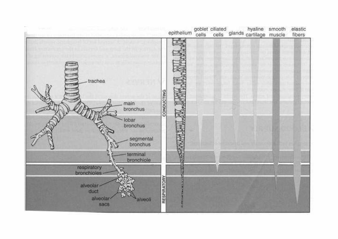
Anatomy and Structure of the
Gaseous Exchange System
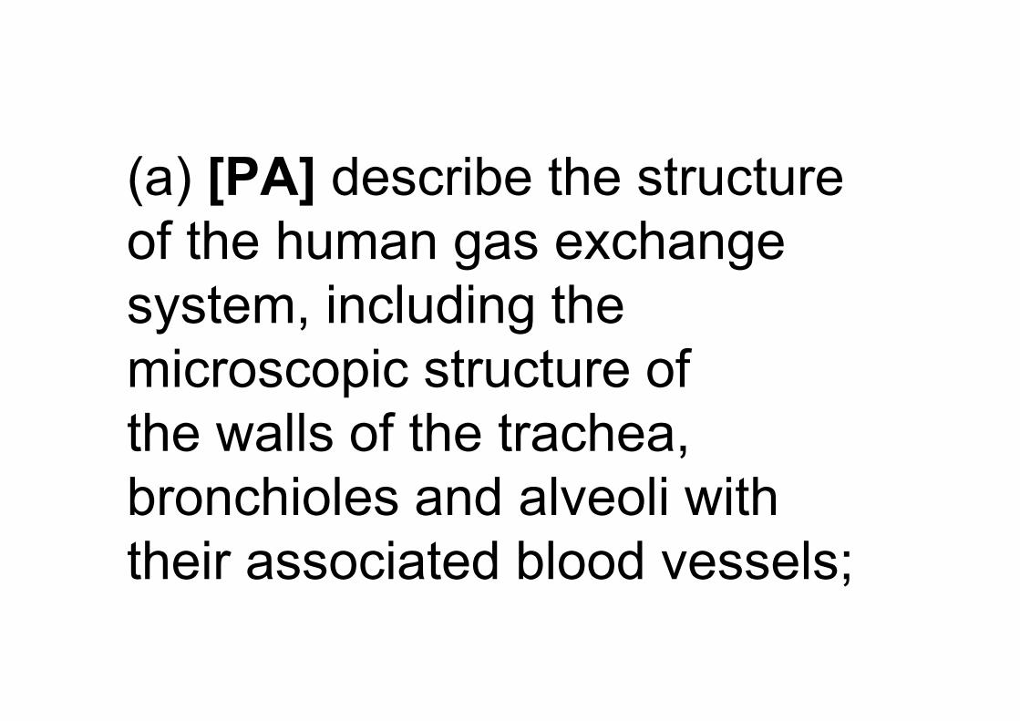
(a) [PA] describe the structure
of the human gas exchange
system, including the
microscopic structure of
the walls of the trachea,
bronchioles and alveoli with
their associated blood vessels;
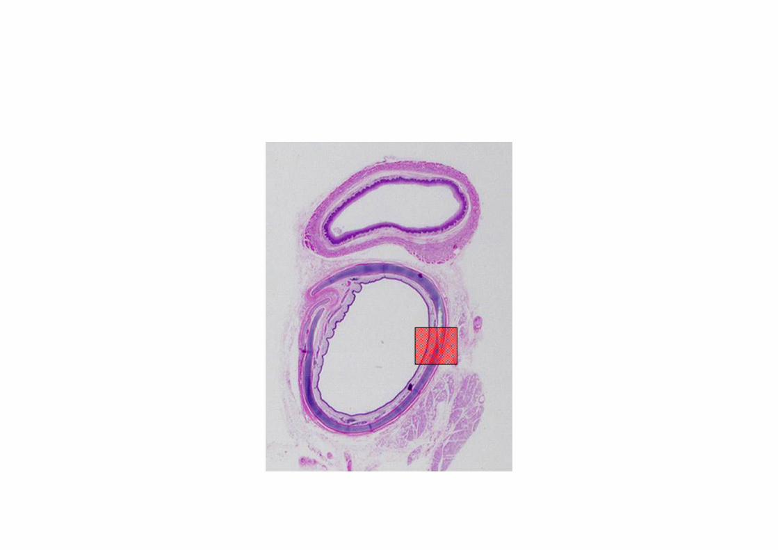
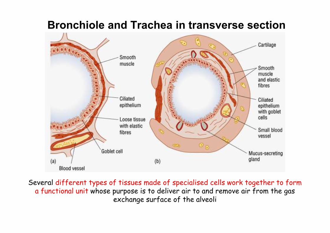
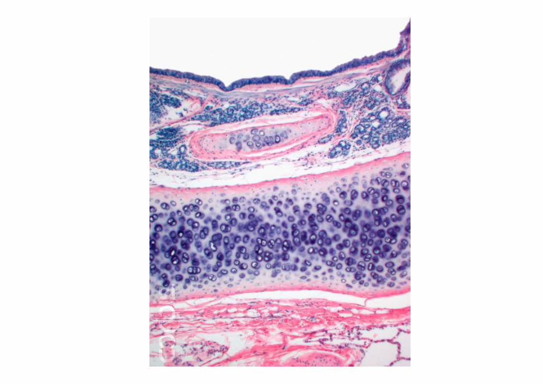
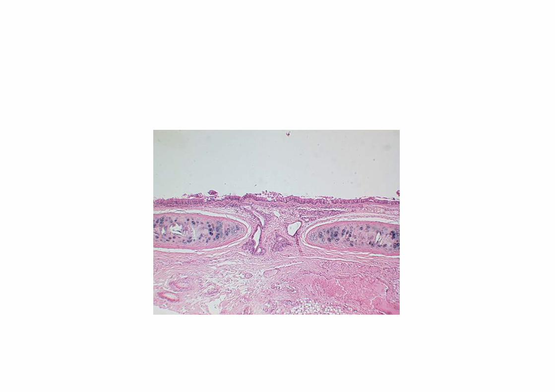
Bronchiole and Trachea in transverse section
Several different types of tissues made of specialised cells work together to form a functional unit whose purpose is to deliver air to and remove air from the gas
exchange surface of the alveoli
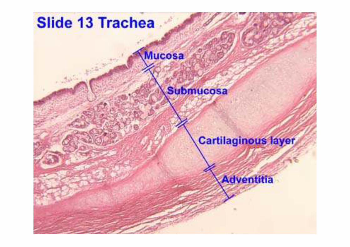
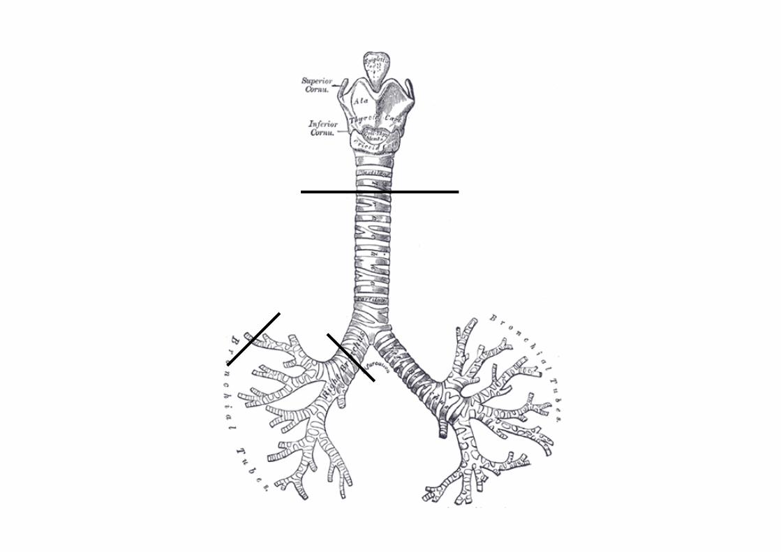
(b) [PA] describe the distribution of
cartilage, ciliated epithelium, goblet
cells and smooth muscle in the
trachea, bronchi and bronchioles;
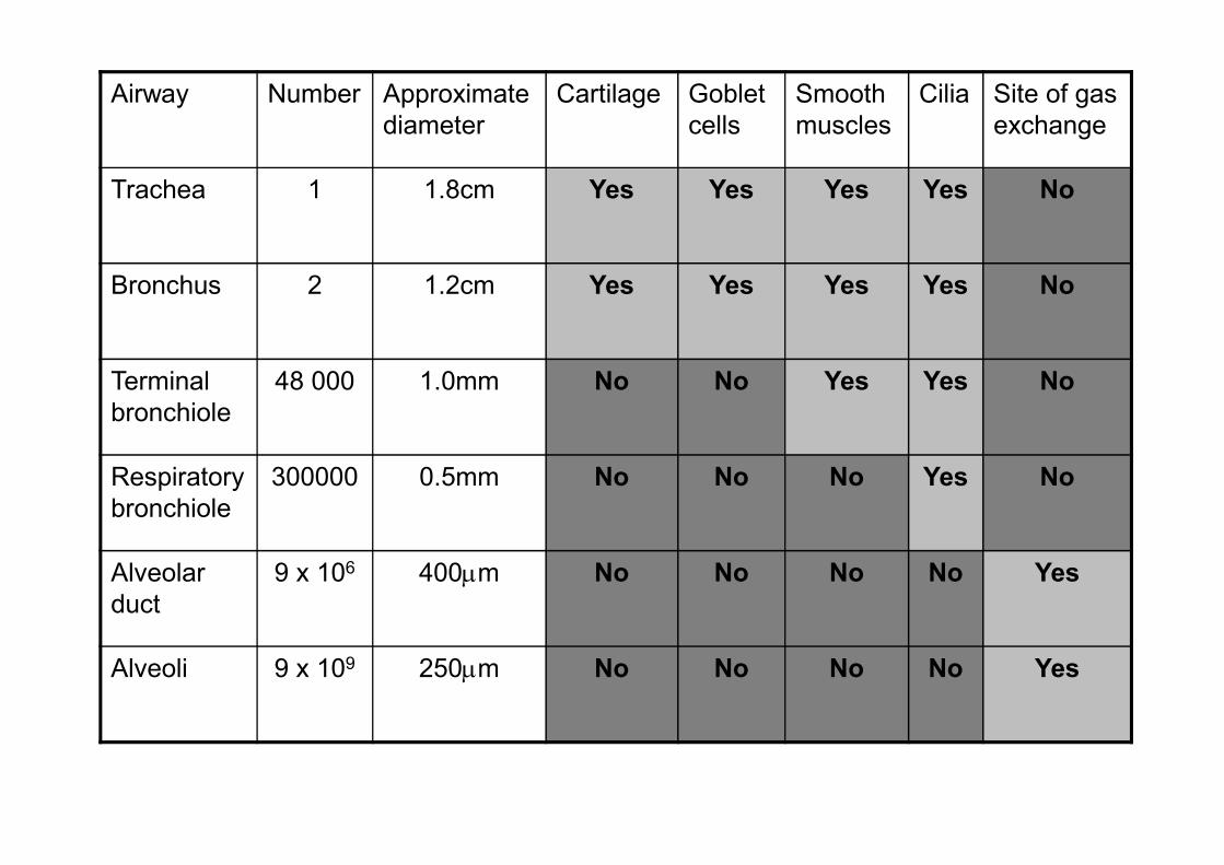
Airway Number Approximate
diameter
Cartilage Goblet
cells
Smooth
muscles
Cilia Site of gas
exchange
Trachea 1 1.8cm Yes Yes Yes Yes No
Bronchus 2 1.2cm Yes Yes Yes Yes No
Terminal
bronchiole
48 000 1.0mm No No Yes Yes No
Respiratory
bronchiole
300000 0.5mm No No No Yes No
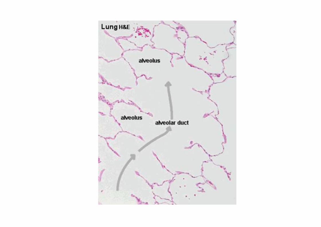
Alveolar
duct
9 x 10⁶ 400µm No No No No Yes
Alveoli 9 x 10⁹ 250µm No No No No Yes
(c) describe the functions of cartilage,
cilia, goblet cells, smooth muscle and
elastic fibres in the gas
exchange system;
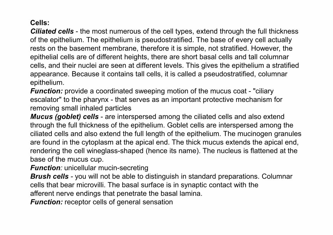
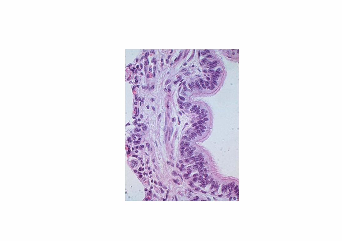
Cells:
Ciliated cells - the most numerous of the cell types, extend through the full thickness
of the epithelium. The epithelium is pseudostratified. The base of every cell actually
rests on the basement membrane, therefore it is simple, not stratified. However, the
epithelial cells are of different heights, there are short basal cells and tall columnar
cells, and their nuclei are seen at different levels. This gives the epithelium a stratified
appearance. Because it contains tall cells, it is called a pseudostratified, columnar
epithelium.
Function: provide a coordinated sweeping motion of the mucus coat - "ciliary
escalator" to the pharynx - that serves as an important protective mechanism for
removing small inhaled particles
Mucus (goblet) cells - are interspersed among the ciliated cells and also extend
through the full thickness of the epithelium. Goblet cells are interspersed among the
ciliated cells and also extend the full length of the epithelium. The mucinogen granules
are found in the cytoplasm at the apical end. The thick mucus extends the apical end,
rendering the cell wineglass-shaped (hence its name). The nucleus is flattened at the
base of the mucus cup.
Function: unicellular mucin-secreting
Brush cells - you will not be able to distinguish in standard preparations. Columnar
cells that bear microvilli. The basal surface is in synaptic contact with the
afferent nerve endings that penetrate the basal lamina.
Function: receptor cells of general sensation
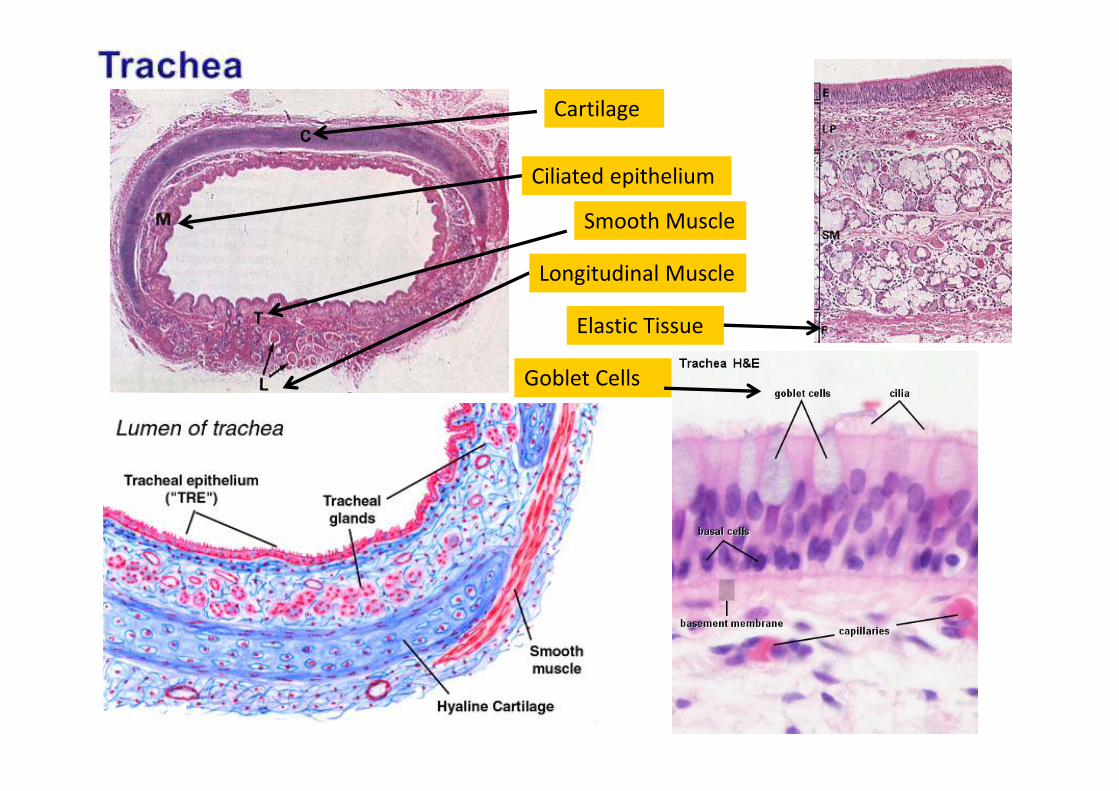
Cartilage
Ciliated epithelium
Smooth Muscle
Longitudinal Muscle
Elastic Tissue
Goblet Cells
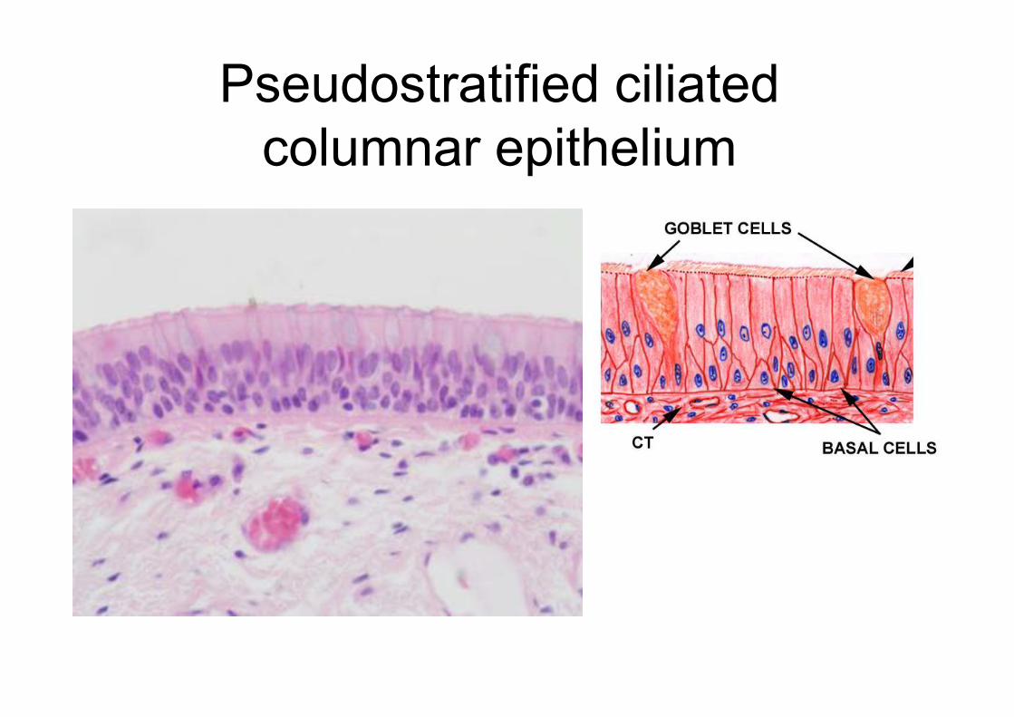
Pseudostratified ciliated
columnar epithelium
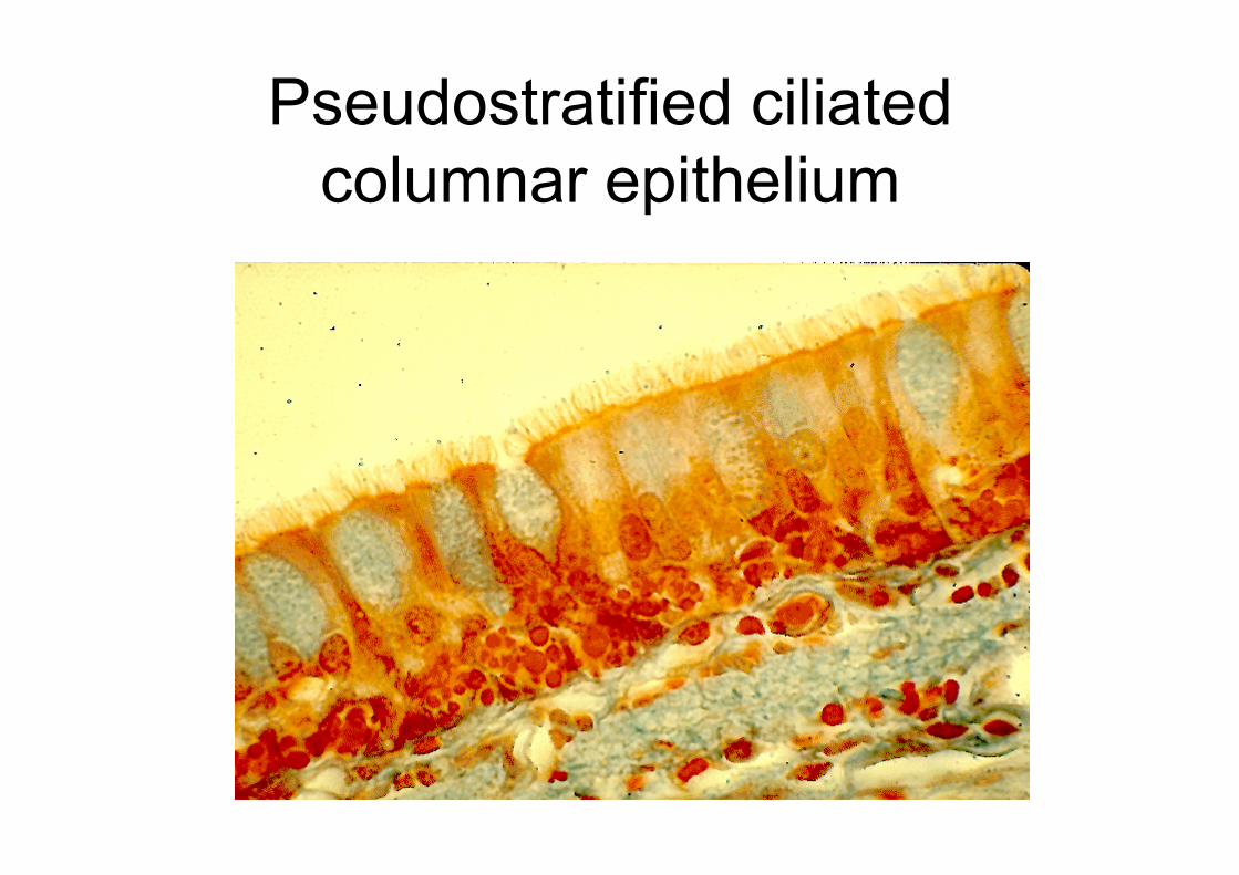
Pseudostratified ciliated
columnar epithelium
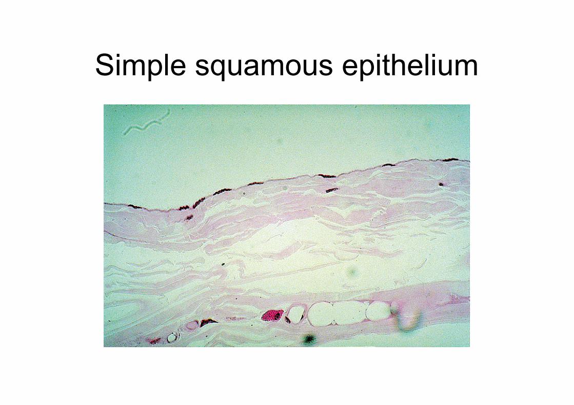
Simple squamous epithelium
Submucosa
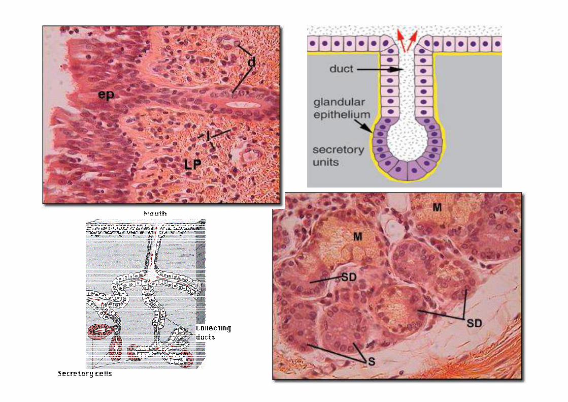
- serous and mucous glands in the submucosa of the
trachea.
- The serous glands secrete a watery proteinaceous product,
while the mucous glands secrete a viscous, heavier product
called mucus.
- The ducts of the glands pass through the layer(submucosa)
and epithelium to empty into the lumen (surface)
- The lumen is obscured as it approaches the epithelium.
(This is a frequent phenomenon of sectioning.)
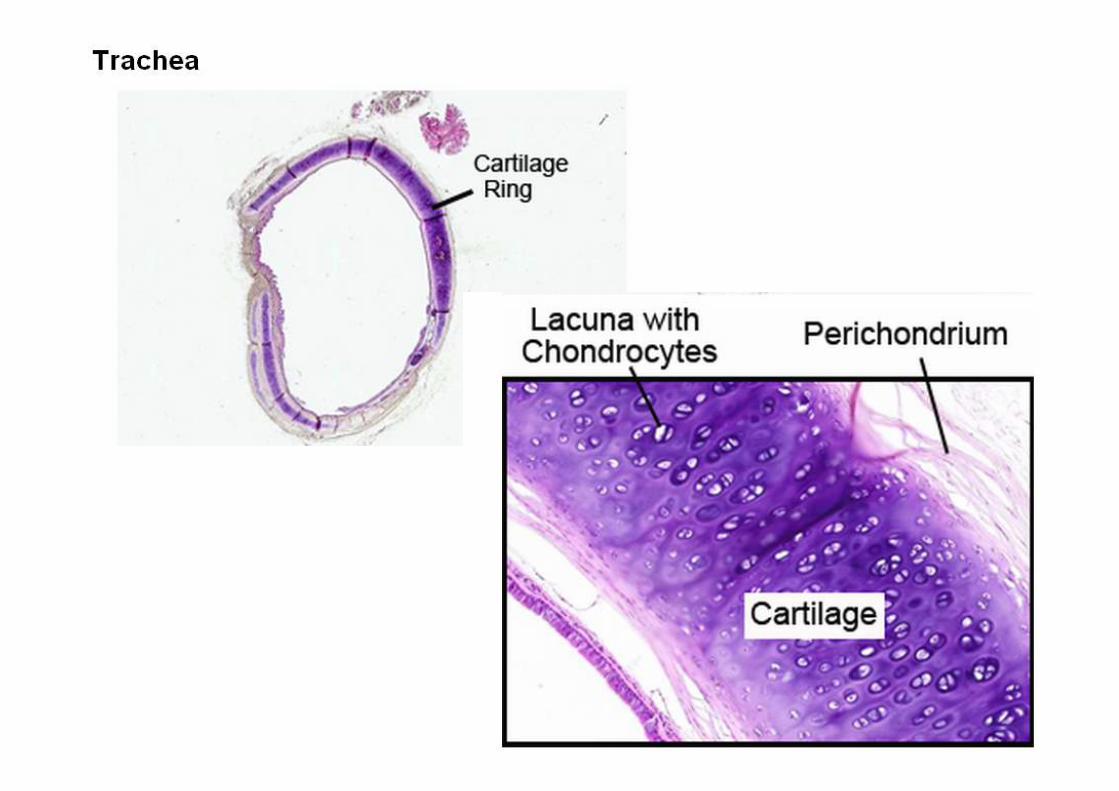
Cartilage- The matrix of cartilage typically stains a purplish color
(depending on the preparation).
- Staining is most intense around the cartilage cells, which are
called chondrocytes and sit in spaces called lacunae.
- Chondrocytes are often found in clusters.
- The chondrocytes occupy the whole lacuna, but they
frequently shrink during preparation, and the lacunae appear
as spaces around cells.
- Function: provide flexibility to the tracheal pipe and maintain
the lumen of being opened.
- The trachealis muscle is smooth muscle. Its functions to
narrow the tracheal lumen so when you cough, the narrower
the trachea, the faster the air moves and can propel
whatever is making you cough out of the trachea.
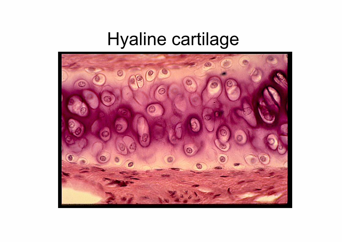
Hyaline cartilage
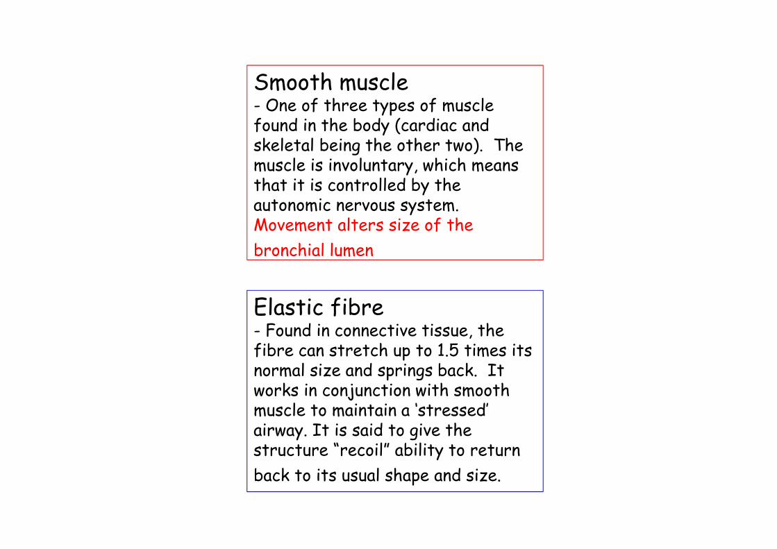
Smooth muscle- One of three types of muscle found in the body (cardiac and skeletal being the other two). The muscle is involuntary, which means that it is controlled by the autonomic nervous system. Movement alters size of the
bronchial lumen
Elastic fibre- Found in connective tissue, the fibre can stretch up to 1.5 times its normal size and springs back. It works in conjunction with smooth muscle to maintain a ‘stressed’ airway. It is said to give the structure “recoil” ability to return
back to its usual shape and size.
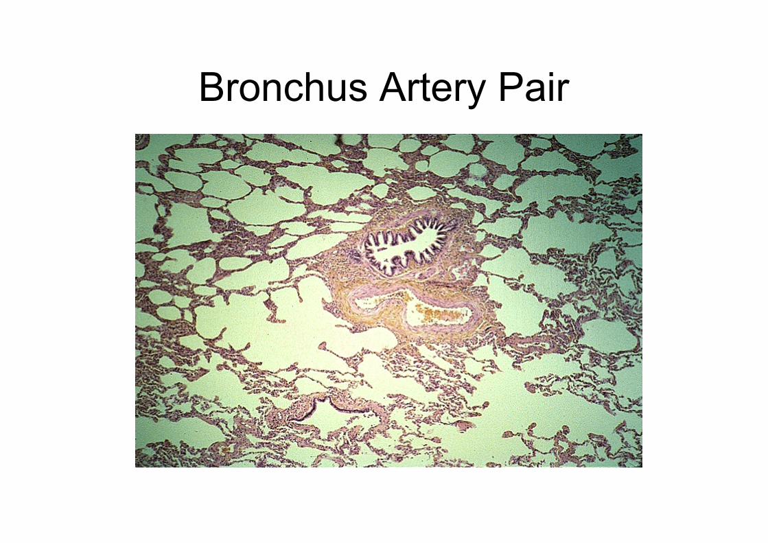
Bronchus Artery Pair
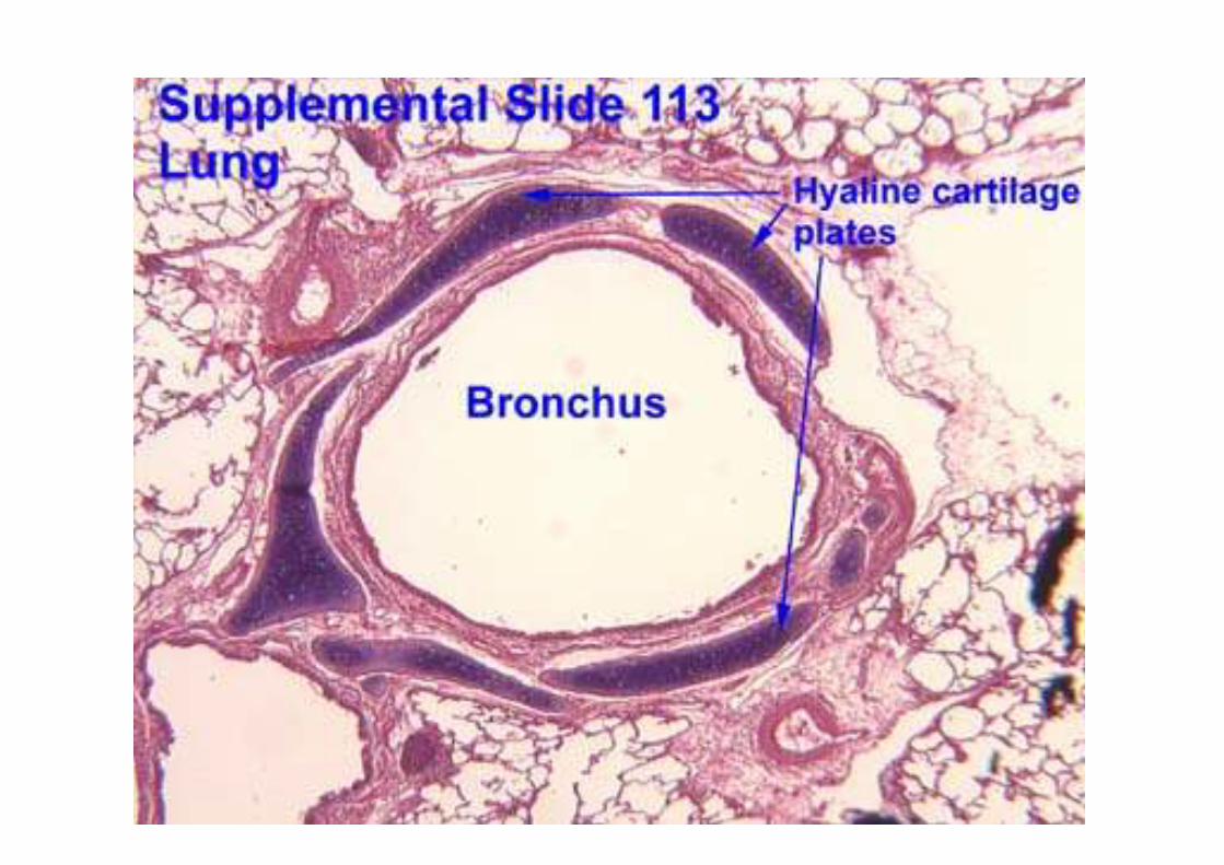
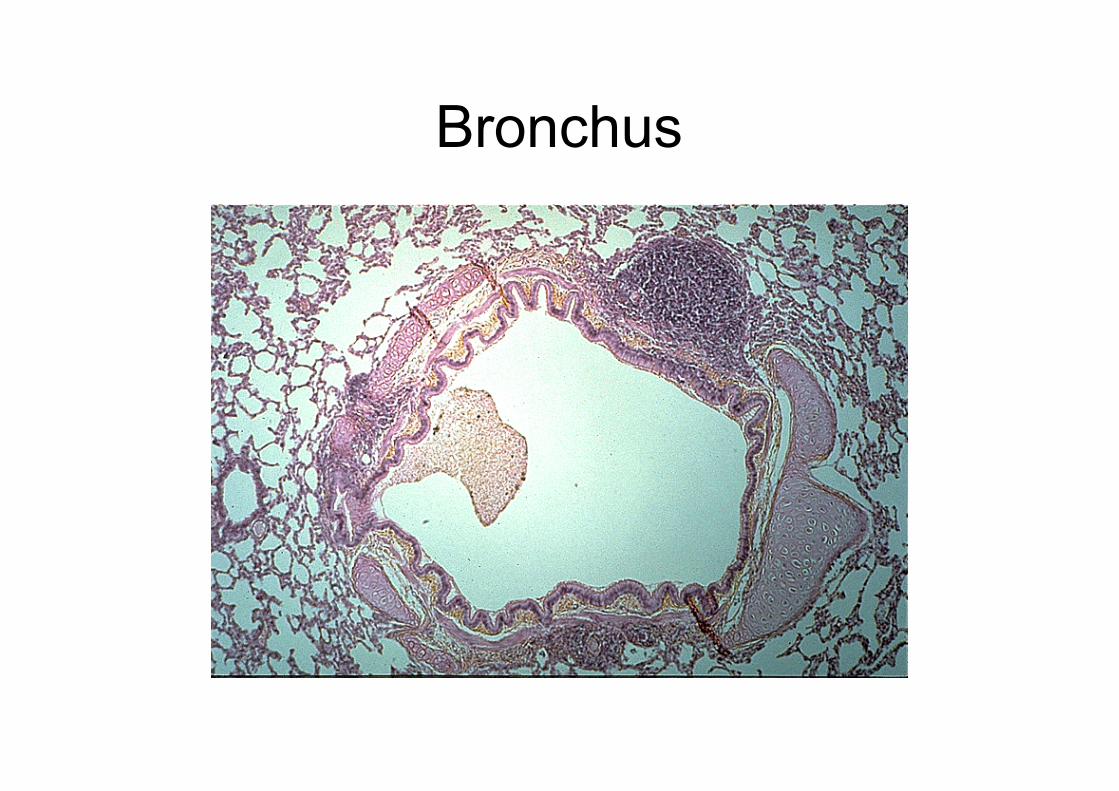
Bronchus
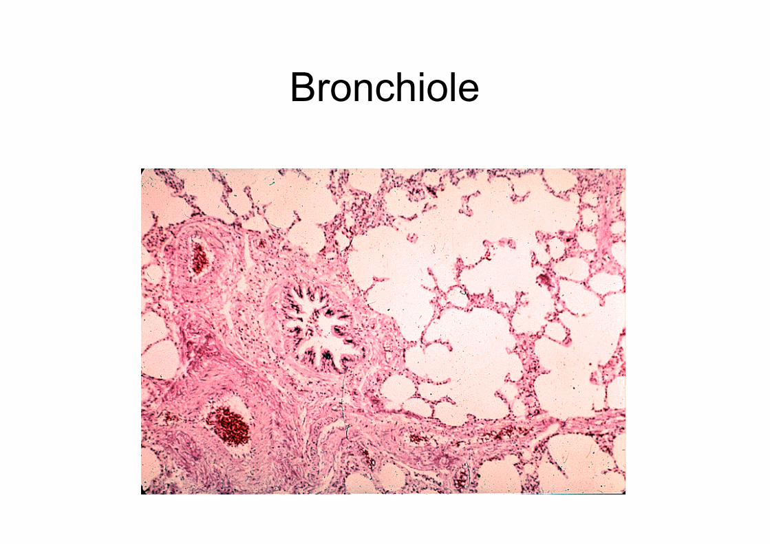
Bronchiole
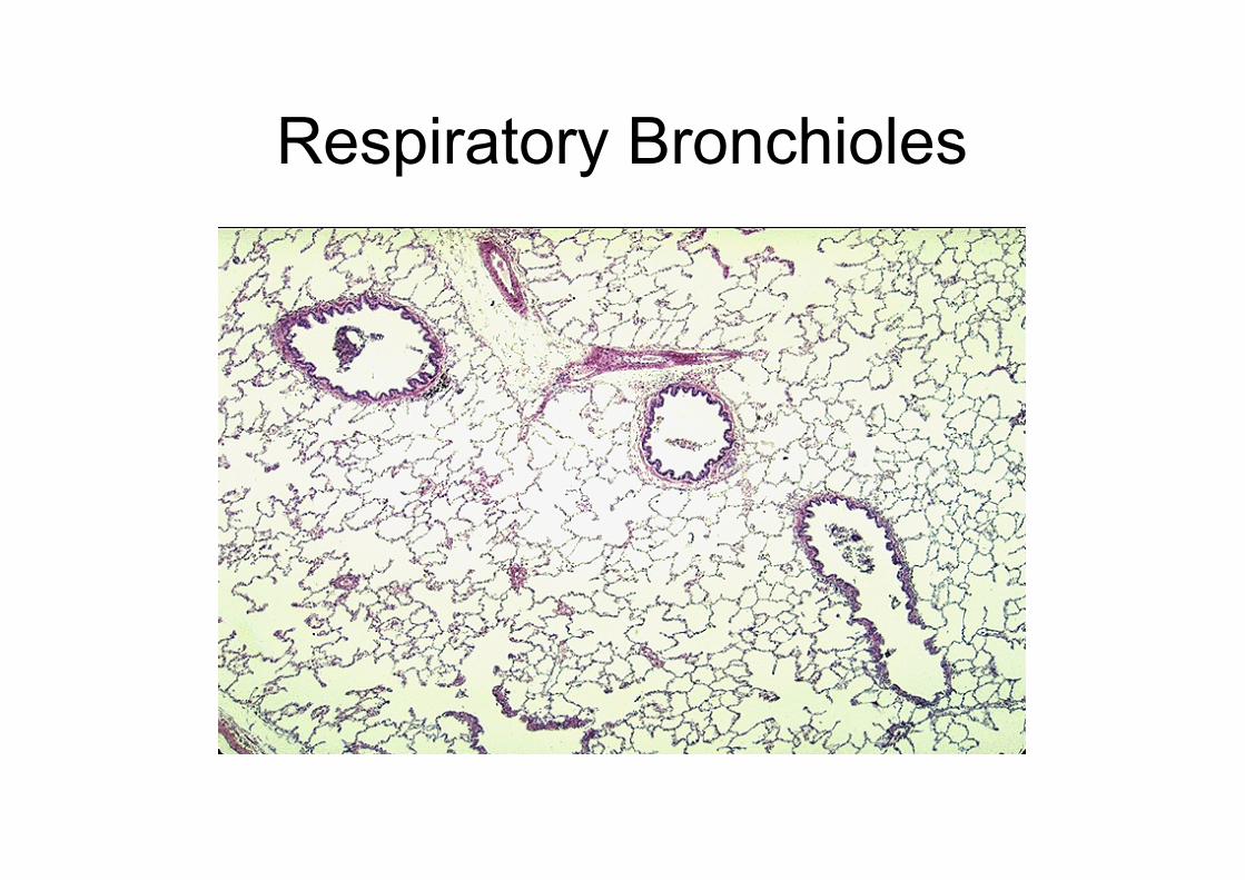
Respiratory Bronchioles
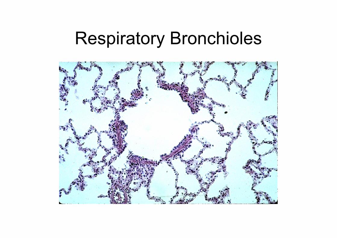
Respiratory Bronchioles
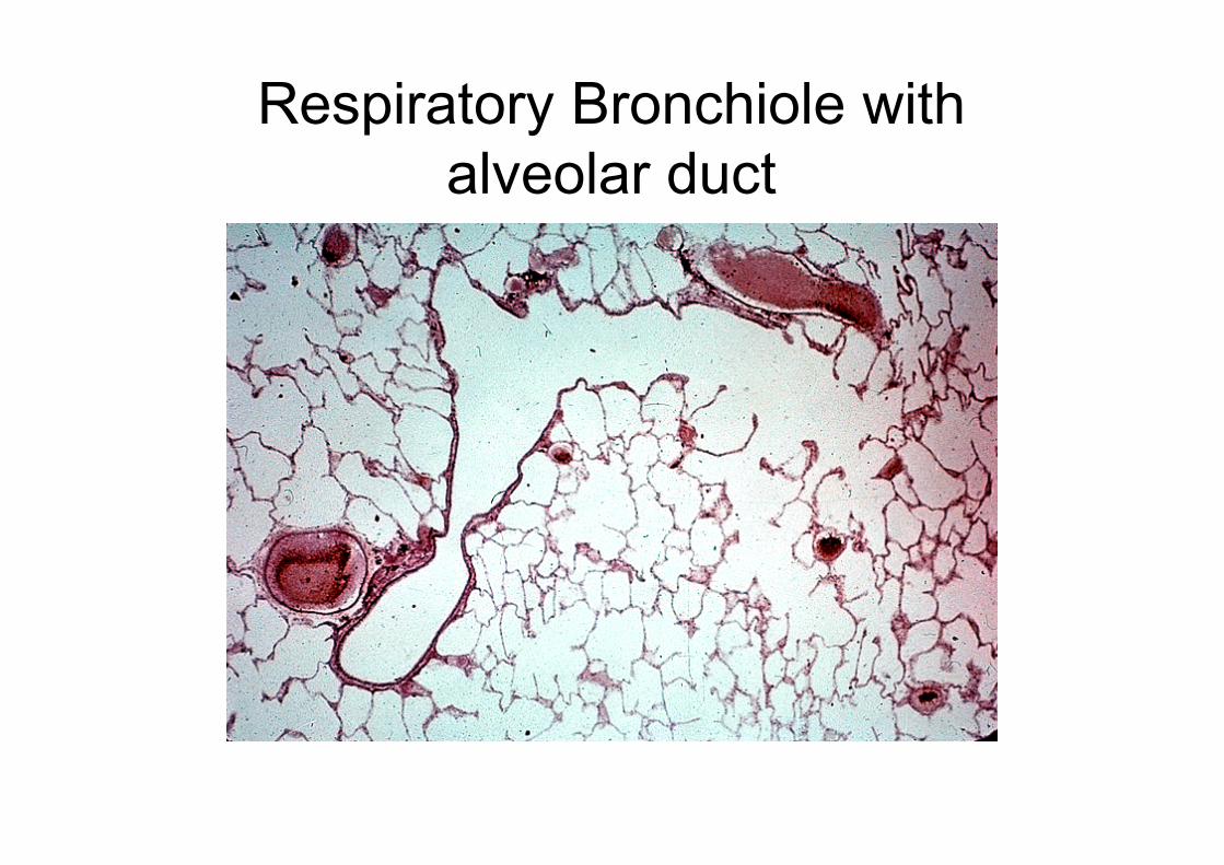
Respiratory Bronchiole with
alveolar duct
(d) describe the process of gas
exchange between air in the
alveoli and the blood;
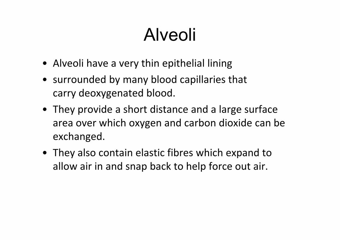
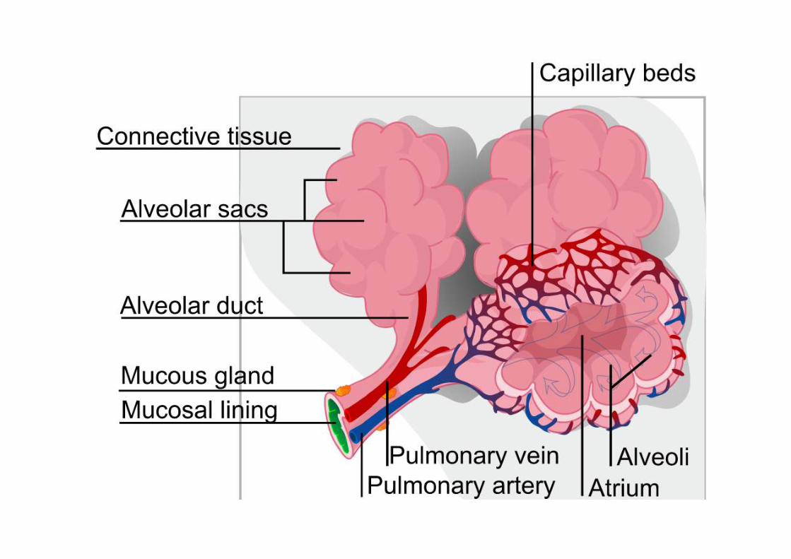
Alveoli
• Alveoli have a very thin epithelial lining
• surrounded by many blood capillaries that
carry deoxygenated blood.
• They provide a short distance and a large surface
area over which oxygen and carbon dioxide can be
exchanged.
• They also contain elastic fibres which expand to
allow air in and snap back to help force out air.
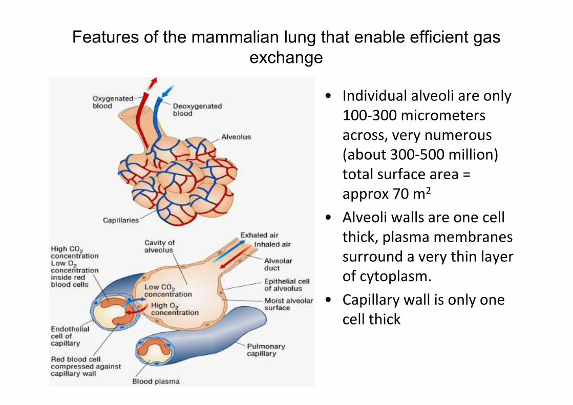
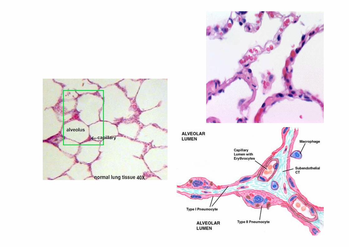
Features of the mammalian lung that enable efficient gas
exchange
• Individual alveoli are only
100-300 micrometers
across, very numerous
(about 300-500 million)
total surface area =
approx 70 m2
• Alveoli walls are one cell
thick, plasma membranes
surround a very thin layer
of cytoplasm.
• Capillary wall is only one
cell thick

Features of the mammalian lung that enable efficient gas
exchange
• Cells are squamous,
flattened
• Capillaries in close
contact with alveolus
wall
• Capillaries very narrow
so RBCs are squeezed
close to the walls and
so close to the air in the
alveoli
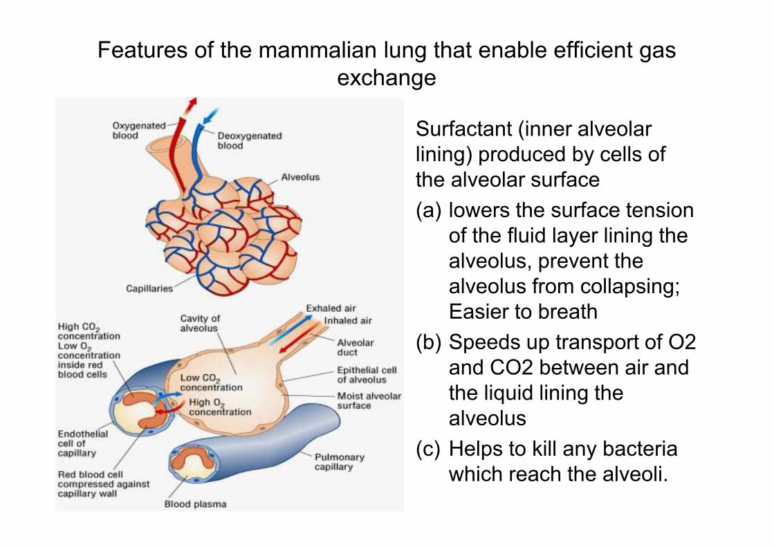
Features of the mammalian lung that enable efficient gas
exchange
Surfactant (inner alveolar
lining) produced by cells of
the alveolar surface
(a) lowers the surface tension
of the fluid layer lining the
alveolus, prevent the
alveolus from collapsing;
Easier to breath
(b) Speeds up transport of O2
and CO2 between air and
the liquid lining the
alveolus
(c) Helps to kill any bacteria
which reach the alveoli.
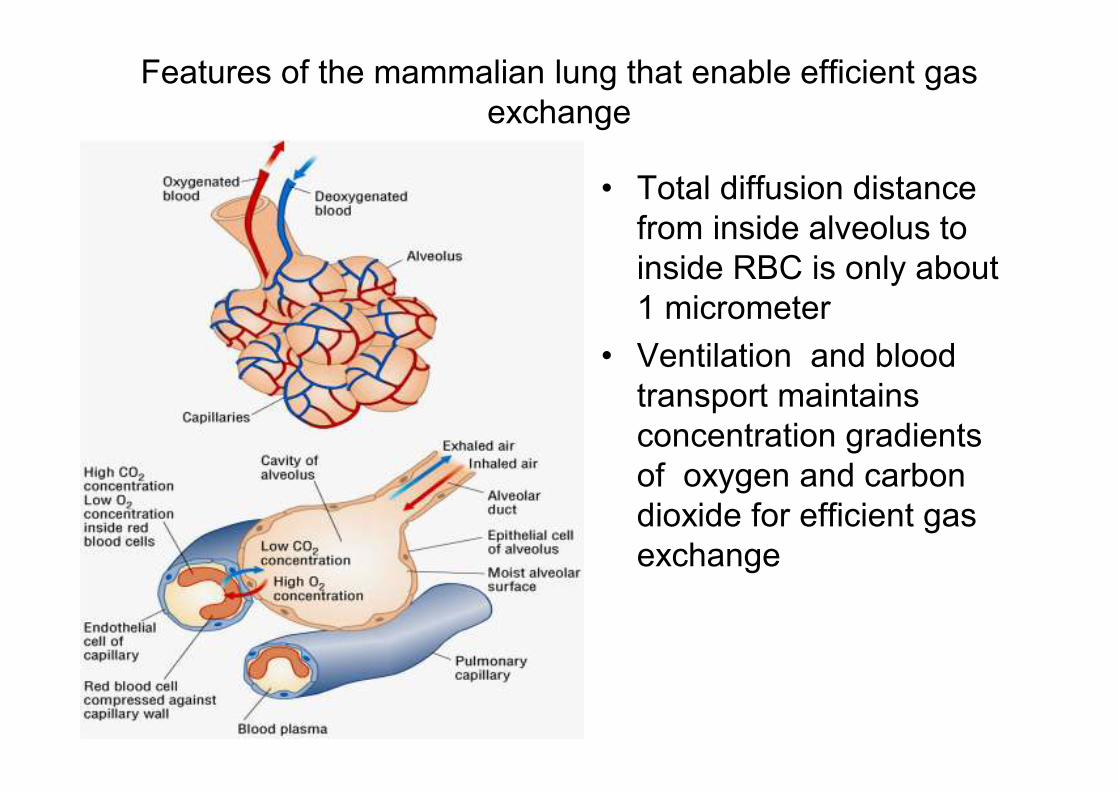
Features of the mammalian lung that enable efficient gas
exchange
• Total diffusion distance
from inside alveolus to
inside RBC is only about
1 micrometer
• Ventilation and blood
transport maintains
concentration gradients
of oxygen and carbon
dioxide for efficient gas
exchange
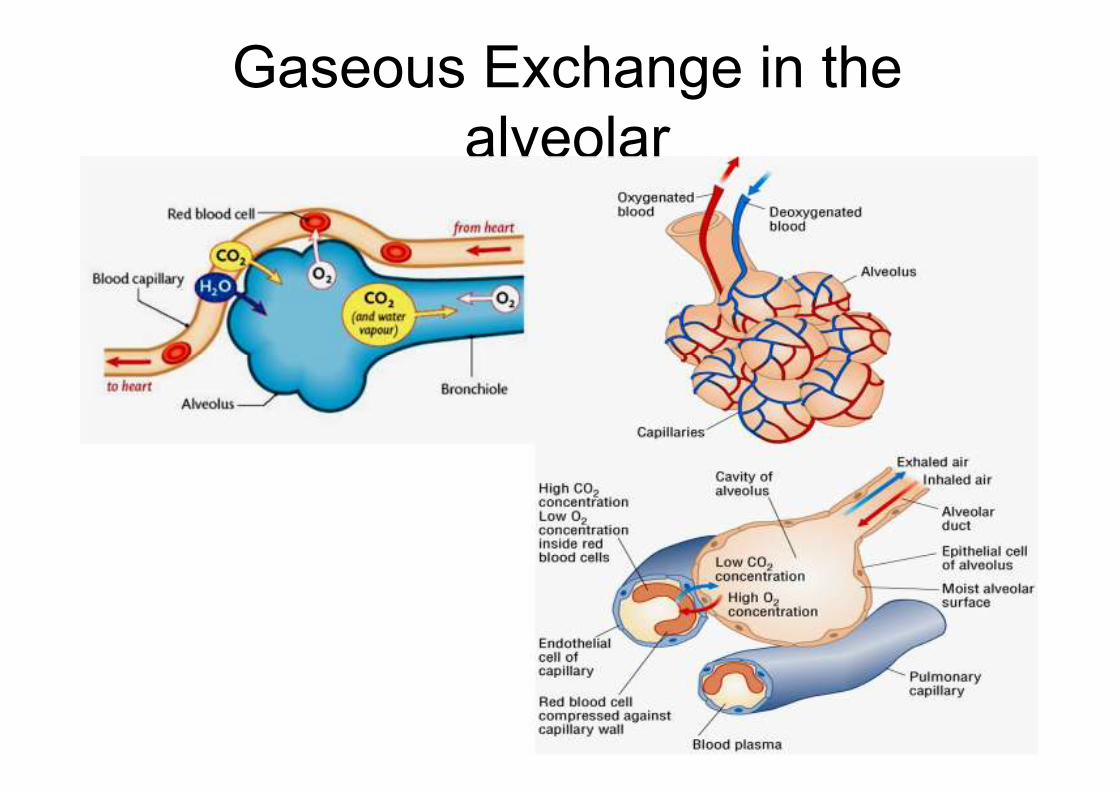
Gaseous Exchange in the
alveolar
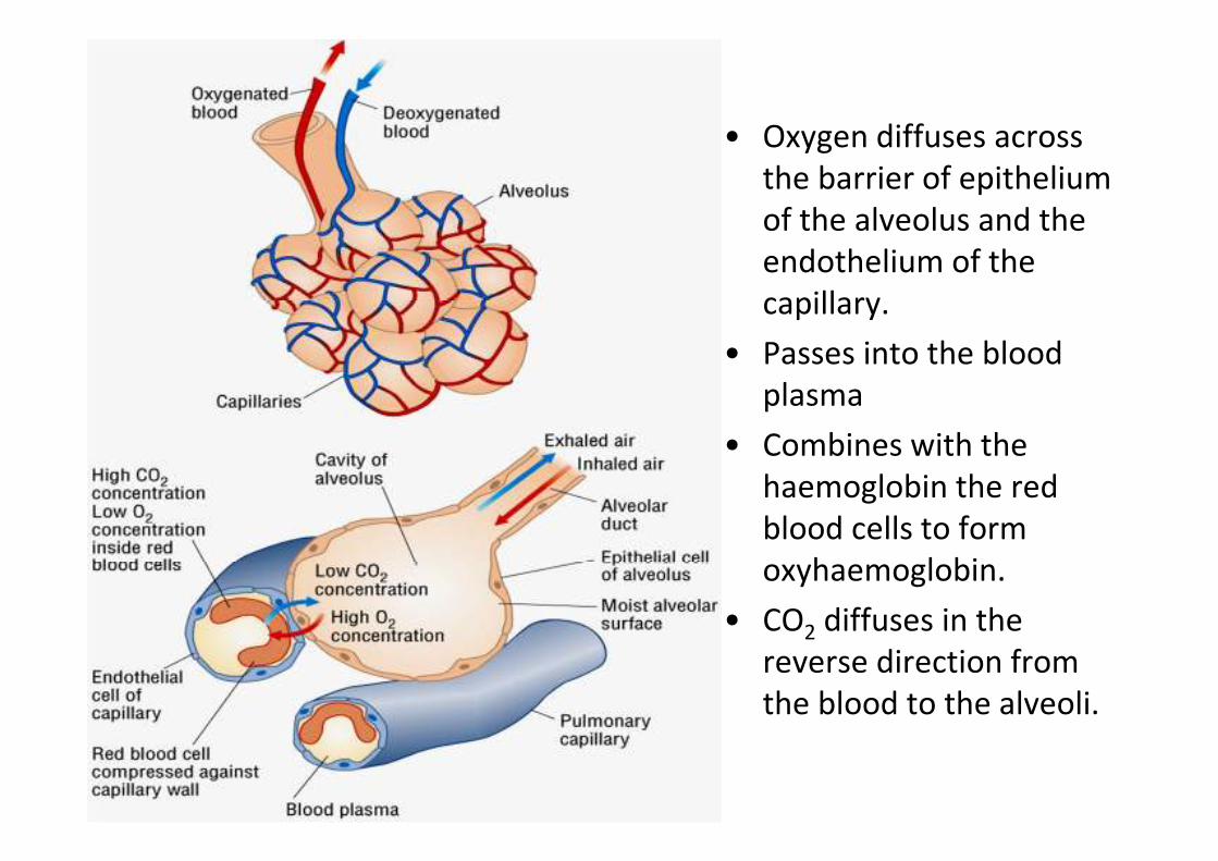
• Oxygen diffuses across
the barrier of epithelium
of the alveolus and the
endothelium of the
capillary.
• Passes into the blood
plasma
• Combines with the
haemoglobin the red
blood cells to form
oxyhaemoglobin.
• CO2 diffuses in the
reverse direction from
the blood to the alveoli.
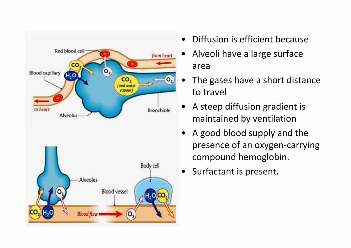
• Diffusion is efficient because
• Alveoli have a large surface
area
• The gases have a short distance
to travel
• A steep diffusion gradient is
maintained by ventilation
• A good blood supply and the
presence of an oxygen-carrying
compound hemoglobin.
• Surfactant is present.
(e) describe the effects of tar
and carcinogens in tobacco
smoke on the gas exchange
system;
(g) describe the effects of
nicotine and carbon monoxide
on the cardiovascular systems;
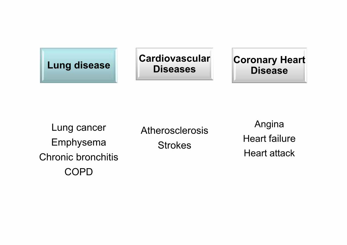
Lung diseaseCardiovascular
DiseasesCoronary Heart
Disease
Lung cancer
Emphysema
Chronic bronchitis
COPD
Atherosclerosis
Strokes
Angina
Heart failure
Heart attack
Tobacco smoke
• There are three main components that are
hazardous to health.
(i) Nicotine
(ii) Carbon monoxide
(iii)Tar
• Nicotine - is the addictive element of
cigarettes, stimulates the nervous system
to reduce arteriole diameter and release
adrenaline –
• increasing heart rate and blood
pressure.
• Causes increased stickiness of blood
platelets, which increases the risk of
blood clotting.
• Carbon monoxide - combines irreversibly
with haemoglobin meaning that oxygen
cannot bind effectively.
• This causes a strain on the heart muscle
because it must pump more to provide the
same amount of oxygen
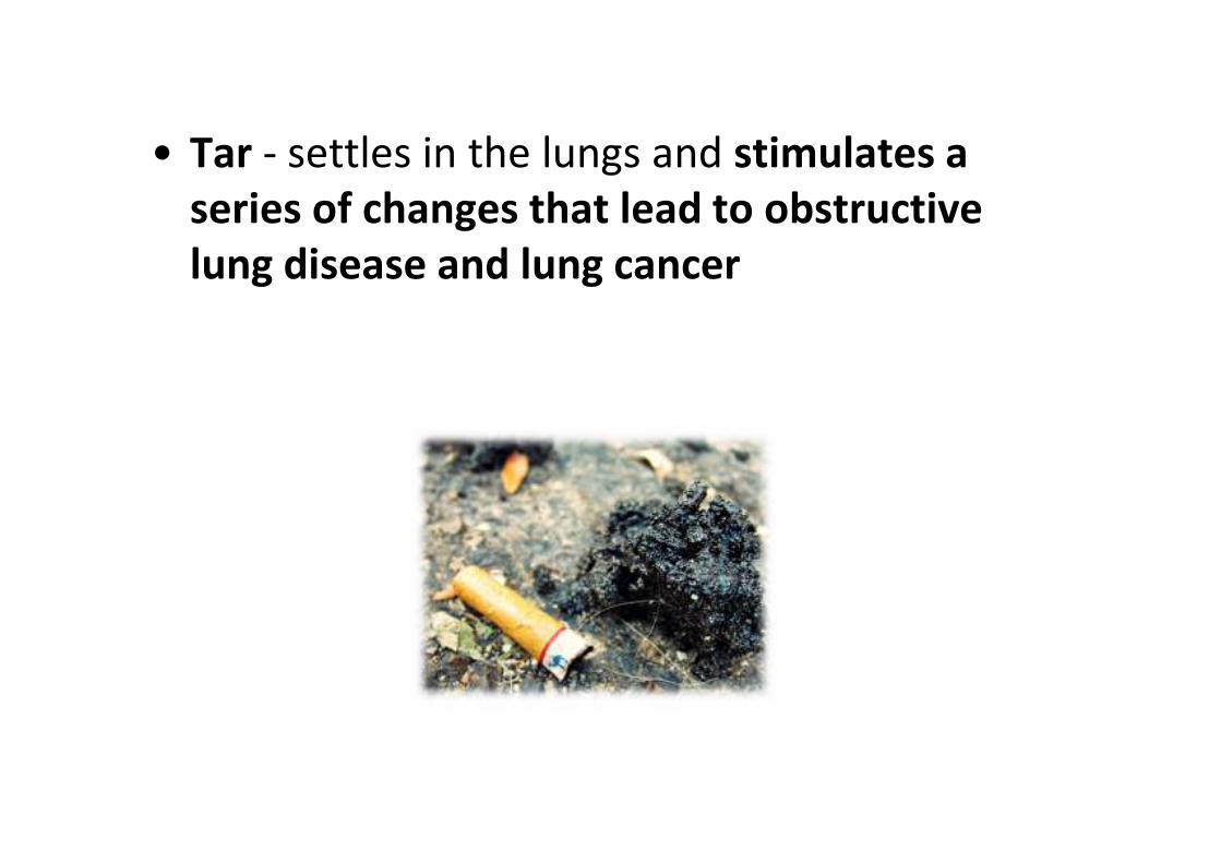
• Tar - settles in the lungs and stimulates a
series of changes that lead to obstructive
lung disease and lung cancer
(f) describe the signs and
symptoms of lung cancer and
chronic obstructive pulmonary
disease (emphysema and
chronic bronchitis);
(h) explain the link between
smoking and atherosclerosis,
coronary heart disease and
strokes;
Lung Cancer
• tar in tobacco smoke contains several
carcinogens.
• These can make DNA in epithelial cells lining
the lungs mutate, which is the first step
towards a malignant tumour.
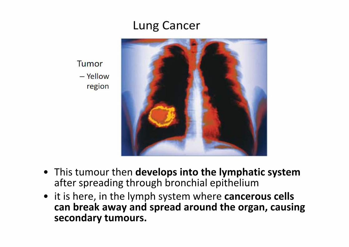
• This tumour then develops into the lymphatic systemafter spreading through bronchial epithelium
• it is here, in the lymph system where cancerous cells can break away and spread around the organ, causing secondary tumours.

Chronic Obstructive Pulmonary Disease
(a) Chronic bronchitis
- Inflammation of the lining of the air
passages and may be chronic or acute.
- Acute bronchitis usually lasts a few days
only and is a side effect of an infection like a
cold.
- Chronic bronchitis has a gradual onset and
last for a longer duration than its acute
counterpart.
• Most commonly caused by smoking and to a lesser extent air pollution.
• Tars in the cigarette smoke causes the inflammation
• Secretion of excess mucus from the goblet cells in response to the irritation
• Smoking destroys/paralyses the cilia which normally sweep away the nucleus – inhibits the cleaning action of the cilia.
• Frequent coughing of greenish-yellow sputum.
• Breathlessness increase as damaged epithelia are replaced by scar tissues narrowing the bronchi and bronchioles.

(b) Emphysema
• Inflammation of the constantly infected lungs
causes phagocytes to leave blood and line the
airways.
• To reach the lining of the lungs from the
capillaries, phagocytes release elastase
• This enzyme destroys elastin in the walls of
the alveoli
• Elastin is responsible for the recoil of the
alveoli when we breathe out
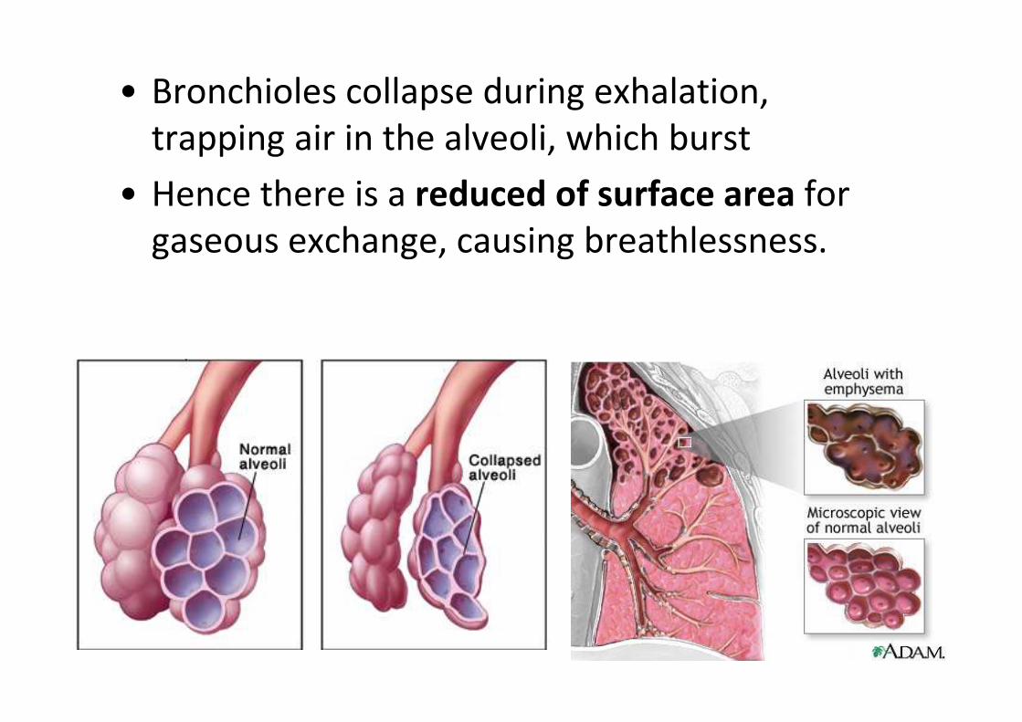
• Bronchioles collapse during exhalation,
trapping air in the alveoli, which burst
• Hence there is a reduced of surface area for
gaseous exchange, causing breathlessness.
Cardiovascular Disease
• Cardiovascular diseases are degenerative
diseases of the heart and circulatory system
• And are caused by many factor -
multifactorial
• diseases that involve the heart or blood
vessels (arteries and veins)
(a) Artherosclerosis
(b) Coronary heart disease
(c) Strokes
• Related to nicotine and carbon monoxide
found in cigarette smoke.
Nicotine and Cardiovascular Disease
• NICOTINE is a CENTRAL NERVOUS SYSTEM
stimulant that acts on nerves throughout the
body.
• In the cardiovascular system, nicotine
stimulates the nerves that regulate smooth
MUSCLE tissue, causing smooth muscle cells
to contract.
• This constricts blood vessels, notably arteries,
reducing the channel for blood flow.
• Nicotine further stimulates the baroreflexsensors (clusters of NERVE cells in the major arteries and the heart that sense the flow and pressure of blood).
• These actions result in increased blood pressure, HEART RATE, and cardiac workload.
• Nicotine further acts as an irritant within the arteries, causing INFLAMMATION of the inner layer of the arterial wall.
• Researchers believe such inflammation may be the foundation for atherosclerosis.
CO and cardiovascular disease
• Carbon monoxide is a poison. It has a greater
affinity than oxygen for HAEMOGLOBIN and
binds with hemoglobin to form
carboxyhaemoglobin, blocking haemoglobin
from carrying oxygen.
• This reduces the amount of oxygen that
enters the bloodstream from the LUNGS.
CO and cardiovascular disease
• By the end of a cigarette, a smoker can have
concentrations of carbon monoxide as high as
7%; 10% is the level at which symptoms of
carbon monoxide poisoning begin to become
apparent.
• Carbon monoxide in the bloodstream deprives
cells in the BRAIN and heart, which rely on
oxygen for fuel
Atherosclerosis
• Starts with accumulation of fatty materials in
artery walls.
• Reduces flow of blood to tissues and may also
increase the chance of blood clots forming
within the artery, obstructing the flow of
blood entirely.
• Build up which contains cholesterol, fibres,
dead muscle cells and platelets is called
atheroma.
• Once fibers are deposited in the cholesterol,
and these often start to calcify and become
hard, a process known as arteriosclerosis.
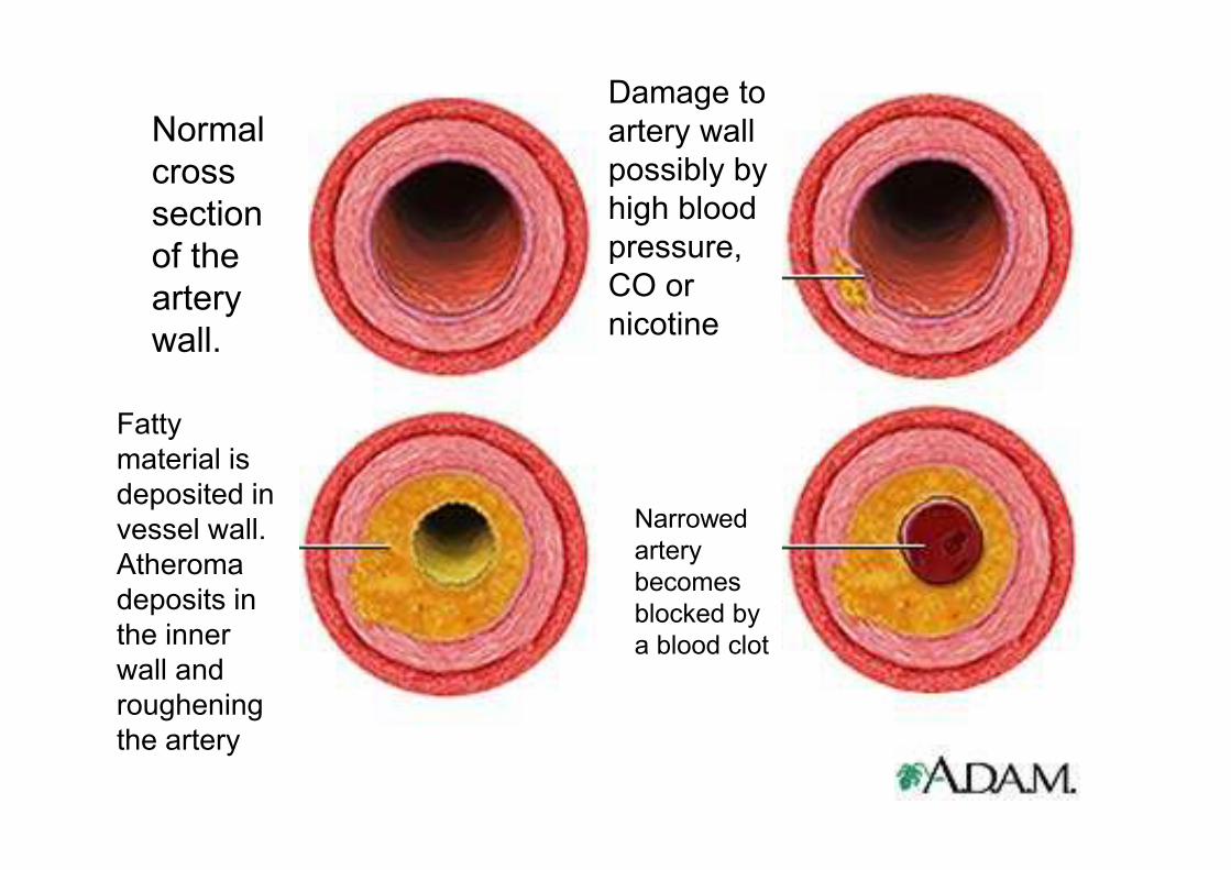
Normal
cross
section
of the
artery
wall.
Damage to
artery wall
possibly by
high blood
pressure,
CO or
nicotine
Fatty
material is
deposited in
vessel wall.
Atheroma
deposits in
the inner
wall and
roughening
the artery
Narrowed
artery
becomes
blocked by
a blood clot
Stroke
• A stroke occurs when an artery in the brain
(a) Bursts so that blood leaks into brain tissues
(brain haemorrhage)
(b) Is blocked due to artherosclerosis or a
thrombus.
Stroke
• The brain tissue in the area supplied by the
artery is starved of oxygen and dies (cerebral
infarction)
• A stroke may be fatal or cause mild or severe
disability.
• Depending on how large the area of brain
infected.
Coronary Heart Disease
• Two coronary arteries branch from the aorta
to supply all the muscles of the atria and the
ventricles.
• Coronary heart disease is a disease of these
arteries that causes damage to or malfunction
of the heart.
• Three forms of coronary heart disease:
(a) Angina pectoris
(b) Heart attack
(c) Heart failure
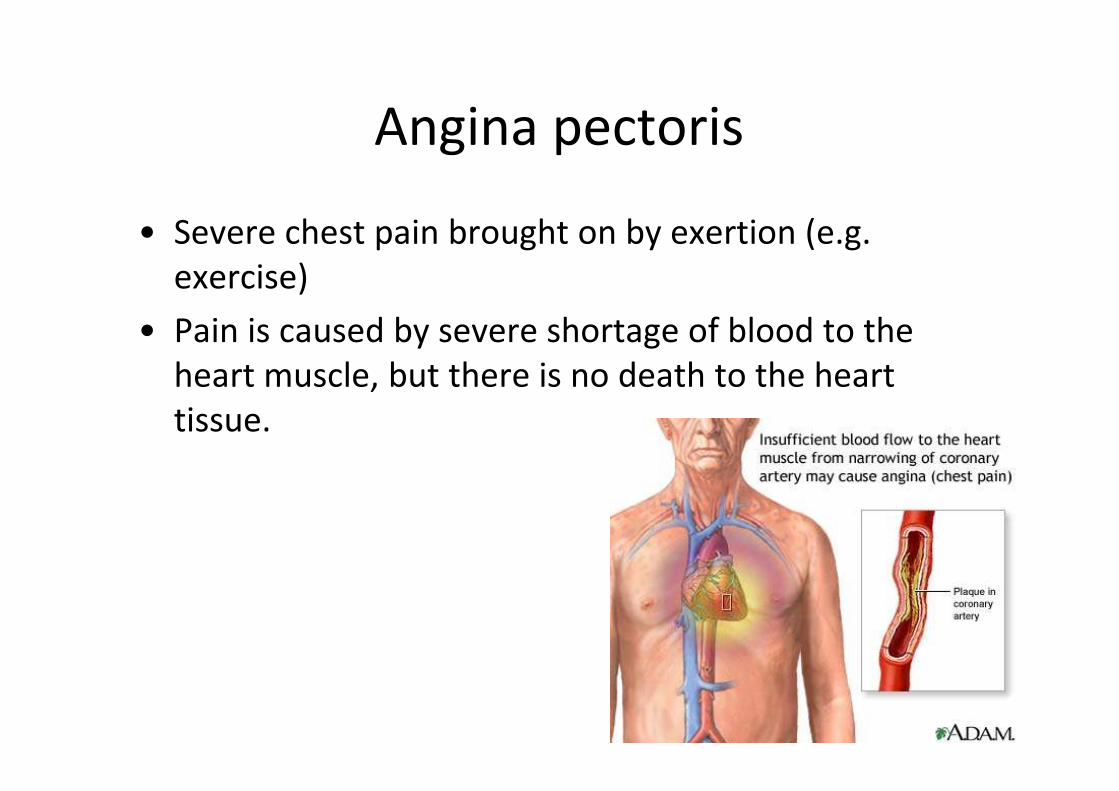
Angina pectoris
• Severe chest pain brought on by exertion (e.g.
exercise)
• Pain is caused by severe shortage of blood to the
heart muscle, but there is no death to the heart
tissue.
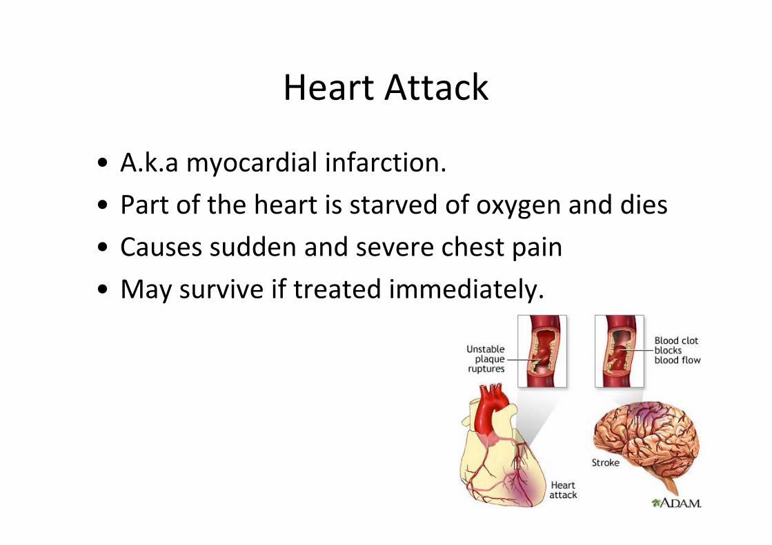
Heart Attack
• A.k.a myocardial infarction.
• Part of the heart is starved of oxygen and dies
• Causes sudden and severe chest pain
• May survive if treated immediately.
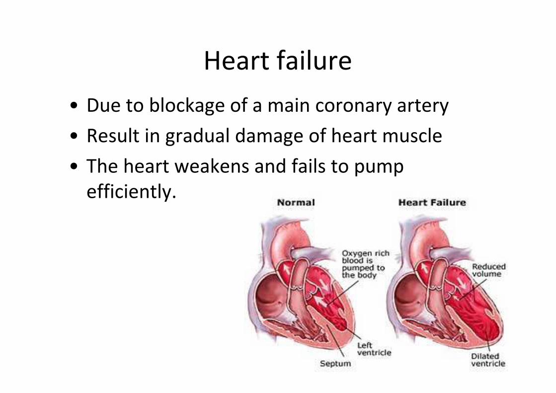
Heart failure
• Due to blockage of a main coronary artery
• Result in gradual damage of heart muscle
• The heart weakens and fails to pump
efficiently.
(i) evaluate the epidemiological
and experimental evidence
linking cigarette smoking to
disease and early
death;
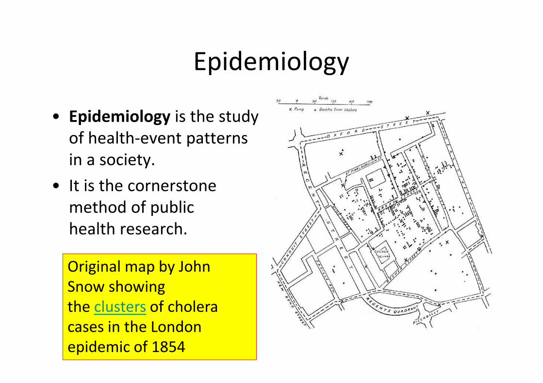
Epidemiology
• Epidemiology is the study
of health-event patterns
in a society.
• It is the cornerstone
method of public
health research.
Original map by John
Snow showing
the clusters of cholera
cases in the London
epidemic of 1854
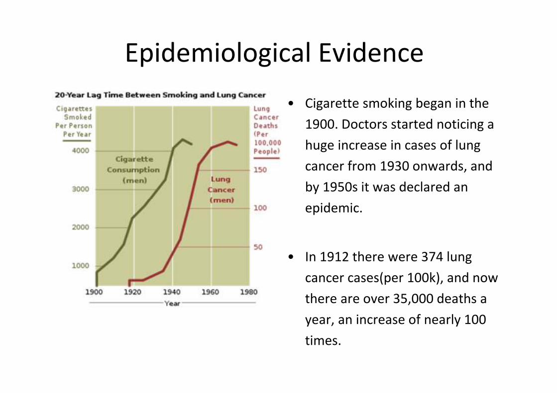
Epidemiological Evidence
• Cigarette smoking began in the
1900. Doctors started noticing a
huge increase in cases of lung
cancer from 1930 onwards, and
by 1950s it was declared an
epidemic.
• In 1912 there were 374 lung
cancer cases(per 100k), and now
there are over 35,000 deaths a
year, an increase of nearly 100
times.
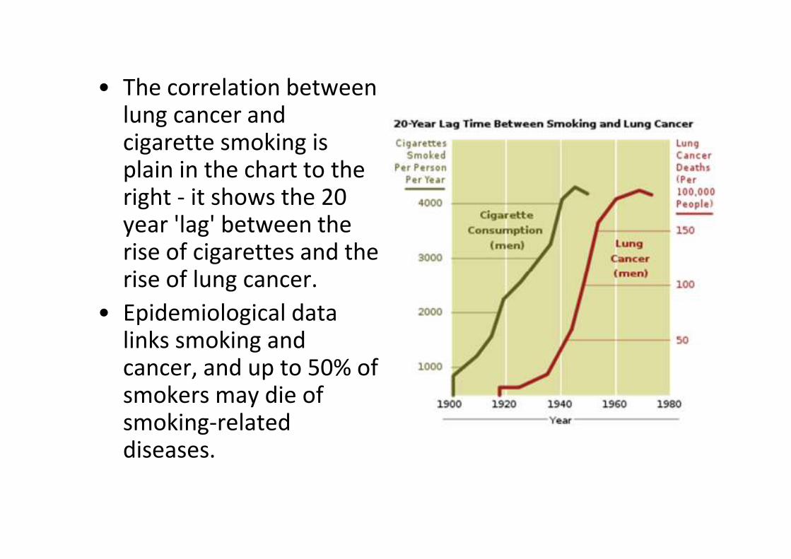
• The correlation between lung cancer and cigarette smoking is plain in the chart to the right - it shows the 20 year 'lag' between the rise of cigarettes and the rise of lung cancer.
• Epidemiological data links smoking and cancer, and up to 50% of smokers may die of smoking-related diseases.
• About 1/3 of cancer deaths are as a result of
cigarette smoking, and a quarter of smokers die
of lung cancer.
• Chronic obstructive pulmonary disease is very
rare in non smokers, less than 10% of victims are
non-smokers, and less than 2% of people with
emphysema are non-smokers.
• Approx. 1/5 of smokers suffer from emphysema,
and as a result deaths from pneumonia and
influenza are twice as high amongst smokers.
• Epidemiological studies have ruled out other
factors, that is to say that they cannot find any
other factor with a close correlation with
smoking.
• Smoking has been found to have a direct link
with lung cancer, as smoking is the common
fact in almost all cases.
• Smoking also contributes to many other
cancers of the mouth, larynx, bladder, kidney
and pancreas.
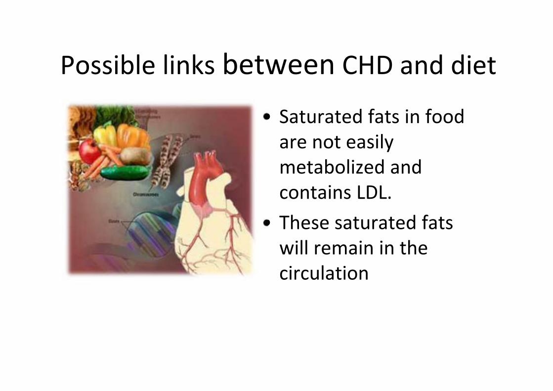
Possible links between CHD and diet
• Saturated fats in food
are not easily
metabolized and
contains LDL.
• These saturated fats
will remain in the
circulation
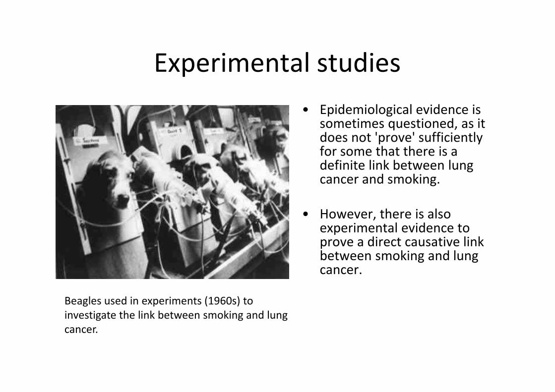
Experimental studies
• Epidemiological evidence is sometimes questioned, as it does not 'prove' sufficiently for some that there is a definite link between lung cancer and smoking.
• However, there is also experimental evidence to prove a direct causative link between smoking and lung cancer.
Beagles used in experiments (1960s) to
investigate the link between smoking and lung
cancer.
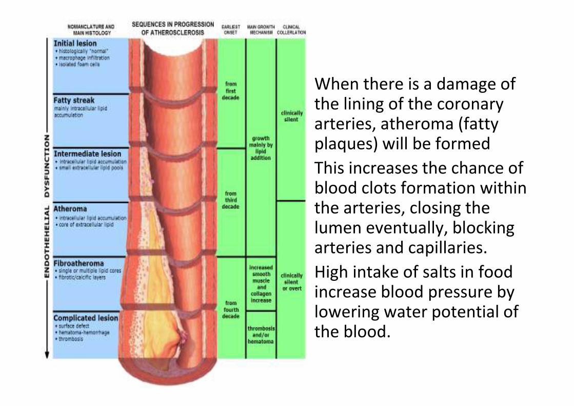
• When there is a damage of the lining of the coronary arteries, atheroma (fatty plaques) will be formed
• This increases the chance of blood clots formation within the arteries, closing the lumen eventually, blocking arteries and capillaries.
• High intake of salts in food increase blood pressure by lowering water potential of the blood.
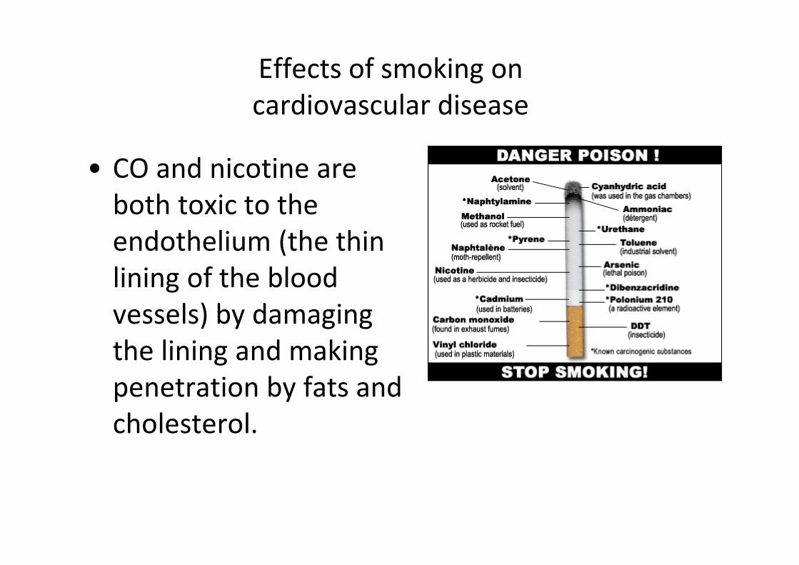
Effects of smoking on
cardiovascular disease
• CO and nicotine are
both toxic to the
endothelium (the thin
lining of the blood
vessels) by damaging
the lining and making
penetration by fats and
cholesterol.
• CO combines with Hb and reduces the O2
transport. Oxygen deficiency is a cause of
angina and may induce heart attack.
• Nicotine increases blood pressure, heart rate
and constriction of blood vessels.
• Cigarette smokers produce more fibrinogen
(blood clotting protein) and reduced levels of
the enzymes involved in removing blood clots.
• Smoking greatly stimulates the sticking of
blood platelets to the surface of the
endothelium and these are involved in blood
clotting.
• Nicotine has a direct effect on raising blood
fat levels.
(j) discuss the difficulties in
achieving a balance between
preventions and cure with
reference to coronary
heart disease, coronary by-pass
surgery and heart transplant
surgery;
Treatment to coronary heart
disease (CHD)• Drugs.
• Coronary artery by-pass surgery.
• Heart Transplant.
• Angioplasty.
Drugs
• Used to lower blood pressure, decrease risk of blood clotting,
prevent abnormal heart rhythms, reduce retention of fluids and
decrease the cholesterol
– ACE (angiotensin-converting enzyme) inhibitors. ACE inhibitors
are commonly used to treat high blood pressure. Examples
include captopril and enalapril.
– If you have a high blood cholesterol level, cholesterol-lowering
medicine called statins may be prescribed. Examples include
simvastatin, pravastatin and atorvastatin. They work by blocking
the formation of cholesterol and increasing the number of LDL
receptors in the liver, which help to remove the LDL cholesterol
from your blood
Coronary artery bypass surgery
• Invasive surgery involves replacing the
damaged artery by blood vessels from the
leg.
• The bypass carries blood from the aorta to
a place on the heart beyond the blockage
in the coronary artery. Sometimes two or
three bypass is necessary.
Heart Transplant
• Very high cost
• Difficulties in finding suitable donor
• Make sure that the host do not reject donor’s tissue.
• Drugs are used to suppress the immune system
after the transplant which may produce unpleasant
side effects and may not entirely prevent rejection.
• There are strict rules on deciding who should
receive transplant. Smokers who didn’t listen to
advice will be rejected from being in the transplant
list.
Angioplasty
• Less invasive procedure.
• Involves stretching the coronary arteries
by inserting a deflated balloon in the
femoral artery in the leg, positioning it in
the narrowed coronary artery and
expanding the balloon several times.
Prevention of CHD
• Prevention is more cost effective than
treatment.
• Two major ways;
– Screen for population at risk
– Adopting healthy lifestyle


![Anatomy and Structure of the Gaseous Exchange Systemfixurscore.com/wp-content/uploads/2015/06/H1-Gas-Exchange.pdf · Anatomy and Structure of the Gaseous Exchange System (a) [PA]](https://img.pdfslide.us/doc/110x75/5abad16e7f8b9a8f058bc79a/anatomy-and-structure-of-the-gaseous-exchange-and-structure-of-the-gaseous-exchange.jpg)