Embed Size (px)
Citation preview
1
Effect of Microencapsulated Hepatocytes Transplantation on Copper Metabolism of Rat Model with
Hepatolenticular Degeneration
H Lin1, C Xia
1, X Chen
1, L Huang
2, C Lu
1
ABSTRACT
Objective: To observe the effect of microencapsulated hepatocytes after intraperitoneal
transplantation of hepatolenticular degeneration (HDL) of model rats’ serum copper and
copper metabolism in liver.
Methods: Rat HLD model was made by copper load diet. To prepare microencapsulated hepatocytes
by the airflow method, and the method of intraperitoneal transplantation were used with naked cells or
the microencapsulated cell transplantation, respectively, compared with blank control group group I),
model group(group II), Bare hepatocytes after intraperitoneal transplantation group (group III) and
microencapsulated hepatocyte transplantation group(group IV). Determination of HLD rat model of
ALT, AST, albumin levels, copper of liver tissue, copper level in serum.
Results: II, III, IV groups of rats at each time point of ALT, AST, serum copper, liver copper values
were significantly higher than group I rats increased (each P < 0.05), synthesis of albumin levels
decreased significantly (each P < 0.05).
Keywords: Copper metabolism, hepatocyte, hepatolenticular degeneration
___________________________________________________________________________________
From: Pediatric infection, The Second affiliated Hospital of Wenzhou Medical University 109thth
Xueyuanxi Road Wenzhou, Zhejiang Province, China.
Correspondence: Dr C Lu, Department of Pediatric Infection, The Second Affiliated Hospital of
Wenzhou Medical University, 109th
Xueyuanxi Road, Wenzhou, Zhejiang Province, China,
e:mail:[email protected]
West Indian Med J 2015 DOI: 10: 7727/wimj.2015.433

Lin et al
2
Conclusion: Microencapsulated hepatocytes intraperitoneal transplantation can significantly reduce the
liver copper deposition of HLD rat, accelerate metabolism of serum copper, may become a new method
for cell transplantation in the treatment of HLD. The ALT, AST, serum copper and liver copper levels
of group IV were significantly lower than that in group III after 7-14d (each P < 0.05), the lowest value
respectively with 85.1±7.0 U/L, 87.9±22.7 U/L, 2.44±0.18μg/ml, 26.73±3.22μg/g, and albumin in 14d
than that in group III, the most high up to 38.36±1.52g/L

Encapsulated hepatocytes transplantation
3
INTRODUCTION
Hepatolenticular degeneration (HLD) is a genetic disease with copper metabolic
disorder characterized by multiple organ involvement, occur in adolescents, often
early involvement of liver (1), which is relatively common in China (2). Hepatocyte
transplantation has been becoming one of the treatment scheme of HLD (3, 4).
Hepatocyte microencapsulation technology enables allogeneic cell transplantation
becomes possible, which can effectively avoid immune rejection and prolong the
lifespan of liver cells (5,6). In this artical, to study its effect on copper metabolism of
HLD rats through the comparison of HLD microencapsulated rat hepatocytes after
intraperitoneal transplantation and naked hepatocyte transplantation.
Materials and methods
Materials: Trypsin, Type I collagenase, EGTA, Sodium alginate, Poly lysine,
Percoll(Sigma company, USA), DMEM culture medium (glucose, Hyclone company,
USA), EDTA(Amresco company, USA), Trypan blue(Beijing Suolaibao company).
Isolation and culture of primary rat hepatocytes
Liver tissue from adult male Wistar rats (provided by the experimental animal
center of Wenzhou Medical University, approved by the ethics committee of Wenzhou
medical university), hepatocyte isolated by EDTA-collagenase in situ two steps
perfusion method(7), The method is as follows: Rats were anesthetized, with
D-Hank's solution for portacaval bypass perfusion, perfusion of about 30ml/min,

Lin et al
4
about 7 minutes, waiting for the liver was soft opened, vena cava cannula outflow
liquid turbid, stop infusion. Remove the liver, in DMEM medium to tear the liver
capsule, hepatic cells gently shake off into the medium, followed by repeated
centrifugation washing 2 times, liquid into cell suspension cultured with DMEM,
Percoll separation of liquid specific gravity 1.096 purified hepatocytes. Obtained cell
contained by DMEM complete culture medium of 10% fetal bovine serum and 1%
penicillin streptomycin, cell counted and determined the activity after dyeing 4%
trypan blue, each rat hepatocyte yield can reach more than 108
cells, when the cell
activity of >85% preparing for microencapsulation.
Cell Microencapsulation
The preparation of microcapsules use of one-step process microcapsule-forming
method with sodium alginate barium chloride(8). Centrifugal collection of
hepatocytes, and mixed 1.5% purified sodium alginate solution into the air-flow
method, microcapsule generator homemade, gas flow rate of 4L/min, sodium alginate
solution drop speed is 50ml/h, drip into the 25mmol/L BaCl2 solution. Sodium
alginate and Ba2+ cross-linked into capsules, and static 15min. Washed by D-Hanks
liquid for 3 times to remove the excess BaCl2, into DMEM culture medium for
culturing.
Establishment and experimental grouping of rat HLD model
Rat HLD model was established by copper loading method(9). Took 120 male

Encapsulated hepatocytes transplantation
5
Wistar rats of 3 months old, according to the requirements of the experiment were
randomly divided into model group (II group), bare hepatocytes after intraperitoneal
transplantation group (group III) and microencapsulated hepatocyte transplantation
group (group IV), divided into 5 time points with 3 days, 7 days, 14 days, 21 days, 28
days (copper load for ninth weeks as starting 0 days), each group 8 rats for per time
point, another 8 rats as the blank control group (group I). All rats were fed for 12
weeks according to the standard, group II, group III and group IV were fed with feed
containing copper sulfate 1g/kg and 0.185% copper sulfate water also, a total of 12
weeks. From ninth weeks of feeding, took the rat median abdominal incision 5mm,
group II, group III and group IV according to the experimental requirements were
respectively injected 0.9% sodium chloride solution 2ml, bare hepatocytes and hepatic
cell microcapsules, with 12 gauge needle. Each rat of group III and group IV was
peritoneal injected with approximately 1×10⁷ liver cells. After operation strict
disinfection of wounds was made.
Index Determination
ALT, AST, albumin determination were taken at different time points: the serum
samples of Wistar rats, the value of ALT, AST, albumin was determined by automatic
biochemical analyzer.
Determination of the content of serum copper: serum copper to be detected to
measure serum 1:10 diluted with deionized water mixing, with atomic absorption
Lin et al
6
spectrophotometry measurement. Liver tissue copper determination: first with 0.9%
saline repeatedly washed out of the liver tissue, then clean and dry with filter paper
and weighed 500mg, concentrated nitric acid (analytical) 10ml with low temperature
heating digestion, and completely dissolved to yellow clear transparent, the
determination of copper content by atomic absorption spectrometry.
The Statistical Method
Data processing and statistical analysis using SPSS 16 statistical software
package, samples compared with single factor analysis of variance and
Newman-Keuls test.
Results
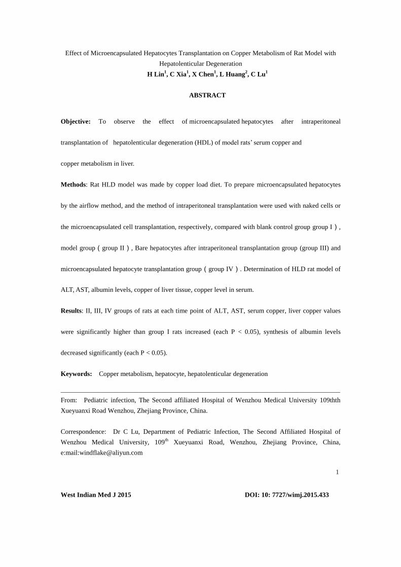
Primary hepatocytes and cells in microencapsules
Isolated Primary hepatocytes were cultured for 2d when viability of >85% preparing
for microencapsulation.(Figure 1,2)

Encapsulated hepatocytes transplantation
7
Figure 1- Isolated Primary hepatocytes
Hepatocytes yield >2.5×10⁸/rat, when cell viability of >85% preparing for
microencapsulation.
Figure 2- Cells in microencapsules
The microencapsule diameter of 0.4 ~ 0.8 mm include hepatocytes.
Serum ALT, AST and albumin variations
II, III, IV groups of rats at each time point of ALT, AST were higher than that in
normal group rats (each P<0.05), synthesis of albumin levels decreased significantly
Lin et al
8
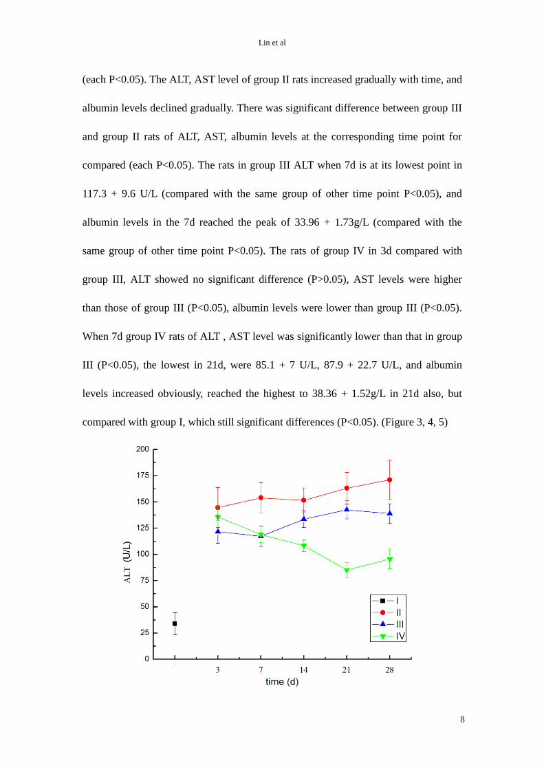
(each P<0.05). The ALT, AST level of group II rats increased gradually with time, and
albumin levels declined gradually. There was significant difference between group III
and group II rats of ALT, AST, albumin levels at the corresponding time point for
compared (each P<0.05). The rats in group III ALT when 7d is at its lowest point in
117.3 + 9.6 U/L (compared with the same group of other time point P<0.05), and
albumin levels in the 7d reached the peak of 33.96 + 1.73g/L (compared with the
same group of other time point P<0.05). The rats of group IV in 3d compared with
group III, ALT showed no significant difference (P>0.05), AST levels were higher
than those of group III (P<0.05), albumin levels were lower than group III (P<0.05).
When 7d group IV rats of ALT , AST level was significantly lower than that in group
III (P<0.05), the lowest in 21d, were 85.1 + 7 U/L, 87.9 + 22.7 U/L, and albumin
levels increased obviously, reached the highest to 38.36 + 1.52g/L in 21d also, but
compared with group I, which still significant differences (P<0.05). (Figure 3, 4, 5)

Encapsulated hepatocytes transplantation
9
Figure 3- ALT value of each group at different time points
II, III, IV groups of rats at each time point of ALT was higher than that in normal
group rats (each P<0.05). There was significant difference between group III and
group II of ALT levels at the corresponding time point (each P<0.05). Group IV in 3d
compared with group III, ALT showed no significant difference (P>0.05), When 7d
group IV of ALT level was significantly lower than that in group III (P<0.05).
Figure 4- AST value of each group at different time points
II, III, IV groups of rats at each time point of AST was higher than that in normal
group rats (each P<0.05). Significant difference between group III and group II of
AST levels at the corresponding time point for compared (each P<0.05). The rats of
group IV AST levels were higher than those time point of group III (P<0.05).
Lin et al
10
Figure 5- Albumin value of each group at different time points
II, III, IV groups of rats at each time point of albumin levels decreased
significantly (each P<0.05). There was significant difference between group III and
group II of albumin level (each P<0.05). Group IV in 3d compared with group III,
albumin levels were lower (P<0.05).
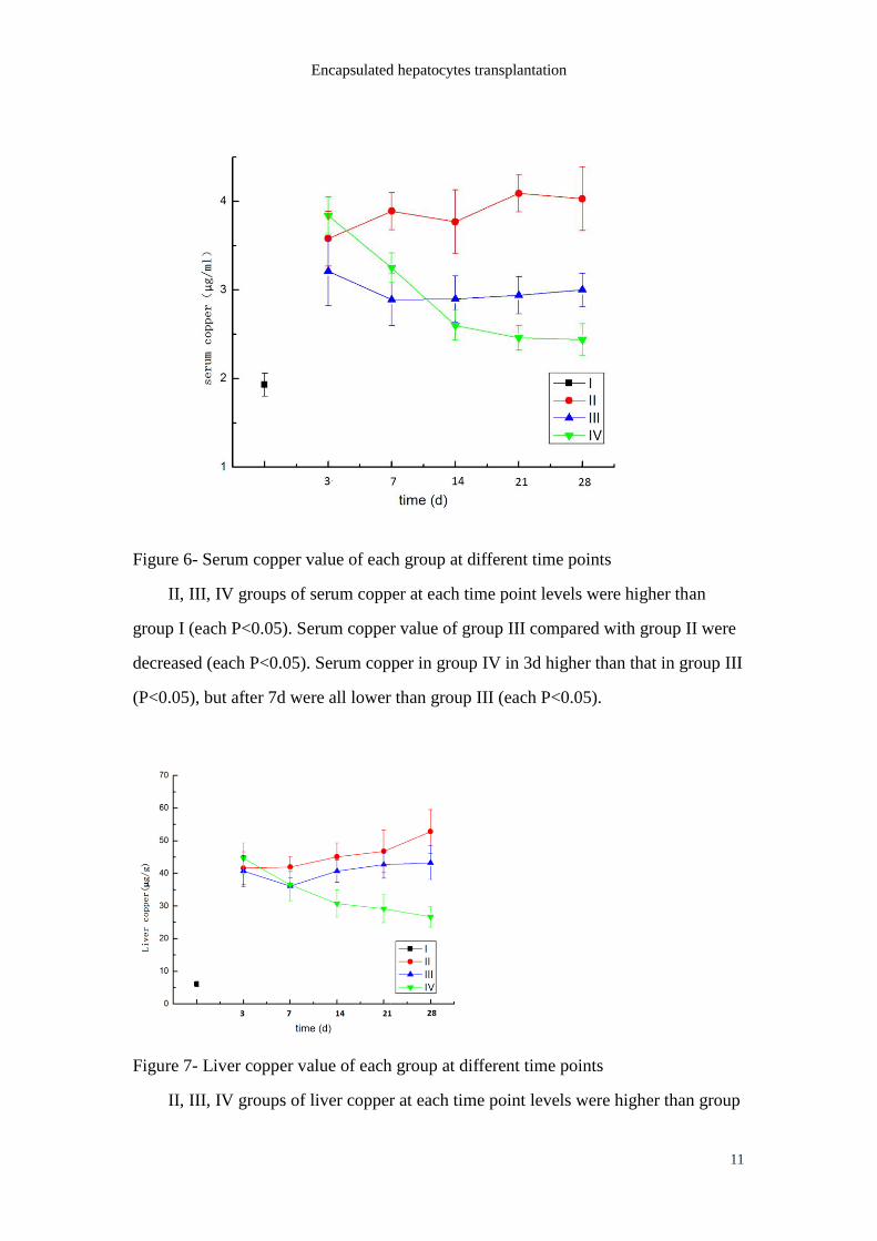
Variations of serum copper, copper content in the liver tissue
II, III, IV groups of rats’ serum copper, liver copper at each time point levels
were higher than group I which significantly increasing (each P<0.05). The rats in
group II, the liver copper content and serum copper increased gradually with the time
point. Serum copper value of group III compared with group II at the corresponding
time points were decreased (each P<0.05), and the liver copper content in 7d, 14d and
28d lower than that of group II (each P<0.05). Serum copper value of group III
didn’t get significant fluctuations after 7d (P>0.05), and the liver copper values in the
7d reaches the lowest to 36.10 + 4.4μg/g and thereafter gradually upward trend.
Serum copper in group IV in 3d higher than that in group III (P<0.05), but after 7d
were all lower than group III (each P<0.05), the minimum to 2.44 + 0.18μg/ml, but
still significantly higher than those in group I (P<0.05). Liver copper content of group
IV in 3d, 7d compared with group III showed no difference (each P>0.05), but the
14d decreased (P<0.05), the minimum to 26.73 + 3.22μg/g, were significantly higher
than that of group I (P<0.05) . (Figure 6,7)
Encapsulated hepatocytes transplantation
11
Figure 6- Serum copper value of each group at different time points
II, III, IV groups of serum copper at each time point levels were higher than
group I (each P<0.05). Serum copper value of group III compared with group II were
decreased (each P<0.05). Serum copper in group IV in 3d higher than that in group III
(P<0.05), but after 7d were all lower than group III (each P<0.05).
Figure 7- Liver copper value of each group at different time points
II, III, IV groups of liver copper at each time point levels were higher than group
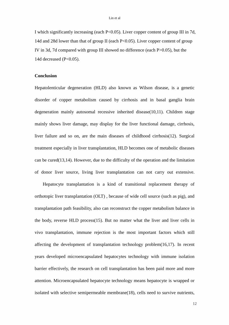
Lin et al
12
I which significantly increasing (each P<0.05). Liver copper content of group III in 7d,
14d and 28d lower than that of group II (each P<0.05). Liver copper content of group
IV in 3d, 7d compared with group III showed no difference (each P>0.05), but the
14d decreased (P<0.05).
Conclusion
Hepatolenticular degeneration (HLD) also known as Wilson disease, is a genetic
disorder of copper metabolism caused by cirrhosis and in basal ganglia brain
degeneration mainly autosomal recessive inherited disease(10,11). Children stage
mainly shows liver damage, may display for the liver functional damage, cirrhosis,
liver failure and so on, are the main diseases of childhood cirrhosis(12). Surgical
treatment especially in liver transplantation, HLD becomes one of metabolic diseases
can be cured(13,14). However, due to the difficulty of the operation and the limitation
of donor liver source, living liver transplantation can not carry out extensive.
Hepatocyte transplantation is a kind of transitional replacement therapy of
orthotopic liver transplantation (OLT) , because of wide cell source (such as pig), and
transplantation path feasibility, also can reconstruct the copper metabolism balance in
the body, reverse HLD process(15). But no matter what the liver and liver cells in
vivo transplantation, immune rejection is the most important factors which still
affecting the development of transplantation technology problem(16,17). In recent
years developed microencapsulated hepatocytes technology with immune isolation
barrier effectively, the research on cell transplantation has been paid more and more
attention. Microencapsulated hepatocyte technology means hepatocyte is wrapped or
isolated with selective semipermeable membrane(18), cells need to survive nutrients,

Encapsulated hepatocytes transplantation
13
oxygen, metabolites and secretion of bioactive substances through a semipermeable
membrane access, but host immune cells, immune globulin and complement can not
through a semipermeable membrane, so hepatocyte in capsule will not suffer from
host immune rejection and get long-term survival, exerts its biological function, to
achieve the purpose of treatment. At present in the treatment of fulminant hepatic
failure have got a high embodiment of value(19,20). The current HLD treatment has
formed the hepatocyte transplantation in spleen transplantation method as the main
pathway(21). But cause of the size which restricted to microencapsulated hepatocytes
(300 microns), spleen transplantation is obviously difficult to achieve, therefore this
research selected peritoneal transplant operation- the most common pathway for
microencapsulated transplantation.
Copper load model is currently the most common HLD animal model, can better
reflect the similar HLD liver copper injury(22). In this study, high concentrations of
copper fed rats for up to 12 weeks in the given, the serum levels of ALT, AST and
albumin levels showed a similar hepatitis even liver dysfunction performance, which
is consistent with the liver damage after HLD copper deposition(23). Liver copper
and serum copper level is the direct reflection of liver copper deposition to lead liver
damage extent, meanwhile, monitoring of copper level in liver tissue and serum helps
to understand the effect of hepatocyte transplantation therapy. In this experiment, the
treatment group (naked hepatocyte transplantation group and microencapsulated
hepatocyte transplantation group) liver aminopherase levels in rats after
transplantation than copper loaded rats have decreased, liver copper and serum copper

Lin et al
14
levels also decreased, and the liver synthesis of albumin levels have also been
enhanced. However, the naked hepatocyte transplantation group whether liver
aminopherases, liver copper and serum copper level was decreased, or albumin
synthesis recovery degree, or the effect of the consolidation time, compared
microencapsulated hepatocyte transplantation group to the existence of significant
differences. This may be due to immune bare hepatocytes can not tolerate receptor
rejection. In this study, the decreasing degree of naked hepatocyte transplantation
group liver aminopherase levels, copper levels were lowest in about 7 days, and 14
days is obviously aggravated, suggest that the function of the liver cells difficult to
last more than 1 weeks. The microencapsulated hepatocyte transplantation rats liver
aminopherases, serum copper, liver copper levels were significantly lower than the
naked hepatocyte transplantation group, 28 days before took on a declining trend,
while the synthesis of albumin levels increased significantly, at the same time
prolonging holding time effect. This comprehensive experimental results showed
microencapsulated hepatocyte transplantation can improve copper metabolism level
of HLD rats, alleviate the hepatic copper deposition, accelerate the serum copper
metabolism, improve liver function, indicates that the research prospect in hepatocyte
transplantation in the treatment of HLD. But HLD is a chronic disease, how to further
improve the time to maintain the function of hepatocyte transplantation will be the
next focus of research work.

Encapsulated hepatocytes transplantation
15
Acknowledgements
This work was supported by Medical science and technology project of Zhejiang
Province, China (No. 201339321).
Conflict of Interest
We declare that we have no financial and personal relationships with other people or
organizations that can inappropriately influence our work, there is no professional or
other personal interest of any nature or kind in any product, service and/or company
that could be construed as influencing the position presented in, or the review of, the
manuscript entitled,

Lin et al
16
REFERENCES
1 Schilsky ML. Wilson disease:current status and the future. Biochimie J 2009;
91:1278-1281.
2 Abuduxikuer K, Wang JS. Zinc mono-therapy in pre-symptomatic Chinese
children with Wilson disease: a single center, retrospective study. PLoS One J
2014 Jan 24;9(1):e86168. doi: 10.1371/journal.pone.0086168. eCollection 2014.
3 Rosencrantz R, Schilsky M. Wilson disease: pathogenesis and clinical
considerations in diagnosis and treatment. Semin Liver Dis J 2011
Aug;31(3):245-259.
4 Filippi C, Dhawan A. Current status of human hepatocyte transplantation and
its potential for Wilson's disease. Ann N Y Acad Sci J 2014 May;1315:50-55.
5 Mei J, Sgroi A, Mai G, Baertschiger R, Gonelle-Gispert C, Serre-Beinier V et al.
Improved survival of fulminant liver failure by transplantation of
microencapsulated cryopreserved porcine hepatocytes in mice. Cell Transplant J
2009;18(1):101-110.
6 Sgroi A, Mai G, Morel P, Baertschiger RM, Gonelle-Gispert C, Serre-Beinier V et
al. Transplantation of encapsulated hepatocytes during acute liver failure
improves survival without stimulating native liver regeneration. Cell Transplant J
2011;20(11-12):1791-1803.
7 Seglen PO. Preparation of rat liver cells. I. Effect of Ca2+
on enzymatic
dispersion of isolated, perfused liver. Exp Cell Res J 1972;74(2):450-454.
8 Kang IK, Moon JS, Jeon HM, Meng W, Kim YI, Hwang YJ et al. Morphology
and metabolism of Ba-alginate encapsulated hepatocytes with galactosylated
poly(allyl amine) and poly(vinyl alcohol) as extracellular matrices. Mater Sci
Mater Med J 2005;16(6):533-539.
9 Pulverer BJ, Kakis JM, Avrueh J, Nikolakaki E, Woodgett JR et al.
Phosphorylation of c-junmediated by MAP kinases. Nature J 199l; 353:670-674.
10 Kitzberger R, Madl C, Ferenci P. Wilson disease. Metab Brain Dis J
2005;20(4):295-302.
11 Rosen JM, Kuntz N, Melin-Aldana H, Bass LM. Spasmodic muscle cramps and
weakness as presenting symptoms in Wilson disease. Pediatrics J
2013;132(4):1039-1042.
12 Hahn SH. Population screening for Wilson's disease. Ann N Y Acad Sci J
2014;1315:64-69.
13 Narumi S, Umehara M, Toyoki Y, Ishido K, Kudo D, Kimura N et al. Liver
transplantation for Wilson's disease in pediatric patients: decision making and
timing. Transplant Proc J 2012;44(2):478-480.
14 Schilsky ML. Liver transplantation for Wilson's disease. Ann N Y Acad Sci J
2014;1315:45-49.
15 Malhi H, Irani AN, Volenherg I, Schilsky ML, Gupta S. Early cell
transplantation in LEC rats modeling Wilson disease eliminates hepatic copper
with re-versa]of liver disease. Gastroenterology J 2002;122(2) :438-447.
16 Pons JA, Revilla-Nuin B, Ramírez P, Baroja-Mazo A, Parrilla P. Development

Encapsulated hepatocytes transplantation
17
of immune tolerance in liver transplantation. Gastroenterol Hepatol J
2011;34(3):155-169.
17 Sood S, Testro AG. Immune monitoring post liver transplant. World J
Transplant J 2014 24;4(1):30-39.
18 Orive G, Hernandez RM, Rodriguez Gascon A, Calafiore R, Chang TM, de Vos
P et al. History, challenges and perspectives of cell microencapsulation. Trends
Biotechnol J 2004;22(2):87-92.
19 Nyberg SL, Peshwa MV, Payne WD, Hu WS, Cerra FB. Evolution of the
bioartificial liver: the need for randomized clinical trials. Am J Surg J
1993;166(5):512-521.
20 Mai G, Nguyen TH, Morel P, Mei J, Andres A, Bosco D et al. Treatment of
fulminant liver failure by transplantation of microencapsulated primary or
immortalized xenogeneic hepatocytes. Xenotransplantation J 2005;12(6):457-64.
21 Raper SE, Wilson JM. Cell transplantation in liver-directed gene therapy. Cell
Transplant J 1993;2(5):381-400.
22 Suzuki KT, Kanno S, Misawa S, Aoki Y. Copper metabolism leading to and
following acute hepatitis in LEC rats. Toxicology J 1995; 97:8l-92.
23 Friendman L S, Yarze J C. Zinc in the Treatment of Wilson's Disease: How It
Works. Gastroenterology J 1993; 4(5) :1566.