Embed Size (px)
Citation preview

9 Apr 2003 13:41 AR AR184-PP54-16.tex AR184-PP54-16.sgm LaTeX2e(2002/01/18)P1: GJB10.1146/annurev.arplant.54.031902.134823
Annu. Rev. Plant Biol. 2003. 54:403–30doi: 10.1146/annurev.arplant.54.031902.134823
Copyright c© 2003 by Annual Reviews. All rights reserved
HOW DO CELLS KNOW WHAT THEY WANT TO
BE WHEN THEY GROW UP? Lessons fromEpidermal Patterning in Arabidopsis
John C. Larkin1, Matt L. Brown1, and John Schiefelbein21Department of Biological Sciences, Louisiana State University, Baton Rouge, Louisiana70803; email: [email protected], [email protected] of Molecular, Cellular, and Developmental Biology, University of Michigan,Ann Arbor, Michigan 48109; email: [email protected]
Key Words pattern formation, cell differentiation, lateral inhibition, MYB, basichelix-loop-helix (bHLH), WD-repeat, trichome, root hair, stomata
■ Abstract Because the plant epidermis is readily accessible and consists of fewcell types on most organs, the epidermis has become a well-studied model for celldifferentiation and cell patterning in plants. Recent advances in our understanding ofthe development of three epidermal cell types, trichomes, root hairs, and stomata, allowa comparison of the underlying patterning mechanisms. In Arabidopsis, trichome de-velopment and root epidermal patterning use a common mechanism involving closelyrelated cell fate transcription factors and a similar lateral inhibition signaling pathway.Yet the resulting patterns differ substantially, primarily due to the influence of a prepat-tern derived from subepidermal cortical cells in root epidermal patterning. Stomatalpatterning uses a contrasting mechanism based primarily on control of the orientationof cell divisions that also involves an inhibitory signaling pathway. This review focuseson comparing and contrasting these patterning pathways to identify and illustrate gen-eral themes that may be broadly applicable to other systems. Where these pathwaysoccur in the same tissue, interaction and competition between these pathways is alsodiscussed.
CONTENTS
INTRODUCTION . . . . . . . . . . . . . . . . . . . . . . . . . . . . . . . . . . . . . . . . . . . . . . . . . . . . . 404THE EPIDERMIS . . . . . . . . . . . . . . . . . . . . . . . . . . . . . . . . . . . . . . . . . . . . . . . . . . . . . 405TRICHOMES . . . . . . . . . . . . . . . . . . . . . . . . . . . . . . . . . . . . . . . . . . . . . . . . . . . . . . . . 405
Cell Fate Transcription Factors. . . . . . . . . . . . . . . . . . . . . . . . . . . . . . . . . . . . . . . . . 406Interactions Among Cell Fate Transcription Factors. . . . . . . . . . . . . . . . . . . . . . . . . 407Local Signaling in Trichome Development. . . . . . . . . . . . . . . . . . . . . . . . . . . . . . . . 408A Case of Transdetermination?. . . . . . . . . . . . . . . . . . . . . . . . . . . . . . . . . . . . . . . . . 410Tissue-Specificity and Hormone Regulationof Trichome Production. . . . . . . . . . . . . . . . . . . . . . . . . . . . . . . . . . . . . . . . . . . . . . 411
The Role of the Cell Cycle in Trichome Formation. . . . . . . . . . . . . . . . . . . . . . . . . 411
1040-2519/03/0601-0403$14.00 403

9 Apr 2003 13:41 AR AR184-PP54-16.tex AR184-PP54-16.sgm LaTeX2e(2002/01/18)P1: GJB
404 LARKIN ¥ BROWN ¥ SCHIEFELBEIN
ROOT EPIDERMAL PATTERNING . . . . . . . . . . . . . . . . . . . . . . . . . . . . . . . . . . . . . . 412Cell Fate Transcription Factors. . . . . . . . . . . . . . . . . . . . . . . . . . . . . . . . . . . . . . . . . 413Local Signaling in Root Epidermal Patterning. . . . . . . . . . . . . . . . . . . . . . . . . . . . . 414Prepattern and Hormone Signaling. . . . . . . . . . . . . . . . . . . . . . . . . . . . . . . . . . . . . . 415The Cell Cycle in Root Epidermal Patterning. . . . . . . . . . . . . . . . . . . . . . . . . . . . . . 415
TRICHOMES AND ROOT HAIRS: ONEMECHANISM, TWO PATTERNS . . . . . . . . . . . . . . . . . . . . . . . . . . . . . . . . . . . . . . . 416
ADDITIONAL CELL TYPES AND PATHWAYS EMPLOYINGMYB/bHLH PROTEINS: A CONSERVED REGULATORYMODULE? . . . . . . . . . . . . . . . . . . . . . . . . . . . . . . . . . . . . . . . . . . . . . . . . . . . . . . . . . 416
STOMATA . . . . . . . . . . . . . . . . . . . . . . . . . . . . . . . . . . . . . . . . . . . . . . . . . . . . . . . . . . . 417Initiation of Stomatal Development. . . . . . . . . . . . . . . . . . . . . . . . . . . . . . . . . . . . . . 418Local Signaling in Stomatal Development. . . . . . . . . . . . . . . . . . . . . . . . . . . . . . . . 418Tissue-Specific and Environmental Influences on Stomatal Patterning. . . . . . . . . . 420The Cell Cycle in Stomatal Patterning. . . . . . . . . . . . . . . . . . . . . . . . . . . . . . . . . . . 421
INTERACTIONS AMONG CELL-FATE PATHWAYS . . . . . . . . . . . . . . . . . . . . . . . . 421Integration of Trichome, Stomatal, and Pavement Cell DifferentiationDuring Leaf Development. . . . . . . . . . . . . . . . . . . . . . . . . . . . . . . . . . . . . . . . . . . . 421
Patterning of Stomata in Hypocotyl Uses theRoot Epidermal Patterning Mechanism. . . . . . . . . . . . . . . . . . . . . . . . . . . . . . . . . . 422
CONCLUSION: TOWARD A GENERAL THEORYOF CELL FATE SPECIFICATION?. . . . . . . . . . . . . . . . . . . . . . . . . . . . . . . . . . . . . . 423
INTRODUCTION
Plants, like other multicellular eukaryotes, develop from a single-celled zygotethat ultimately gives rise to the many specialized cell types of the adult organ-ism. Cell patterning is when cells are guided to their appropriate differentiatedfate at the correct time and place in the developing organism. Although pattern-ing mechanisms are very heterogeneous in detail, some common themes emergerepeatedly in a wide variety of cell types and organisms. First, commitment to aspecific cell fate is typically regulated by the combinatorial interaction of severaltranscription factors. Second, the timing and location of differentiation is oftenregulated by both long-range hormonal signals and local cell-cell signaling. Third,differentiating cells often either stop cycling mitotically or enter a modified cellcycle.
The plant epidermis is an excellent tissue for studying cell patterning. In bothshoots and roots, the epidermis consists of few cell types (20). The epidermis is alsoreadily accessible to observation and developmental manipulation. In geneticallywell-studied plants such asArabidopsis thaliana, numerous mutants affecting epi-dermal cell types have been isolated and studied. This review focuses on recentwork that provides significant insight into the mechanisms involved in the pattern-ing of three epidermal cell types in Arabidopsis: trichomes, root hairs, and stomatalguard cells. Work on other cell types and plant species is discussed where appropri-ate. Individually, these cell types have been the focus of numerous recent reviews

9 Apr 2003 13:41 AR AR184-PP54-16.tex AR184-PP54-16.sgm LaTeX2e(2002/01/18)P1: GJB
EPIDERMAL PATTERNING 405
(17, 67, 82, 91). However, this review focuses on comparing and contrasting thesepatterning pathways in an attempt to identify and illustrate general themes thatmay be broadly applicable to other systems. Where these pathways occur in thesame tissue, we also discuss the interaction and competition between them.
THE EPIDERMIS
The migration of plants onto land in the Ordovician resulted in new selectivepressures acting on the surface tissues of early land plants (78). The requirementsfor gas exchange were changed, and both dehydration and UV damage becamesevere threats. Soon, defenses were needed against new types of herbivores andpathogens, and new opportunities arose for the dispersal of spores and gametes.In response, a wide variety of epidermal specializations arose, including a waxycuticle and many different specialized types of cells. These specialized cells includestomatal guard cells, root hairs, trichomes (shoot hairs), various secretory cells ofglands and nectaries, the enlarged bulliform cells of monocot leaves, and ordinaryepidermal pavement cells, among many others (20).
The epidermis of angiosperms has three different developmental origins. Theepidermis of the embryonic organs, the cotyledons and the hypocotyl, originatesdirectly from the divisions establishing the embryo tissues (20). After germination,the shoot’s epidermis originates from the outer or L1 layer of the shoot apicalmeristem (20, 87), and the root’s epidermis originates from the root apical meristemas well as from the meristems of lateral roots. At the root tips, the meristemand developing epidermis (protoderm) are covered by the root cap, which is alsoproduced by the root meristem. The shoot and root meristems differ significantlyin the way tissues derived from the meristem are produced, and lateral organs(leaves and lateral roots) are produced much differently in the shoot and root (20).As a consequence of these differences, the root protoderm typically has a moreorderly relationship with the underlying cortical tissue than the leaf protoderm haswith the underlying mesophyll. These differences have interesting consequencesfor the patterning of shoot and root epidermal cell types (see below).
TRICHOMES
Trichomes (shoot epidermal hairs) are present on the aerial surfaces of most plants(trachaeophytes), ranging from ferns to angiosperms. The term trichome is derivedfrom trichos, the Greek root meaning hair. Trichomes exist in a wide varietyof morphologies, from single celled to multicellular, and include both glandularsecretory hairs and nonglandular hairs (20, 95). A wide variety of functions havebeen ascribed to trichomes in various plants, including resisting insect herbivores,reducing transpiration, increasing freezing tolerance, and protecting plants fromUV light (40). In addition to their roles in the plant, some types of trichomes havesignificant economic importance; for instance, cotton fibers are trichomes isolatedfrom the epidermis ofGossypiumovules.

9 Apr 2003 13:41 AR AR184-PP54-16.tex AR184-PP54-16.sgm LaTeX2e(2002/01/18)P1: GJB
406 LARKIN ¥ BROWN ¥ SCHIEFELBEIN
One of the most thoroughly studied plant cell differentiation pathways is thedevelopment of Arabidopsis trichomes (91). Ecological studies suggest that Ara-bidopsis trichomes protect plants from insect herbivores (60). These trichomesare single cells that protrude from the epidermis of leaves and stems. On leaves,these cells have an unusual branched shape resulting from a dramatic programof cellular morphogenesis. On stems, trichomes are generally unbranched. Thesingle nucleus of a wild-type trichome continues to replicate its genomic DNAduring differentiation, reaching average nuclear DNA levels of 20C–32C (34, 63),a process known as either endoreplication or endoreduplication (19). Endorepli-cation is a common variant of the cell cycle in which mitosis and cytokinesis aresuppressed, but cycles of DNA replication continue. The ready accessibility oftrichomes on the leaves of Arabidopsis has facilitated the isolation of mutationsidentifying genes involved in all stages of trichome development (56).
Trichome development begins near the distal tips of leaves when they are ap-proximately 100-µm long, and proceeds basipetally (48). Trichomes are foundadjacent to one another much less frequently than would be expected by chance,suggesting that an active mechanism exists to govern trichome spacing (34, 48).Epidermal cells that have entered the trichome pathway can first be identified byan increase in cell and nuclear volume. The nuclear enlargement in developing tri-chomes is correlated with the start of endoreplication (34, 104). The trichome thenbegins to expand out of the plane of the epidermis and branching ensues, followedby elaboration of the thickened secondary cell wall of the mature trichome.
Cell Fate Transcription Factors
Several genes that directly control trichome initiation and development have beenidentified. Null alleles of theGLABRA1(GL1) gene result in plants with no or veryfew trichomes.GL1encodes an R2-R3 MYB transcription factor with two repeatsof the MYB DNA-binding domain (72), typical of a large family of plant MYBs(43). The GL1 protein is localized to the nucleus (89). The highest levels ofGL1transcripts are found in early stages of developing trichomes, but low levels ofGL1expression are found throughout the developing epidermis (46). The increasein GL1expression appears to precede detectable expansion of trichome precursorcells. This observation suggests that commitment to the trichome fate involves apositive feedback loop regulatingGL1expression.
Null alleles of TRANSPARENT TESTA GLABRA(TTG) result in a hairlessphenotype as well as reduced anthocyanin pigmentation, absence of seed coatmucilage, and an increase in the number of root hairs (22, 42). The various aspectsof the pleiotropicttg mutant result from a defect in a single biochemical function,rather than from multiple independent functional domains in the protein (47).TTGencodes a small protein with four or five WD repeats (103). WD repeats appearto function as protein interaction domains in a wide variety of processes, and noWD-repeat protein has either enzymatic activity or a DNA-binding domain (68).
A third gene that plays a central role in trichome initiation isGLABRA3(GL3).Loss-of-functiongl3 mutants produce a reduced number of trichomes that aresmaller and have fewer branches than wild type (34). The nuclear DNA content

9 Apr 2003 13:41 AR AR184-PP54-16.tex AR184-PP54-16.sgm LaTeX2e(2002/01/18)P1: GJB
EPIDERMAL PATTERNING 407
of gl3 mutant trichomes is also reduced (34). The recent demonstration thatGL3encodes a basic helix-loop-helix (bHLH) protein closely related to the maizeRgene (74) clears up a lingering mystery. The maizeRgene was previously shown tosuppress the mutant defects ofttgwhen constitutively expressed in Arabidopsis, aswell as cause ectopic production of trichomes, especially when coexpressed withGL1, but until the identification ofGL3, no Arabidopsis gene encoding a bHLHprotein had been implicated in trichome development (45, 54). Payne et al. (72)also shed light on why even apparentgl3 null alleles produce some trichomes.They showed that the Arabidopsis genome contains a close homolog ofGL3 thatmay be functionally redundant withGL3.
Recent work suggests that theGLABRA2(GL2) gene may also play a role intrichome initiation (36, 71). Loss-of-functiongl2mutants produce small trichomeswith reduced branching and aberrant expansion, in addition to the pleiotropic phe-notypes of extra root hairs and absence of seed coat mucilage (16, 79).GL2encodesa homeodomain-leucine zipper protein (16, 79) that is expressed at high levels indeveloping trichomes and is localized to the nucleus of developing trichomes (79,89). Because altered cell expansion is the primary effect ofgl2 mutations on tri-chome development,GL2 has been proposed to regulate polar cell expansion ofdeveloping trichomes (34, 79). Constitutive overexpression ofGL2under control ofthe Cauliflower mosaic virus35S promoter appears to be lethal (71). However, vi-able transgenic lines exhibiting moderate overexpression ofGL2 were identified,and these plants produce increased numbers of trichomes, as well as increasednumbers of trichomes adjacent to another trichome (trichome clusters), stronglysuggesting a role forGL2 in trichome initiation (71).
Several other genes have been implicated in the initiation of trichome develop-ment. TheREDUCED TRICHOME NUMBER(RTN) locus was discovered as aquantitative trait locus (QTL) variant between the Columbia and Landsbergerectaecotypes ofArabidopsis(48). This locus affects the number of trichomes pro-duced on leaves by controlling the time during leaf development that the initiationof new trichomes ends. This study also described two other potential QTLs thatmay influence the trichome density on leaves (48). The recently describedTRANS-PARENT TESTA-GLABRA2(TTG2) gene, which encodes a WRKY transcriptionfactor, may also play a role in trichome initiation (39). Finally, several genes regu-late trichome development via local or hormonal signaling; the roles of these genesare described below in separate sections. Two other loci,FIDDLEHEAD (FDL)(113) andINCREASED CHALCONE SYNTHASE EXPRESSION(ICX) (37, 101)also affect the number of trichomes per leaf.
Interactions Among Cell Fate Transcription Factors
Initial experiments in whichGL1 and the maizeR gene were expressed constitu-tively from theCauliflower mosaic virus35S promoter suggested thatGL1 waslikely to function at the same genetic step in trichome development as an Arabidop-sisR homolog (44, 45). With the demonstration thatGL3 encodes a homolog ofR, these interactions were confirmed using the endogenous Arabidopsis genes(74). Indeed, co-overexpression ofGL1 andGL3 results in increased numbers of
9 Apr 2003 13:41 AR AR184-PP54-16.tex AR184-PP54-16.sgm LaTeX2e(2002/01/18)P1: GJB
408 LARKIN ¥ BROWN ¥ SCHIEFELBEIN
trichomes and ectopic trichomes, and can bypass the need forTTG function. Thelatter result suggests thatTTG is functionally upstream ofGL1andGL3.
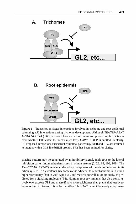
There is strong evidence that the products of these three genes interact basedprimarily on a careful yeast two-hybrid study (Figure 1a) (74). First, both GL1 andGL3 contain transcription activation domains, GL1 in its C terminus and GL3 inits N terminus. The GL1 activation domain may correspond to an essential regionof 34 amino acids near the C terminus that is rich in acidic amino acids (21, 31).Second, GL3 can homodimerize via a C-terminal region containing the bHLHdomain. Third, GL1 and GL3 interact strongly via the N-terminal MYB domainrepeats of GL1, and at least the first 96 amino acids of GL3 (74). GL1 and the maizeR protein also interact in vitro, although in these experiments the N terminus ofGL1 alone was not sufficient for interaction with R (89). Interaction between GL1and GL3 may explain the curious observation that overexpression ofGL1 inhibitstrichome formation (45), if it is assumed that GL3 is limiting, and that GL1 that isnot bound to GL3 inhibits transcription from downstream promoters. Consistentwith this hypothesis, overexpression of bothGL1 andGL3 together rescues theinhibitory effect ofGL1overexpression (74).
Finally, Payne et al. (74) demonstrated that, in yeast, TTG interacts with theN-terminal region of GL3 distinct from that to which GL1 binds. TTG does notbind to GL1. Although genetic interactions betweengl1 andttg alleles were ini-tially interpreted as evidence that GL1 and TTG interact directly, these geneticinteractions are also consistent with these two proteins being part of the samecomplex, but not in physical contact (47). Although TTG interacts with GL3, it isunclear whether TTG actually enters the nucleus. A closely related homolog frompetunia, AN11, appears to be cytoplasmic (15), andAN11can rescuettg mutationswhen expressed in Arabidopsis (74). At this point, while TTG may be part of thetranscription complex with GL1 and GL3 (Figure 1a), it may also act solely in thecytoplasm to stabilize homo- or heterodimer formation, or be involved in activatingthe GL1/GL3 complex in some way, perhaps by stabilizing its interaction with amodifying enzyme such as a protein kinase.
The only candidate gene so far identified as a likely target regulated by GL1 andGL3 isGL2(Figure 1a). Overexpression ofGL1together with the maizeRgene re-sults in ectopic expression of aGL2reporter construct throughout the plant, includ-ing tissues that normally do not expressGL2 (89), although it has not been deter-mined whether this expression is independent of new protein synthesis, as predictedfor a direct transcriptional target (102). This ectopic expression was dependent on aregion of theGL2promoter required forGL2expression in developing trichomes.It is also unlikely thatGL2 is the only target of this transcription complex.
Local Signaling in Trichome Development
As noted above, at the time of initiation, trichomes are spaced nonrandomly, withan average distance of three cells between developing trichomes (34, 48). Thisminimum-distance spacing pattern (111) cannot be explained by a specialized celllineage based on a stereotyped pattern of cell divisions (48), suggesting that the
9 Apr 2003 13:41 AR AR184-PP54-16.tex AR184-PP54-16.sgm LaTeX2e(2002/01/18)P1: GJB
EPIDERMAL PATTERNING 409
Figure 1 Transcription factor interactions involved in trichome and root epidermalpatterning. (A) Interactions during trichome development. AlthoughTRANSPARENTTESTA GLABRA(TTG) is shown here as part of the transcription complex, it is un-clear whether TTG enters the nucleus (see text).CAPRICE(CPC) omitted for clarity.(B) Proposed interactions during root epidermal patterning. WER and TTG are assumedto interact with a GL3-like bHLH protein. TRY has been omitted for clarity.
spacing pattern may be generated by an inhibitory signal, analogous to the lateralinhibition patterning mechanisms seen in other systems (2, 26, 88, 108, 109). TheTRIPTYCHON(TRY) gene encodes a key component of the trichome lateral inhi-bition system. Intry mutants, trichomes arise adjacent to other trichomes at a muchhigher frequency than in wild type (34), andtry acts noncell autonomously, as pre-dicted for a signaling molecule (84). Homozygoustry mutants that also constitu-tively overexpressGL1and maizeRhave more trichomes than plants that just over-express the two transcription factors (84). ThusTRYcannot be solely a repressor

17 Apr 2003 14:8 AR AR184-PP54-16.tex AR184-PP54-16.sgm LaTeX2e(2002/01/18)P1: GJB
410 LARKIN ¥ BROWN ¥ SCHIEFELBEIN
of GL1transcription, but must inhibit trichome development by interfering with theability of GL1andGL3 to activate transcription of downstream genes (Figure 1a).Consistent with this possibility,try mutations can allowGL1 overexpression topartially bypass the need forTTG for trichome initiation (84). Additionally,constitutive expression of TRY eliminates production of trichomes (81).
TRYencodes a single-repeat MYB protein with no apparent transcription acti-vation domain (81). The nature of the predicted TRY protein is consistent with thehypothesis that it acts as a transcriptional repressor of targets of theGL1/GL3com-plex. The sequence ofTRYis similar to that of theCAPRICE(CPC) gene (100),an important negative regulator of root epidermal patterning, as described below.However,CPCalso plays at least a minor role in trichome patterning parallel tothat of TRY. Constitutive expression ofCPC in leaves eliminates trichome pro-duction (100), andcpcmutants have an increased number of trichomes, especiallyin combination withtry (81). BothTRYandCPCare expressed most strongly indeveloping trichomes and not in other epidermal cells, even though their functionis to inhibit neighboring cells from developing as trichomes (81). However, planttranscription factors can move between cells via plasmodesmata (55), and a rea-sonable model is that these proteins are synthesized in the developing trichomeand move to neighboring cells to inhibit them from developing as trichomes.
Figure 2 shows a model for selecting trichome precursors in the observedminimum-distance spacing pattern based on a lateral inhibition mechanism. Lat-eral inhibition is a patterning mechanism whereby cells taking on a particular fatesend an inhibitory signal to their neighbors, preventing them from adopting thesame fate (2, 26, 88, 108, 109). Initially, all cells in the protoderm are competentto develop as trichomes (Figure 2a). These cells produce low levels of GL1 andpresumably GL3 and TTG as well. Together, these proteins comprise a transcrip-tional activator that induces transcription of the downstream genesGL2, TRY, andCPC, among other genes (Figure 2a). The GL2 protein promotes the trichome cellfate, while TRY and CPC are transported to neighboring cells, where they inhibitexpression ofGL1, GL2, TRY, CPC, and perhapsGL3 or TTG. At first, the cellsare locked in mutual inhibition (Figure 2a). This state is metastable, and owing tostocastic variations in gene expression levels, some cells will have higher levelsof the transcriptional acitvators (Figure2b). These cells will then produce higherlevels of TRY and CPC, and due to the feedback loops between cells, these cellswill ultimately become committed to the trichome fate and succeed in inhibitingtheir neighbors from doing the same. Although direct evidence for the intercellularfeedback loops postulated here has not yet been demonstrated for trichome devel-opment, such feedback loops have now been clearly demonstrated for the closelyrelated mechanism described below that patterns the root epidermis (50).
A Case of Transdetermination?
There is one result that does not seem to fit the proposed model based on lateralinhibition. The trichome clusters intry cpc double mutants are large, consistingof 20–30 adjacent trichomes (81). During the formation of these clusters, all cells

9 Apr 2003 13:41 AR AR184-PP54-16.tex AR184-PP54-16.sgm LaTeX2e(2002/01/18)P1: GJB
EPIDERMAL PATTERNING 411
immediately adjacent to a developing trichome begin to expand from the epidermisand develop as trichomes. Their neighbors repeat this process for several rounds,resulting in a large trichome cluster. In wild type, cells adjacent to a trichomeexpand within the epidermal plane and become trichome accessory cells. Acces-sory cell differentiation is induced by a trichome late in its development. Perhapsthe accessory cell program is a modification of the trichome program, andTRYandCPC act to shunt these cells into the accessory cell fate by repressing partof the trichome program. In this case, as is observed, a trichome developing ona try cpc mutant would induce trichome formation in its neighbors, rather thanaccessory cell development, and these trichomes would induce further trichomes,etc., ultimately producing a large cluster.
Tissue-Specificity and Hormone Regulationof Trichome Production
Several genes are involved in restricting trichome expression to appropriate tis-sues.TRYhas in determining the tissue-specificity of trichome formation. Over-expression ofGL1 in a try mutant produces large numbers of ectopic trichomeson cotyledons, the inflorescence, and floral organs (85, 92). In addition, subepi-dermal tissues in these plants produce trichomes (85, 92).TRYmay also have adirect role in subepidermal tissues other than regulating trichome development,because leaves oftry mutants have an increased number of mesodermal cells perunit of leaf area relative to wild type (92).COTYLEDON TRICHOME1(COT1)(90) andCONSTITUTIVE PHOTOMORPHOGENESIS1(COP1) are also poten-tially involved in regulating tissue and organ specificity of trichome formation(64). Finally, ectopic trichomes are formed on cotyledons ofleafy cotyledon(lec)andfusca3(fus3) mutants due to apparent homeotic conversion of cotyledons intotrue leaves (62, 106).
In addition to these tissue-specific regulatory factors, trichome formation ispositively regulated by long-day photoperiods and by gibberellins (GAs) (11).GA-deficient plants grown under short-day conditions lack trichomes, and appli-cation of exogenous GA restores trichome production (11). Overexpression ofGL1 and maizeR overcomes this GA requirement, and GA regulatedGL1 ex-pression (76). The production of trichomes on the abaxial surfaces of leaves isparticularly sensitive to GA. The abaxial surfaces of the first two leaves of wild-type plants never form trichomes, and neither long days nor added GA can inducetrichome formation on these surfaces (94), indicating that these abaxial tissues arenot competent to respond to either signal.
The Role of the Cell Cycle in Trichome Formation
Trichomes are the earliest differentiated cells to appear in the Arabidopsis epider-mis, and at the time trichomes initiate development, the surrounding protodermalcells are rapidly dividing. Thus, the trichome cell fate decision is a choice betweencontinued mitotic division and trichome development, rather than a choice between

9 Apr 2003 13:41 AR AR184-PP54-16.tex AR184-PP54-16.sgm LaTeX2e(2002/01/18)P1: GJB
412 LARKIN ¥ BROWN ¥ SCHIEFELBEIN
one differentiated state and another. Approximately 11 rounds of cell division arerequired to generate the adaxial epidermis of the Arabidopsis first leaf (48). Tri-chome development begins during the fifth cell cycle and continues for about fourcell cycles. During this time, the trichome nucleus increases its DNA content byendoreplication to approximately 20C–32C, or approximately four rounds of en-doreplication (34, 63). The first recognizable event in trichome development isenlargement of the nucleus, indicative of the onset of endorepliciation (34, 104).These results suggest that one of the earliest steps in trichome development is thesuppression of mitosis and a switch to an endoreplication cell cycle (endo cycle).These endo cycles might then continue to be coupled to general growth signalspresent in the developing leaf until cycling ceases with the terminal differentiation.Indeed, the primary purpose of endoreplication in developing trichomes may bethat mitosis suppression coupled with continued cell growth allows production ofa large cell suitable for modification as a defense against herbivores, rather than adirect need for an increased DNA content.
Several genes involved in the trichome cell fate decision are involved in en-doreplication during trichome development. Trichomes ongl3 mutants exhibitreduced endoreplication, as well as reduced size and branching (34). However,trymutant trichomes exhibit increased levels of endoreplication, as well as increasedsize and branching (34). The role ofGL1 is less clear; one group reported thatGL1overexpression results in increased endoreplication in trichomes (85), whereas an-other group reported thatGL1overexpression results in decreased endoreplication(92). In one author’s laboratory, there was no convincing effect ofGL1overexpres-sion on endoreplication (J.D. Walker & J.C. Larkin, unpublished observations).Recently, mutations in theSIAMESE(SIM) gene were found to result in multi-cellular trichomes with a reduced DNA content per nucleus, indicating that thisgene normally suppresses mitosis during the switch from mitotic cell cycles toendocycles (104). Taken together, these results suggest that among the targets ofthe trichome cell fate transcription factors are genes whose products promote theswitch to endocycling. These downstream genes are likely to include both factorsthat promote S phase and factors that inhibit mitosis, as has been found for othercases of endoreplication (19, 29).
ROOT EPIDERMAL PATTERNING
The root epidermis of most angiosperms is composed of only two cell types: cellsbearing long cylindrical hairs (root-hair cells) and cells lacking these appendages(nonhair cells). Root hairs increase the surface area of the root and are likely toaid in nutrient acquisition, anchorage, and microbe interactions (33). The densityof root hairs can vary dramatically in different plant species and under differentenvironmental conditions.
The mechanisms that govern the patterning of hair and nonhair cells in an-giosperms can be divided into three major categories (12). In one group of plants,every root epidermal cell appears capable of differentiating into a root-hair cell,
9 Apr 2003 13:41 AR AR184-PP54-16.tex AR184-PP54-16.sgm LaTeX2e(2002/01/18)P1: GJB
EPIDERMAL PATTERNING 413
and the final distribution of hair and nonhair cells depends primarily on the environ-mental conditions. In a second group, which includes many monocots, a terminalasymmetric division generates a smaller daughter cell that becomes a root-hair celland a larger daughter that differentiates to a nonhair cell. Some plants, includingArabidopsis and other members of the family Brassicaceae, generate a position-dependent pattern of hair/nonhair cells whereby epidermal cells located in a cleftbetween two underlying cortical cells develop as hair cells while epidermal cellsoutside a single cortical cell become mature nonhair cells (5, 8, 14, 18, 22). Thislatter mechanism has been studied in detail at the molecular level, and it is thefocus of the remainder of this section.
The simple correlation between cell position and cell type differentiation im-plies that cell-cell communication events are crucial for establishing the position-dependent pattern of root epidermal cells. Furthermore, it is clear that this pat-terning mechanism must be operating at an early stage in epidermis developmentbecause immature epidermal cells destined to become root-hair cells (trichoblasts)can be distinguished cytologically in many ways from immature nonhair cells (atri-choblasts) prior to the formation of the hair itself (6, 18, 22, 57).
Cell Fate Transcription Factors
Researchers have identified several mutants that possess a disrupted pattern ofroot epidermal cell types in Arabidopsis. Three mutants,werewolf(wer), ttg, andgl2, produce root hairs on essentially every root epidermal cell, which implies thatthe normal role of theWER, TTG, andGL2 genes is either to promote nonhaircell differentiation or to repress root-hair cell differentiation (16, 22, 49, 57).These mutations differ in their specific effects on nonhair cell differentiation; thewer andttg mutations alter all aspects of nonhair differentiation whereas thegl2mutations only affect the final cell morphology and do not affect the immaturephenotypes (6, 22, 49, 57). This suggests thatWERandTTG are required at anearlier developmental stage thanGL2.
TheWERgene encodes a MYB transcription factor of the R2-R3 type (49). Itis preferentially expressed in developing nonhair epidermal cells, which are thesame cells whose fate is mis-specified in thewer mutant.WERexpression is firstobserved in the epidermal stem cells (initial cells) in the root core meristem andit persists through the meristematic and early elongation stages of development(49). Overexpression of the WER gene has little effect on the epidermal pattern(49), possibly due to the presence of efficient feedback loops that are not easilyovercome (see below). Promoter swapping experiments have demonstrated that theWER MYB protein is functionally equivalent to the GL1 MYB protein, despitethe fact that they share only 57% amino acid identity (49).
As already discussed, theTTG gene encodes a small protein with WD40 re-peats (103). Although the protein sequence does not provide a clear mechanisticunderstanding ofTTG’s role, it is known thatttg mutations can be functionallycomplemented by expression of the maizeR cDNA (under the control of thestrongCauliflower mosaic virus35S promoter) (22, 54). This may mean thatTTG

17 Apr 2003 14:8 AR AR184-PP54-16.tex AR184-PP54-16.sgm LaTeX2e(2002/01/18)P1: GJB
414 LARKIN ¥ BROWN ¥ SCHIEFELBEIN
activates an Arabidopsis homolog of the maize R, equivalent to GL3 in trichomes,to specify the nonhair cell fate.
TheGL2gene encodes a homeodomain transcription factor protein (16, 79), andit is preferentially expressed in the differentiating nonhair epidermal cells withinthe meristematic and elongation regions of the root (57). Transcription of theGL2gene is influenced by theWERandTTG genes, withwer mutations effectivelyabolishingGL2 promoter activity andttg mutations causing a reduction inGL2promoter activity (35, 49, 52). Because the position-dependentGL2expression pat-tern is maintained in thettgmutant, but not thewermutant, it is likely thatWER(butnotTTG) mediates the positionalGL2expression. Taken together,WER, TTG, andan R-like bHLH protein appear to begin to positively regulate the expression ofGL2(and perhaps other unidentified genes) in a cell position-dependent manner to spec-ify the nonhair cell type at an early stage in embryonic development (Figure 1b).
In contrast toWER,TTG, andGL2, two other previously mentioned Arabidopsisgenes,CPCandTRY, help specify the root-hair cell fate (81, 100). Mutations inCPC reduce the hair frequency considerably, and, althoughtry mutants have littleeffect, thecpc trydouble mutant lacks hair cells altogether (81). This implies thatCPCandTRYare positive regulators of the hair cell fate. Interestingly, theGL2gene is ectopically expressed incpcmutant roots, which suggests thatCPCacts as anegative regulator ofGL2transcription (50). As previously discussed, theCPCandTRYgenes encode related proteins with a single MYB-like DNA binding domainbut without a typical transcriptional activation domain (81, 100). Thus, CPC andTRY may promote the hair cell fate indirectly by interfering with WER-dependentactivation ofGL2 transcription in the developing hair cells (Figure 1b).
Local Signaling in Root Epidermal Patterning
Recently, a detailed study of the regulation ofWER, CPC, andGL2uncovered anintercellular transcriptional feedback loop that is critical for patterning (50). TheWERgene is required for positive regulation ofCPCtranscription in the developingnonhair cells, and CPC acts as a negative regulator ofWER, GL2, and its own genein the developing hair cells. Thus, CPC appears to act in a cell-nonautonomousfashion as part of a lateral inhibition pathway to indirectly promote the hair cellfate. In addition, Wada et al. (99a) found that whileCPCis expressed primarily inthe nonhair cells, a CPC::GUS fusion protein expressed from theCPCpromoteris found equally in both hair and nonhair cells. These results indicate that thenonautonomous action of CPC may be mediated by movement of CPC proteinfrom nonhair cells to hair cells.
These findings also suggest a simple model for the origin of the cell pattern(Figure 3). In this model, the specification of a particular cell type, hair or non-hair, relies on the relative activity of two competing transcription factors, WERand CPC. In heart-stage embryos, adjacent cell files are balanced in mutual in-hibition (Figure3a). The pattern is established by positional cues from the un-derlying tissue that break the symmetry of the inhibition and cause greaterWERtranscription in the cells overlying a single cortical cell (Figure3b). This leadsto a high level ofGL2 and CPC expression (and probably other genes) and

9 Apr 2003 13:41 AR AR184-PP54-16.tex AR184-PP54-16.sgm LaTeX2e(2002/01/18)P1: GJB
EPIDERMAL PATTERNING 415
to the nonhair cell fate. In the alternate cell position, the CPC protein producedby the developing nonhair cell is proposed to accumulate by virtue of its cell-celltrafficking, and it repressesGL2, WER, andCPCexpression, permitting hair celldifferentiation to proceed. Although TRY involvement has not been rigorouslyexamined, it is likely that CPC and TRY act in a partially redundant fashion in thelateral inhibition mechanism.
The molecular basis of the positional cues from underlying cells that establishthe root cell pattern is not understood. Several mutants exist that may help clarifythis issue. These include theroothairlessmutantsrhl1, rhl2, and rhl3; and theectopic root hairmutantserh1, erh2/pom1, anderh3(83); as well as thetornadomutantstrn1 andtrn2 (13). Each mutant alters the early differentiation features ofthe hair and nonhair cells, indicating that they affect cell specification rather thana later root-hair morphogenesis process.
Prepattern and Hormone Signaling
Two different reporter gene fusions show position-dependent expression in theimmature root and hypocotyl epidermis during Arabidopsis embryogenesis (5, 13,52, 57). AGL2 promoter::GFP reporter exhibits the earliest position-dependentexpression, beginning at the early heart stage. Thus, it appears that positionalinformation is provided during embryonic root development to establish a prepat-tern of gene activities that ultimately leads to appropriate postembryonic cell typedifferentiation.
The prepattern setup during embryogenesis may be modified postembryoni-cally by hormone action. Results from numerous pharmacological and geneticexperiments indicate that ethylene and auxin help promote root-hair cell differen-tiation in Arabidopsis. For example, aminoethoxyvinylglycine (AVG, an ethylenebiosynthesis inhibitor) or Ag+ (an inhibitor of ethylene perception) blocks root-hairformation (58, 93) and 1-amino-cyclopropane-1-carboxylic acid (ACC, an ethy-lene precursor) induces some ectopic root-hair cells in Arabidopsis (93). Althoughthese hormones are involved in root-hair development, results from epistasis testsand reporter gene analyses show that the ethylene/auxin pathway does not regulatetheTTG/GL2gene pathway (59). In addition, studies of the developmental timingof the hormone effects indicate that the ethylene and auxin pathways promote root-hair outgrowth after epidermal cell-type characteristics have developed (10, 59).Taken together, the results suggest that the initial patterning involving the tran-scription factor genes acts upstream of, or independently from, the ethylene/auxinpathway to define the pattern of cell types in the root epidermis.
The Cell Cycle in Root Epidermal Patterning
Immature root-hair and nonhair cells in Arabidopsis resemble stem cells in that thefate-specifying process acts before division ceases, and each file produces multipledifferentiated cells of the appropriate type. Well before morphological differentia-tion begins, the distinction between the two cell types is apparent in the cell divisionrate, with the developing hair cells displaying a higher rate of cell division than the

9 Apr 2003 13:41 AR AR184-PP54-16.tex AR184-PP54-16.sgm LaTeX2e(2002/01/18)P1: GJB
416 LARKIN ¥ BROWN ¥ SCHIEFELBEIN
developing nonhair cells, as illustrated in Figure 3b (6). This may be due to nonhaircells exiting the cell cycle earlier or progressing more slowly through the cell cycle.However, the hair and nonhair cell types do not exhibit a significant difference intheir extent of endoreplication (M.T. Hauser, personal communication).
TRICHOMES AND ROOT HAIRS: ONEMECHANISM, TWO PATTERNS
As we have seen, trichome patterning and root epidermal patterning share a com-mon mechanism based on lateral inhibition, and even share some of the samecomponents. In spite of these parallels, the patterns resulting from lateral inhibi-tion in these two systems are quite different. Trichomes obey a minimum-distancespacing pattern at the time of formation, but are otherwise randomly arranged. Incontrast, root-hair and nonhair cells are arranged in orderly files. This difference isthe result of differences in the role of cells underlying the epidermis in providing aprepattern. In the shoot, the only prepattern seems to be an inhibition of trichomeformation in subepidermal tissues, maintained by expression ofTRY, and lateralinhibition is the sole patterning mechanism within the epidermis (Figure 2). Inthe root epidermis, underlying cortical cells provide a prepattern that biases thewinners of the mutual inhibition battle (Figure 3). On top of these local pattern-ing mechanisms, there are various tissue-specific controls, as well as hormonaland environmental cues that can modify the basic pattern to suit the organism’simmediate needs.
Although our current models are compelling, several aspects remain unknownor unclear. Theoretical models of lateral inhibition pathways predict that cell fatedecisions triggered by the inhibitory signals acting between cells should be rein-forced by positive feedback loops acting within the cells (61, 97). Although theexpression patterns ofGL1 andWERhint at the existence of such positive feed-back loops, the molecular components have not been defined. There is also nodirect evidence that TRY and CPC are the actual molecules carrying the lateralinhibition signal. The nature of the signals from the root cortex that generate theroot prepattern also are unknown.
ADDITIONAL CELL TYPES AND PATHWAYSEMPLOYING MYB/bHLH PROTEINS: ACONSERVED REGULATORY MODULE?
One striking similarity of the trichome and root-hair patterning pathways is theinvolvement of a related set of MYB, bHLH, and WD-repeat proteins. Interestingly,other plant pathways are regulated by the combinatorial action of MYB/bHLHproteins, and in at least one case, a WD-repeat protein. Together, these suggestthat a WD/MYB/bHLH protein cassette or complex has been widely employed inplant gene regulation.

9 Apr 2003 13:41 AR AR184-PP54-16.tex AR184-PP54-16.sgm LaTeX2e(2002/01/18)P1: GJB
EPIDERMAL PATTERNING 417
A well-studied example of transcriptional control by MYB/bHLH proteins isflavonoid biosynthetic gene expression [reviewed in (65)]. In Arabidopsis, theTT8 gene encodes a bHLH protein necessary for anthocyanin production in ad-dition to theTTG WD-repeat protein (69), although no MYB partner has beendefined. InPetunia hybrida, bHLH, MYB, and WD-repeat proteins required forregulation of anthocyanin production have all been identified (65, 86). Althoughthere is no known cell-patterning aspect to Arabidopsis seed-coat development,the genes controlling this process are closely related to trichome and root-hairpatterning genes, and includeGL2, TTG, and an R2-R3 MYB gene,MYB61(75, 79, 107, 110). Other processes in which R2-R3 MYBs were implicatedinclude conical cell formation on petals (3, 70, 98), formation of multicellu-lar trichomes inNicotiana tobacum, (28, 73), and the response of Arabidopsisplants to abscisic acid (1). Recently, an R2-R3 MYB gene was also implicatedin a late stage of stomatal development, as described below (F. Sack, personalcommunication).
The Arabidopsis genome contains more than 90 genes encoding the plant-specific R2-R3 MYB protein family (43) and appears to be typical of most an-giosperm genomes in this respect. An R2-R3 MYB gene was also found in abryophyte (51). Most of the pathways controlled by the MYB/bHLH/WD-repeatmodule discussed here are epidermal, and all are specific to land plants. It is in-triguing to speculate that diversification of the R2-R3 gene family may have beendriven by the challenges faced by the first land plants.
STOMATA
Stomata are pores on the leaf surface that regulate gas exchange between the plantand the atmosphere. They consist of two cells, called guard cells, that surroundan opening in the epidermis and regulate its size (67). In many dicots, stomataappear to have no regular arrangement on the leaf surface, but stomata are almostnever found next to one another on the mature leaf (9, 67, 80). This minimum-distance spacing pattern was initially proposed to be maintained by inhibitorysignals (9). It was later pointed out that formation of dicot stomata typically in-volves a stereotypical cell-lineage pattern that might explain all or most of thepattern (41, 80). However, Geisler and coworkers showed that the major factor ingenerating the spacing pattern in Arabidopsis is control of the orientation of celldivisions in the stomatal lineage by inhibitory signals emanating from stomatalprecursors (23).
The first event in stomatal differentiation in Arabidopsis is the asymmetric divi-sion of a protodermal cell, called a meristemoid mother cell (MMC). Asymmetricdivision of an MMC produces a smaller triangular meristemoid and a larger sis-ter cell. Meristemoids can either differentiate immediately into an oval-shapedguard mother cell (GMC) or can continue to divide asymmetrically, producinganother meristemoid at each division. The sister cell of a meristemoid may be-come an MMC and divide asymmetrically to produce a new meristemoid. These

9 Apr 2003 13:41 AR AR184-PP54-16.tex AR184-PP54-16.sgm LaTeX2e(2002/01/18)P1: GJB
418 LARKIN ¥ BROWN ¥ SCHIEFELBEIN
meristemoids formed from cells that were already part of a stomatal lineage arecalled satellite meristemoids (44). In Arabidopsis, a majority of the stomata on themature leaf appear to be formed from satellite meristemoids (23). There is no pro-hibition against MMCs forming adjacent to one another, or against meristermoidsforming adjacent to one another (23). Furthermore, most stomatal complexes haveat least one neighbor that is not clonally related (23). Thus, neither cell lineage norinhibitory signals preventing MMC or meristemoid formation can be a significantpatterning mechanism. Instead, the primary patterning mechanism is controllingthe plane of MMC division such that a satellite meristemoid always forms on theside away from an adjacent guard cell, GMC, or meristemoid (23).
Recent advances in understanding the molecular mechanisms underlying stom-atal patterning in Arabidopsis allow a fruitful comparison of this unique mecha-nism to the trichome and root epidermal patterning mechanisms described above.The focus will be on the way in which this comparison can illuminate issues in-volved in all patterning events. We will not attempt a complete review of stomataldevelopment; recent reviews are available or in progress (44, 67).
Initiation of Stomatal Development
Nothing is known about the regulatory mechanism by which the first MMCs be-come committed to the stomatal pathway, or about how this commitment is main-tained through repeated cell divisions. No stomatal patterning mutant isolated todate plays a positive role in committing cells to the stomatal pattern. Only onegene identified as part of the patterning mechanism,FOUR LIPS(FLP), encodesa transcription factor (F. Sack, personal communication), and it seems to act fardownstream in the process. Although stomata are likely essential for plant growth,even mutants with no stomata could probably germinate and be detected. Perhapssuch genes are also involved in other pathways, and the mutants are lethal or other-wise unrecognizable. However, few alleles are available for the stomatal signalingmutants described below, and it is likely that many genes that could mutate to astomatal patterning phenotype remain undetected.
Local Signaling in Stomatal Development
Most current knowledge about stomatal patterning involves the signaling mech-anism controlling the division plane of MMCs and the progression of cells frommeristemoids to GMCs. Two genes,TOO MANY MOUTHS(TMM) andSTOM-ATAL DENSITY AND DISTRIBUTION1(SDD1), were described that affect thisaspect of stomatal patterning. A third gene,FLP, appears to restrict divisions ofGMCs, and is discussed below.
Loss-of-functiontmm mutants have a stomatal clustering phenotype (112).These clusters vary in shape and contain up to 26 adjacent guard cells. Loss ofTMMfunction randomizes the orientation of asymmetric cell divisions that occur adja-cent to preexisting stomata, or stomatal precursors (23). Positional cloning iden-tified TMM as a leucine-rich repeat-containing receptor-like protein (LRR-RLP)

9 Apr 2003 13:41 AR AR184-PP54-16.tex AR184-PP54-16.sgm LaTeX2e(2002/01/18)P1: GJB
EPIDERMAL PATTERNING 419
that is related to disease-resistance receptors andCLAVATA2(CLV2) (66). Cellsexpressing aTMM reporter construct showed the strongest expression in meriste-moids, as well as expression in many cells that neighbor meristemoids, GMCs, orguard cells, but not all neighbor cells express this construct. Expression is highestin the smallest neighboring cells, which are most likely to divide asymmetrically.Thus,TMM appears is an early marker of commitment to the stomatal pathway.
Mutants with defects in theSDD1locus show increased stomatal density and amuch higher frequency of adjacent stomata than wild-type (4). Almost half of theplant’s stomata are in clusters and almost every epidermal cell is in contact with atleast one stoma. However, individual clusters contain far fewer stomata than thoseof tmmplants. There is also a nearly twofold increase in the proportion of cells thatadopt the stomatal fate compared to wild-type cells. Dental impression replicasof sdd1mutant epidermis made during leaf development show that all stomatalclusters come from satellite meristemoids. Thus,SDD1acts to properly orient theasymmetric divisions of satellite meristemoids, similar to the role ofTMM. SDD1encodes a subtilisin-like serine protease (99). In situ hybridization and expressionof a reporter gene construct show thatSDD1is highly expressed in meristemoidsand GMCs, but shows little, if any, expression in neighboring cells. In addition,the gene is weakly expressed in mesophyll cells of developing rosette leaves andin all layers of the SAM (99). Preliminary studies indicate thatSDD1 may beassociated with the extracellular side of the plasma membrane. TheSDD1promoteris negatively feedback regulated bySDD1in all cells except for meristemoids andGMCs (99). Overexpression ofSDD1in wild-type plants caused a two- to threefolddecrease in stomatal density. Thetmmmutant phenotype was epistatic to thisSDD1overexpression phenotype, consistent with the possibility that these two genes actin the same pathway (99). A simplified but plausible interpretation of the geneticrelationships and function ofSDDandTMM in stomatal development is shown inFigure 4.
The demonstration that TMM is similar to CLAVATA2, a protein involvedin signaling during shoot apical meristem development (38), suggests a signal-ing pathway for stomatal patterning. CLAVATA2 interacts with CLAVATA1, aleucine-rich repeat receptor-like kinase (LRR-RLK). Together, they bind to thesmall peptide produced by theCLAVATA3gene and activate a signal transductioncascade in response to this ligand (96).TMM is expressed in meristemoids andneighbor cells that are potential MMCs, exactly the cells that need to receive asignal altering cell-division orientation.SDD1 is expressed in meristemoids andGMCs, the same cells that must produce the signal. It is tempting to speculate thatthe SDD1 protein is involved in cleaving a protein that is necessary to produce thesignal controlling orientation of neighbor cell divisions, and that TMM is part ofthe receptor for this signal. It is interesting to note that monocots have apparentTMM homologs (66). Although monocot stomata are typically arranged in rows,and are often assumed to be patterned by lineage, clonal analysis shows that rowsof stomata in maize leaves are not clonal, consistent with the need for TMM-likesignaling in maize (32).

9 Apr 2003 13:41 AR AR184-PP54-16.tex AR184-PP54-16.sgm LaTeX2e(2002/01/18)P1: GJB
420 LARKIN ¥ BROWN ¥ SCHIEFELBEIN
Figure 4 Model of stomatal patterning. A meristemoid mother cell (MMC) producesa merstemoid (M) and a neighbor cell (N) by asymmetric division. SDD1 generates,and TMM receives an inhibitory signal that prevents neighbor cells from dividing toproduce a satellite meristemoid (S) adjacent to M, preventing formation of clusteredstomata. SDD1 and TMM are probably also involved in inhibiting cells from becomingMMCs or M cells from becoming GMCs if they are adjacent to a meristemoid.
Tissue-Specific and Environmental Influenceson Stomatal Patterning
Stomatal density varies widely on different epidermal surfaces of shoots of Ara-bidopsis and other plants. The phenotypes oftmmandflp mutants also vary widelyamong tissues (24). For example, atmm mutation virtually eliminates stomatafrom the adaxial surface of sepals, but nearly doubles the number of stomata onthe abaxial surface of sepals (24). How this can be explained by the simple signaling

9 Apr 2003 13:41 AR AR184-PP54-16.tex AR184-PP54-16.sgm LaTeX2e(2002/01/18)P1: GJB
EPIDERMAL PATTERNING 421
model presented above (Figure 4) is not clear. In any case, this example shows thatstomatal patterning responds to tissue-specific cues.
Stomatal density is also altered by environmental conditions, the best studiedof which is CO2 concentration. Growing wild-type plants in closed containersproduces stomatal clusters similar to those found intmmandflpmutants by growingthem in closed containers, perhaps due to increased CO2 or ethylene. These extrastomata come from neighbor cells from the lineage of preexisting stomata. Also,loss of HIGH CARBON DIOXIDE (HIC) function causes abnormal perceptionof CO2 concentration, which results in increased stomatal density during timesof elevated CO2 (30). This is the inverse of the wild-type response to CO2. HICencodes a 3-ketoacyl coenzyme A synthase (KCS) that is involved in the synthesisof waxes, cutin, and other complex fat-containing compounds.HIC appears to beexpressed only in guard cells (30).
The Cell Cycle in Stomatal Patterning
The stomatal lineage resembles a stem cell population, committed to a particularcell fate but continuing to divide and amplify the cell population. In this respect,stomatal development resembles root-hair development, where cells become com-mitted to the root-hair fate but continue to divide for some time. How this stem cellstate is initiated, maintained, and terminated in plant cells is unknown. One genethat may play a role in terminating divisions in the stomatal cell lineage isFLP.Plants withflp mutations produce clusters of stomata (112), but unlike the clustersseen intmmandsdd1, each cluster develops from a single GMC (44; F. Sack,personal communication). Thus, FLP does not act in stomatal initiation. Clusterscan have unpaired guard cells and range in size from one to eight guard cells. FLPencodes an R2-R3 MYB protein (F. Sack, personal communication). The role ofFLP is to limit GMC division, as part of the guard-cell differentiation process.
INTERACTIONS AMONG CELL-FATE PATHWAYS
Integration of Trichome, Stomatal, and Pavement CellDifferentiation During Leaf Development
Coordination among alternative cell-differentiation programs is essential for opti-mal functioning of a tissue. Although little is known about how this coordinationis achieved in any system, the examination of several different cell-fate pathwaysgives some insight into this process in the leaf and root epidermis. The mature leafepidermis consists of three primary cell types: trichomes, stomatal guard cells, andepidermal pavement cells. As described above, trichomes are the first differenti-ated cell type to develop in the leaf epidermis. Trichome development proceedsbasipetally from the distal tip of the leaf. New trichomes continue to develop be-tween the maturing trichomes, indicating that the protoderm remains competent toinitiate trichome formation for a period of time. Eventually, trichome development

9 Apr 2003 13:41 AR AR184-PP54-16.tex AR184-PP54-16.sgm LaTeX2e(2002/01/18)P1: GJB
422 LARKIN ¥ BROWN ¥ SCHIEFELBEIN
stops, first at the tip of the leaf and then toward the base. An experiment using a mod-ified version of the maize R protein expressed in Arabidopsis that can be activatedby the mammalian steroid hormone analog dexamethasone clearly demonstratesthat beyond a certain developmental stage, protodermal cells are no longer compe-tent to develop as trichomes (53). In this experiment, activation of R could rescueloss ofttg function and restore trichome formation. However, if R was activatedlate in leaf development, no trichomes were formed on distal regions of the leaf,and there was a sharp boundary between the cells competent to respond and thosethat were not competent. Stomatal development also proceeds basipetally, and itis tempting to speculate that the cells no longer competent to develop as trichomesare those that have entered the stomatal pathway. The signals that coordinate tri-chome development and stomatal development with the growth of the leaf areunknown. Glover et al. (28) showed that when the number of multicellular tri-chomes in tobacco is increased by expression of a heterologous Antirhinum MYBgene, the number of stomata formed is proportionately decreased. This suggeststhat, at least in tobacco, these two cell-differentiation programs can compete forthe pool of uncommitted cells.
The remaining cell type in the epidermis is the epidermal pavement cell. Liketrichomes, the majority of these cells undergo endoreplication of their nuclearDNA, and expand roughly in proportion to their DNA content, adopting a charac-teristic jigsaw-puzzle shape (63). Some pavement cells remain diploid; it has beensuggested that these cells may act as stem cells allowing the epidermis to continueto divide, for example, under short-day conditions (63). Approximately 50% ofleaf pavement cells, and 80% of all epidermal cells, are the product of stomatallineages (23). It is unknown why some epidermal cells enlarge and endoreplicatemore than others, but it seems possible that this decision may be controlled byinhibitory signals regulating passage through the stomatal pathway, as well as theoverall growth signal regulating leaf development.
Patterning of Stomata in Hypocotyl Uses theRoot Epidermal Patterning Mechanism
The patterning of stomata in the Arabidopsis hypocotyl is similar to the patterningof the nonhair root epidermal cells. Although hypocotyl epidermal cells do notproduce root hairs, there are two epidermal cell types in the Arabidopsis hypocotylthat arise in a position-dependent manner (7, 25, 35, 105). The stomatal cells arepreferentially located in the cleft between underlying cortical cells, equivalent tothe position of hair cells in the root epidermis. The nonstomatal cells tend to belocated in a position equivalent to the nonhair root epidermal cells. This means thatcells of the hypocotyl epidermis and the root epidermis undergo position-dependentcell differentiation to generate a common pattern of cell types throughout theapical-basal axis of the Arabidopsis seedling.
The similarity in cell specification in the root and hypocotyl epidermis is alsoapparent in the molecular components employed. Thewer, ttg, andgl2 mutations
9 Apr 2003 13:41 AR AR184-PP54-16.tex AR184-PP54-16.sgm LaTeX2e(2002/01/18)P1: GJB
EPIDERMAL PATTERNING 423
significantly alter the patterning of the hypocotyl cell types, causing a greaterproportion of ectopic stomata (stomata located outside a periclinal cell wall) (7,35, 49). Furthermore, theWER, GL2, and J2301 enhancer-trap GFP reporter genesare preferentially expressed in epidermal cells located outside the periclinal corti-cal cell wall of the root and hypocotyl (7, 35, 49). The similar pattern of specializedand nonspecialized epidermal cells in the root and hypocotyl is initiated duringembryogenesis, as demonstrated by similar marker gene expression beginning atthe heart stage (7, 52). The parallel pattern of gene activity indicates that theWER/TTG/GL2 pathway is employed in both organs of the seedling beginningduring embryogenesis to establish the similar cell-type pattern. Furthermore, al-though the role of CPC and TRY in the hypocotyl has not been demonstrated, it istempting to speculate that the same lateral inhibition mechanism identified in theroot epidermis is also employed to pattern these stomata.
CONCLUSION: TOWARD A GENERAL THEORYOF CELL FATE SPECIFICATION?
Substantial information is now available about three different epidermal patterningsystems in plants. Two of these systems, trichomes and root hairs, are patternedby essentially the same mechanism, but due to external factors produce differentpatterns of differentiated cells. The third patterning system, controlling stomataldevelopment, uses a much different patterning mechanism based on control ofasymmetric divisions. Some common themes recur in all three mechanisms. Allthree mechanisms involve local inhibitory signaling that regulates competitionbetween adjacent cells for a particular cell fate. All three mechanisms involvemodification of the mitotic cell cycle, although the way in which the cell cycleis altered varies in each case. All three mechanisms respond to tissue-specificand environmental or hormonal cues. Both trichome and root-hair formation arecontrolled by transcription factors, and it is likely that transcription factors willultimately be identified controlling entry into the stomatal pathway.
Other general lessons pertaining to developmental biology as a whole havealso emerged from these comparisons. As developmental biology has moved fromdiscussion of fields and patterns to focusing on specific molecular details, it hassometimes seemed as if we are drowning in details and that each system must beanalyzed on its own terms, without any general rules or guidelines. The examplesdescribed here make it clear that this is not so. At a basic level, the same regulatorycomponents are often recycled by evolution to generate quite different patterns, asseen here for the two pathways controlled by the MYB/bHLH/WD-repeat module.But on a more fundamental level, developmental decisions as different as the choicebetween lysis and lysogeny in bacteriophageλ (77), heterocyst differentiation inblue-green algae (109, 114), and macrochaete differentiation in Drosophila (2, 26)involve alternative positive feedback loops coupled by inhibitory pathways. Thiscoupling of positive and negative regulatory interactions is at the heart of many

9 Apr 2003 13:41 AR AR184-PP54-16.tex AR184-PP54-16.sgm LaTeX2e(2002/01/18)P1: GJB
424 LARKIN ¥ BROWN ¥ SCHIEFELBEIN
classic theoretical models of pattern formation (61, 97). Indeed, this may be oneof a limited number of ways in which a biological system can be caused to choosebetween two alternatives; the positive feedback loops stabilize the alternative pos-sibilities, and the inhibitory interactions render the system metastable and force achoice to be made. Furthermore, this type of mechanism is flexible enough to giverise to alternative patterns in response to different initial conditions, as we haveseen for trichome and root epidermal patterning.
At least for the root-hair patterning system, some of these feedback loops havebeen clearly demonstrated, and while the biochemical mechanism of patterningin this system is utterly different from that used by Drosophila macrochaetes, theregulatory interactions are parallel. A careful reading of the literature on trichomeand root-hair patterning reveals that investigators in these fields have been guidedby analogies from Drosophila andCaenorhabditis elegansdevelopment for nearlya decade (34, 45, 49). Experiments based on these analogies have led directly tothe discovery of a biochemically novel patterning mechanism. On the strength ofthis success, it is reasonable to predict that the positive feedback loops within thesecell types expected on theoretical grounds will be discovered, and that mutuallyinhibitory feedback loops connecting neighboring cells will be detected regulatingstomatal development.
ACKNOWLEDGMENTS
We thank Marcia Duggan for preparation of Figures 2 and 3 and Ginger Brinin-stool, Dave Oppenheimer, Fred Sack, and Jan Nadeau for critical reading of themanuscript. This work was supported by a National Science Foundation grantto J.C.L. (IBN0110418) and a National Science Foundation grant to J.S. (IBN-0077900).
The Annual Review of Plant Biologyis online at http://plant.annualreviews.org
LITERATURE CITED
1. Abe H, Yamaguchi-Shinozaki K, Urao T,Iwasaki T, Hosokawa D, Shinozaki K.1997. Role of Arabidopsis MYC andMYB homologs in drought- and abscisicacid-regulated gene expression.PlantCell 9:1859–68
2. Artavanis-Tsakonas S, Rand MD, LakeRJ. 1999. Notch signaling: cell fate con-trol and signal integration in development.Science284:770–76
3. Avila J, Nieto C, Canas L, Benito MJ, Paz-Ares J. 1993.Petunia hybridagenes re-lated to the maize regulatory C1 gene and
to animal myb proto-oncogenes.Plant J.3:553–62
4. Berger D, Altmann T. 2000. A subtilisin-like serine protease involved in the reg-ulation of stomatal density and distribu-tion in Arabidopsis thaliana. Genes Dev.14:1119–31
5. Berger F, Haseloff J, Schiefelbein J, DolanL. 1998. Positional information in rootepidermis is defined during embryogene-sis and acts in domains with strict bound-aries.Curr. Biol. 8:421–30
6. Berger F, Hung CY, Dolan L, Schiefelbein

9 Apr 2003 13:41 AR AR184-PP54-16.tex AR184-PP54-16.sgm LaTeX2e(2002/01/18)P1: GJB
EPIDERMAL PATTERNING 425
J. 1998. Control of cell division in the rootepidermis ofArabidopsis thaliana. Dev.Biol. 194:235–45
7. Berger F, Linstead P, Dolan L, HaseloffJ. 1998. Stomata patterning on the hypo-cotyl ofArabidopsis thalianais controlledby genes involved in the control of rootepidermis patterning.Dev. Biol.194:226–34
8. Bunning E. 1951. Uber die Differenzier-ungsvorgange in der Cruciferenwurzel.Planta39:126–53
9. Bunning E, Sagromsky H. 1948. Die Bil-dung des Spalt¨offnungsmusters in derBlattepidermis.Z. Naturforsch. Teil B3:203–16
10. Cao XF, Linstead P, Berger F, Kieber J,Dolan L. 1999. Differential ethylene sen-sitivity of epidermal cells is involved inthe establishment of cell pattern in theArabidopsis root.Physiol. Plant106:311–17
11. Chien JC, Sussex IM. 1996. Differentialregulation of trichome formation on theadaxial and abaxial leaf surfaces by gib-berellins and photoperiod inArabidop-sis thaliana (L.) Heynh. Plant Physiol.111:1321–28
12. Clowes L. 2000. Pattern in root meristemdevelopment in angiosperms.New Phytol.146:83–94
13. Cnops G, Wang X, Linstead P, Van Mon-tagu M, Van Lijsebettens M, Dolan L.2000. Tornado1 and tornado2 are requiredfor the specification of radial and circum-ferential pattern in the Arabidopsis root.Development127:3385–94
14. Cormack RGH. 1935. The developmentof root hairs byElodea canadensis. NewPhytol.34:19–25
15. de Vetten N, Quattrocchio F, Mol J,Koes R. 1997. Thean11locus controllingflower pigmentation in petunia encodesa novel WD-repeat protein conserved inyeast, plants, and animals.Genes Dev.11:1422–34
16. Di Cristina M, Sessa G, Dolan L, LinsteadP, Baima S, et al. 1996. The Arabidopsis
Athb-10 (GLABRA2) is an HD-Zip pro-tein required for regulation of root hairdevelopment.Plant J.10:393–402
17. Dolan L, Costa S. 2001. Evolution and ge-netics of root hair stripes in the root epi-dermis.J. Exp. Bot.52(Suppl.):413–17
18. Dolan L, Duckett C, Grierson C, LinsteadP, Schneider K, et al. 1994. Clonal rela-tionships and cell patterning in the rootepidermis ofArabidopsis. Development120:2465–74
19. Edgar BA, Orr-Weaver TL. 2001. En-doreplication cell cycles: more for less.Cell 105:297–306
20. Esau K. 1965.Plant Anatomy. New York:Wiley. 2nd ed.
21. Esch JJ, Oppenheimer DG, Marks MD.1994. Characterization of a weak allele ofthe GL1 gene of Arabidopsis thaliana.Plant Mol. Biol.24:203–7
22. Galway ME, Masucci JD, Lloyd AM,Walbot V, Davis RW, Schiefelbein JW.1994. TheTTGgene is required to specifyepidermal cell fate and cell patterning intheArabidopsisroot.Dev. Biol.166:740–54
23. Geisler M, Nadeau J, Sack FD. 2000. Ori-ented asymmetric divisions that generatethe stomatal spacing pattern in Arabidop-sis are disrupted by thetoo-many-mouthsmutation.Plant Cell12:2075–86
24. Geisler M, Yang M, Sack FD. 1998.Divergent regulation of stomatal initia-tion and patterning in organ and subor-gan regions of the Arabidopsis mutantstoo-many-mouthsand four lips. Planta205:522–30
25. Gendreau E, Trass J, Thierry D, Grand-jean O, Caboche M, H¨ofte H. 1997. Cellu-lar basis of hypocotyl growth inArabidop-sis thaliana. Plant Physiol.114:295–305
26. Ghysen A, Dambly-Chaudi`ere C, Jan LY,Jan Y-N. 1993. Cell interactions and geneinteractions in peripheral neurogenesis.Genes Dev.7:723–33
27. Deleted in proof28. Glover BJ, Perez-Rodriguez M, Martin C.
1998. Development of several epidermal
9 Apr 2003 13:41 AR AR184-PP54-16.tex AR184-PP54-16.sgm LaTeX2e(2002/01/18)P1: GJB
426 LARKIN ¥ BROWN ¥ SCHIEFELBEIN
cell types can be specified by the sameMYB-related plant transcription factor.Development125:3497–508
29. Grafi G, Larkins BA. 1995. Endoredu-plication in maize endosperm: involve-ment of M phase-promoting factor inhi-bition and induction of S phase-relatedkinases.Science269:1262–64
30. Gray JE, Holroyd GH, van der Lee FM,Bahrami AR, Sijmons PC, et al. 2000. TheHIC signalling pathway links CO2 per-ception to stomatal development.Nature408:713–16
31. Hauser MT, Harr B, Schlotterer C. 2001.Trichome distribution in Arabidopsisthalianaand its close relativeArabidopsislyrata: molecular analysis of the candidategeneGLABROUS1. Mol. Biol. Evol. 18:1754–63
32. Hernandez ML, Passas HJ, Smith LG.1999. Clonal analysis of epidermal pat-terning in maize.Dev. Biol.216:646–58
33. Hofer R-M. 1991. Root hairs. InPlantRoots The Hidden Half, ed. Y Waisel, AEshel, U Kafkafi, pp. 129–48. New York:Marcel Dekker
34. Hulskamp M, Misera S, J¨urgens G. 1994.Genetic dissection of trichome cell devel-opment in Arabidopsis.Cell 76:555–66
35. Hung CY, Lin Y, Zhang M, Pollock S,Marks MD, Schiefelbein J. 1998. A com-mon position-dependent mechanism con-trols cell-type patterning and GLABRA2regulation in the root and hypocotyl epi-dermis of Arabidopsis.Plant Physiol.117:73–84
36. Imajuku Y, Ohashi Y, Aoyama T, GotoK, Oka A. 2001. An upstream regionof the Arabidopsis thalianaCDKA;1(CDC2aAt) gene directs transcriptionduring trichome development.Plant Mol.Biol. 46:205–13
37. Jackson JA, Fuglevand G, Brown BA,Shaw MJ, Jenkins GI. 1995. Isolation ofArabidopsis mutants altered in the light-regulation of chalcone synthase gene ex-pression using a transgenic screening ap-proach.Plant J.8:369–80
38. Jeong S, Trotochaud AE, Clark SE. 1999.The Arabidopsis CLAVATA2 gene en-codes a receptor-like protein required forthe stability of the CLAVATA1 receptor-like kinase.Plant Cell11:1925–34
39. Johnson CS, Kolevski B, SmythDR. 2002. TRANSPARENT TESTAGLABRA2, a trichome and seed coat de-velopment gene of Arabidopsis, encodesa WRKY transcription factor.Plant Cell14:1359–75
40. Johnson HB. 1975. Plant pubescence: anecological perspective.Bot. Rev.41:233–58
41. Kagan ML. 1992. Variable cell lineagesform the functional pea epidermis.Ann.Bot.69:303–12
42. Koornneef M. 1981. The complex syn-drome of ttg mutants.Arabidopsis Inf.Serv.18:45–51
43. Kranz HD, Denekamp M, Greco R, JinH, Leyva A, et al. 1998. Towards func-tional characterisation of the membersof the R2R3-MYB gene family fromArabidopsis thaliana. Plant J. 16:263–76
44. Larkin JC, Marks MD, Nadeau J, Sack F.1997. Epidermal cell fate and patterningin leaves.Plant Cell9:1109–20
45. Larkin JC, Oppenheimer DG, Lloyd A,Paparozzi ET, Marks MD. 1994. Theroles of GLABROUS1and TRANSPAR-ENT TESTA GLABRAgenes in Arabidop-sis trichome development.Plant Cell6:1065–1076
46. Larkin JC, Oppenheimer DG, Pol-lock S, Marks MD. 1993. Arabidop-sis GLABROUS1gene requires down-stream sequences for function.Plant Cell5:1739–48
47. Larkin JC, Walker JD, Bolognesi-Winfield AC, Gray JC, Walker AR.1999. Allele-specific interactions be-tweenttg andgl1 during trichome devel-opment inArabidopsis thaliana. Genetics151:1591–604
48. Larkin JC, Young N, Prigge M, MarksMD. 1996. The control of trichome

9 Apr 2003 13:41 AR AR184-PP54-16.tex AR184-PP54-16.sgm LaTeX2e(2002/01/18)P1: GJB
EPIDERMAL PATTERNING 427
spacing and number inArabidopsis. De-velopment122:997–1005
49. Lee MM, Schiefelbein J. 1999. WERE-WOLF, a MYB-related protein in Ara-bidopsis, is a position-dependent regula-tor of epidermal cell patterning.Cell 99:473–83
50. Lee MM, Schiefelbein J. 2002. Cell pat-tern in the Arabidopsis root epidermis de-termined by lateral inhibition with feed-back.Plant Cell14:611–18
51. Leech MJ, Kammer W, Cove DJ, MartinC, Wang TL. 1993. Expression ofmyb-related genes in the mossPhyscomitrellapatens. Plant J.3:51–61
52. Lin Y, Schiefelbein J. 2001. Embryoniccontrol of epidermal cell patterning in theroot and hypocotyl of Arabidopsis.Devel-opment128:3697–705
53. Lloyd AM, Schena M, Walbot V, DavisRW. 1994. Epidermal cell fate determina-tion in Arabidopsis: patterns defined by asteroid-inducible regulator.Science266:436–39
54. Lloyd AM, Walbot V, Davis RW. 1992.Arabidopsis and Nicotiana antho-cyanin production activated by maizeanthocyanin-specific regulators,R andC1. Science258:1773–75
55. Lucas WJ, Bouche-Pillon S, Jackson DP,Nguyen L, Baker L, et al. 1995. Selectivetrafficking of KNOTTED1 homeodomainprotein and its mRNA through plasmod-esmata.Science270:1980–83
56. Marks MD. 1997. Molecular genetic anal-ysis of trichome development in Ara-bidopsis.Annu. Rev. Plant Physiol. PlantMol. Biol. 48:137–63
57. Masucci JD, Rerie WG, Foreman DR,Zhang M, Galway ME, et al. 1996. Thehomeobox geneGLABRA2 is requiredfor position-dependent cell differentia-tion in the root epidermis ofArabidop-sis thaliana. Development 122:1253–60
58. Masucci JD, Schiefelbein JW. 1994. Therhd6mutation ofArabidopsis thalianaal-ters root hair initiation through and auxin-
and ethylene-associated process.PlantPhysiol.106:1335–46
59. Masucci JD, Schiefelbein JW. 1996. Hor-mones act downstream of TTG and GL2to promote root hair outgrowth during epi-dermis development in the Arabidopsisroot.Plant Cell8:1505–17
60. Mauricio R, Rausher MD. 1997. Experi-mental manipulation of putative selectiveagents provides evidence for the role ofnatural enemies in the evolution of plantdefense.Evolution51:1435–44
61. Meinhardt H, Gierer A. 1974. Applica-tions of a theory of biological pattern for-mation based on lateral inhibition.J. CellSci.15:321–46
62. Meinke D, Franzmann L, Nickle T, Ye-ung E. 1994.Leafy Cotyledonmutants ofArabidopsis.Plant Cell6:1049–64
63. Melaragno J, Mehrota B, Coleman A.1993. Relationship between endoploidyand cell size in epidermal tissue of Ara-bidopsis.Plant Cell5:1661–68
64. Misera S, Muller A, Weiland-HeideckerU, Jurgens G. 1994. TheFUSCAgenes ofArabidopsis: negative regulators of lightresponses.Mol. Gen. Genet.244:242–52
65. Mol J, Grotewold E, Koes R. 1998. Howgenes paint flowers and seeds.TrendsPlant Sci.3:212–17
66. Nadeau JA, Sack FD. 2002. Control ofstomatal distribution on the Arabidopsisleaf surface.Science296:1697–700
67. Nadeau JA, Sack FD. 2002. Stomatadevelopment in Arabidopsis. InTheArabidopsis Book, ed. C Sommerville,E. Meyerowitz. http://www.aspb.org/publications/arabidopsis/index.cfm
68. Neer EJ, Schmidt CJ, Nambudripad R,Smith TF. 1994. The ancient regulatory-protein family of WD-repeat proteins.Na-ture371:297–300
69. Nesi N, Debeaujon I, Jond C, PelletierG, Caboche M, Lepiniec L. 2000. TheTT8gene encodes a basic helix-loop-helixdomain protein required for expressionof DFR and BAN genes in Arabidopsissiliques.Plant Cell12:1863–78
9 Apr 2003 13:41 AR AR184-PP54-16.tex AR184-PP54-16.sgm LaTeX2e(2002/01/18)P1: GJB
428 LARKIN ¥ BROWN ¥ SCHIEFELBEIN
70. Noda K, Glover B, Linstead P, MartinC. 1994. Flower colour intensity dependson specialized cell shape controlled by aMyb-related transcription factor.Nature369:661–64
71. Ohashi Y, Oka A, Ruberti I, Morelli G,Aoyama T. 2002. Entopically additiveexpression of GLABRA2 alters the fre-quency and spacing of trichome initiation.Plant J.29:359–69
72. Oppenheimer DG, Herman PL, Sivaku-maran S, Esch J, Marks MD. 1991. Amybgene required for leaf trichome differenti-ation in Arabidopsis is expressed in stip-ules.Cell 67:483–93
73. Payne T, Clement J, Arnold D, LloydA. 1999. Heterologousmyb genes dis-tinct from GL1 enhance trichome pro-duction when overexpressed inNicotianatabacum. Development126:671–82
74. Payne TC, Zhang F, Lloyd AM. 2000.GL3 encodes a bHLH protein that regu-lates trichome development in Arabidop-sis through interaction with GL1 andTTG1.Genetics156:1349–62
75. Penfield S, Meissner RC, Shoue DA,Carpita NC, Bevan MW. 2001. MYB61 isrequired for mucilage deposition and ex-trusion in the Arabidopsis seed coat.PlantCell 13:2777–91
76. Perazza D, Vachon G, Herzog M. 1998.Gibberellins promote trichome formationby up-regulating GLABROUS1 in ara-bidopsis.Plant Physiol.117:375–83
77. Ptashne M. 1986.A Genetic Switch: GeneControl and Phageλ. Cambridge, MA:Cell Press
78. Raven PH, Evert RF, Eichhorn SE. 1999.Biology of Plants. New York: Freeman.6th ed.
79. Rerie WG, Feldmann KA, Marks MD.1994. The GLABRA2 gene encodes ahomeodomain protein required for nor-mal trichome development inArabidop-sis. Genes Dev.8:1388–99
80. Sachs T. 1978. The development of spac-ing patterns in the leaf epidermis. InTheClonal Basis of Development, ed. S Sub-
telny, IM Sussex, pp. 161–83. New York:Academic
81. Schellmann S, Schnittger A, Kirik V,Wada T, Okada K, et al. 2002.TRIP-TYCHON and CAPRICE mediate lat-eral inhibition during trichome and roothair patterning inArabidopsis. EMBO J.21:5036–46
82. Schiefelbein JW. 2000. Constructing aplant cell. The genetic control of root hairdevelopment.Plant Physiol124:1525–31
83. Schneider K, Wells B, Dolan L, RobertsK. 1997. Structural and genetic analy-sis of epidermal cell differentiation inArabidopsis primary roots.Development124:1789–98
84. Schnittger A, Folkers U, Schwab B,Jurgens G, H¨ulskamp M. 1999. Genera-tion of a spacing pattern: the role ofTRIP-TYCHONin trichome patterning in Ara-bidopsis.Plant Cell11:1105–16
85. Schnittger A, J¨urgens G, H¨ulskamp M.1998. Tissue layer and organ specificityof trichome formation are regulated byGLABRA1 and TRIPTYCHONin Ara-bidopsis.Development125:2283–89
86. Spelt C, Quattrocchio F, Mol JNM, KoesR. 2000.anthocyanin1of Petunia encodesa basic helix-loop-helix protein that di-rectly activates transcription of structuralanthocyanin genes.Plant Cell 12:1619–31
87. Steeves TA, Sussex IM. 1989.Patternsin Plant Development. New York: Cam-bridge Univ. Press. 2nd ed.
88. Sternberg PW. 1993. Falling off the knifeedge.Curr. Biol. 3:763–65
89. Szymanski DB, Jilk RA, Pollock SM,Marks MD. 1998. Control ofGL2expres-sion inArabidopsisleaves and trichomes.Development125:1161–71
90. Szymanski DB, Klis DA, Larkin JC,Marks MD. 1998.cot1: a regulator ofArabidopsis trichome initiation.Genetics149:565–77
91. Szymanski DB, Lloyd AM, Marks MD.2000. Progress in the molecular ge-netic analysis of trichome initiation and

9 Apr 2003 13:41 AR AR184-PP54-16.tex AR184-PP54-16.sgm LaTeX2e(2002/01/18)P1: GJB
EPIDERMAL PATTERNING 429
morphogenesis inArabidopsis. TrendsPlant Sci.5:214–19
92. Szymanski DB, Marks MD. 1998.GLABROUS1overexpression andTRIP-TYCHON alter the cell cycle and tri-chome cell fate in Arabidopsis.Plant Cell10:2047–62
93. Tanimoto M, Roberts K, Dolan L. 1995.Ethylene is a positive regulator of roothair development inArabidopsis thaliana.Plant J.8:943–48
94. Telfer A, Bollman KM, Poethig RS.1997. Phase change and the regulationof trichome distribution inArabidopsisthaliana. Development124:645–54
95. Theobald WL, Krahulik JL, Rollins RC.1979. Trichome description and classifi-cation. InAnatomy of the Dicotyledons,ed. CR Metcalfe, L Chalk, 1:40–53. Ox-ford: Clarendon
96. Trotochaud AE, Jeong S, Clark SE. 2000.CLAVATA3, a multimeric ligand for theCLAVATA1 receptor-kinase.Science289:613–17
97. Turing AM. 1952. The chemical basis ofmorphogenesis.Philos. Trans. R. Soc. Ser.B 641:37–72
98. van Houwelingen A, Souer E, Spelt K,Kloos D, Mol J, Koes R. 1998. Analy-sis of flower pigmentation mutants gener-ated by random transposon mutagenesisin Petunia hybrida. Plant J.13:39–50
99. Von Groll U, Berger D, Altmann T.2002. The subtilisin-like serine proteaseSDD1 mediates cell-to-cell signaling dur-ing Arabidopsis stomatal development.Plant Cell14:152739
99a. Wada T, Kurata T, Tominaga R, Koshino-Kimura Y, et al. 2002. Role of a posi-tive regulator of root hair development,CAPRICE, in Arabidopsisroot epidermaldifferentiation. Development129:5409-19
100. Wada T, Tachibana T, Shimura Y, OkadaK. 1997. Epidermal cell differentiationin Arabidopsis determined by a Myb ho-molog, CPC.Science277:1113–16
101. Wade HK, Bibikova TN, Valentine WJ,
Jenkins GI. 2001. Interactions within anetwork of phytochrome, cryptochromeand UV-B phototransduction pathwaysregulate chalcone synthase gene expres-sion in Arabidopsis leaf tissue.Plant J.25:675–85
102. Wagner D, Sablowski RWM, MeyerowitzEM. 1999. Transcriptional activation ofAPETALA1 by LEAFY. Science285:582–84
103. Walker AR, Davison PA, Bolognesi-Winfield AC, James CM, Srinivasan N,et al. 1999. TheTransparent Testa Gla-bra1 locus, which regulates trichome dif-ferentiation and anthocyanin biosynthesisin Arabidopsis, encodes a WD40 repeatprotein.Plant Cell11:1337–49
104. Walker JD, Oppenheimer DG, ConcienneJ, Larkin JC. 2000.SIAMESE, a gene con-trolling the endoreduplication cell cycle inArabidopsis thalianatrichomes.Develop-ment127:3931–40
105. Wei N, Kwok SF, von Arnim AG, LeeA, McNellis TW, et al. 1994. Arabidop-sis COP8, COP10, and COP11 genesare involved in repression of photomor-phogenic development in darkness.PlantCell 6:629–43
106. West MAL, Yee KM, Danao J, Zimmer-man JL, Fischer RL, et al. 1994.LeafyCotyledonis an essential regulator of lateembryogenesis and cotyledon identity inArabidopsis.Plant Cell6:1731–45
107. Western TL, Burn J, Tan WL, Skinner DJ,Martin-McCaffrey L, et al. 2001. Isola-tion and characterization of mutants de-fective in seed coat mucilage secretorycell development in Arabidopsis.PlantPhysiol.127:998–1011
108. Wigglesworth VB. 1940. Local and gen-eral factors in the development of “pat-tern” in Rhodnius prolixus(Hemiptera).J. Exp. Biol.17:180–201
109. Wilcox M, Mitichison GJ, Smith RJ.1973. Pattern formation in the blue-greenalga, Anabaena. J. Cell Sci. 12:707–23
110. Windsor JB, Symonds VV, Mendenhall J,

9 Apr 2003 13:41 AR AR184-PP54-16.tex AR184-PP54-16.sgm LaTeX2e(2002/01/18)P1: GJB
430 LARKIN ¥ BROWN ¥ SCHIEFELBEIN
Lloyd AM. 2000. Arabidopsis seed coatdevelopment: morphological differentia-tion of the outer integument.Plant J.22:483–93
111. Wolpert L, Stein WD. 1984. Positional in-formation and pattern formation. InPat-tern Formation: A Primer in Develop-mental Biology, ed. GM Malacinski, SVBryant, pp. 3–21. New York: Macmillan
112. Yang M, Sack FD. 1995. Thetoo-many-mouths and four lips mutations affect
stomatal production in Arabidopsis.PlantCell 7:2227–39
113. Yephremov A, Wisman E, Huijser P, Hui-jser C, Wellesen K, Saedler H. 1999. Char-acterization of theFIDDLEHEADgene ofArabidopsis reveals a link between adhe-sion response and cell differentiation inthe epidermis.Plant Cell11:2187–201
114. Yoon HS, Golden JW. 1998. Heterocystpattern formation controlled by a dif-fusible peptide.Science282:935–38
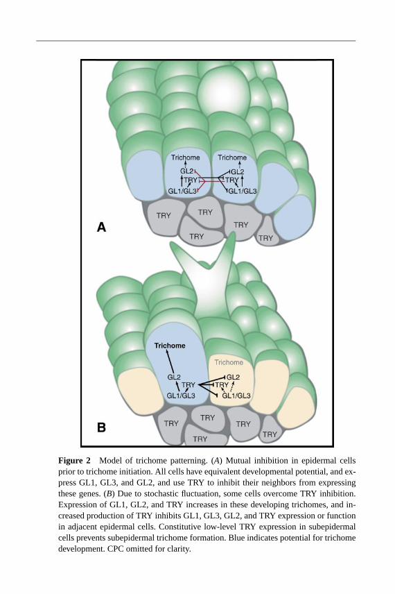
17 Apr 2003 16:54 AR AR184-16-COLOR.tex AR184-16-COLOR.SGM LaTeX2e(2002/01/18)P1: GDL
Figure 2 Model of trichome patterning. (A) Mutual inhibition in epidermal cellsprior to trichome initiation. All cells have equivalent developmental potential, and ex-press GL1, GL3, and GL2, and use TRY to inhibit their neighbors from expressingthese genes. (B) Due to stochastic fluctuation, some cells overcome TRY inhibition.Expression of GL1, GL2, and TRY increases in these developing trichomes, and in-creased production of TRY inhibits GL1, GL3, GL2, and TRY expression or functionin adjacent epidermal cells. Constitutive low-level TRY expression in subepidermalcells prevents subepidermal trichome formation. Blue indicates potential for trichomedevelopment. CPC omitted for clarity.

17 Apr 2003 16:54 AR AR184-16-COLOR.tex AR184-16-COLOR.SGM LaTeX2e(2002/01/18)P1: GDL
Figure 3 Model of root epidermal patterning. (A) Mutual inhibition in heart-stageembryos. All cells have equivalent developmental potential, express WER and GL2,and use CPC to inhibit their neighbors from expressing these genes. A bHLH geneis probably also expressed. (B) Growing root. Position cues from underlying cellsgenerate a bias in WER expression due either to inhibition of WER in H cells oractivation of WER in N-cells. This bias is enhanced by increased CPC levels, whichcarry an inhibitory signal from N-cells to H-cells. N-cells: Cells in files that willproduce nonhair cells (blue). H-cells: Cells in files that will produce hair cells (brown).Note that cells continue to divide after fate is determined, producing a file of cells thatdifferentiate.


![MINHO J , TARAS MAKSYMYUK, RODRIGO L. …iot.korea.ac.kr/Member/Vol.21,No.6,pp54-62, Dec.2014-A...ent unlicensed spectrum bands (Wi-Fi, Bluetooth, etc.) for relay transmission [4],](https://img.pdfslide.us/doc/110x75/5e97a7390444007af646f796/minho-j-taras-maksymyuk-rodrigo-l-iotkoreaackrmembervol21no6pp54-62.jpg)