Embed Size (px)
Citation preview
Gyriform Calcifications in Tuberous Sclerosis Simulating the Appearance of Sturge-Weber Disease
G. Wilms, 1 E. Van Wijck , 1 Ph. Demaerel, 1 M.-H. Smet, 1 C. Plets,2 and J. M . Brucher3
Summary: Two cases of tuberous sclerosis are presented. Extensive superficial occipital calcifications were found as classically described in Sturge-Weber syndrome. Other radiologic signs of tuberous sclerosis, such as subependymal calcifications in both patients and surgically proved giant cell astrocytoma in one patient, were present. At pathologic examination, the calcifications appeared to be located in extensive cortical tubers.
Index terms: Phakomatoses; Sclerosis, tuberous; Brain, calcification
Sturge-Weber syndrome and tuberous sclerosis are both phakomatoses. In both neurocutaneous syndromes, intracranial calcifications are classically described with a typical appearance and localization that allow radiologic diagnosis (1). In Sturge-Weber syndrome the calcifications are tram-line with parieto-occipital cortical localization (1-4). In tuberous sclerosis, nodular calcifications with subependymal localization are present (1, 5-7). We describe two patients who, in addition to the typical subependymal calcifications, had superficial parieto-occipital calcifications resembling the radiologic appearance of Sturge-Weber syndrome.
Case 1
After an uneventful pregnancy, a baby girl was delivered by forceps extraction in 1974. At 6 weeks of age, she developed several grand mal type epileptic seizures. Clinical neurologic examination was normal. Some pigmented nevi were found on the lower limbs beside other depigmented spots.
Plain radiographs of the skull (Fig. 1A) showed extensive calcifications in the temporo-occipital region. Selective angiography of the left internal carotid artery confirmed
the infrasylvian localization of the lesion but without evidence of neoplastic vessels. Because of the young age of the patient, no surgical intervention was planned. Differential diagnosis was proposed between a posttraumatic hematoma, secondary to the forceps extraction, and a congenital tumor, such as a teratoma. In 1976, at age 2 , she was readmitted because of an increase in frequency of the epileptic seizures. A computed tomography (CT) scan was now available and confirmed the extensive calcificat ion of the temporo-occipital area.
At surgery, the cortex in the left occipital region appeared thin, with evidence of a firm tumoral mass in the subcortical area. Biopsy of the lesion showed a mixed astrocytoma, with giant and fibrillar cells, characteristic for tuberous sclerosis. Several follow-up CT scans with higher resolution confirmed the extensive calcifications and demonstrated typical subependymal calcifications near the foramen of Monro, as typically seen in tuberous sclerosis (Fig. 1 B). At the present time, this girl is an adolescent. She sti ll suffers from frequent epileptic insults, severe psychomotor retardation , cerebral palsy, and choreoathetosis. The patient receives antiepileptic drugs, but her epilepsy is poorly controlled.
Case 2
In 1974, a baby boy was born after uneventful pregnancy and delivery. At 5 weeks of age, the patient presented with epileptic seizures. At clinical neurologic examination, a right hemiparesis was noted with ptosis of the right eyelid. Multiple depigmentation spots were found over the abdomen and under both limbs. Angiography and air encephalography indicated a space-occupying lesion in the left temporal area. At surgery, the sylvian fissure was markedly elevated, but the temporal convolutions were preserved. One centimeter beneath the cortex, a grayishpurple tissue was found with firm and calcified consistency. An anterior temporal lobectomy was performed. Anato-
Received February 20, 1991 ; accepted and revision requested May 29; revision received July 30. 1 Department of Radiology, University Hospitals K. U. Leuven, Herestraat 49, B-3000 Leuven, Belgium. Address reprint requests to G. Wilms. 2 Department of Neurosurgery, University Hospitals, K. U. Leuven, Leuven, Belgium. 3 Department of Neuropathology, Cliniques Universitaires Saint-Luc, Brussels, Belgium.
AJNR 13:295-298, Jan/ Feb 1992 0195-6108/ 1301-0295 © American Society of Neuroradiology
295
296 AJNR: 13, January / February 1992
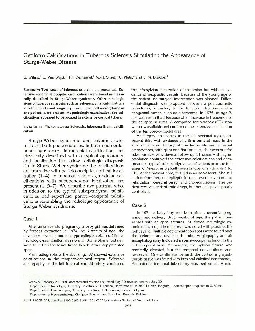
Fig. 1. A, Patient 1: Lateral skull radiograph. Extensive parieto-occipital calcifications suggestive of Sturge-Weber disease.
a, CT scan before contrast. Confirms the location of the calcification over the left temporo-occipital cortex . Notice typical subependymal calcifications in the wall of the lateral ventricles.
A
A
. ,. ,. , .... ,
-~ .. tJ • f
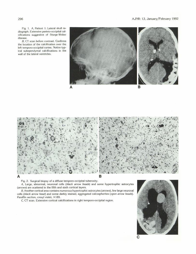
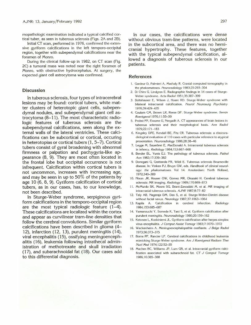
Fig. 2. Surgical biopsy of a diffuse temporo-occipital tuberosity.
8
A, Large, abnormal , neuronal cells (black arrow heads) and some hypertrophic astrocytes (arrows) are scattered in the fifth and sixth cortical layers.
a, Another cortical area contains numerous hypertrophic astrocytes (arrows) , few large neuronal cells (black arrow head) and some darkly stained, aggregated calcospherites (open arrow heads). Paraffin section , cresyl violet, X 189.
C, CT scan. Extensive cortical calcifications in right temporo-occipital region.
8
c
AJNR : 13, January/February 1992
m opathologic examination indicated a t ypical calcified cortical tuber, as seen in tuberous sclerosis (Figs. 2A and 26 ).
Initial CT scan, performed in 1976, conf irmed the extensive gyriform calcifications in the left temporo-occipital region , together with subependymal calcif ications near the foramen of Monro.
During the clinical follow-up in 1982, on CT scan (Fig. 2C) a tumoral mass was noted near the right foramen of Monro, with obstructive hydrocephalus. At surgery , the expected giant cell astrocytoma was confirmed.
Discussion
In tuberous sclerosis, four types of intracerebral lesions may be found: cortical tubers, white matter clusters of heterotopic giant cells, subependymal nodules, and subependymal giant cell astrocytoma (8-11 ). The most characteristic radiologic features of tuberous sclerosis are the subependymal calcifications, seen along the external walls of the lateral ventricles. These calcifications can be subcortical or cortical, occuring in heterotopias or cortical tubers ( 1, 5-7). Cortical tubers consist of gyral broadening with abnormal firmness or palpation and pachygyria-like appearance (8, 9). They are most often located in the frontal lobe but occipital occurrence is not infrequent. Calcification within cortical tubers is not uncommon, increases with increasing age, and may be seen in up to 50% of the patients by age 10 (6, 8 , 9). Gyriform calcification of cortical tubers, as in our cases, has, to our knowledge, not been described.
In Sturge-Weber syndrome, serpiginous gyriform calcifications in the temporo-occipital region are the most typical radiologic feature (1-4). These calcifications are localized within the cortex and appear as curvilinear tram-line densities that follow the cerebral convolutions. Similar gyriform calcifications have been described in glioma (4-12), infarction (12, 13), purulent meningitis (14), viral encephalitis (15), ossifying meningoencephalitis (16), leukemia following intrathecal administration of methotrexate and skull irradiation (17), and subarachnoidal fat (18). Our cases add to this differential diagnosis.
297
In our cases , the calcifications were dense without obvious tram-line patterns, were located in the subcortical area , and there was no hemicrania! hypertrophy. These features , together with the typical subependymal calcification, allowed a diagnosis of tuberous sclerosis in our patients.
References
1. Gardeur D. Palm ieri A , Mashaly R. Crania l computed tomography in
the phakomatoses. Neuroradiology 1983;25:293-304 2. Di Chiro G. Lindgren E. Radiographic findings in 14 cases of Sturge
Weber syndrome. Acta Radio/1951 ;35:387-399 3. Boltshauser E, Wilson J , Hoare RD. Sturge-Weber syndrome with
bilateral intracranial calc ification. Neurol Neurosurg Psychiatry
1976;39:429-435 4. Coulam CM, Brown LR, Reese DF. Sturge-Weber syndrome. Semin
Roentgeno/ 1976; 11 :55-59 5. Probst FP, Erasme U, Nergadh A . CT appearances of brain lesions in
tuberous sclerosis and their morphologica l basis. Ann Radio/
1979;22:1 71-183 6. Kingsley DPE, Kendall BE, Fitz CR. Tuberous sclerosis: a cl inicora
diological evaluation of 110 cases with particu lar reference to atypical presentation. Neuroradio/ogy 1986;28:38-46
7. Legge M, Sauerbrei E, MacDonald A . Intracranial tuberous sclerosis
in infancy. Radiology 1984;153:667-668 8. Bender BL, Yunis EJ. T he pathology of tuberous sclerosis. Pathol
Ann 1982; 17:339-382 9. Donegani G, Grattarola FR. Wildi E. Tuberous sclerosis Bourneville
disease. In: Vinken PJ. Bruyn GW. eds. Handbook of clinical neurol
ogy: the phakomatoses. Vol 14. Amsterdam: North Holland,
1972;340- 389 10. Nixon JR. Houser OW, Gomez MR. Okazaki H. Cerebra l tuberous
sclerosis: MR imaging. Radiology 1989; 170:869-873 11 . McMurdo SK, Moore SG, Brant-Zawadzki M, et al. MR imaging of
intracranial tuberous sclerosis. AJNR 1987;8:77-82 12. Taly AB. Nagaraja DM, Das S, et al. Sturge-Weber-Dimitri disease
without facial nevus. Neurology 1987;37: 1063-1064 13. Kapila A. Calcification in cerebral infarction. Radiology
1984; 153:685- 687 14. Yamanouchi Y. Someda K. TaniS, et al. Gyriform calci fication after
purulent meningitis. Neuroradiology 1980;20: 159- 162 15. Ketonen L, Koskiniemi JL. Gyriform calcification after herpes simplex
virus encephalitis . J Comput Assist Tomogr 1983;7: 1070-1072 16. Wackenheim A . Meningoencephalopathie ossifiante. J Beige Radio/
1973;56:373-375 17. Borns PF, Rancier LF. Cerebra l calc if ications in childhood leukemia
mimick ing Sturge-Weber syndrome. Am J Roentgenol Radium Ther
Nuc/ fried 1974; 122:52-55 18. Machen BC, Will iams JP. Lum GB, et al. Intracranial gyri form calci
fication associated with subarachnoid fat. CT J Comput Tomogr
1986; 10:385-388