Embed Size (px)
Citation preview

RESEARCH ARTICLE Open Access
GWAS and enrichment analyses of non-alcoholic fatty liver disease identify newtrait-associated genes and pathways acrosseMERGE NetworkBahram Namjou1,2*, Todd Lingren2,3, Yongbo Huang1, Sreeja Parameswaran1, Beth L. Cobb1, Ian B. Stanaway4,John J. Connolly5, Frank D. Mentch5, Barbara Benoit6, Xinnan Niu7, Wei-Qi Wei7, Robert J. Carroll7,Jennifer A. Pacheco8, Isaac T. W. Harley9, Senad Divanovic9, David S. Carrell10, Eric B. Larson10, David J. Carey11,Shefali Verma12, Marylyn D. Ritchie12, Ali G. Gharavi13, Shawn Murphy14, Marc S. Williams15, David R. Crosslin4,Gail P. Jarvik16, Iftikhar J. Kullo17, Hakon Hakonarson5,18, Rongling Li19, The eMERGE Network19,Stavra A. Xanthakos20 and John B. Harley1,2,21
Abstract
Background: Non-alcoholic fatty liver disease (NAFLD) is a common chronic liver illness with a geneticallyheterogeneous background that can be accompanied by considerable morbidity and attendant health carecosts. The pathogenesis and progression of NAFLD is complex with many unanswered questions. Weconducted genome-wide association studies (GWASs) using both adult and pediatric participants from the ElectronicMedical Records and Genomics (eMERGE) Network to identify novel genetic contributors to this condition.
Methods: First, a natural language processing (NLP) algorithm was developed, tested, and deployed at each site toidentify 1106 NAFLD cases and 8571 controls and histological data from liver tissue in 235 available participants. Theseinclude 1242 pediatric participants (396 cases, 846 controls). The algorithm included billing codes, text queries, laboratoryvalues, and medication records. Next, GWASs were performed on NAFLD cases and controls and case-only analyses usinghistologic scores and liver function tests adjusting for age, sex, site, ancestry, PC, and body mass index (BMI).
Results: Consistent with previous results, a robust association was detected for the PNPLA3 gene cluster in participantswith European ancestry. At the PNPLA3-SAMM50 region, three SNPs, rs738409, rs738408, and rs3747207, showedstrongest association (best SNP rs738409 p = 1.70 × 10− 20). This effect was consistent in both pediatric (p = 9.92 × 10− 6)and adult (p = 9.73 × 10− 15) cohorts. Additionally, this variant was also associated with disease severity and NAFLD ActivityScore (NAS) (p = 3.94 × 10− 8, beta = 0.85). PheWAS analysis link this locus to a spectrum of liver diseases beyond NAFLDwith a novel negative correlation with gout (p = 1.09 × 10− 4). We also identified novel loci for NAFLD disease severity,including one novel locus for NAS score near IL17RA (rs5748926, p = 3.80 × 10− 8), and another near ZFP90-CDH1 forfibrosis (rs698718, p = 2.74 × 10− 11). Post-GWAS and gene-based analyses identified more than 300 genes that were usedfor functional and pathway enrichment analyses.
(Continued on next page)
© The Author(s). 2019 Open Access This article is distributed under the terms of the Creative Commons Attribution 4.0International License (http://creativecommons.org/licenses/by/4.0/), which permits unrestricted use, distribution, andreproduction in any medium, provided you give appropriate credit to the original author(s) and the source, provide a link tothe Creative Commons license, and indicate if changes were made. The Creative Commons Public Domain Dedication waiver(http://creativecommons.org/publicdomain/zero/1.0/) applies to the data made available in this article, unless otherwise stated.
* Correspondence: [email protected] for Autoimmune Genomics and Etiology, Cincinnati Children’sHospital Medical Center (CCHMC), Cincinnati, OH, USA2College of Medicine, University of Cincinnati, 3333 Burnet Avenue,Cincinnati, OH 45229, USAFull list of author information is available at the end of the article
Namjou et al. BMC Medicine (2019) 17:135 https://doi.org/10.1186/s12916-019-1364-z
Beyond Big Data to new Biomedical and Health DataScience: moving to next century precision health

(Continued from previous page)
Conclusions: In summary, this study demonstrates clear confirmation of a previously described NAFLD risk locus andseveral novel associations. Further collaborative studies including an ethnically diverse population with well-characterizedliver histologic features of NAFLD are needed to further validate the novel findings.
Keywords: NAFLD, Fatty liver, Genetic polymorphism, GWAS, PheWAS, Polygenic risk score
BackgroundNonalcoholic fatty liver disease (NAFLD) is one of themost common chronic liver diseases, found in 17–30%of the population in Western countries [1]. NAFLD, de-fined as greater than 5% fatty acid content of liver byweight, includes not only simple and benign steatosisbut also the more serious nonalcoholic steatohepatitis(NASH), which may progress to cirrhosis and liver fail-ure in 8 to 26% of adults with NASH [2]. NASH is de-fined histologically by the presence of macrovesicularsteatosis, lobular inflammation, and hepatocellular bal-looning. The pathology is often indistinguishable fromalcoholic fatty liver disease; therefore, the diagnosis canonly be made in the absence of significant alcohol use[3]. NAFLD is now recognized as a common metabolicdisorder globally as a result of ongoing obesity pan-demic. It also increases risk of adverse long-term conse-quences including death from liver cirrhosis andcardiovascular disease. In fact, NASH is now the secondmost common indication for liver transplantation in theUSA after chronic hepatitis C [4].Growing evidence has shown that NAFLD can also
occur in 10–20% of non-obese population, most often inassociation with central adiposity, recent weight gain,dietary factors, or genetic risk alleles [5]. In East Asiancountries, for example, the incidence and prevalence ofNAFLD are increasing with time despite lower rates ofobesity compared to Western countries [6]. Hence, it isimportant to identify the natural course of NAFLD andthe contributing factors for the development and main-tenance or regression of this disease. The underlying eti-ology is believed to be multifactorial with a substantialgenetic component. The heritability estimates of NAFLDgenerally range from 20 to 70%, depending on the studydesign, ethnicity, and the methodology used [7]. Like-wise, for indices of disease severity, the heritability esti-mates in a twin study for hepatic steatosis was 0.52(based on MRI proton-density fat fraction) and for liverfibrosis (based on liver stiffness) 0.5 [8]. In addition, her-itability risk for NAFLD may be independent of bodymass index heritability. For example, family studies showthat while fatty liver can be present in 17% of siblingsand 37% of parents of overweight children withoutNAFLD, it was significantly more common in siblings(59%) and parents (78%) of children with NAFLD [9].
To date, several genome-wide association studies(GWAS) have been published for this condition mainlyin adult cohorts [10–12]. One of the established effectsis in the PNPLA3 (patatin-like phospholipase domain–containing 3) gene with consistent results across studiesin which the rs738409 C>G variant (resulting in anamino acid substitution of methionine for isoleucine atposition 148 (I148M)) is strongly associated with thistrait. The PNPLA3 protein exerts lipase activity andplays a role in the hydrolysis of glycerolipids, with max-imum enzymatic activity against triglycerides, diacylglyc-erol, and monacylglycerol [13]. Structural modelingsuggests that this substitution may occlude access ofsubstrates to the catalytic dyad [14]. However, the exactunderlying mechanisms remain unclear.The electronic medical record (EMR) is a rich source
of clinical information. Natural language processing(NLP) techniques have demonstrated successes withinthe clinical domain and have been tested for transferabil-ity to another institution [15]. The electronic MEdicalRecords and GEnomics (eMERGE) Network, founded in2007, is a consortium of multiple adult and pediatric in-stitutions developed to explore the utility of DNA biore-positories linked to EMRs as well as establishing andvalidating specific algorithms with and without NLP formany common phenotypes [16]. In this study, we inves-tigated the genetic variants associated with NAFLD/NASH in children and adults using phenotypic measuresextracted from medical records in a collection of alreadygenotyped samples from more than 80,000 eMERGEparticipants to replicate prior studies and identify add-itional genetic loci.
MethodsStudy participants and phenotypeData for this study were collected from the eMERGENetwork [17]. Protocols for this study were approved bythe Institutional Review Boards (IRBs) at the institutionswhere participants were recruited; all included partici-pants provided written informed consent prior for inclu-sion in the study. The population comprised 9677unrelated European ancestry participants (1106 casesand 8571 controls). A natural language processing (NLP)algorithm was deployed in each site to identify NAFLDcases and controls. These include logic concepts using
Namjou et al. BMC Medicine (2019) 17:135 Page 2 of 19

billing codes, laboratory values, text queries, and medi-cation records to identify true cases and controls at eachsite. A rules-based NLP algorithm was developed usingstructured and unstructured data from Cincinnati Chil-dren’s Hospital and Medical Center (CCHMC) and sec-ondarily validated in Children’s Hospital of Philadelphia(CHOP) with high precision. The eMERGE protocol in-cludes development of an algorithm at a primary siteand the implementation and validation at a secondarysite. The secondary site serves as a testing ground forthe purposes of mitigating overfitting concerns and en-suring portability. Expert validation includes manualchart review at each site by a physician for both casesand controls. After obtaining a validated positive predict-ive value of 95% for cases and controls at both the pri-mary (CCHMC) and secondary sites (CHOP), thealgorithm has been implemented across network. Theexclusion and inclusion criteria for NAFLD were derivedaccording to recommendation from the American Asso-ciation for the Study of Liver Diseases (AASLD) practicalguideline for NAFLD [18]. Case inclusion and exclusioncriteria, list of excluded medications, and the number ofparticipants per eMERGE site can be found in Add-itional file 1: Table S1. We processed the pathology andradiology reports from encounters with diagnosis codesby searching with regular expressions for specific relatedterms as shown in Additional file 1: Table S1. TheNegEx multilingual lexicon was used to assess positiveand negative condition for each term [19]. In addition,NAFLD disease severity was assessed based on availableliver enzyme and histopathologic grade using theNAFLD Activity Score (NAS). NAS score is a standardmethod used to score NAFLD disease activity and ori-ginally has been developed as a tool to measure diseaseprognosis and changes in NAFLD during therapeutic tri-als [20]. The NAS is derived from an unweighted sum ofscores of liver steatosis (0–3), lobular inflammation (0–3), and hepatocellular ballooning (0–2), ranging between0 and 8. Coexistent fibrosis also has a separate scoringrange of 0–4. This consists of no fibrosis (0), perisinusoi-dal or periportal (1), portal (2), bridging fibrosis (3), andcirrhosis (4). The NAS classification scoring system isshown in Additional file 1: Table S1. We obtained thesevalues from pathology reports using NLP processingfor 235 of our NAFLD case participants. In addition,for each case, the highest level of liver enzyme valuesfor aspartate aminotransferase (AST U/L) and alanineaminotransferase (ALT U/L) was obtained for associ-ation testing.
Genotyping and imputationGenetic data for the eMERGE Network is available fromthe coordinating center and can be accessed throughdbGAP (phs000888.v1.p1) which is annually updated.
High-throughput SNP genotyping was carried out previ-ously in each contributing medical center. A series ofstandard quality control (QC) measures has been appliedbefore and after imputation. These measures have beendeveloped by the eMERGE Genomics Workgroup [21,22]. The standard QC process included sample call rates,sample relatedness, and population stratification, sexinconsistency as well as marker quality (i.e., marker callrate, minor allele frequency (MAF), and Hardy-Weinberg equilibrium (HWE). In this study, all analyseswere limited to participants with call rates > 98%, SNPswith call rates > 99%, and SNPs with MAF > 1% andHWE p > 0.0001 in controls. The details of imputationprocess and principal component (PC) analyses havebeen included in Additional file 2 [23–25].
Statistical analysesLogistic (case-control) and quantitative linear (case-only)regression analyses were performed using an additivegenetic model adjusting for 10 medical centers; PCs 1, 2,and 3; sex; and age. In addition, since NAFLD is closelylinked to obesity, we included the most recent BMI foreach subject as another covariate and remove all missingparticipants from analyses. Traditionally absolute BMI(kg/m2) is used for adults, while age- and sex-specificBMI-z scores and percentiles apply in children and ado-lescents to account for their continuing growth. In com-bined analyses, we therefore transformed all BMI into 6classes: underweight (< 18.5 or < 5th percentile), normal(18.50–24.99 or 5th to < 85th percentile), overweight(25.00–29.99 or 85th to < 95th percentile), and obese:class 1 (30–34.99 or 95th to < 120% of the 95th percent-ile), class 2 (35–39.99 or 120% to < 140% of the 95thpercentile), and class 3 (≥ 40 or ≥ 140% of the 95thpercentile). The percentage of BMI ≥ 95% in pediatricsparticipants was estimated using the CDC-based onlineresource [27]. The distribution of participants that wereceived from the network also varied per site (seeAdditional file 1: Table S1); we therefore adjust for 10study sites. Adjusting for too many covariates maysometimes cause the standard logistic regression to failto converge especially for less-frequent variants. Firth’spenalized likelihood approach, available in second gener-ation of PLINK, is a method of addressing issues of sep-aration and bias of the parameter estimates in which weused in regression analyses when necessary [26]. Forliver enzymes (AST, ALT), we used the highest value U/L per subject. All quantitative phenotypes including liverenzymes and NAS score were standardized to mean ofzero and variance 1 using PLINK.Further conditional analyses and pairwise SNP × SNP
interactions were also performed using “epistasis” optionin PLINK. In this study, we only analyzed the pairwiseinteraction effect of one known SNP (rs738409) in
Namjou et al. BMC Medicine (2019) 17:135 Page 3 of 19
PNPLA3 gene against the genome. The slower “—epista-sis” command was used to test for epistasis using logisticregression which is the most accurate test to define SNP ×SNP interactions [26]. Interactions were excluded if twoSNPs were located within 1Mb of each other to avoidspurious evidence of interaction due to linkage disequilib-rium (LD). Narrow-sense heritability was also estimatedusing an SNP-based approach available in the GCTA pro-gram [28] which evaluates the proportion of phenotypicvariance explained by all SNPs. Briefly, the GCTA analysisconsists of two steps. First, all SNPs are used to calculatethe genetic relationship matrix (GRM) among participantsusing the observed low-level genetic similarity in SNP datafrom individuals who are not directly related. This meas-ure is then used as a predictor in a mixed linear modelwith a trait as the response to estimate h2 [28]. Theweighted genetic risk score (GRS) was also calculatedusing PLINK-score function by multiplying each β-coeffi-cient of highly significant SNPs with the number of corre-sponding risk alleles (0, 1, or 2) and then summing theproducts ( [26]. For known variants, β-coefficients wereobtained from the GWAS catalog [29]. The performanceof the obtained GRS score for disease diagnosis and pre-diction accuracy were evaluated using receiver operatingcharacteristic (ROC) curve, using MedCalc software [30].Finally, to estimate the level of heterogeneity betweenpediatrics and adult cohorts, Cochran’s Q test statisticswas applied using PLINK2 [26].
PheWAS analysesA phenome-wide association study (PheWAS) was alsoperformed in order to evaluate pleotropic effects of theknown GWAS variant (rs738409) as well as other noveleffects in this study with any other trait in children oradults. The trait definition in PheWAS approach ismainly based on billing International Classification ofDiseases (ICD) codes; therefore, it is less conservative.The detail of this approach has been described previ-ously [31]. We used the PheWAS package in R version
3.5.1 [32]. Briefly, in the PheWAS process, first the ICD-9 codes are collapsed into PheWAS codes according toPheWAS map [32]. Then, cases and controls are deter-mined according to the code under study. In these ana-lyses, a case was defined as having at least twooccurrences of the PheWAS code on different days andthe controls with no instances. Additionally, we used athreshold of at least 20 cases for the code to be used inthe model. Next, for each PheWAS code, a logistic re-gression model was created and adjusted for age, sex,BMI, site of genotyping, and PCs similar to GWASstudy. A false discovery rate (FDR) of 0.05 using theBenjamini–Hochberg procedure implemented in Phe-WAS was then used to correct the threshold for mul-tiple hypotheses testing.
Post-GWAS analyses and data visualizationThe details of post-GWAS analyses including functionalannotation, prioritization, and interpretation of GWASresults based on functional mapping are included inAdditional file 2 [33–41].
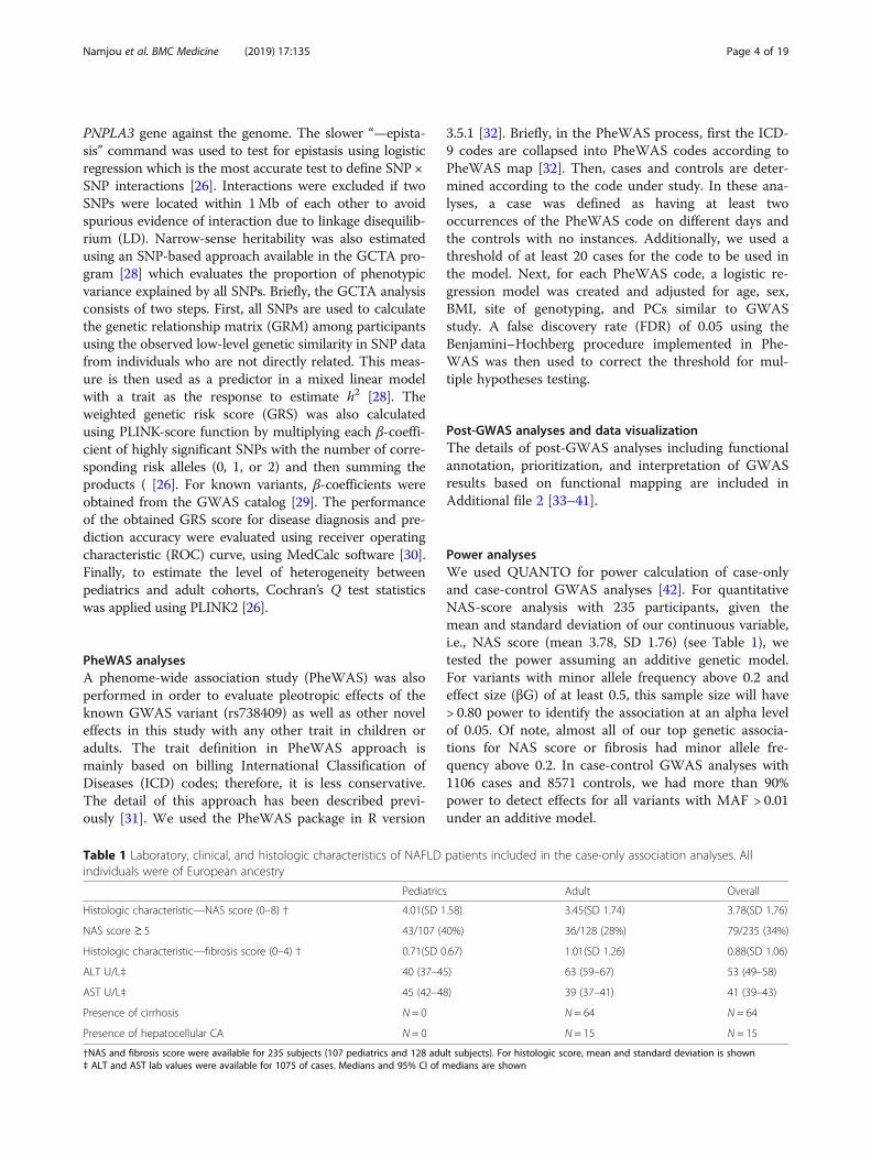
Power analysesWe used QUANTO for power calculation of case-onlyand case-control GWAS analyses [42]. For quantitativeNAS-score analysis with 235 participants, given themean and standard deviation of our continuous variable,i.e., NAS score (mean 3.78, SD 1.76) (see Table 1), wetested the power assuming an additive genetic model.For variants with minor allele frequency above 0.2 andeffect size (βG) of at least 0.5, this sample size will have> 0.80 power to identify the association at an alpha levelof 0.05. Of note, almost all of our top genetic associa-tions for NAS score or fibrosis had minor allele fre-quency above 0.2. In case-control GWAS analyses with1106 cases and 8571 controls, we had more than 90%power to detect effects for all variants with MAF > 0.01under an additive model.
Table 1 Laboratory, clinical, and histologic characteristics of NAFLD patients included in the case-only association analyses. Allindividuals were of European ancestry
Pediatrics Adult Overall
Histologic characteristic—NAS score (0–8) † 4.01(SD 1.58) 3.45(SD 1.74) 3.78(SD 1.76)
NAS score ≥ 5 43/107 (40%) 36/128 (28%) 79/235 (34%)
Histologic characteristic—fibrosis score (0–4) † 0.71(SD 0.67) 1.01(SD 1.26) 0.88(SD 1.06)
ALT U/L‡ 40 (37–45) 63 (59–67) 53 (49–58)
AST U/L‡ 45 (42–48) 39 (37–41) 41 (39–43)
Presence of cirrhosis N = 0 N = 64 N = 64
Presence of hepatocellular CA N = 0 N = 15 N = 15
†NAS and fibrosis score were available for 235 subjects (107 pediatrics and 128 adult subjects). For histologic score, mean and standard deviation is shown‡ ALT and AST lab values were available for 1075 of cases. Medians and 95% CI of medians are shown
Namjou et al. BMC Medicine (2019) 17:135 Page 4 of 19

ResultsThe results reported below consist of overall NAFLDcase-control GWAS and four additional case-only GWAquantitative studies for NAS score, fibrosis, and ASTand ALT liver enzymes.
NAFLD case-control GWASTable 2 shows demographic characteristics of patientsand controls included in this study. The mean age was63.5 (±16.86 SD) for adult participants (N = 8435) and13.05 (±5.41 SD) for pediatric participants (N = 1242).The number of participants per site is included inAdditional file 1: Table S1. In this study, 47% of pediatricparticipants and 42% of adults were males. A total of9677 unrelated European ancestry participants (1106cases and 8571 controls) and 7,263,501 autosomal vari-ants were evaluated for this GWAS analysis.
Associations of previously reported SNPsConsistent with previous reports, we identified strong gen-etic signals at the PNPLA3 locus at 22q13. Figure 1a showsa Manhattan plot with one main peak located on chromo-some 22 that was associated with NAFLD. The Q–Q plotof this GWAS is also shown in Fig. 1b. The overall low in-flation rate of λ = 1.001 indicated no major populationstratification. At the PNPLA3-SAMM50 region, three proxySNPs (r2 > 0.95), rs738409, rs738408, and rs3747207, lo-cated in the PNPLA3 gene showed the strongest associa-tions (best SNP rs738409 p = 1.70 × 10− 20, OR = 1.79 (95%CI = 1.58–2.02)) (Table 3, Fig. 2a). This effect was consist-ent in both pediatric (p = 9.92 × 10− 6, OR = 1.76 (95% CI =1.37–2.27)) and adult (9.73 × 10− 15, OR = 1.79 (95%CI = 1.55–2.08)) cohorts and with no evidence ofheterogeneity (Cochran’s Q = 0.78, I2 = 0). Consistentwith previous results, another coding variantrs2294918 (E434K) in PNPLA3 gene was associated ata weaker level (p = 1.90 × 10− 5). The SNPs with themost significant evidence for association are summa-rized in Table 3, and all results with p < 10− 5 are in-cluded in Additional file 1: Table S2.Apart from the main effect at the PNPLA3 locus, pre-
vious genetic studies identified several minor effects atother chromosomal loci, including GCKR at 2p23, andGATAD2A, NCAN, and TM6SF2 at 19p12 [43, 44]. Weexamined whether or not the associations were repro-duced in our cohorts by extracting genotype information
of SNP markers corresponding to these loci. None ofthese effects reached genome-wide significance. In ourpediatric cohorts, the association of rs1260326 andrs780094 in GCKR was borderline significant (p = 0.006,OR = 1.40, 95% CI 1.1–1.78). However, the associationwas lost when examined in the adult cohort (seeAdditional file 1: Table S3). At the 19p12 region(GATAD2A, NCAN,TM6SF2), the association with knownSNP rs4808199 was also detected using all cohorts (p =0.004, OR = 1.22, 95% CI 1.06–1.40)). Of note, the knownTM6SF2 missense variant rs58542926 (E167K) produceda p = 0.03 (OR = 1.23, 95% CI 1.01–1.52) in our cohort. Inthis region, we also found other unreported downstreammarkers with stronger associations (best effect for SNPrs56408111 p = 5.26 × 10− 6) (see Additional file 1: TableS2; Fig. 2b). The observed effect for rs4808199 orrs58542926 disappeared after conditioning on rs56408111(p = 0.71, p = 0.17 respectively) suggesting that the associ-ation in this region mostly derives from rs56408111. TheLD between these two known markers (rs4808199,rs58542926) and the best variant in this study,rs56408111, was modest (r2 = 0.25, r2 = 0.40 respectively).We also confirmed an effect at 8q24 near the
TRIB1 gene that previously associated with NAFLDin the Japanese population [45]. In their population,rs2954021 produced p = 4.5 × 10− 5. In our Europeanancestry population and for the first time, this vari-ant as well as a cluster of variants nearby was asso-ciated with NAFLD with the best marker rs2980888(p = 5.98 × 10− 7, OR = 1.36 95% CI = 1.20–1.53) (seeTable 3, Fig. 2c). Conditional analyses suggest thatrs2980888 is the most informative variant in this re-gion in European ancestry. These two markers re-sided in one risk haplotype in European ancestrywith (r2 = 0.45, D’ = 0.97) (Fig. 2c).
Controlling for the main effects at PNPLA3We used logistic regression models conditioned on themain effect at PNPLA3 as well as testing for epistaticinteraction between the known SNP at PNPLA3rs738409 and the rest of genome. For conditional ana-lysis, the genotype data of rs738409 in dosage format (0,1, 2) was included as another covariate in addition toage, sex, PCs, BMI, and sites of genotyping. While thisvariant controlled all effects at PNPLA3 indicating noother independent effects at this locus, no major changes
Table 2 The demographic distribution of EMR-linked eMERGE cohorts
Case_EA Control_EA Mean age ♂/♀ Mean BMI, kg/m2
Pediatrics* 396 846 13.05 (SD 5.41) 693/549 22.70 (SD 7.87) †
Adults 710 7725 63.50 (SD 16.86) 3810/4625 32.64 (SD 8.21)
Total 1106 8571
*Defined as ≤ 21 years old†The average BMI-for-age z score in pediatric cohorts was 1.16 (95% CI = 1.03–1.20, SD = 1.39)
Namjou et al. BMC Medicine (2019) 17:135 Page 5 of 19

have been detected in other loci. Next, in a separate ana-lysis, the epistatic effect of the known SNP rs738409 withthe rest of the genome was evaluated (see “Methods”).Several suggestive results were detected across the gen-ome with only one effect at 16p12 that passed the signifi-cance threshold of p < 0.0001 (SNP rs2188761, case-onlyPepistasis = 2.47 × 10− 7, case-control Pepistasis = 7.32 × 10− 6,OR of interaction = 1.50). Several proxy markers in this re-gion (16p12) such as rs7499477, rs2188760, andrs6497497 (r2 > 0.95 with rs2188761) also interact withrs738409 in PNPLA3. In addition, this novel epistatic ef-fect was consistent in both pediatrics and adult cohorts(OR of interaction = 1.57 and 1.43 for pediatrics andadults, respectively). As mentioned above, all thesemarkers had passed QC and were in HWE. However,none of these markers at the 16p12 region were genome-wide significant in GWAS analyses (0.1 > p > 0.02).
Because NAFLD is closely linked to obesity, we alsoexplored the specific SNP × SNP interaction of the majorobesity locus, FTO (rs1421085) and PNPLA3 (rs738409).We did not find any significant SNP × SNP interaction(p = 0.72). Of note, the GWAS effect for FTO(rs1421085) in this study was p = 0.25 after controllingfor BMI. However, by relaxing the model and removingthe BMI as a covariate, this effect in FTO increased insignificance (p = 9.26 × 10− 6).
Case-only GWA studiesImpact of SNPs on the severity of NAFLDWe next investigated the associations of the SNPs withNAFLD disease severity based on available histopatho-logic grade, namely, NAS and liver enzymes (see“Methods”). Because liver biopsy usually is not indicatedfor NAFLD diagnosis, we were able to identify and score
Fig. 1 a, b Manhattan plot (a) and Q–Q plot (b) of genome-wide markers for NAFLD in European ancestry (1106 cases and 8571 controls). A totalof 1106 cases of NAFLD and 8571 controls were analyzed after quality control. Logistic regression analysis was performed for 7,261,527 variantswith MAF > 1% assuming an additive genetic model, adjusted for age, sex, BMI, genotyping site, and genetic ancestry (principal components 1through 3). Results are plotted as –log10 p values on the y-axis by position in chromosome (x-axis) (NCBI build 37)
Namjou et al. BMC Medicine (2019) 17:135 Page 6 of 19

only 235 participants using EMR data from the total of1106 NAFLD cases that includes 107 pediatric and 128adult cases (Table 1). Liver function tests (AST U/L,ALT U/L) were available for 1075 of case participants. Inaddition to the main case-control study, for disease se-verity index, we performed GWAS for each quantitative
trait (NAS score, fibrosis, ALT, AST) using linear regres-sion method adjusting for age, sex, BMI, PCs, and site ofgenotyping. These case-only analyses showed several lociwith significant associations. The SNPs with the mostsignificant evidence are summarized in Table 3 and allresults with p < 10− 5 are provided in Additional file 1:
Fig. 2 a–c LocusZoom plot of the associations signals in three previously known regions for NAFLD. a Confirmation at 22q13 for PNPLA3. SNPrs738409 is a missense variation (I148M) in PNPLA3 produced the best effect (p = 1.70 × 10− 20). b Detected signal at 19p12 (GATAD2A, NCAN, TM6SF2)region. The best marker in this study was rs56408111 (p = 5.26 × 10− 6). The linkage disequilibrium (LD) between rs56408111 and previously knownSNP rs4808199 was r2 = 0.24, D’ = 0.74. c Detected signal at 8q24 (TRIB1) genetic region. The best marker in this study (rs2980888) is shown (see alsoAdditional file 1: Table S2). Estimated recombination rates (from HapMap) are plotted in cyan to reflect the local LD structure. The SNPs surroundingthe most significant variant are color-coded to reflect their LD with the index SNP (taken from pairwise r2 values from the HapMap CEU database,www.hapmap.org). Regional plots were generated using LocusZoom (http://csg.sph.umich.edu/locuszoom)
Namjou et al. BMC Medicine (2019) 17:135 Page 7 of 19

Table S2. Consistent with previous reports, index SNPrs738409 at PNPLA3 showed a significant associationwith disease severity NAS score (p = 3.94 × 10− 8, beta =0.85) (Table 3). Indeed, if we consider a binary outcomein which NAS score ≥ 5 as case versus the remainingcases as control (79 cases versus 156 controls), an OR =2.72, 95% OR (1.83–4.04), and p = 4.27 × 10− 7 can be ob-tained for this marker. As shown in Table 1, 34% of ourparticipants had a NAS score ≥ 5. Figure 3 also showsthe mean of NAS score and fibrosis together (0–12)stratified by PNPLA3 index SNP rs738409-genotype(GG/GC/CC) in which a beta of 1.07 (SE = 0.20) can beobtained. This is almost equal to one unit increase inNAS scores per risk allele.Apart from the main effect at the PNPLA3 locus that
also was associated with disease activity, previous genetic
studies also identified several effects for histologic NASscore, fibrosis, and liver enzyme in NAFLD cases [10,46]. In particular, Chalasani et al. evaluated 236 well-characterized NAFLD European ancestry female casesusing 324,623 SNP markers for the histologic traits.After extracting genotype information of SNP markerscorresponding to these loci, none of these effectsreached genome-wide significance in our cohort. How-ever, an effect for SNP rs1227756 at COL13A1 was asso-ciated (p = 0.008) with the NAS score (Additional file 1:Table S3). Another published effect was at chromosome8 for SNP rs2645424 near FDFT1 for NAS score; how-ever, a subsequent study failed to confirm it [47]. Whileassociation with this marker was weak in our cohort(best p = 0.15 for fibrosis), several nearby markers in thisregion were suggestively associated including eQTL
Table 3 Major SNP association results with NAFLD (case-control), and 4 quantitative case-only GWA studies (NAS score, fibrosis, liverenzymes ALT and AST) in the eMERGE Network. All results adjusted for age, gender, site of genotyping, 3 first principal components,and BMI. For more details and results with p < 10− 5, see Additional file 1: Table S2
NAFLD-GWAS
SNP CHR Positiona Gene Minor allele MAFb OR L95 U95 p
rs738409 22 44,324,727 PNPLA3 G 0.23 1.79 1.58 2.02 1.70 × 10⁻²⁰
rs738408 22 44,324,730 PNPLA3 T 0.23 1.79 1.58 2.02 1.93 × 10⁻²⁰
rs3747207 22 44,324,855 PNPLA3 A 0.23 1.78 1.58 2.02 2.63 × 10⁻²⁰
rs2294915 22 44,340,904 PNPLA3 T 0.25 1.75 1.55 1.97 1.40 × 10⁻¹⁹
rs2980888 8 126,507,308 TRIB1 T 0.31 1.36 1.20 1.53 5.98 × 10−07
rs2954038 8 126,507,389 TRIB1 C 0.31 1.35 1.20 1.52 8.30 × 10−07
NAS score
SNP CHR Position Gene Minor allele MAF Beta SE p
rs5748926 22 17,649,774 IL17RA T 0.34 0.91 0.16 3.81 × 10−08
rs738409 22 44,324,727 PNPLA3 G 0.41 0.85 0.15 3.94 × 10−08
Fibrosis
SNP CHR Position Gene Minor allele MAF Beta SE p
rs698718 16 68,560,185 ZFP90-CDH1 A 0.23 0.83 0.12 2.74 × 10⁻¹¹
rs1645976 16 68,563,509 ZFP90-CDH1 T 0.23 0.83 0.12 2.79 × 10⁻¹¹
rs72943235 2 88,500,646 FABP1 A 0.01 2.38 0.43 8.18 × 10−08
ALT liver enzyme
SNP CHR Position Gene Minor allele MAF Beta SE p
rs206833 2 31,708,616 XDH A 0.17 0.26 0.05 3.41 × 10−07
rs2294915 22 44,340,904 PNPLA3 T 0.34 0.20 0.04 4.04 × 10−07
rs738409 22 44,324,727 PNPLA3 G 0.33 0.20 0.04 4.68 × 10−07
AST liver enzyme
SNP CHR Position Gene Minor allele MAF Beta SE p
rs10272006 7 21,520,132 SP4 G 0.33 0.25 0.04 5.83 × 10−09
rs7796796 7 21,499,857 SP4 A 0.32 0.25 0.04 6.29 × 10− 09
rs62141163 2 31,663,114 XDH A 0.11 0.34 0.07 2.30 × 10−07
Abbreviations: MAF minor allele frequency, OR odds ratio, and 95% confidence interval (CI), Beta change in quantitative case-only phenotypes (NAS score, fibrosis(235 cases), ALT and AST liver enzymes (1075 cases)) per copy of minor allele (direction of beta is for minor alleles, SE standard error of beta; aPosition = GRch37/hg19; bThe direction of all effects is for the minor allele. The minor allele frequency for case-only GWA results is for cases
Namjou et al. BMC Medicine (2019) 17:135 Page 8 of 19

A
B
C
Fig. 3 (See legend on next page.)
Namjou et al. BMC Medicine (2019) 17:135 Page 9 of 19

variant rs1908814 (best p = 1.49 × 10− 4) for the sametrait but with low LD with the previously publishedmarker (r2 = 0.01) (see Additional file 1: Table S3). A re-cent study also identified an association of a splice vari-ant in HSD17B13 (rs72613567:TA insertion) withreduced risk of NAFLD (p = 1.3 × 10− 5) [48]. In ourmain case-control GWAS analysis, while we detected atrend of association with this indel in the same direction,it was not significant after adjusting for covariates(Additional file 1: Table S3). Of note, another reportedmissense variant in this gene (rs62305723) which en-codes a P260S substitution, was weakly associated in thepediatric only cohort (p = 0.05) (Additional file 1: TableS3) [49]. Additionally, in NAS score analysis in this re-gion, we detected a novel eQTL marker for HSD17B13(rs3923441) that was nominally significant with NASscore (p = 0.008, beta = 0.55) (Additional file 1: Table S3)and produced a PheWAS effect for abnormal liver en-zyme levels (see the “PheWAS approach” section). Ofnote, the LD between these markers was weak (r2 < 0.1).We also evaluate whether any of the implicatedHSD17B13 allele modifies the risk of liver injury associ-ated with PNPLA3 rs738409 by SNP × SNP interactionanalyses. While all results were suggestive, we observeda nominally significant interaction effects betweenrs3923441 and rs738409 with AST level (p = 0.01, betainteraction = 0.19) as well as ALT level (p = 0.03, betainteraction = 0.16). Of note, these two effects wereimproved if we included only obese persons (for ASTp = 0.002, beta interaction = 0.24, and for ALT p = 0.02,beta interaction = 0.18 respectively).
Novel effectsAcross the genome, we identified several new effects thathave not been reported previously and evaluated thenearby functional markers at r2 > 0.6 (Fig. 4a–c). Indeed,a few of them reached genome-wide significance levels(p < 5.0 × 10− 8), including a novel effect for NAS scoreat 22p13 in which a cluster of SNPs near IL17RA wereassociated; best SNP = rs5748926, p = 3.81 × 10− 8, beta =0.91 (Fig. 4a, Table 3). For fibrosis, a novel effect was de-tected at 16q22 near the ZFP90 locus (best SNPrs698718, p = 2.74 × 10− 11, beta = 0.83) (Fig. 4b, Table 3).There was no evidence of heterogeneity betweenpediatrics and adult for these two new effects (Cochran’sQ = 0.24 and 0.37) respectively. Of note, the SNP × SNPinteraction effects between rs738409 (PNPLA3) and ei-ther rs5748926 (IL17RA region) or rs698718, (ZFP90 re-gion) were suggestive or not significant (p = 0.02 and
p = 0.61 respectively). Another significant effect was de-tected on the short arm of chromosome 2 near theFABP1 gene. The best marker, rs72943235, produced ap = 8.18 × 10− 8 for fibrosis and p = 3.17 × 10− 8 for NASscore plus fibrosis (Fig. 4c, Table 3); however, most ofthe variants in this cluster were rare in the European an-cestry participants (1% <MAF < 5%, see Additional file 1:Table S2). More common markers in this region such asrs4618056 had a weaker GWAS effect (p = 0.0004) anddid not show significant LD with rs72943235 (r2 = 0.05).We also evaluated transaminase test (AST and ALT)
levels as a surrogate quantitative biomarker for NAFLDdisease activity. The median values of AST and ALTwere 41 and 53 respectively among NAFLD cases (seeTable 1). As expected, higher values of the NAS scorewere associated with higher levels of ALT and AST in216 overlap participants (p = 0.001, correlation coeffi-cient r = 0.23). Analyses of AST or ALT levels in 1075cases showed a robust association at PNPLA3 (best ef-fect was for ALT rs738409 p = 4.68 × 10− 7, beta = 0.20,Table 3) again indicating the importance of PNPLA3 fordisease severity and higher liver enzyme levels. Inaddition, a common novel effect at 2p22 near the XDHgene can be detected for both AST and ALT. The bestvariant rs62141163 produced a p = 2.30 × 10− 7, beta =0.34 for AST (Fig. 4d, Table 3). Some of the effects weremore specific to individual liver enzyme (AST or ALT).An effect at 7p15 in the SP4 transcription factor (bestmarker rs10272006 p = 5.83 × 10− 9, beta = 0.25, Table 3)was observed for the AST enzyme level, and an effectnear SDC1 (rs6531222, p = 5.16 × 10− 6, beta = 0.18,Additional file 1: Table S2) was identified for the ALTliver enzyme. We summarized all suggestive genetic ef-fects regarding disease severity, i.e., NAS score, fibrosis,and liver enzymes (p < 10− 5) in Additional file 1: Table S2.End-stage liver disease is another measure of disease se-
verity and outcome. In this cohort, there were 64 adultparticipants with liver cirrhosis (15 of them with hepato-cellular cancer, see Table 1). As expected, a higher effectsize for rs738409 at PNPLA3 was obtained when onlyNAFLD plus presence of cirrhosis were compared withhealthy controls (OR = 2.0, 95% CI 1.38–2.86, p = 0.0001).
Gene-based and pathway analysesWe annotated the most significant variants in this study(including SNPs in LD), for cis-eQTL effect and otherregulatory functions and report in brief in Additional file 1:Table S4. We also provide the average direction of geneexpression based on the risk alleles in several related
(See figure on previous page.)Fig. 3 a Means and standard deviations of NAS and fibrosis score (0–12) stratified by genotype of rs738409 at PNPLA3 in 235 NAFLD cases. Theresults are plotted as the sum of NAS and fibrosis score (0–12) (y-axis) against the three genotypes of rs738409 C>G polymorphism (x-axis). Theresults are further sub-divided by age groups (pediatrics, adult, and all). Results for IL17RA (b) and ZFP90 (c) also are shown
Namjou et al. BMC Medicine (2019) 17:135 Page 10 of 19

tissues including blood, skin fibroblast, adipocytes, liver,and gastrointestinal tissues according to GTEx (v7). InAdditional file 1: Table S4, other regulatory functions fromRoadmap Epigenomics including enhancer, motif change,DNAse hypersensitivity, protein bounding effects, andchromatin marks specific for the liver have been shown.For pathway enrichment analyses, first, we performedgene-based analyses using MAGMA that results in 4genes with significant (gene-based threshold of 2.72 × 10⁻⁶) and 39 genes with suggestive results (p ≤ 10− 3). Add-itional file 1: Table S5 shows all MAGMA gene-based
result for NAFLD case-control GWAS at p < 0.05. Sincesome lead SNPs are quite remote from the associated genetranscripts, we also separately annotated and identified allfunctional SNPs with GWAS p ≤ 10− 5 and assigned a geneto a locus if the index SNP or linked variants (r2 > 0.6)have any functional effect on that gene (see “Methods”).We combined this gene list with MAGMA gene-based re-sults mentioned above for a total of 79 genes to be evalu-ated for pathway enrichment. Gene sets available in theMolecular Signatures Database (MSigDB) that are dividedinto 8 major collections (C1-C8) were primarily used for
Fig. 4 a–d Regional association plots of best effects in case-only linear regression analyses for continuous traits of NAS score, fibrosis, and ALTliver enzyme, respectively. a The best observed effect near the IL17RA region for NAS score. b The most significant effects at 16q22 near ZFP90gene for fibrosis. c The effect near FABP1 locus for fibrosis. d An effect at 2p22 near XDH for AST liver enzyme
Namjou et al. BMC Medicine (2019) 17:135 Page 11 of 19

pathway-based analyses. After Bonferroni correction, sev-eral pathways were enriched including Intrleukin-1 recep-tor binding genes (p = 8.05 × 10− 17) in GO molecularfunctions C5 (MsigDB c5) and genes in mitochondrial as-sembly (GO cellular components) (p = 4.51 × 10− 5). Sinceseveral genes in the IL-1 receptor pathway were co-located at the same genomic region in chromosome 2,more restricted LD pruning (r2 > 0.2) was also applied toavoid potential inflation in enrichment analyses and re-sults for this pathway still remained significant (p = 7.76 ×10− 15). At the 22q region, PNPLA3 incorporate mostly inthe phospholipid metabolism and lipase activity pathways,SAMM50 enriched in the mitochondrial assembly path-way (GO cellular components), and PARVB enriched inthe liver cancer pathway (see Additional file 1: Table S6).We followed the above approach for GWAS of NAS
score, fibrosis, and liver enzymes, identified nominatedgenes for each group, and provide significant pathway en-richment results in each group and all combined for a totalof 349 genes. All nominated genes by GWAS are listed inAdditional file 1: Table S6. Of note, gene sets for the TGFBsignaling pathway particularly showed enrichment for fibro-sis and NAS score (p = 1.62 × 10− 4) and IL17RA wasenriched in GO_receptor binding (MsigDB c5) (p = 1.49 ×10− 4) and immunologic signature (MsigDB c7) (p = 1.71 ×10− 3) (Additional file 1: Table S6). Furthermore, in order totest the relationships between tissue-specific gene expres-sion profiles and NAFLD-gene association results,MAGMA gene-property analysis was performed usingGTEx (v7) as a reference. As shown in Fig. 5, this approachparticularly revealed a specific gene expression enrich-ment of liver tissue when applying NAFLD case-con-trol GWAS. Similarly, according to HaploReg (V4.1),
GWAS loci at p < 10− 5 as reported in Additional file 1:Table S2 were enriched with enhancer regulatory elementsin liver and adipose tissue (p = 1.30 × 10− 5 for liver andp = 1.50 × 10− 5 in adipocyte). For other GWAS related tosub-phenotypes, this liver enrichment was not detected.Next, TF-enrichment analysis was applied using Regula-
tory Element Locus Intersection (RELI) (see “Methods”), anovel algorithmic approach to nominating candidate regu-latory variants based on LD pattern and CHIP-Seq data.In contrast to other enrichment analyses that start with alist of nominated genes, this algorithm takes a list of asso-ciated risk SNPs as an input. We applied this method forall SNPs with p < 10− 5. The list of TF for NAFLD case-control GWAS that survived the multiple test correctionsis included in Additional file 1: Table S6. The top-rankedTFs include NFIL3 (p = 2.95 × 10− 16), PPARG (p = 3.36 ×10− 11), SPI1 (p = 1.30 × 10− 07), and FLI1 (p = 6.52 × 10− 04). Moreover, in these analyses, when we limit the ChIP-seqdatasets to only liver cells, a liver specific TF, CEPBA, wasat the border of significance (see Additional file 1: TableS6). PPARG TF enrichment can also be detected for sub-phenotypes including NAS score with less magnitude(data not shown).As part of FUMA module, GWAS catalog (release
e89) was also used as a source to determine the geneticsharing and enrichment of all nominated genes in thisstudy with other traits. As expected, metabolic traits,NAFLD, liver enzyme, and obesity-related traits were inthe top list and provided in Additional file 1: Table S6.
Heritability estimateAs mentioned above, heritability estimates of NAFLDrange from 20 to 70% in different family studies [7, 8].
Fig. 5 NAFLD case-control gene-based results using MAGMA as a base and tissue-specific gene expression (GTEx v7 with 30 general tissue types)as a source produced specific enrichment in liver (see “Methods”). List of all MAGMA gene-based results (P < 0.05) is shown in Additional file 1:Table S5
Namjou et al. BMC Medicine (2019) 17:135 Page 12 of 19

Using SNP-based approach, and with the estimatedprevalence of 0.3 of this trait in the general popula-tion, we obtained a narrow-sense heritability of h2 =0.24, (SE = 0.03) in our cohort adjusting for allcovariates including BMI. This approach, however,needs a large number of participants for accurate esti-mation, and therefore, standard errors were higher forsmaller group-studies such as pediatrics-only partici-pants, though with higher heritability estimate in ourcohort (h2 = 0.53 (SE = 0.27)).
PheWAS approachWe also applied PheWAS to evaluate the pleotropic ef-fect of the known PNPLA3 variant rs738409 as well asnovel variants in this study against available traits in alleMERGE Network participants. PheWAS is a less con-servative approach in terms of phenotype definition andmainly based on ICD-9 and ICD-10 disease classificationcodes but provides more statistical power. The detail ofmethodology described in “Methods” and previous pub-lications. All results were adjusted for the abovemen-tioned covariates, and multiple hypotheses testing usinga false discovery rate (FDR < 0.05) was implemented. Inthis approach, 17 traits satisfied the FDR criteria(Additional file 1: Table S6). Almost all of the significanttraits were related to the spectrum of liver diseases in-cluding NAFLD, liver cirrhosis, alcoholic fatty liver con-dition, esophageal bleeding, and hepatocellular livercancer. Unexpectedly, we found a negative correlationbetween PNPLA3 variant rs738409 with gout or goutyarthropathy (p = 1.09 × 10− 4, beta = − 0.12, SE = 0.03)(Additional file 1: Table S6). Interestingly, this inverseassociation with gout remained significant after condi-tioning for NAFLD disease status as another covariateindicating an independent effect (p = 4.67 × 10− 5, beta =− 0.14, SE = 0.03). Of note, ICD9 codes related to viral orchronic hepatitis or psychological alcohol dependencedid not show association with PNPLA3 (hepatitis C p =0.07, alcohol dependence p = 0.39).In addition, PheWAS evaluation of novel variants in
this study results in two significant findings: one formarker rs2980888 at TRIB1 gene that was associatedwith disorders of lipoid metabolism (p = 8.63 × 10− 7) andanother for novel eQTL variant rs3923441 nearHSD17B13 that was associated with an abnormal liverfunction test (p = 3.74 × 10− 6, see Additional file 1: TableS6). Moreover, these two effects remain significant afterconditioning on NAFLD status with p = 2.60 × 10− 6 andp = 3.19 × 10− 6 respectively.
Genetic risk score (GRS) for disease predictionWe also calculated weighted GRS based on the known riskSNPs for NAFLD to evaluate the efficiency of this ap-proach in eMERGE cohorts. For this purpose, we selected
SNPs from previous publication in which we could alsoconfirm at the level of p < 0.05 in this study and that werenot in complete proxy with each other (r2 < 0.99). As a re-sult of this criteria, ten variants (GRS-10) from genesPNPLA3 (rs738409, rs3747207, rs2294915, rs2294918),GCKR (rs1260326, rs780094), TM6SF2 (rs4808199,rs58542926), COL13A1 (rs1227756), and TRIB1(rs2954021) were used to profile our case and control par-ticipants. Using this initial information, we generatedROC curves which provide a measure for the diagnosticpower for both disease and disease severity. Figure 6ashows the ROC plot for prediction of overall NAFLDdiagnosis (1106 cases and 8571 controls) using 10-SNPs(GRS-10) in which the area under the ROC curve (AUC)of 60% was obtained. In addition, when weighted 10-SNPGRS values were distributed according to quantiles(Fig. 6d), the prevalence of NAFLD significantly increasedby increasing quantiles with a 2.2-fold increase in NAFLDrisk when the highest to the lowest GRS quantiles werecompared (OR = 2.16, 95% CI = 1.81–2.58, p < 0.0001)(Fig. 6d).The same set of SNPs however had better performance
for predicting disease severity, defined here as NASscore above and below 5 (79 cases above NAS score ≥ 5versus 156 controls with score < 5) (AUC = 72%)(Fig. 6b). This was equal to > 8-fold increase in diseaseseverity when the highest to the lowest GRS quantileswere compared (OR = 8.50, 95% CI 3.45–20.96) (Fig. 6d).As expected, adding novel findings from this study canimprove the area under the curve respectively, andtherefore, this needs to be verified in an independentpopulation; in particular, adding only one SNPrs5748926 near IL17RA for NAS score (GRS-11) im-proved the AUC to 76% and the difference was statisti-cally significant (difference between areas = 0.035 (SE0.012), p = 0.004)) (Fig. 6c).
DiscussionNAFLD has become the most common chronic liver dis-ease worldwide, but currently, only limited therapiesexist. A better understanding of the genetic biomarkersfor this epidemic may help inform the development ofnovel therapeutics. The objective of this project was todevelop an NLP algorithm for the NAFLD/NASHphenotype, identify cases and controls with high predict-ive values, and perform GWAS using data from theeMERGE Network. We demonstrate that DNA biobankslinked to EMRs can be used to identify true cases andcontrols for NAFLD as well as disease severity index. Byusing this approach, we confirmed the association ofPNPLA3 and two nearby genes (SAMM50 and PARVB)for NAFLD. We also detected an additive relationshipbetween index SNP rs738409 and disease severity inwhich presence of the risk allele can increase the NAS
Namjou et al. BMC Medicine (2019) 17:135 Page 13 of 19

A
C
B
D
Fig. 6 a–d ROC graphical plot that illustrates the diagnostic ability of the binary classifier NAFLD (cases and controls) and NAS score (above andbelow 5) using weighted GRS score of ten previously published SNPs (GRS-10, see “Results”). The sensitivity and specificity and AUC measures foreach plot are also shown. a ROC curve for NAFLD-1106 cases and 8571 controls. b ROC curve for NAS score (79 cases above NAS score ≥ 5 versus 156controls with score < 5). c Adding SNP rs5748926 near IL17RA improved the ROC curves for NAS score (GRS_11); difference between areas 0.035 (SE =0.012, p = 0.004). d Distribution of quantiles of weighted 10-SNP GRS in NAFLD (cases and controls) and NAS score (above and below 5); percentage ofNAFLD risk increases by increasing GRS quantiles; for NAFLD (cases and controls) from 17% in Q1 to 36% in Q4 (OR = 2.16, 95% CI = 1.81–2.58, p < 0.0001); for NAS score above 5 (defined as case) from 10% in Q1 to 43% in Q4 (OR = 8.50, 95% CI 3.45–20.96). The weighted 10-SNP GRS was calculated by multiplying the sum of the number of risk alleles (0, 1, 2) with the allele-specific effect sizes (beta coefficients) obtainedfrom previous publications (see “Methods”)
Namjou et al. BMC Medicine (2019) 17:135 Page 14 of 19

severity score approximately one unit per risk allele.This result was noted in both adult and pediatric partici-pants and with no heterogeneity (Fig. 3). Furthermore,the epistatic effect of the known SNP rs738409 with therest of the genome produced at least one suggestive ef-fect near the ACSM5 at chromosome 16p12. Acyl-CoAsynthetase medium chain family member 5 (ACSM5) isa mitochondrial gene belonging to a family of mediumchain acyl-CoA synthetases, mostly expressed in liverand fat tissues with key roles in energy storage and me-tabolism. Further studies are needed to validate or refutethis suggestive novel epistatic effect. In this study, wedid not find any significant interaction between the FTOalpha-ketoglutarate-dependent dioxygenase (FTO) vari-ants (rs1421085) and PNPLA3 (rs738409) indicating thatthe effect of FTO on pathogenesis of NAFLD is not dir-ectly dependent on PNPLA3 genotypes but more likelyby means of increasing BMI-set point.In PheWAS analyses, we found that the SNP rs738409
is associated with a wide spectrum of liver pathologiesincluding not only NAFLD, but also alcoholic fatty livercondition, hepatocellular liver cancer, and liver cirrhosis.In addition, this effect tends to be independent of viralhepatitis or psychological alcohol dependence. This Phe-WAS also indicates an inverse association between thePNPLA3 risk allele and presence of gout. The relation-ship between uric acid and PNPLA3 either in diseasestate or normal population has not been described previ-ously. There has been a clear correlation between higherserum uric acid and NAFLD disease severity [50]. Al-though this relationship seems to be contradictory, how-ever, uric acid is also a powerful anti-oxidant [51] andlower serum uric acid might reinforce the oxidativestress especially on early disease stages.This study, for the first time in European ancestry
participants identified an effect at 8q24 near TRIB1gene for NAFLD that was previously reported in theJapanese population [45]. TRIB1 (tribbles pseudoki-nase 1) is highly expressed in bone marrow and liverand regulates activation of MAPK kinases and in-volves in regulating proliferation, apoptosis, andcytokine production. Indeed, modulation of TRIB1expression affects hepatic lipogenesis and glycogen-esis through multiple molecular interactions [52].Several GWAS effects have been attributed to thisgene for other metabolic traits including serum adi-ponectin level, liver enzyme, lipid traits, and re-sponse to statin therapy [53–55]. Of note, the bestmarker in our European ancestry study, rs2980888,has enhancer histone mark properties in liver andseveral tissues (Additional file 1: Table S4). Indeed,in PheWAS analysis, we also detected an independ-ent effect of rs2980888 for disorder of lipoid metab-olism (p = 8.63 × 10− 7, Additional file 1: Table S6).
Gene-based and enrichment pathway analyses for themain NAFLD GWAS indicate an IL1 pathway as a po-tentially important pathway (see “Results”; adjusted p =7.76 × 10− 15, Additional file 1: Table S6). The IL-1 familymembers are released upon cell death by necrosis andinduce a cascade of proinflammatory cytokines resultingin sterile inflammation, a feature of NAFLD. These cyto-kines are also critically involved in liver inflammation,steatosis, fibrosis, and cancer development [56]. In fact,concentrations of proinflammatory IL-1 members are in-creased in patients with severe obesity [57].In this study, we also identified novel variants associ-
ated with NAFLD disease severity, in particular an effectnear the IL17RA locus for NAS score and another effectat the ZPF90-CDH1 locus for fibrosis. IL-17RA is ubiqui-tously expressed on a wide range of tissues (liver, intes-tine, lung, adipose tissue) and cell types (endothelial andimmune cells). Indeed, previous published findingsclearly established that the IL-17 axis plays an importantrole in NAFLD pathogenesis in multiple NAFLD murinemodels [58–60] including a role in a recently described,more human-like experimental model of NAFLD [61].Notably, our novel data reinforce these findings inhumans with a promising novel genetic biomarker(rs5748926, see Additional file 1: Table S4). As shown inAdditional file 1: Table S4, a decreased expression of thisgene is predicted given the haplotype risk allele in ourcohort. Similarly, il-17ra−/− mice exhibit increasedobesity and hepatic steatosis when fed an obesogenicdiet although they are protected from downstream in-flammatory damage [59]. Because of the high correlationof the NAS score sub-components in human liver hist-ology, additional samples are necessary to fully elucidatedeeper relationships between each component of NAShistologic criteria and this variant, such as the presenceof only steatosis without lobular inflammation or pres-ence of inflammation without significant steatosis. Con-sistent with the murine findings, our data indicate thatthis effect is mainly related to steatosis-driven NASscore rather than fibrosis, and the result remained sig-nificant after conditioning on fibrosis state as additionalcovariate (p = 9.38 × 10− 7). The functional consequenceof ZFP90 in the context of NAFLD fibrosis however isless clear. It has been previously shown that the zinc fin-ger protein 90 (zfp90) transgenic mice had significantlyincreased body weight, and retroperitoneal, mesenteric,and subcutaneous fat mass [62]. In addition, genome-wide association studies have identified this regionZFP90-CDH1 among ulcerative colitis risk loci [63].Cadherin 1 (CDH1) encodes E cadherin, a transmem-brane glycoprotein with a key function in intercellularadhesion in the intestinal epithelium; it also acts as atumor suppressor protein and involved in the TGF-betasignaling pathway in which we found the nominally
Namjou et al. BMC Medicine (2019) 17:135 Page 15 of 19

significant enrichment result in our fibrosis GWA study(see Additional file 1: Table S6). Another effect for fibro-sis was near FABP1 (see Fig. 4c). Most of the associatedvariants in this cluster however were rare in Europeanancestry participants (1% <MAF < 5%, see Additionalfile 1: Table S2 and 4). Fatty acid-binding protein (FABP)family members are involved in intracellular lipid metab-olism and play roles in nuclear receptor regulation.FABP1 is mainly expressed in the liver and at very highlevels found in the cytoplasm of hepatocytes. In murinestudies, fabp deletion attenuates both diet-induced hep-atic steatosis and fibrogenesis [64]. Indeed, in humanstudies, serum liver fatty acid-binding protein has showna positive correlation with NAS score (p = 0.03, r =0.312) and fibrosis (p = 0.02, r = 0.324) [65]. A recentstudy also identified an association of a splice variant inone of the 17β-HSD family members, HSD17B13(rs72613567:TA insertion) with reduced risk of NAFLD[48]. This family of proteins plays an important role inlipid metabolism [48]. While this effect was weak in ourcohort, we detected another eQTL marker forHSD17B13 (rs3923441) that was nominally significantwith NAS score (p = 0.008, beta = 0.55), and it alsoshowed a PheWAS effect for abnormal liver enzymelevels (p = 3.74 × 10− 6, see Additional file 1: Table S6).We also observed a nominally significant interaction ef-fect between rs3923441 and rs738409 in PNPLA3 withAST and ALT levels especially if we included only obesepersons (for AST p = 0.002, beta interaction = 0.24, andfor ALT p = 0.02, beta interaction = 0.18 respectively).Interestingly, the similar findings has been reported be-tween rs72613567:TA insertion and PNPLA3 (rs738409)for liver transaminases [48].In case-only GWAS analyses using standardized liver
enzyme as a quantitative phenotype, a robust effect atPNPLA3 (best effect for ALT rs738409 p = 4.68 × 10− 7)was noted indicating the association of the PNPLA3 riskallele with higher ALT levels, a biomarker for disease se-verity. This is also consistent with a previous publication[66]. Another common novel effect at 2p22 near theXDH (xanthine dehydrogenase) gene was detected forboth AST and ALT. Xanthine dehydrogenase is involvedin the oxidative metabolism of purines and is highlyexpressed in the liver. This enzyme catalyzes the oxida-tion of hypoxanthine to xanthine and xanthine to uricacid. Uric acid and reactive oxygen species (ROS), pro-duced by XDH, therefore, could cause inflammation andoxidative stress. Indeed, it is recently been shown thatthe serum level of xanthine dehydrogenase is correlatedwith obesity-related metabolic indexes in blood such astriglycerides, cholesterol, and glucose [67]. An effect at7p15 in the SP4 transcription factor gene was also ob-served for AST enzyme level. SP transcription factorsare overexpressed in many different cancer cell lines
including hepatocellular carcinoma [68]. Among sug-gestive effects for liver enzyme levels, an effect nearSyndecan-1 (CD138, SDC1) is noteworthy as it is atransmembrane heparan sulfate proteoglycan expressedhighly in the liver and exert metabolic effects. Indeed,the serum syndecan-1 level has been shown to beincreased among NAFLD patients [69]. Furthermore,transcription factor enrichment analyses using RELInominate TF such as PPARG (peroxisome proliferator-activated receptor gamma) which is a master regulatorof adipocyte differentiation that trans-activates multipletarget genes involved in lipid metabolic pathways andinflammation. These targets include PNPLA3 andSAMM50 two nearby genes that we found the most sig-nificant results [70]. When we limit CHIP_seq experi-ments only to liver cells, another liver-specific TF(CEPBA) was enriched. Likewise, CEPBA (CCAAT/en-hancer binding protein alpha (C/EBP)) is essential forthe regulation of hepatogenesis, adipogenesis, andhematopoiesis. Overall, our post-GWAS associationstrategy combined with enrichment analyses invokesseveral novel associations that require further studies toelucidate the biological basis for these initial findings.
Strengths and limitationsThe major strengths of our study include stringent qual-ity control in both genotypic and phenotypic data andminimal population stratification. In genomic analyses,we explored both case-control and case-only GWA stud-ies for NAFLD and nominate more than 300 genes. Weattempted to increase emphasis toward functional anno-tation and downstream genomic dissection using add-itional bioinformatics tools available in public resources.Another strength of our study is that the eMERGE co-horts represent many geographic areas in USA and in-clude both adolescents and adults. Indeed, all of themain results in this study consisted in both adolescentand adult cohorts. However, other ancestry groups areunder-represented in the eMERGE Network, especiallyafter NLP processing and sub-phenotyping. Electronicmedical records have a potential for unintended healtherrors in billing codes, lab measures, and clinical diagno-ses. We have controlled and removed outliers and excludeconfounding medical diagnoses using NLP processingsuch as alcoholic liver condition, viral hepatitis, and othersto avoid potential biases. Nonetheless, the quantitativetraits such as circulating levels of liver enzymes that arewidely used as indicators of liver disease are not specific,and the results we provide here need to be replicated inlarger cohorts in the context of NAFLD. The associationswith PNPLA3 for both NAFLD and disease severity werehighly consistent with previous publications that have re-cruited well-characterized participants, thus serving asvalidation of our overall approach.
Namjou et al. BMC Medicine (2019) 17:135 Page 16 of 19

ConclusionIn summary, we report genome-wide significant loci as-sociated with NAFLD and disease severity index in aGWAS analysis of 9677 European ancestry individualsfrom 10 eMERGE study cohorts. Apart from thePNPLA3 effect, the GWAS implicates IL17RA and otherbiologically informative genes as important contributorsto disease severity of NAFLD. The results also highlightstrong overlap of PNPLA3 in the genetics of NAFLDand other liver pathologies and metabolic traits in thepopulation, indicating a spectrum of conditions.
Additional files
Additional file 1: One excel file with 6 master tables divided into 18table-sheets. (XLSX 227 kb)
Additional file 2: Additional methodology. (DOC 53 kb)
AbbreviationsAASLD: American Association for the Study of Liver Diseases; ACSM5: Acyl-CoA Synthetase Medium chain family member 5 (gene); ALT: Alanineaminotransferase; AST: Aspartate aminotransferase; AUC: Area under the ROCcurve; BMI: Body mass index; CADD: Combined Annotation-Dependent De-pletion; CDH1: Cadherin 1 (gene); CEPBA: CCAAT/enhancer binding proteinalpha (C/EBP) (gene); COL13A1: Collagen type XIII alpha 1 chain (gene);eMERGE: The electronic MEdical Records and GEnomics Network;EMR: Electronic medical record; FABP1: Fatty acid-binding protein 1 (gene);FDR: False discovery rate; FLI1: Fli-1 proto-oncogene ETS transcription factor(gene); FTO: FTO alpha-ketoglutarate dependent dioxygenase (gene);FUMA: Functional mapping and annotation of genome-wide associationstudies; GATAD2A: GATA zinc finger domain containing 2A (gene);GCKR: Glucokinase regulator (gene); GRS: Genetic risk score; GTEX: Genotype-Tissue Expression; GWAS: Genome-wide association study; HRC: HaplotypeReference Consortium; HSD17B13: Hydroxysteroid 17-beta dehydrogenase 13(gene); HWE: Hardy-Weinberg equilibrium; ICD: International Classification ofDiseases; IL17RA: Interleukin 17 receptor A (gene); LD: Linkage disequilibrium;MAF: Minor allele frequency; MIS: Michigan Imputation Server;MSigDB: Molecular Signatures Database; NAFLD: Nonalcoholic fatty liverdisease; NAS: NAFLD Activity Score; NASH: Nonalcoholic steatohepatitis;NCAN: Neurocan (gene); NFIL3: Nuclear factor interleukin 3 regulated (gene);NLP: Natural language processing; PARVB: Parvin beta (gene); PC: Principalcomponent; PheWAS: Phenome-wide association study; PNPLA3: Patatin-likephospholipase domain–containing 3 (gene); PPARG: Peroxisome proliferator-activated receptor gamma (gene); QC: Quality control; RELI: RegulatoryElement Locus Intersection; ROC: Receiver operating characteristic curve;SAMM50: SAMM50 sorting and assembly machinery component (gene);SDC1: Syndecan 1 (gene); SPI1: Spi-1 proto-oncogene (gene);TM6SF2: Transmembrane 6 superfamily member 2 (gene); TRIB1: Tribblespseudokinase 1 (gene); XDH: Xanthine dehydrogenase (gene); ZFP90: ZFP90zinc finger protein (gene)
Acknowledgements“Not applicable”.
Authors’ contributionsAll authors agree to be accountable for all aspects of the work and read andapproved the final manuscript. BN, JBH, and SAX contributed to the studyconcept and design, data review and comments, interpretation of data,study supervision, and manuscript preparation. BN, TL YH, SP, IBS, DRC, XN,WW, and BB contributed to the data acquisition, data preparation, and dataanalyses. BN, JJC, FDM, RJC, JAP, ITWH, SD, DSC, EBL, DJC, SV, MDR, AGG, SM,MSW, DRC, GPJ, IJK, HH, and RL contributed to the organization, datapreparation, and manuscript review and critique.
FundingIn eMERGE Network (Phase 3 ascertainment), this phase of the eMERGENetwork was initiated and funded by the NHGRI through the followinggrants: U01HG008666 (Cincinnati Children’s Hospital Medical Center);U01HG008657 (Kaiser Washington/University of Washington); U01HG008685(Brigham and Women’ s Hospital); U01H00G8672 (Vanderbilt UniversityMedical Center); U01HG006379 (Mayo Clinic); U01HG008679 (GeisingerClinic); U01HG008680 (Columbia University Health Sciences); U01HG008684(Children’s Hospital of Philadelphia); U01HG008673 (Northwestern University);U01HG008701 (Vanderbilt University Medical Center serving as theCoordinating Center); U01HG00676 (Partners Health-care/Broad Institute);and U01HG008664 (Baylor College of Medicine). In eMERGE Network (Phase1 and 2 ascertainment), the eMERGE Network was initiated and funded byNHGRI through the following grants: U01HG006828 (Cincinnati Children sHospital Medical Center/Boston Children’s Hospital); U01HG006830 (Children’sHospital of Philadelphia); U01HG006389 (Essentia Institute of Rural Health,Marshfield Clinic Research Foundation and Pennsylvania State University);U01HG006382 (Geisinger Clinic); U01HG006375 (Group Health (now KaiserPermanente Washington Health Research Institute)/University of Washington;U01HG006379 (Mayo Clinic); U01HG006380 (Icahn School of Medicine at MountSinai); U01HG006388 (Northwestern University); U01HG006378 (VanderbiltUniversity Medical Center); and U01HG006385 (Vanderbilt University MedicalCenter serving as the Coordinating Center) with U01HG004438 (CIDR) andU01HG004424 (the Broad Institute) serving as Genotyping Centers. This projectwas also supported by NIH R01DK099222 (SD), and U01AI130830, P30AR070549,R01AI024717, the US Department of Veterans Affairs (I01BX001834) (JBH).
Availability of data and materialsGenetic data for the eMERGE Network is available from the coordinatingcenter and can be accessed through dbGAP (phs000888.v1.p1).
Ethics approval and consent to participateProtocols for this study were approved by the Institutional Review Boards(IRBs) at the institutions where participants were recruited; all includedparticipants provided written informed consent prior to inclusion in thestudy.
Consent for publication“Not applicable”.
Competing interestsThe authors declare that they have no competing interests.
Author details1Center for Autoimmune Genomics and Etiology, Cincinnati Children’sHospital Medical Center (CCHMC), Cincinnati, OH, USA. 2College of Medicine,University of Cincinnati, 3333 Burnet Avenue, Cincinnati, OH 45229, USA.3Division of Biomedical Informatics, Cincinnati Children’s Hospital MedicalCenter, Cincinnati, OH, USA. 4Department of Biomedical Informatics MedicalEducation, School of Medicine, University of Washington, Seattle, WA, USA.5Center for Applied Genomics, Children’s Hospital of Philadelphia, Bethesda,MD, USA. 6Research IS and Computing, Partners HealthCare, HarvardUniversity, Somerville, MA, USA. 7Departments of Biomedical Informatics andMedicine, Vanderbilt University, Nashville, TN, USA. 8Center for GeneticMedicine, Northwestern University Feinberg School of Medicine, Chicago, IL,USA. 9Division of Immunobiology, Department of Pediatrics, CincinnatiChildren’s Hospital Research Foundation and the University of CincinnatiCollege of Medicine, Cincinnati, OH, USA. 10Kaiser Permanente WashingtonHealth Research Institute (Formerly Group Health Cooperative-Seattle), KaiserPermanente, Seattle, WA, USA. 11Department of Molecular and FunctionalGenomics, Geisinger, Danville, PA, USA. 12Department of Genetics, Universityof Pennsylvania, Philadelphia, PA, USA. 13Department of Medicine, ColumbiaUniversity, New York City, NY, USA. 14Research Information Science andComputing, Partners HealthCare, Boston, MA, USA. 15Genomic MedicineInstitute (M.S.W.), Geisinger, Danville, PA, USA. 16Departments of Medicine(Medical Genetics) and Genome Sciences, University of Washington MedicalCenter, Seattle, WA, USA. 17Department of Cardiovascular Diseases, MayoClinic, Rochester, MN, USA. 18Perelman School of Medicine at the Universityof Pennsylvania, Philadelphia, PA, USA. 19National Human Genome ResearchInstitute, National Institutes of Health, Bethesda, MD, USA. 20Division ofGastroenterology, Hepatology and Nutrition, Department of Pediatrics,
Namjou et al. BMC Medicine (2019) 17:135 Page 17 of 19

Cincinnati Children’s Hospital Medical Center, University of Cincinnati Schoolof Medicine, Cincinnati, OH, USA. 21U.S. Department of Veterans AffairsMedical Center, Cincinnati, OH, USA.
Received: 19 February 2019 Accepted: 11 June 2019
References1. Loomba R, Sanyal AJ. The global NAFLD epidemic. Nat Rev Gastroenterol
Hepatol. 2013;10:686–90.2. Masuoka HC, Chalasani N. Nonalcoholic fatty liver disease: an emerging
threat to obese and diabetic individuals. Ann N Y Acad Sci. 2013;1281:106–22.
3. Sanyal AJ, Brunt EM, Kleiner DE, Kowdley KV, Chalasani N, Lavine JE, et al.Endpoints and clinical trial design for nonalcoholic steatohepatitis. Hepatol.2011;54:344–53.
4. Anstee QM, Targher G, Day CP. Progression of NAFLD to diabetes mellitus,cardiovascular disease or cirrhosis. Nat Rev Gastroenterol Hepatol. 2013;10:330–44.
5. Younes R, Bugianesi E. NASH in lean individuals. Semin Liver Dis. 2019;39:86–95.
6. Kim HJ, Kim HJ, Lee KE, Kim DJ, Kim SK, Ahn CW, et al. Metabolicsignificance of nonalcoholic fatty liver disease in nonobese, nondiabeticadults. Arch Intern Med. 2004;164:2169–75.
7. Sookoian S, Pirola CJ. Genetic predisposition in nonalcoholic fatty liverdisease. Clin Mol Hepatol. 2017;23:1–12.
8. Loomba R, Schork N, Chen CH, Bettencourt R, Bhatt A, Ang B, et al.Heritability of hepatic fibrosis and steatosis based on a prospective twinstudy. Gastroenterology. 2015;149:1784–93.
9. Schwimmer JB, Celedon MA, Lavine JE, Salem R, Campbell N, Schork NJ, etal. Heritability of nonalcoholic fatty liver disease. Gastroenterology. 2009;136:1585–92.
10. Chalasani N, Guo X, Loomba R, Goodarzi MO, Haritunians T, Kwon S,Nonalcoholic Steatohepatitis Clinical Research Network, et al. Genome-wide association study identifies variants associated with histologicfeatures of nonalcoholic fatty liver disease. Gastroenterology. 2010;139:1567–76 1576.e1–6.
11. Kawaguchi T, Shima T, Mizuno M, Mitsumoto Y, Umemura A, Kanbara Y, etal. Risk estimation model for nonalcoholic fatty liver disease in the Japaneseusing multiple genetic markers. PLoS One. 2018;1:e0185490.
12. Kitamoto T, Kitamoto A, Yoneda M, Hyogo H, Ochi H, Nakamura T, et al.Genome-wide scan revealed that polymorphisms in the PNPLA3, SAMM50,and PARVB genes are associated with development and progression ofnonalcoholic fatty liver disease in Japan. Hum Genet. 2013;132:783–92.
13. Huang Y, Cohen JC, Hobbs HH. Expression and characterization of aPNPLA3 protein isoform (I148M) associated with nonalcoholic fatty liverdisease. J Biol Chem. 2011;28:37085–93.
14. He S, McPhaul C, Li JZ, Garuti R, Kinch L, Grishin NV, et al. A sequencevariation (I148M) in PNPLA3 associated with nonalcoholic fatty liver diseasedisrupts triglyceride hydrolysis. J Biol Chem. 2010;26:6706–15.
15. Wei WQ, Denny JC. Extracting research-quality phenotypes from electronichealth records to support precision medicine. Genome Med. 2015;7:41.
16. McCarty CA, Chisholm RL, Chute CG, Kullo IJ, Jarvik GP, Larson EB, et al. TheeMERGE Network: a consortium of biorepositories linked to electronicmedical records data for conducting genomic studies. BMC Med Genet.2011;4:13.
17. eMERGE Network. https://emerge.mc.vanderbilt.edu. 2007. Accessed 15 May2018.
18. Chalasani N, Younossi Z, Lavine JE, Charlton M, Cusi K, Rinella M, et al. Thediagnosis and management of nonalcoholic fatty liver disease: practiceguidance from the American Association for the Study of Liver Diseases.Hepatology. 2018;67:328–57.
19. Chapman WW, Bridewell W, Hanbury P, Cooper GF, Buchanan BG. A simplealgorithm for identifying negated findings and diseases in dischargesummaries. J Biomed Inform. 2001;34:301–10.
20. Kleiner DE, Brunt EM, Van Natta M, Behling C, Contos MJ, Cummings OW, etal. Design and validation of a histological scoring system for nonalcoholicfatty liver disease. Hepatology. 2005;41:1313–21.
21. Turner S, Armstrong LL, Bradford Y, Carlson CS, Crawford DC, Crenshaw AT,et al. Quality control procedures for genome-wide association studies. CurrProtoc Hum Genet. 2011;68:1–19.
22. Zuvich RL, Armstrong LL, Bielinski SJ, Bradford Y, Carlson CS, Crawford DC,et al. Pitfalls of merging GWAS data: lessons learned in the eMERGE networkand quality control procedures to maintain high data quality. GenetEpidemiol. 2011;35:887–98.
23. Stanaway IB, Hall TO, Rosenthal EA, Palmer M, Naranbhai V, Knevel R, et al.The eMERGE genotype set of 83,717 subjects imputed to ~40 millionvariants genome wide and association with the herpes zoster medicalrecord phenotype. Genet Epidemiol. 2019;43:63–81.
24. Das S, Forer L, Schönherr S, Sidore C, Locke AE, Kwong A, et al. Next-generation genotype imputation service and methods. Nat Genet. 2016;48:1284–7.
25. Price AL, Patterson NJ, Plenge RM, Weinblatt ME, Shadick NA, Reich D.Principal components analysis corrects for stratification in genome-wideassociation studies. Nat Genet. 2006;38:904–9.
26. Chang CC, Chow CC, Tellier LC, Vattikuti S, Purcell SM, Lee JJ. Second-generation PLINK: rising to the challenge of larger and richer datasets.Gigascience. 2015;4:7.
27. PediTools, Clinical tools for pediatric providers. 2012. https://peditools.org.Accessed 20 Aug 2018.
28. Yang J, Lee SH, Goddard ME, Visscher PM. GCTA: a tool for genome-widecomplex trait analysis. Am J Hum Genet. 2011;88:76–82.
29. MacArthur J, Bowler E, Cerezo M, Gil L, Hall P, Hastings E, et al. The newNHGRI-EBI catalog of published genome-wide association studies (GWAScatalog). Nucleic Acids Res. 2017;4:D896–901.
30. MedCalc Statistical Software version 18.6 (MedCalc Software bvba, Ostend,Belgium). 2018. http://www.medcalc.org. Accessed 10 Aug 2018.
31. Denny JC, Ritchie MD, Basford MA, Pulley JM, Bastarache L, Brown-Gentry K,et al. PheWAS: demonstrating the feasibility of a phenome-wide scan todiscover gene-disease associations. Bioinformatics. 2010;26:1205–10.
32. Carroll RJ, Bastarache L, Denny JC. R PheWAS: data analysis and plottingtools for phenome-wide association studies in the R environment.Bioinformatics. 2014;30:2375–6.
33. Watanabe E, Taskesen A, Bochoven v, Posthuma D. Functional mapping andannotation of genetic associations with FUMA. Nat Commun. 2017;8:1826.
34. Carithers LJ, Ardlie K, Barcus M, Branton PA, Britton A, Buia SA, et al. A novelapproach to high-quality postmortem tissue procurement: the GTEx project.Biopreserv Biobank. 2015;13:311–9.
35. Ward LD, Kellis M. HaploReg: a resource for exploring chromatin states,conservation, and regulatory motif alterations within sets of geneticallylinked variants. Nucleic Acids Res. 2012;40:D930–4.
36. Zhbannikov IY, Arbeev K, Ukraintseva S, Yashin AI. haploR: an R package forquerying web-based annotation tools. Version 2. F1000Res. 2017;6:97.
37. De Leeuw CA, Mooij JM, Heskes T, Posthuma D. MAGMA: generalized gene-set analysis of GWAS data. PLoS Comput Biol. 2015;11:e1004219.
38. Harley JB, Chen X, Pujato M, Miller D, Maddox A, Forney C, et al.Transcription factors operate across disease loci, with EBNA2 implicated inautoimmunity. Nat Genet. 2018;50:699–707.
39. Fang H, Knezevic B, Burnham KL, Knight JC. XGR software for enhancedinterpretation of genomic summary data, illustrated by application toimmunological traits. Genome Med. 2016;8:129.
40. Golden Helix GenomeBrowse1 visualization tool (SNP and variationsuite” Version 8.8.1). Bozeman, MT: Golden Helix, Inc. (http://www.goldenhelix.com)). Accessed 5 Aug 2018.
41. Pruim RJ, Welch RP, Sanna S, Teslovich TM, Chines PS, Gliedt TP, et al.LocusZoom: regional visualization of genome-wide association scan results.Bioinformatics. 2010;26:2336–7.
42. Gauderman WJ, Morrison, JM. QUANTO 1.1: a computer program for powerand sample size calculations for genetic-epidemiology studies. 2006; http://hydra.usc.edu/gxe.
43. Speliotes EK, Yerges-Armstrong LM, Wu J, Hernaez R, Kim LJ, Palmer CD, etal. Genome-wide association analysis identifies variants associated withnonalcoholic fatty liver disease that have distinct effects on metabolic traits.PLoS Genet. 2011;7:e1001324.
44. Wang X, Liu Z, Wang K, Wang Z, Sun X, Zhong L, et al. Additive effects ofthe risk alleles of PNPLA3 and TM6SF2 on non-alcoholic fatty liver disease(NAFLD) in a Chinese population. Front Genet. 2016;7:140.
45. Kitamoto A, Kitamoto T, Nakamura T, Ogawa Y, Yoneda M, Hyogo H, et al.Association of polymorphisms in GCKR and TRIB1 with nonalcoholic fattyliver disease and metabolic syndrome traits. Endocr J. 2014;61:683–9.
46. Larrieta-Carrasco E, Flores YN, Macías-Kauffer LR, Ramírez-Palacios P, QuiterioM, Ramírez-Salazar EG, et al. Genetic variants in COL13A1, ADIPOQ and
Namjou et al. BMC Medicine (2019) 17:135 Page 18 of 19

SAMM50, in addition to the PNPLA3 gene, confer susceptibility to elevatedtransaminase levels in an admixed Mexican population. Exp Mol Pathol.2018;104:50–8.
47. Stättermayer AF, Rutter K, Beinhardt S, Wrba F, Scherzer TM, Strasser M, et al.Role of FDFT1 polymorphism for fibrosis progression in patients withchronic hepatitis C. Liver Int. 2014;34:388–95.
48. Abul-Husn NS, Cheng X, Li AH, Xin Y, Schurmann C, Stevis P, et al. Aprotein-truncating HSD17B13 variant and protection from chronic liverdisease. N Engl J Med. 2018;378:1096–106.
49. Ma Y, Belyaeva OV, Brown PM, Fujita K, Valles K, Karki S, et al. 17-Betahydroxysteroid dehydrogenase 13 is a hepatic retinol dehydrogenaseassociated with histological features of nonalcoholic fatty liver disease.Hepatology. 2019;69:1504–19.
50. Darmawan G, Hamijoyo L, Hasan I. Association between serum uric acidand non-alcoholic fatty liver disease: a meta-analysis. Acta Med Indones.2017;49:136–47.
51. Sautin YY, Johnson RJ. Uric acid: the oxidant-antioxidant paradox.Nucleosides Nucleotides Nucleic Acids. 2008;27:608–19.
52. Ishizuka Y, Nakayama K, Ogawa A, Makishima S, Boonvisut S, Hirao A, et al.TRIB1 downregulates hepatic lipogenesis and glycogenesis via multiplemolecular interactions. J Mol Endocrinol. 2014;52:145–58.
53. Dastani Z, Hivert MF, Timpson N, Perry JR, Yuan X, Scott RA, et al. Novel locifor adiponectin levels and their influence on type 2 diabetes and metabolictraits: a multi-ethnic meta-analysis of 45,891 individuals. PLoS Genet. 2012;8:e1002607.
54. Chambers JC, Zhang W, Sehmi J, Li X, Wass MN, Van der Harst P, et al.Genome-wide association study identifies loci influencing concentrations ofliver enzymes in plasma. Nat Genet. 2011;43:1131–8.
55. Kraja AT, Borecki IB, Tsai MY, Ordovas JM, Hopkins PN, Lai CQ, et al. Geneticanalysis of 16 NMR-lipoprotein fractions in humans, the GOLDN study.Lipids. 2013;48:155–65.
56. Tilg H, Diehl AM. Cytokines in alcoholic and nonalcoholic steatohepatitis. NEngl J Med. 2000;343:1467–76.
57. Moschen AR, Molnar C, Enrich B, Geiger S, Ebenbichler CF, Tilg H. Adiposeand liver expression of interleukin (IL)-1 family members in morbid obesityand effects of weight loss. Mol Med. 2011;17:840–5.
58. Chackelevicius CM, Gambaro SE, Tiribelli C, Rosso N. Th17 involvement innonalcoholic fatty liver disease progression to non-alcoholic steatohepatitis.World J Gastroenterol. 2016;22:9096–103.
59. Harley IT, Stankiewicz TE, Giles DA, Softic S, Flick LM, Cappelletti M, et al. IL-17 signaling accelerates the progression of nonalcoholic fatty liver diseasein mice. Hepatology. 2014;59:1830–9.
60. Giles DA, Moreno-Fernandez ME, Stankiewicz TE, Cappelletti M, Huppert SS,Iwakura Y, et al. Regulation of inflammation by IL-17A and IL-17F modulatesnon-alcoholic fatty liver disease pathogenesis. PLoS One. 2016;11:e0149783.
61. Giles DA, Moreno-Fernandez ME, Stankiewicz TE, Graspeuntner S, CappellettiM, Wu D, et al. Thermoneutral housing exacerbates nonalcoholic fatty liverdisease in mice and allows for sex-independent disease modeling. Nat Med.2017;23:829–38.
62. Yang X, Deignan JL, Qi H, Zhu J, Qian S, Zhong J, et al. Validation ofcandidate causal genes for obesity that affect shared metabolic pathwaysand networks. Nat Genet. 2009;41:415–23.
63. Genetics Consortium UKIBD, Barrett JC, Lee JC, Lees CW, Prescott NJ,Anderson CA, et al. Genome-wide association study of ulcerative colitisidentifies three new susceptibility loci, including the HNF4A region. NatGenet. 2009;41:1330–4.
64. Mukai T, Egawa M, Takeuchi T, Yamashita H, Kusudo T. Silencing of FABP1ameliorates hepatic steatosis, inflammation, and oxidative stress in micewith nonalcoholic fatty liver disease. FEBS Open Bio. 2017;7:1009–16.
65. Özenirler S, Degertekin CK, Erkan G, Elbeğ Ş, Tuncer C, Kandilc U, et al.Serum liver fatty acid binding protein shows good correlation with liverhistology in NASH. Hepatogastroenterology. 2013;60:1095–100.
66. Li Q, Qu HQ, Rentfro AR, Grove ML, Mirza S, Lu Y, et al. PNPLA3polymorphisms and liver aminotransferase levels in a Mexican Americanpopulation. Clin Invest Med. 2012;35:E237–45.
67. Battelli MG, Bortolotti M, Polito L, Bolognesi A. The role of xanthineoxidoreductase and uric acid in metabolic syndrome. Biochim Biophys ActaMol basis Dis. 2018;1864:2557–65.
68. Gandhy SU, Imanirad P, Jin UH, Nair V, Hedrick E, Cheng Y, et al. Specificityprotein (Sp) transcription factors and metformin regulate expression of thelong non-coding RNA HULC. Oncotarget. 2015;6:26359–72.
69. Yilmaz Y, Eren F, Colak Y, Senates E, Celikel CA, Imeryuz N. Hepaticexpression and serum levels of syndecan 1 (CD138) in patients withnonalcoholic fatty liver disease. Scand J Gastroenterol. 2012;47:1488–93.
70. Fang L, Zhang M, Li Y, Liu Y, Cui Q, Wang N. PPARgene: a database ofexperimentally verified and computationally predicted PPAR target genes.PPAR Res. 2016;2016:6042162.
Publisher’s NoteSpringer Nature remains neutral with regard to jurisdictional claims inpublished maps and institutional affiliations.
Namjou et al. BMC Medicine (2019) 17:135 Page 19 of 19