Embed Size (px)
Citation preview

Article
Gut Microbiota Regulate Motor Deficits and
Neuroinflammation in a Model of Parkinson’sDiseaseGraphical Abstract
Highlights
d Gut microbes promote a-synuclein-mediated motor deficits
and brain pathology
d Depletion of gut bacteria reduces microglia activation
d SCFAs modulate microglia and enhance PD
pathophysiology
d Human gut microbiota from PD patients induce enhanced
motor dysfunction in mice
Sampson et al., 2016, Cell 167, 1469–1480December 1, 2016 ª 2016 Elsevier Inc.http://dx.doi.org/10.1016/j.cell.2016.11.018
Authors
Timothy R. Sampson,
Justine W. Debelius, Taren Thron, ...,
Pernilla Wittung-Stafshede, Rob Knight,
Sarkis K. Mazmanian
[email protected] (T.R.S.),[email protected] (S.K.M.)
In Brief
Signals from gut microbes are required
for the neuroinflammatory responses as
well as hallmark gastrointestinal and
a-synuclein-dependent motor deficits in
a model of Parkinson’s disease.

Article
Gut Microbiota Regulate Motor Deficitsand Neuroinflammationin a Model of Parkinson’s DiseaseTimothy R. Sampson,1,* Justine W. Debelius,2 Taren Thron,1 Stefan Janssen,2 Gauri G. Shastri,1 Zehra Esra Ilhan,3
Collin Challis,1 Catherine E. Schretter,1 Sandra Rocha,4 Viviana Gradinaru,1 Marie-Francoise Chesselet,5
Ali Keshavarzian,6 Kathleen M. Shannon,7,9 Rosa Krajmalnik-Brown,3 Pernilla Wittung-Stafshede,4 Rob Knight,2,8
and Sarkis K. Mazmanian1,10,*1Division of Biology & Biological Engineering, California Institute of Technology, Pasadena, CA 91125, USA2Department of Pediatrics, University of California, San Diego, San Diego, CA 92110, USA3Swette Center for Environmental Biotechnology, Biodesign Institute, Arizona State University, Tempe, AZ 85287, USA4Biology and Biological Engineering Department, Chalmers University of Technology, Gothenburg 41296, Sweden5Department of Neurology, The David Geffen School of Medicine at UCLA, Los Angeles, CA 90095, USA6Department of Internal Medicine, Division of Gastroenterology, Rush University Medical Center, Chicago, IL 60612, USA7Department of Neurological Sciences, Section of Movement Disorders, Rush University Medical Center, Chicago, IL 60612, USA8Department of Computer Science and Engineering, University of California, San Diego, San Diego, CA 92093, USA9Present address: Department of Neurology, University of Wisconsin-Madison, Madison, WI 53705, USA10Lead Contact
*Correspondence: [email protected] (T.R.S.), [email protected] (S.K.M.)http://dx.doi.org/10.1016/j.cell.2016.11.018
SUMMARY
The intestinal microbiota influence neurodevelop-ment, modulate behavior, and contribute to neuro-logical disorders. However, a functional link betweengut bacteria and neurodegenerative diseases re-mains unexplored. Synucleinopathies are character-ized by aggregation of the protein a-synuclein(aSyn), often resulting in motor dysfunction as exem-plified by Parkinson’s disease (PD). Using mice thatoverexpress aSyn, we report herein that gut micro-biota are required formotor deficits, microglia activa-tion, and aSyn pathology. Antibiotic treatmentameliorates, while microbial re-colonization pro-motes, pathophysiology in adult animals, suggestingthat postnatal signaling between the gut and thebrain modulates disease. Indeed, oral administrationof specific microbial metabolites to germ-free micepromotes neuroinflammation and motor symptoms.Remarkably, colonization of aSyn-overexpressingmice with microbiota from PD-affected patients en-hances physical impairments compared to micro-biota transplants from healthy human donors. Thesefindings reveal that gut bacteria regulate movementdisorders in mice and suggest that alterations inthe humanmicrobiome represent a risk factor for PD.
INTRODUCTION
Neurological dysfunction is the basis of numerous human dis-
eases. Behavioral, psychiatric, and neurodegenerative disorders
often display hallmark neuropathologies within the central ner-
vous system (CNS). One neuropathology, amyloidosis, results
from aberrant aggregation of specific neuronal proteins that
disrupt many cellular functions. Affected tissues often contain
insoluble aggregates of proteins that display altered conforma-
tions, a feature believed to contribute to an estimated 50 distinct
human diseases (Sacchettini and Kelly, 2002). Neurodegenera-
tive amyloid disorders, including Alzheimer’s, Huntington’s,
and Parkinson’s diseases (PD), are each associated with a
distinct amyloid protein (Brettschneider et al., 2015). PD is the
second most common neurodegenerative disease in the United
States, affecting an estimated 1 million people and 1% of the US
population over 60 years of age (Nalls et al., 2014). Worldwide,
about 3 million patients and caregivers suffer from the often-
debilitating symptoms of PD, which involve motor deficits
including tremors, muscle rigidity, bradykinesia, and impaired
gait. It is a multifactorial disorder that has a strong environmental
component, as less than 10%of cases are hereditary (Nalls et al.,
2014). Aggregation of a-synuclein (aSyn) is thought to be patho-
genic in a family of diseases termed synucleinopathies, which in-
cludes PD, multiple system atrophy, and Lewy body disease
(Brettschneider et al., 2015; Luk et al., 2012; Prusiner et al.,
2015). aSyn aggregation is a stepwise process, leading to oligo-
meric species and intransient fibrils that accumulate within
neurons. Dopaminergic neurons of the substantia nigra pars
compacta (SNpc) appear particularly vulnerable to effects of
aSyn aggregates. Dopamine modulators are a first-line thera-
peutic in PD; however, treatments can carry serious side effects
and often lose effectiveness (Jenner, 2008). Discovery of safe
and effective therapeutics are needed to address the increasing
burden of PD in an ever-aging population, a paradoxical conse-
quence of mankind’s achievements in increased lifespan.
Although neurological diseases have been historically studied
within theCNS, peripheral influences have been implicated in the
Cell 167, 1469–1480, December 1, 2016 ª 2016 Elsevier Inc. 1469
onset and/or progression of diseases that impact the brain
(Dinan andCryan, 2015). Indeed, emerging data suggest bidirec-
tional communication between the gut and the brain in anxiety,
depression, nociception, and autism spectrum disorder (ASD),
among others (Mayer et al., 2014; Schroeder and Backhed,
2016; Sharon et al., 2016). Gastrointestinal (GI) physiology and
motility are influenced by signals arising both locally within the
gut and from the CNS. Neurotransmitters, immune signaling,
hormones, and neuropeptides produced within the gut may, in
turn, impact the brain (Selkrig et al., 2014; Wall et al., 2014).
Research into how the gut-brain axis influences neurological
conditions may reveal insights into disease etiology.
The human body is permanently colonized by microbes on
virtually all environmentally exposed surfaces, the majority of
which reside within the GI tract (Ley et al., 2006). Increasingly,
research is beginning to uncover the profound impacts that the
microbiota can have on neurodevelopment and the CNS (Sharon
et al., 2016). Germ-free (GF)mice and antibiotic-treated specific-
pathogen-free (SPF) mice are altered in hippocampal neurogen-
esis, resulting in impaired spatial and object recognition (Mohle
et al., 2016). The microbiota regulate expression of the 5-hy-
droxytryptamine receptor (5-HT1A), brain-derived neurotropic
factor (BDNF), and NMDA receptor subunit 2 (NR2A) (Bercik
et al., 2011; Diaz Heijtz et al., 2011; Sudo et al., 2004). GF mice
have altered cortical myelination and impaired blood-brain bar-
rier function (Braniste et al., 2014; Hoban et al., 2016). Addition-
ally, the microbiota promotes enteric and circulating serotonin
production in mice (Yano et al., 2015) and affects anxiety, hyper-
activity, and cognition (Clarke et al., 2013; Diaz Heijtz et al., 2011;
Neufeld et al., 2011; Selkrig et al., 2014). To augment mouse
models, dysbiosis (alterations to the microbial composition) of
the humanmicrobiome has been reported in subjects diagnosed
with several neurological diseases (Schroeder and Backhed,
2016). For example, fecal and mucosa-associated gut microbes
are different between individuals with PD and healthy controls
(Hasegawa et al., 2015; Keshavarzian et al., 2015; Scheperjans
et al., 2015; Unger et al., 2016). Yet, how dysbiosis arises and
whether this feature contributes to PD pathogenesis remains
unknown.
Gut bacteria control the differentiation and function of immune
cells in the intestine, periphery, and brain (Erny et al., 2015; Mat-
covitch-Natan et al., 2016; Rooks and Garrett, 2016). Intrigu-
ingly, subjects with PD exhibit intestinal inflammation (Devos
et al., 2013), and GI abnormalities such as constipation often
precede motor defects by many years (Braak et al., 2003; Ver-
baan et al., 2007). Braak’s hypothesis posits that aberrant
aSyn accumulation initiates in the gut and propagates via the
vagus nerve to the brain in a prion-like fashion (Del Tredici and
Braak, 2008). This notion is supported by pathophysiologic evi-
dence: aSyn inclusions appear early in the enteric nervous sys-
tem (ENS) and the glossopharyngeal and vagal nerves (Braak
et al., 2003; Shannon et al., 2012), and vagotomized individuals
are at reduced risk for PD (Svensson et al., 2015). Further, injec-
tion of aSyn fibrils into the gut tissue of healthy rodents is suffi-
cient to induce pathology within the vagus nerve and brainstem
(Holmqvist et al., 2014). However, the notion that aSyn aggrega-
tion initiates in the ENS and spreads to the CNS via retrograde
transmission remains controversial (Burke et al., 2008), and
1470 Cell 167, 1469–1480, December 1, 2016
experimental support for a gut microbial connection to PD is
lacking.
Based on the common occurrence of GI symptoms in PD,
dysbiosis among PD patients, and evidence that the microbiota
impacts CNS function, we tested the hypothesis that gut bacte-
ria regulate the hallmark motor deficits and pathophysiology of
synucleinopathies. Herein, we report that the microbiota is
necessary to promote aSyn pathology, neuroinflammation, and
characteristic motor features in a validated mouse model. We
identify specific microbial metabolites that are sufficient to
promote disease symptoms. Remarkably, fecal microbes from
PD patients impair motor function significantly more than micro-
biota from healthy controls when transplanted into mice.
Together, these results suggest that gut microbes may play a
critical and functional role in the pathogenesis of synucleinopa-
thies such as PD.
RESULTS
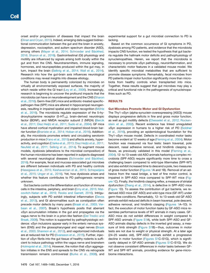
Gut Microbes Promote Motor and GI DysfunctionThe Thy1-aSyn (alpha-synuclein-overexpressing [ASO]) mouse
displays progressive deficits in fine and gross motor function,
as well as gut motility defects (Chesselet et al., 2012; Rocken-
stein et al., 2002). Recent evidence has linked unregulated
aSyn expression in humans to a higher risk of PD (Soldner
et al., 2016), providing an epidemiological foundation for the
Thy1-aSyn mouse model. Defects in coordinated motor tasks
become evident at 12 weeks of age (Fleming et al., 2004). Motor
function was measured via four tests: beam traversal, pole
descent, nasal adhesive removal, and hindlimb clasping re-
flexes, as previously validated in this model (Fleming et al.,
2004). 12- to 13-week-old ASO animals harboring a complex mi-
crobiota (SPF-ASO) require significantly more time to cross a
challenging beam compared to wild-type littermates (SPF-WT)
and also exhibit increased time to descend a pole, twomeasures
of gross motor function (Figures 1A and 1B). Removal of an ad-
hesive from the nasal bridge, a test of fine motor control, is
impaired in SPF-ASO mice compared to SPF-WT mice (Fig-
ure 1C). Finally, the hindlimb clasping reflex, ameasure of striatal
dysfunction (Zhang et al., 2014), is defective in SPF-ASO mice
(Figure 1D). To assess the contribution of gut bacteria, we re-
derived ASO mice (GF-ASO) and wild-type mice (GF-WT) under
germ-free conditions. Remarkably, 12- to 13-week-old GF-ASO
animals exhibit reduced deficits in beam traversal, pole descent,
adhesive removal, and hindlimb clasping (Figures 1A–1D). In
fact, the execution of motor function tasks by GF-ASO mice re-
sembles performance levels of WT animals in many cases. GF-
ASO mice do not exhibit differences in weight compared to
SPF-ASO animals (Figure S1A), while both SPF-ASO and GF-
ASO animals display defects in the inverted grid assay, a mea-
sure of limb strength (Figure S1B)—thus, outcomes in motor
tests are not due to weight or physical strength. At a later age
(24–25 weeks old), SPF-ASO animals exhibit a progressive
decline in motor function (Figures S1C–S1G), which is signifi-
cantly delayed in GF-ASO animals (Figures S1C–S1G). We do
not observe consistent differences in motor tasks between GF-
WT and SPF-WT animals, providing evidence for gene-micro-
biome interactions.

A B E
C D F
Figure 1. Gut Microbes Promote Motor and Gastrointestinal
Dysfunction
(A) Time to traverse beam apparatus.
(B) Time to descend pole.
(C) Time to remove adhesive from nasal bridge.
(D) Hind-limb clasping reflex score.
(E) Time course of fecal output in a novel environment over 15 min.
(F) Total fecal pellets produced in 15 min.
Animals were tested at 12–13 weeks of age. n = 4–6, error bars represent the
mean and standard error from three trials per animal. Data are representative
of two experiments. *p % 0.05; **p % 0.01; ***p % 0.001; ****p % 0.0001.
Abbreviations: SPF, specific-pathogen-free; GF, germ-free; WT, wild-type;
ASO, Thy1-a-synuclein genotype. See also Figure S1.
As in PD, motor dysfunction in this mouse model co-occurs
with decreased GI function and constipation (Verbaan et al.,
2007; Wang et al., 2012). In SPF-ASO animals, we observe a
marked decrease in the total output of fecal pellets, at both
12–13 weeks and 24–25 weeks of age, while fecal output is un-
altered in GF-ASO animals (Figures 1E, 1F, S1H, and S1I).
Further, fecal pellets produced by SPF-ASO mice contain
reduced water content compared to GF-ASO mice (Figure S1J),
together revealing reduced GI defects in GF animals. Indeed,
compilation of all motor phenotypes into a principal-component
analysis (PCoA) displays a striking segregation by the SPF-ASO
group, while GF-ASO animals cluster more similarly to WT mice
(Figure S1K). Together, these data demonstrate that the pres-
ence of gut microbes promote the hallmark motor and intestinal
dysfunction in a preclinical model of PD.
The Gut Microbiota Is Required for aSyn PathologyMotor deficits in PD coincide with the aggregation of aSyn. Uti-
lizing an antibody that recognizes only conformation-specific
aSyn aggregates and fibrils, we performed immunofluores-
cence microscopy to visualize aSyn inclusions in the brains
of mice. Under SPF conditions, we observe notable aggrega-
tion of aSyn in the caudoputamen (CP) and substantia nigra
(SN) of ASO animals (Figures 2A and 2B), brain regions of the
nigrostriatal pathway affected in both mouse models and hu-
man PD (Brettschneider et al., 2015). Surprisingly, GF-ASO
mice display appreciably fewer aSyn aggregates (Figures 2A
and 2B). To quantify aSyn aggregation, we performed western
blots of brain extracts (Figure 2C). We reveal significantly less
insoluble aSyn in brains of GF-ASO animals (Figures 2C–2E).
To further confirm these findings, we performed dot blot anal-
ysis for aggregated aSyn in the CP and inferior midbrain, where
the SN is located, and observe similarly decreased aSyn aggre-
gation in GF-ASO animals (Figures S2A–S2C). Interestingly, we
observe regional specificity of aSyn aggregation: in the frontal
cortex (FC), GF-ASO animals harbor fewer aSyn aggregation
than SPF animals, while in the cerebellum (CB), we observe
nearly equal quantities of aSyn in SPF and GF mice (Figures
S2D–S2H). To ensure that these findings do not reflect differ-
ences in transgene expression, we report similar levels of
aSyn transcript and protein in the inferior midbrain and the
CP between SPF- and GF-ASO animals (Figures 2F and 2G).
These data suggest that the microbiota regulates pathways
that promote aSyn aggregation and/or prevent the clearance
of insoluble protein aggregates.
aSyn-Dependent Microglia Activation by the MicrobiotaThe microbiota modulates immune development in the CNS
(Erny et al., 2015; Matcovitch-Natan et al., 2016), and aSyn ag-
gregates activate immune cells, including brain-resident micro-
glia (Kim et al., 2013; Sanchez-Guajardo et al., 2013). Microglia
undergo significant morphological changes upon activation,
transitioning from thin cell bodies with numerous branched ex-
tensions to round, amoeboid cells with fewer branches (Erny
et al., 2015). In situ 3D reconstructions of individual microglia
cells from confocal fluorescence microscopy reveals that wild-
type GF animals harbor microglia that are distinct from SPF
animals. Within the CP and SN, microglia in GF-WT mice display
increased numbers and total lengths of microglia branches
compared to SPF-WT animals (Figures 3A–3C). These morpho-
logical features are indicative of an arrest in microglia maturation
and/or a reduced activation state in GF animals, corroborating a
recent report that gut bacteria affect immune cells in the brain
(Erny et al., 2015).
Extending these observations to a disease model, microglia
from SPF-ASO mice display significant increases in cell
body diameter, along with fewer processes of shorter length
compared to GF-ASO mice (Figures 3A–3C). Tissue homoge-
nates from the CP and inferior midbrain of SPF-ASO mice
contain a marked increase in the pro-inflammatory cytokines tu-
mor necrosis factor-a (TNF-a) and interleukin-6 (IL-6) compared
to GF-ASO mice (Figures 3D and 3E). Both cytokines are
elevated in the brains of PD patients (Mogi et al., 1994a,
1994b). Gene expression analysis of RNA from enriched
CD11b+ cells (primarily microglia) reveals increased Tnfa and
Il6 expression in SPF-ASO animals, which is nearly absent in
GF animals (Figure 3F). Neuroprotective Bdnf and the cell
cycle marker Ddit4 levels are upregulated in GF animals (Fig-
ure S2I), as observed in previous studies (Erny et al., 2015; Mat-
covitch-Natan et al., 2016). Neuroinflammatory responses are
region specific with increased in microglia diameter and TNF-a
Cell 167, 1469–1480, December 1, 2016 1471

A C
D EB
F G
Figure 2. aSyn Pathology Is Increased in Mice
Harboring a Gut Microbiota
(A) Representative images of the caudoputamen (CP)
from SPF-ASO or GF-ASO animals stained with
aggregation-specific aSyn antibody (red), Phospho-
Ser129-aSyn antibody (green), and Neurotrace/Nissl
(blue).
(B) Representative images of the substantia nigra
(SN) from SPF-ASO or GF-ASO animals, stained as
above.
(C) Representative western blot of triton soluble and
insoluble brain homogenates, immunostained with
anti-aSyn antibody.
(D and E) Densitometry quantification of anti-aSyn
western blots for (D) all aSyn and (E) ratio of insoluble
to soluble aSyn staining.
(F) qRT-PCR analysis of human aSyn in the CP or
inferior midbrain (Mid).
(G) ELISA analysis of total aSyn present in homoge-
nates from the CP or inferior midbrain (Mid).
Tissues collected from mice at 12–13 weeks of age.
n = 3–4, error bars represent the mean and standard
error. *p % 0.05; **p % 0.01; ***p % 0.001. Abbrevi-
ations: SPF, specific-pathogen-free; GF, germ-free;
WT, wild-type; ASO, Thy1-a-synuclein genotype. See
also Figure S2.
production in the FC but not the CB (Figures 3G and 3H). Overall,
these findings support the hypothesis that gut microbes promote
aSyn-dependent activation of microglia within specific brain re-
gions involved in disease.
Postnatal Microbial Signals Modulate aSyn-DependentPathophysiologyThe microbiota influence neurological outcomes during gesta-
tion, as well as via active gut-to-brain signaling in adulthood.
In order to differentiate between these mechanisms, we treated
SPF animals with an antibiotic cocktail to postnatally deplete
the microbiota (Figure 4A). Conversely, we colonized groups
of 5- to 6-week-old GF mice with a complex microbiota from
SPF-WT animals (Figure 4A). Remarkably, antibiotic-treated
(Abx) animals display little aSyn-dependent motor dysfunction,
closely resembling mice born under GF conditions (Figures 4B–
4E). Postnatal colonization of previously GF animals (Ex-GF)
recapitulates the genotype effect observed in SPF mice,
with mice that overexpress aSyn displaying significant motor
dysfunction (Figures 4B–4E). GI function, as measured by fecal
output, is also significantly improved in Abx-treated animals,
while Ex-GF mice exhibit an aSyn-dependent decrease in total
fecal output (Figures 4F and 4G). Furthermore, in the transgenic
ASO line, microglia from Ex-GF animals have increased cell
body diameters comparable to those in SPF mice (Figures 4H
and 4I). Abx-ASO animals, however, harbor microglia with
diameters similar to GF animals (Figures 4H and 4I). While
not excluding a role for the microbiota during prenatal neuro-
development, modulation of microglia activation during adult-
hood contributes to aSyn-mediated motor dysfunction and
neuroinflammation, suggesting active gut-brain signaling by
the microbiota.
1472 Cell 167, 1469–1480, December 1, 2016
SCFAs Are Sufficient to Promote aSyn-MediatedNeuroinflammationRecently, it was revealed that gut bacteria modulate microglia
activation during viral infection through production of microbial
metabolites, namely short-chain fatty acids (SCFAs) (Erny et al.,
2015). Indeed, we observe lower fecal SCFA concentrations in
GFandAbx-treated animals, compared toSPFmice (Figure S3A;
Smith et al., 2013). To address whether SCFAs impact neuroim-
mune responses in a mouse model of PD, we treated GF-ASO
and GF-WT animals with a mixture of the SCFAs acetate, propi-
onate, and butyrate (while the animals remained microbiologi-
cally sterile) and significantly restored fecal SCFA concentrations
(Figure S3A). Within affected brain regions (i.e., CP and SN), mi-
croglia in SCFA-administered animals display morphology indic-
ative of increased activation compared to untreated mice, and
similar to cells from Ex-GF and SPF mice (Figures 5A, 5B, S3B,
and S3C; see also Figures 3 and 4). Microglia from GF-ASO
mice fed SCFAs (SCFA-ASO) are significantly larger in diameter
than those of GF-WT animals treated with SCFAs (SCFA-WT),
with a concomitant decrease in the length and total number of
branches. Abx-treated animals, however, display microglia
morphology similar to GF animals (Figures 5B, S3B, and S3C;
see also Figures 3 and 4). Changes in microglia diameter are
also observed in the FC, but not the CB, demonstrating region-
specific responses (Figures S3D and S3E).
Corresponding to microglia morphology, we reveal aSyn ag-
gregates in mice administered SCFAs compared to untreated
and Abx-treated mice, and similar to Ex-GF animals (Figures
S3F–S3I). Strikingly, we observe that postnatal signaling by mi-
crobes induces increased aSyn aggregation in the CP and SN
(Figures S3F and S3G), with no observable difference in the FC
and CB (Figures S3H and S3I), confirmed by quantification and

A B
C
D E F
G H
Mid
Figure 3. aSyn-Dependent Microglia Activation by the Microbiota
(A) Representative 3D reconstructions of Iba1-stained microglia residing in the caudoputamen (CP) of SPF-WT, SPF-ASO, GF-WT, and GF-ASO animals.
(B) CP-resident microglia parameters diameter, number of branch points, and total branch length.
(C) Substantia nigra (SN)-resident microglia parameters diameter, number of branch points, and total branch length.
(D) ELISA analysis for TNF-a and IL-6 present in homogenates from the CP.
(E) ELISA analysis for TNF-a and IL-6 present in homogenates from the inferior midbrain (Mid).
(F) qPCR analysis of CD11b+ cells derived from brain homogenate for tnfa and il6.
(G) Diameter of microglia residing in the frontal cortex (FC) or cerebellum (CB).
(H) ELISA analysis for TNF-a present in homogenates from the FC or CB.
Tissues collected from mice at 12–13 weeks of age. n = 3–4 (with 20–60 cells per region per animal analyzed); error bars represent the mean and standard error.
*p % 0.05; **p % 0.01; ***p % 0.001; ****p % 0.0001. Abbreviations: SPF, specific-pathogen-free; GF, germ-free; WT, wild-type; ASO, Thy1-a-synuclein
genotype. See also Figure S2.
western blot (Figures S3J–S3O). SCFAs either singly or in a
mixture, over a range of concentrations, do not expedite the ag-
gregation of human aSyn in vitro (Figures S4A–S4G), nor do they
alter the overall structure of aSyn amyloid fibrils (Figures S4H
and S4I). Though additional studies are needed, it appears that
SCFAs accelerate in vivo aSyn aggregation, albeit independently
of direct molecular interactions.
SCFAs Are Sufficient to Promote Motor DeficitsTo explore a link between microbial metabolites and motor
symptoms in the Thy1-aSyn model, GF animals were treated
with the SCFAmixture beginning at 5–6 weeks of age, andmotor
function was assessed at 12–13 weeks of age. SCFA-ASO mice
display significantly impaired performance in several motor
tasks compared to untreated GF-ASO animals (Figures 5C–
5F), including impairment in beam traversal, pole descent, and
hindlimb reflex (compare GF-ASO to SCFA-ASO mice). All ef-
fects by SCFAs are genotype specific to the Thy1-aSyn mice.
GI deficits are also observed in the SCFA-treated transgenic an-
imals (Figures 5G and 5H). Oral treatment of GF animals with
heat-killed bacteria does not induce motor deficits (Figures
S4J–S4M), suggesting that bacteria need to be metabolically
Cell 167, 1469–1480, December 1, 2016 1473

A
D
B C
E H
F G I
Figure 4. Postnatal Microbial Signals Promote Motor and Gastrointestinal Dysfunction
(A) Time course schema for animal treatment and testing.
(B) Time to traverse beam apparatus.
(C) Time to descend pole.
(D) Time to remove nasal adhesive.
(E) Hindlimb clasping reflex score.
(F) Time course of fecal output in a novel environment over 15 min.
(G) Total fecal pellets produced in 15 min.
(H) Representative 3D reconstructions of Iba1-stained microglia residing in the caudoputamen (CP) of Abx-ASO or Ex-GF-ASO animals.
(I) Diameter of microglia residing in the CP or substantia nigra (SN).
Animals were tested at 12–13 weeks of age. n = 6–12; error bars represent themean and standard error from 3 trials per animal, and compiled from 2 independent
cohorts or 20–60microglia per region analyzed. #0.05 < p < 0.1; *p% 0.05; **p% 0.01; ***p% 0.001; ****p% 0.0001. Abbreviations: SPF, specific-pathogen-free;
GF, germ-free; Abx, antibiotic-treated; Ex-GF, recolonized germ-free animals; WT, wild-type; ASO, Thy1-a-synuclein genotype. See also Figure S3.
active. Additionally, oral treatment of SCFA-fed animals with the
anti-inflammatory compound minocycline is sufficient to reduce
TNF-a production, reduce aSyn aggregation, and improvemotor
function, without altering transgene expression (Figures S5A–
S5H). We propose that the microbiota actively produce metabo-
lites, such as SCFAs, that are required for microglia activation
and aSyn aggregation, contributing to motor dysfunction in a
preclinical model of PD.
Dysbiosis of the PD MicrobiomeGiven recent evidence that PD patients display altered micro-
biomes (Hasegawa et al., 2015; Keshavarzian et al., 2015; Sche-
perjans et al., 2015), we sought to determine whether human gut
microbes affect disease outcomes when transferred into GF
1474 Cell 167, 1469–1480, December 1, 2016
mice. We collected fecal samples from six human subjects diag-
nosedwith PD aswell as sixmatched healthy controls (Table S1).
To limit confounding effects, only new-onset, treatment-naive
PD patients with healthy intestinal histology were chosen,
among other relevant inclusion and exclusion criteria (see
STAR Methods and Table S1).
Fecal microbiota from PD patients or controls were trans-
planted into individual groups of GF recipient animals via oral
gavage. Fecal pellets were collected from ‘‘humanized’’ mice,
bacterial DNA was extracted, and 16S rRNA sequencing was
performed. Sequences were annotated into operational taxo-
nomic units (OTUs), using closed reference picking against the
Greengenes database and metagenome function was predicted
by PICRUSt. Recipient animal groups were most similar to their

GFB
A C D E
H
Figure 5. SCFAs Promote aSyn-Stimulated Microglia Activation and Motor Dysfunction
(A) Representative 3D reconstructions of Iba1-stained microglia residing in the caudoputamen (CP) of wild-type or ASO SCFA-treated animals.
(B) Diameter of microglia residing in the CP or substantia nigra (SN).
(C) Time to traverse beam apparatus.
(D) Time to descend pole.
(E) Time to remove nasal adhesive.
(F) Hindlimb clasping reflex score.
(G) Time course of fecal output in a novel environment over 15 min.
(H) Total fecal pellets produced in 15 min.
Animals were tested at 12–13weeks of age. n = 6–12, error bars represent themean and standard error from 3 trials per animal, and compiled from 2 independent
cohorts or 20–60 microglia per region analyzed. Data are plotted with controls from Figure 4 for clarity. *p % 0.05; **p % 0.01; ***p % 0.001; ****p % 0.0001.
Abbreviations: SPF, specific-pathogen-free; GF, germ-free; SCFA, short-chain fatty acid-treated; WT, wild-type; ASO, Thy1-a-synuclein genotype. See also
Figures S3, S4, and S5.
respective human donor’s profile in unweighted UniFrac (Lozu-
pone and Knight, 2005), based on PCoA (Figures 6A and 6B).
Strikingly, the disease status of the donor had a strong effect
on the microbial communities within recipient mice. Humanized
mouse groups from PD donors are significantly more similar to
each other than to communities transplanted from healthy do-
nors, with this trend persisting when stratified by genetic back-
ground (Figures 6C and 6D). Furthermore, there are significant
differences between the healthy and PDdonors in the ASOback-
ground compared toWT recipients, suggesting genotype effects
on microbial community configuration (Figures 6C and 6D).
We identified a number of genera that are altered in animals
colonized with microbiota derived from PD donors, compared
to healthy controls (Figure 6E), as well as altered KEGG path-
ways between these groups as indicated by Bray-Curtis dis-
tances (Figures S6A–S6C). OTUs increased in abundance in
mice with PD microbiomes include Proteus sp., Bilophila sp.,
and Roseburia sp., with a concomitant loss of members of fam-
ilies Lachnospiraceae, Rikenellaceae, and Peptostreptococca-
ceae, as well as Butyricicoccus sp. (Figure 6E). Interestingly,
some taxa are altered only in ASO animals (e.g., Proteus sp.,
Bilophila sp., and Lachnospiraceae), while others display
significant changes independent of mouse genotype (e.g., Ros-
buria sp., Rikenellaceae, and Enterococcus sp.) (Figure 6E).
Intriguingly, the abundance of three SCFA-producing KEGG
families (K00929, butyrate kinase, and K01034 and K01035, ac-
etate CoA/acetoacetate CoA transferase alpha and beta) are
increased in mice that received fecal microbes derived from
PD donors (Figure S6D). Further, we observe that animals
receiving PD donor-derived microbiota display a significantly
altered SCFA profile, with a lower concentration of acetate
and higher relative abundances of propionate and butyrate,
compared to animals colonized with microbes from healthy con-
trols (Figure S6E). Together, these data indicate that differences
in fecal microbial communities between PDpatients and controls
can be maintained after transfer into mice. Further, aSyn overex-
pression engenders distinct alterations to the gut microbiome
profile after transplantation.
PD-Derived Gut Microbiota Promotes MotorDysfunctionTo assess microbiota function, groups of humanized animals
from each of the donor pairs were tested for motor function.
Consistent among four of the six pairs (pairs #1, 3, 4, and 5), mi-
crobiota derived from individuals with PD promote increased
aSyn-mediated motor dysfunction (Figures 7A–7F). Beam
traversal, pole descent, and nasal adhesive removal are signifi-
cantly impaired in ASO animals colonized with PD microbiota
compared to genotype-matched recipient mice harboring gut
bacteria from healthy controls. Hindlimb reflex scores, on the
other hand, are generally not different between individual do-
nors. Interestingly, microbiota from one pair of samples did not
induce significant genotype effects in the beam traversal and
pole descent tasks (pair #2, Figure 7B), reflecting potential het-
erogeneity in the population that needs to be addressed through
well-powered cohort studies. We observed no notable effects in
motor function by WT recipient animals colonized with micro-
biota from either donor group (Figures 7A–7F). This finding in a
Cell 167, 1469–1480, December 1, 2016 1475

C
A
E
B
D
Figure 6. Microbiome Dysbiosis of PD Patient Samples after
Transplant into Germ-free Mice
(A) Unweighted UniFrac Principle Coordinate Analysis of microbial commu-
nities of human donors (large circles) and recipient mice (small circles). Each
donor and recipient sample are matched by color.
(B) Unweighted and weighted UniFrac analysis of microbial communities in
recipient animals based on donor identity.
(C) Unweighted and weighted UniFrac analysis of microbial communities in
recipient animals based on mouse genotype.
(D) Comparison of unweighted and weighted UniFrac analysis of microbial
communities in recipient animals.
(E) Taxa-level analysis of individual genera altered between PD and healthy
donors as a function of recipient mouse genotype. Left column indicates
percentage with significant differences observed; right column indicates fold
change between PD and healthy donors. Light colors indicate non-statistically
significant differences.
n = 3–6, over 3 time points post-colonization. Error bars represent the mean
and standard error. ***p% 0.001, 999 permutations. Abbreviations: HC, germ-
free mice colonized with fecal microbes from healthy controls; PD, germ-free
mice colonized with fecal microbes from Parkinson’s disease patients; WT,
wild-type; ASO, Thy1-a-synuclein genotype. See also Figure S6.
preclinical mouse model supports the notion the PD microbiota
contributes to disease symptoms in genetically susceptible
hosts. Notably, recipient animals display little alteration to weight
and GI function as measured by fecal output (Figures S7A–S7F).
Compilation of performance data from all groups reveals that mi-
crobiota from PD patients induce increased motor impairment in
ASO animals compared to microbes from healthy controls in
1476 Cell 167, 1469–1480, December 1, 2016
three of four tests used in this study (Figure 7G). In fact, depicting
all motor function by PCoA displays striking global differences
between animals colonized with microbiota from PD donors,
compared to those colonized with gut bacteria derived from
healthy individuals (Figure S7G). The observation that gut bacte-
ria from PD patients compared to healthy controls enhance mo-
tor deficits in a mouse model provides evidence for a functional
contribution by the microbiota to synuclienopathies.
DISCUSSION
Parkinson’s disease represents a growing health concern for an
ever-aging population. While genetic risks have been identified,
environmental factors and gene-environment interactions prob-
ably account for most PD cases (Nalls et al., 2014; Ritz et al.,
2016). We provide evidence that the gut microbiota are required
for postnatal events that promote hallmark motor deficits in
an animal model. Under GF conditions, or when bacteria are
depleted with antibiotics, transgenic animals overexpressing hu-
man aSyn display reduced microglia activation, aSyn inclusions,
and motor deficits compared to animals with a complex micro-
biota. Treatment with microbially produced SCFAs restores all
major features of disease in GFmice, identifying potential molec-
ular mediators involved in gut-brain signaling. Exacerbated
motor symptoms in humanized mice transplanted with a PD
microbiota compared to healthy controls suggest that aSyn
overexpression (genetics) and dysbiosis (environment) combine
to influence disease outcomes in mice. Extrapolation of these
preclinical findings to humans may embolden the concept that
gene-microbiome interactions represent a previously unrecog-
nized etiology for PD.
Mechanisms by which gut bacteria promote aSyn-mediated
pathophysiology are likely complex; herein, we have identified
one potential pathway requiring microbiota-dependent effects
on microglia. Recent studies have demonstrated an active role
for the gut microbiota in promoting full maturation and inflamma-
tory capabilities of microglia through the production of SCFAs
(Erny et al., 2015). Despite a requirement for the SCFA receptor
FFAR2 for microglia maturation, these cells are not known to ex-
press FFAR2, but do express other SCFA-responsive genes
such as the histone deactylases that modulate gene expression
(Erny et al., 2015). SCFAs may cross the BBB and impact the
physiology of cells in the CNS (Mitchell et al., 2011), or they
may have peripheral effects, which indirectly activate and
mature microglia by currently unknown mechanisms (Erny
et al., 2015). Further, insoluble aggregates and oligomeric forms
of aSyn activate microglia (Kim et al., 2013; Sanchez-Guajardo
et al., 2013). Increases in the activation state of microglia and
the production of pro-inflammatory cytokines alter neuronal
function and increase cell death in models of PD and other
neurodegenerative diseases (Kannarkat et al., 2013; Sanchez-
Guajardo et al., 2013). Intriguingly, an inflammatory environment
is known to enhance aSyn aggregation, which may further
activate microglia upon contact and promote a feed-forward
cascade that leads to additional aSyn aggregation and propaga-
tion and progression of disease (Gao et al., 2011). If true,
possible future treatment options may include targeting immune
activation by the microbiota, a notion consistent with research

A
B
C
D
E
F
G
Figure 7. Microbiota from PD Patients Induce Increased aSyn-
Mediated Motor Deficits
(A–F) Time to cross a beam, time to descend the pole, time to remove nasal
adhesive, and hindlimb clasping reflex scores of mice humanized with mi-
crobiota from either PD patients or matched healthy controls.
(G) Compilation of all independent cohorts in each motor task: beam traversal,
pole descent, adhesive removal, and hindlimb clasping reflex score, grouped
by health status of fecal donor.
Animals were tested at 12–13 weeks of age. n = 3–6, error bars represent
the mean and standard error from 3 trials per animal. #0.05 < p < 0.1; *p %
0.05; **p % 0.01; ***p % 0.001; ****p % 0.0001. Abbreviations: HC, germ-
free mice colonized with fecal microbes from healthy controls; PD, germ-
into anti-inflammatory therapeutic modalities for PD (Valera and
Masliah, 2016).
While the microbiota promote microglia maturation, there are
likely other disease-modifying processes that remain undiscov-
ered. These include effects by the microbiota on autophagy
(Lin et al., 2014), a cellular recycling process that is genetically
linked to PD risk and when impaired may lead to reduced clear-
ance of aSyn aggregates (Beilina andCookson, 2015; Nalls et al.,
2014). Additionally, intestinal bacteria have been shown to
modulate proteasome function (Cleynen et al., 2014), which
may also aid in the clearance of aSyn inclusions. The protective
effects of autophagy and the proteasome are not specific to syn-
uclienopathies, and the ability of the microbiota to modulate
these critical cellular functions suggests that other amyloid dis-
orders, such as Alzheimer’s and Huntington’s diseases, may
be impacted by gut bacteria. In fact, recent studies have impli-
cated the gut microbiota in promoting amyloid beta pathology
in a model of Alzheimer’s disease (Minter et al., 2016). Though
we have explored postnatal effects of the microbiota in a model
of neurodegenerative disease, our findings do not address the
likely important role of microbial signals during prenatal neurode-
velopment. Whether gut microbes alter the development of the
dopaminergic system, perhaps by modulating neurogenesis or
neural differentiation in utero or early life, remains unexplored.
Furthermore, gut microbes can produce dopamine and its pre-
cursors from dietary substrates, with almost half of the body’s
dopamine generated in the GI tract (Eisenhofer et al., 1997;
Wall et al., 2014). Deciphering microbiota effects on microglia
activation, cellular protein clearance pathways, neurotransmitter
production, and/or other mechanisms may offer an integrated
approach to understand the pathogenesis of a complex and
enigmatic disorder such as PD.
We reveal that gut bacteria from PD patients promote
enhanced motor impairment compared to microbiota from
healthy controls when transplanted into genetically susceptible
ASO mice. This surprising finding suggests that distinct
microbes associated with PD, rather than general microbial
stimulation, manifest disease symptoms. Several bacterial taxa
are altered in mice receiving fecal transplants from PD
patients compared to healthy controls. Additionally, a number
of bacterial genera are changed specifically in ASO animals,
but not WT mice, receiving microbes from the same donor.
These include depletions in members of family Lachnospiraceae
and Ruminococceae in recipient mice, a notable finding as
these same genera are significantly reduced in fecal samples
directly from PD patients (Keshavarzian et al., 2015). Conversely,
the gut microbiomes in human subjects with PD contain an
increased abundance of Proteobacteria (Hasegawa et al.,
2015; Keshavarzian et al., 2015; Scheperjans et al., 2015; Unger
et al., 2016), remarkably similar to our results in mice. Whether
these specificmicrobes play a role in disease processes remains
unknown. Intriguingly, a recent study demonstrated alterations
in fecal SCFA ratios between patients and healthy controls,
including an elevated relative concentration of butyrate, possibly
free mice colonized with fecal microbes from Parkinson’s disease patients;
WT, wild-type; ASO, Thy1-a-synuclein genotype. See also Figure S7 and
Table S1.
Cell 167, 1469–1480, December 1, 2016 1477

implicating a role for SCFAs in PD (Unger et al., 2016). Accord-
ingly, we observe altered SCFA abundances in animals colo-
nized with PD donor-derived microbiota, and the discovery
that SCFAs are sufficient to generate aSyn-reactive microglia
in the brain is consistent with expansive literature showing that
altered microbial communities impact immune responses in
the gut and periphery (Hooper et al., 2012).
What causes dysbiosis in PD? Physiological functions in
affected individuals, such as altered intestinal absorption,
reduced gastric motility, or dietary habits, represent factors
that may change the microbiome. Epidemiological evidence
has linked specific pesticide exposure to the incidence of PD
(Ritz et al., 2016), with some pesticides known to impact micro-
biome configuration (Gao et al., 2016). Given the structure of
aSyn and its ability to associate with membranes (Jo et al.,
2000), it is tempting to speculate that extracellular aSyn may
act as an antimicrobial, similar to recent observations with amy-
loid beta (Kumar et al., 2016), and shape the PD microbiome.
Whether microbial community alterations are caused by
extrinsic or intrinsic factors, the PD microbiota may be missing
or reduced in protective microbes, harbor increased pathogenic
resident microbes, or both. In turn, dysbiosis will result in differ-
ential production of microbial molecules in the gut. Metabolites
produced by a deranged microbiota may enter the circulation
(or even the brain) and impact neurological function. Identifica-
tion of bacterial taxa or microbial metabolites that are altered
in PD may serve as disease biomarkers or even drug targets,
and interventions that correct dysbiosis may provide safe and
effective treatments to slow or halt the progression of often
debilitating motor symptoms.
Our findings establish that the microbiota are required for
the hallmark motor and GI dysfunction in a mouse model of
PD, via postnatal gut-brain signaling by microbial molecules
that impact neuroinflammation and aSyn aggregation. Coupled
with emerging research that has linked gut bacteria to disorders
such as anxiety, depression, and autism, we propose the pro-
vocative hypothesis that certain neurologic conditions that
have classically been studied as disorders of the brain may
also have etiologies in the gut.
STAR+METHODS
Detailed methods are provided in the online version of this paper
and include the following:
d KEY RESOURCES TABLE
d CONTACT FOR REAGENT AND RESOURCE SHARING
d EXPERIMENTAL MODEL AND SUBJECT DETAILS
147
B Mice
B Human Donor and Criteria
d METHOD DETAILS
B Motor Function and Gastrointestinal Testing
B Immunostaining and Microglia Reconstructions
B CD11b Enrichment and qPCR Analysis
B Cytokine and aSyn ELISAs and Western Blots
B aSyn Aggregation Assays
B SCFA Extraction and Analysis
B Microbiome Profiling
8 Cell 167, 1469–1480, December 1, 2016
d QUANTIFICATION AND STATISTICAL ANALYSIS
d DATA AND SOFTWARE AVAILABILITY
SUPPLEMENTAL INFORMATION
Supplemental Information includes seven figures and one table and can be
found with this article online at http://dx.doi.org/10.1016/j.cell.2016.11.018.
AUTHOR CONTRIBUTIONS
Conceptualization, T.R.S., C.E.S., M.-F.C., and S.K.M.; Formal Analysis,
J.W.D., S.J., and C.C.; Investigation, T.R.S., T.T., G.G.S., Z.E.I., and S.R.;
Resources, A.K. and K.M.S.; Writing - Original Draft, T.R.S. and S.K.M.;
Writing - Review and Editing, all authors; Supervision, V.G., R.K.-B.,
P.W.-S., R.K., and S.K.M.; Funding Acquisition, T.R.S., V.G., M.-F.C., A.K.,
P.W.-S., R.K., and S.K.M.
ACKNOWLEDGMENTS
We thank E. Hsiao, M. Sampson, and the S.K.M. laboratory for helpful cri-
tiques. We are grateful to K. Ly, A. Maskell, and M. Quintos for animal hus-
bandry and Y. Garcia-Flores, G. Ackermann, G. Humphrey, and H. Derderian
for technical support. Imaging and analysis was performed in the Caltech Bio-
logical Imaging Facility, with the support of the Caltech Beckman Institute and
the Arnold and Mabel Beckman Foundation. T.R.S. is a Larry L. Hillblom Foun-
dation postdoctoral fellow. This project was supported by funds from the Knut
and AliceWallenberg Foundation and Swedish Research Council to P.W.-S.; a
gift from Mrs. and Mr. Larry Field to A.K.; the Heritage Medical Research Insti-
tute to V.G. and S.K.M.; and NIH grant NS085910 to S.K.M.
Received: June 29, 2016
Revised: October 12, 2016
Accepted: November 10, 2016
Published: December 1, 2016
REFERENCES
Beilina, A., and Cookson, M.R. (2015). Genes associated with Parkinson’s dis-
ease: regulation of autophagy and beyond. J. Neurochem. 139 (Suppl 1 ),
91–107.
Bercik, P., Denou, E., Collins, J., Jackson, W., Lu, J., Jury, J., Deng, Y., Blen-
nerhassett, P., Macri, J., McCoy, K.D., et al. (2011). The intestinal microbiota
affect central levels of brain-derived neurotropic factor and behavior in mice.
Gastroenterology 141, 599–609, 609.e1–609.e3.
Braak, H., Rub, U., Gai, W.P., and Del Tredici, K. (2003). Idiopathic Parkinson’s
disease: possible routes by which vulnerable neuronal typesmay be subject to
neuroinvasion by an unknown pathogen. J Neural Transm (Vienna) 110,
517–536.
Braniste, V., Al-Asmakh, M., Kowal, C., Anuar, F., Abbaspour, A., Toth, M., Ko-
recka, A., Bakocevic, N., Ng, L.G., Kundu, P., et al. (2014). The gut microbiota
influences blood-brain barrier permeability in mice. Sci. Transl. Med. 6,
263ra158.
Brettschneider, J., Del Tredici, K., Lee, V.M., and Trojanowski, J.Q. (2015).
Spreading of pathology in neurodegenerative diseases: a focus on human
studies. Nat. Rev. Neurosci. 16, 109–120.
Burke, R.E., Dauer, W.T., and Vonsattel, J.P. (2008). A critical evaluation of the
Braak staging scheme for Parkinson’s disease. Ann. Neurol. 64, 485–491.
Caporaso, J.G., Kuczynski, J., Stombaugh, J., Bittinger, K., Bushman, F.D.,
Costello, E.K., Fierer, N., Pena, A.G., Goodrich, J.K., Gordon, J.I., et al.
(2010). QIIME allows analysis of high-throughput community sequencing
data. Nat. Methods 7, 335–336.
Chesselet, M.F., Richter, F., Zhu, C., Magen, I., Watson, M.B., and Subrama-
niam, S.R. (2012). A progressive mouse model of Parkinson’s disease: the
Thy1-aSyn (‘‘Line 61’’) mice. Neurotherapeutics 9, 297–314.

Chorell, E., Andersson, E., Evans, M.L., Jain, N., Gotheson, A., Aden, J.,
Chapman, M.R., Almqvist, F., and Wittung-Stafshede, P. (2015). Bacterial
chaperones CsgE and CsgC differentially modulate human a-synuclein amy-
loid formation via transient contacts. PLoS ONE 10, e0140194.
Clarke, G., Grenham, S., Scully, P., Fitzgerald, P., Moloney, R.D., Shanahan,
F., Dinan, T.G., and Cryan, J.F. (2013). The microbiome-gut-brain axis during
early life regulates the hippocampal serotonergic system in a sex-dependent
manner. Mol. Psychiatry 18, 666–673.
Cleynen, I., Vazeille, E., Artieda, M., Verspaget, H.W., Szczypiorska, M.,
Bringer, M.A., Lakatos, P.L., Seibold, F., Parnell, K., Weersma, R.K., et al.
(2014). Genetic and microbial factors modulating the ubiquitin proteasome
system in inflammatory bowel disease. Gut 63, 1265–1274.
Del Tredici, K., and Braak, H. (2008). A not entirely benign procedure: progres-
sion of Parkinson’s disease. Acta Neuropathol. 115, 379–384.
Devos, D., Lebouvier, T., Lardeux, B., Biraud, M., Rouaud, T., Pouclet, H.,
Coron, E., Bruley des Varannes, S., Naveilhan, P., Nguyen, J.M., et al.
(2013). Colonic inflammation in Parkinson’s disease. Neurobiol. Dis. 50, 42–48.
Diaz Heijtz, R., Wang, S., Anuar, F., Qian, Y., Bjorkholm, B., Samuelsson, A.,
Hibberd, M.L., Forssberg, H., and Pettersson, S. (2011). Normal gutmicrobiota
modulates brain development and behavior. Proc. Natl. Acad. Sci. USA 108,
3047–3052.
Dinan, T.G., and Cryan, J.F. (2015). The impact of gut microbiota on brain and
behaviour: implications for psychiatry. Curr. Opin. Clin. Nutr. Metab. Care 18,
552–558.
Eisenhofer, G., Aneman, A., Friberg, P., Hooper, D., Fandriks, L., Lonroth, H.,
Hunyady, B., and Mezey, E. (1997). Substantial production of dopamine in the
human gastrointestinal tract. J. Clin. Endocrinol. Metab. 82, 3864–3871.
Erny, D., Hrab�e de Angelis, A.L., Jaitin, D., Wieghofer, P., Staszewski, O., Da-
vid, E., Keren-Shaul, H., Mahlakoiv, T., Jakobshagen, K., Buch, T., et al. (2015).
Host microbiota constantly control maturation and function of microglia in the
CNS. Nat. Neurosci. 18, 965–977.
Fleming, S.M., Salcedo, J., Fernagut, P.O., Rockenstein, E., Masliah, E., Lev-
ine, M.S., and Chesselet, M.F. (2004). Early and progressive sensorimotor
anomalies in mice overexpressing wild-type human alpha-synuclein.
J. Neurosci. 24, 9434–9440.
Gao, H.M., Zhang, F., Zhou, H., Kam, W., Wilson, B., and Hong, J.S. (2011).
Neuroinflammation and a-synuclein dysfunction potentiate each other, driving
chronic progression of neurodegeneration in a mouse model of Parkinson’s
disease. Environ. Health Perspect. 119, 807–814.
Gao, B., Bian, X., Mahbub, R., and Lu, K. (2016). Gender-specific effects of
organophosphate diazinon on the gut microbiome and its metabolic functions.
Environ. Health Perspect. Published online May 20, 2016. http://dx.doi.org/10.
1289/EHP202.
Gilbert, J.A., Jansson, J.K., and Knight, R. (2014). The Earth Microbiome proj-
ect: successes and aspirations. BMC Biol. 12, 69.
Hasegawa, S., Goto, S., Tsuji, H., Okuno, T., Asahara, T., Nomoto, K., Shibata,
A., Fujisawa, Y., Minato, T., Okamoto, A., et al. (2015). Intestinal dysbiosis and
lowered serum lipopolysaccharide-binding protein in Parkinson’s disease.
PLoS ONE 10, e0142164.
Hoban, A.E., Stilling, R.M., Ryan, F.J., Shanahan, F., Dinan, T.G., Claesson,
M.J., Clarke, G., and Cryan, J.F. (2016). Regulation of prefrontal cortex myeli-
nation by the microbiota. Transl. Psychiatry 6, e774.
Holmqvist, S., Chutna, O., Bousset, L., Aldrin-Kirk, P., Li, W., Bjorklund, T.,
Wang, Z.Y., Roybon, L., Melki, R., and Li, J.Y. (2014). Direct evidence of Par-
kinson pathology spread from the gastrointestinal tract to the brain in rats. Acta
Neuropathol. 128, 805–820.
Hooper, L.V., Littman, D.R., and Macpherson, A.J. (2012). Interactions be-
tween the microbiota and the immune system. Science 336, 1268–1273.
Jenner, P. (2008). Molecular mechanisms of L-DOPA-induced dyskinesia. Nat.
Rev. Neurosci. 9, 665–677.
Jo, E., McLaurin, J., Yip, C.M., St George-Hyslop, P., and Fraser, P.E. (2000).
alpha-Synuclein membrane interactions and lipid specificity. J. Biol. Chem.
275, 34328–34334.
Kannarkat, G.T., Boss, J.M., and Tansey, M.G. (2013). The role of innate and
adaptive immunity in Parkinson’s disease. J. Parkinsons Dis. 3, 493–514.
Keshavarzian, A., Green, S.J., Engen, P.A., Voigt, R.M., Naqib, A., Forsyth,
C.B., Mutlu, E., and Shannon, K.M. (2015). Colonic bacterial composition in
Parkinson’s disease. Mov. Disord. 30, 1351–1360.
Kim, C., Ho, D.H., Suk, J.E., You, S., Michael, S., Kang, J., Joong Lee, S., Mas-
liah, E., Hwang, D., Lee, H.J., and Lee, S.J. (2013). Neuron-released oligomeric
a-synuclein is an endogenous agonist of TLR2 for paracrine activation of mi-
croglia. Nat. Commun. 4, 1562.
Klucken, J., Ingelsson, M., Shin, Y., Irizarry, M.C., Hedley-Whyte, E.T., Frosch,
M., Growdon, J., McLean, P., and Hyman, B.T. (2006). Clinical and biochem-
ical correlates of insoluble alpha-synuclein in dementia with Lewy bodies. Acta
Neuropathol. 111, 101–108.
Kohman, R.A., Bhattacharya, T.K., Kilby, C., Bucko, P., and Rhodes, J.S.
(2013). Effects of minocycline on spatial learning, hippocampal neurogenesis
and microglia in aged and adult mice. Behav. Brain Res. 242, 17–24.
Kopylova, E., Noe, L., and Touzet, H. (2012). SortMeRNA: fast and accurate
filtering of ribosomal RNAs in metatranscriptomic data. Bioinformatics 28,
3211–3217.
Kumar, D.K., Choi, S.H., Washicosky, K.J., Eimer, W.A., Tucker, S., Ghofrani,
J., Lefkowitz, A., McColl, G., Goldstein, L.E., Tanzi, R.E., andMoir, R.D. (2016).
Amyloid-b peptide protects against microbial infection in mouse and worm
models of Alzheimer’s disease. Sci. Transl. Med. 8, 340ra72.
Langille, M.G., Zaneveld, J., Caporaso, J.G., McDonald, D., Knights, D.,
Reyes, J.A., Clemente, J.C., Burkepile, D.E., Vega Thurber, R.L., Knight, R.,
et al. (2013). Predictive functional profiling of microbial communities using
16S rRNA marker gene sequences. Nat. Biotechnol. 31, 814–821.
Ley, R.E., Peterson, D.A., and Gordon, J.I. (2006). Ecological and evolutionary
forces shaping microbial diversity in the human intestine. Cell 124, 837–848.
Lin, R., Jiang, Y., Zhao, X.Y., Guan, Y., Qian, W., Fu, X.C., Ren, H.Y., and Hou,
X.H. (2014). Four types of Bifidobacteria trigger autophagy response in intes-
tinal epithelial cells. J. Dig. Dis. 15, 597–605.
Lozupone, C., and Knight, R. (2005). UniFrac: a new phylogenetic method for
comparing microbial communities. Appl. Environ. Microbiol. 71, 8228–8235.
Luk, K.C., Kehm, V., Carroll, J., Zhang, B., O’Brien, P., Trojanowski, J.Q., and
Lee, V.M. (2012). Pathological a-synuclein transmission initiates Parkinson-
like neurodegeneration in nontransgenic mice. Science 338, 949–953.
Mandal, S., Van Treuren, W., White, R.A., Eggesbø, M., Knight, R., and Ped-
dada, S.D. (2015). Analysis of composition of microbiomes: a novel method
for studying microbial composition. Microb. Ecol. Health Dis. 26, 27663.
Matcovitch-Natan, O., Winter, D.R., Giladi, A., Vargas Aguilar, S., Spinrad, A.,
Sarrazin, S., Ben-Yehuda, H., David, E., Zelada Gonzalez, F., Perrin, P., et al.
(2016). Microglia development follows a stepwise program to regulate brain
homeostasis. Science 353, aad8670.
Mayer, E.A., Padua, D., and Tillisch, K. (2014). Altered brain-gut axis in autism:
comorbidity or causative mechanisms? BioEssays 36, 933–939.
McDonald, D., Price, M.N., Goodrich, J., Nawrocki, E.P., DeSantis, T.Z.,
Probst, A., Andersen, G.L., Knight, R., and Hugenholtz, P. (2012). An improved
Greengenes taxonomy with explicit ranks for ecological and evolutionary ana-
lyses of bacteria and archaea. ISME J. 6, 610–618.
Minter, M.R., Zhang, C., Leone, V., Ringus, D.L., Zhang, X., Oyler-Castrillo, P.,
Musch, M.W., Liao, F., Ward, J.F., Holtzman, D.M., et al. (2016). Antibiotic-
induced perturbations in gut microbial diversity influences neuro-inflammation
and amyloidosis in a murine model of Alzheimer’s disease. Sci. Rep. 6, 30028.
Mitchell, R.W., On, N.H., Del Bigio, M.R., Miller, D.W., and Hatch, G.M. (2011).
Fatty acid transport protein expression in human brain and potential role in
fatty acid transport across human brain microvessel endothelial cells.
J. Neurochem. 117, 735–746.
Mogi, M., Harada, M., Kondo, T., Riederer, P., Inagaki, H., Minami, M., and Na-
gatsu, T. (1994a). Interleukin-1 beta, interleukin-6, epidermal growth factor and
transforming growth factor-alpha are elevated in the brain from parkinsonian
patients. Neurosci. Lett. 180, 147–150.
Cell 167, 1469–1480, December 1, 2016 1479
Mogi, M., Harada, M., Riederer, P., Narabayashi, H., Fujita, K., and Nagatsu, T.
(1994b). Tumor necrosis factor-alpha (TNF-alpha) increases both in the brain
and in the cerebrospinal fluid from parkinsonian patients. Neurosci. Lett.
165, 208–210.
Mohle, L., Mattei, D., Heimesaat, M.M., Bereswill, S., Fischer, A., Alutis, M.,
French, T., Hambardzumyan, D., Matzinger, P., Dunay, I.R., and Wolf, S.A.
(2016). Ly6C(hi) monocytes provide a link between antibiotic-induced changes
in gut microbiota and adult hippocampal neurogenesis. Cell Rep. 15, 1945–
1956.
Nalls, M.A., Pankratz, N., Lill, C.M., Do, C.B., Hernandez, D.G., Saad, M.,
DeStefano, A.L., Kara, E., Bras, J., Sharma, M., et al.; International Parkinson’s
Disease Genomics Consortium (IPDGC); Parkinson’s Study Group (PSG) Par-
kinson’s Research: The Organized GENetics Initiative (PROGENI); 23andMe;
GenePD; NeuroGenetics Research Consortium (NGRC); Hussman Institute
of Human Genomics (HIHG); Ashkenazi Jewish Dataset Investigator; Cohorts
for Health and Aging Research in Genetic Epidemiology (CHARGE); North
American Brain Expression Consortium (NABEC); United Kingdom Brain
Expression Consortium (UKBEC); Greek Parkinson’s Disease Consortium;
Alzheimer Genetic Analysis Group (2014). Large-scale meta-analysis of
genome-wide association data identifies six new risk loci for Parkinson’s dis-
ease. Nat. Genet. 46, 989–993.
Neufeld, K.M., Kang, N., Bienenstock, J., and Foster, J.A. (2011). Reduced
anxiety-like behavior and central neurochemical change in germ-free mice.
Neurogastroenterol. Motil. 23, 255–264, e119.
Prusiner, S.B., Woerman, A.L., Mordes, D.A., Watts, J.C., Rampersaud, R.,
Berry, D.B., Patel, S., Oehler, A., Lowe, J.K., Kravitz, S.N., et al. (2015). Evi-
dence for a-synuclein prions causing multiple system atrophy in humans
with parkinsonism. Proc. Natl. Acad. Sci. USA 112, E5308–E5317.
Ritz, B.R., Paul, K.C., and Bronstein, J.M. (2016). Of pesticides andmen: a Cal-
ifornia story of genes and environment in Parkinson’s disease. Curr. Environ.
Health Rep. 3, 40–52.
Rockenstein, E., Mallory, M., Hashimoto, M., Song, D., Shults, C.W., Lang, I.,
and Masliah, E. (2002). Differential neuropathological alterations in transgenic
mice expressing alpha-synuclein from the platelet-derived growth factor and
Thy-1 promoters. J. Neurosci. Res. 68, 568–578.
Rooks, M.G., and Garrett, W.S. (2016). Gut microbiota, metabolites and host
immunity. Nat. Rev. Immunol. 16, 341–352.
Sacchettini, J.C., and Kelly, J.W. (2002). Therapeutic strategies for human am-
yloid diseases. Nat. Rev. Drug Discov. 1, 267–275.
Sanchez-Guajardo, V., Barnum, C.J., Tansey, M.G., and Romero-Ramos, M.
(2013). Neuroimmunological processes in Parkinson’s disease and their rela-
tion to a-synuclein: microglia as the referee between neuronal processes
and peripheral immunity. ASN Neuro 5, 113–139.
Scheperjans, F., Aho, V., Pereira, P.A., Koskinen, K., Paulin, L., Pekkonen, E.,
Haapaniemi, E., Kaakkola, S., Eerola-Rautio, J., Pohja, M., et al. (2015). Gut
microbiota are related to Parkinson’s disease and clinical phenotype. Mov.
Disord. 30, 350–358.
Schroeder, B.O., and Backhed, F. (2016). Signals from the gut microbiota to
distant organs in physiology and disease. Nat. Med. 22, 1079–1089.
Selkrig, J., Wong, P., Zhang, X., and Pettersson, S. (2014). Metabolic tinkering
by the gut microbiome: Implications for brain development and function. Gut
Microbes 5, 369–380.
1480 Cell 167, 1469–1480, December 1, 2016
Shannon, K.M., Keshavarzian, A., Dodiya, H.B., Jakate, S., and Kordower, J.H.
(2012). Is alpha-synuclein in the colon a biomarker for premotor Parkinson’s
disease? Evidence from 3 cases. Mov. Disord. 27, 716–719.
Sharon, G., Sampson, T.R., Geschwind, D.H., and Mazmanian, S.K. (2016).
The central nervous system and the gut microbiome. Cell 167, 915–932.
Smith, P.M., Howitt, M.R., Panikov, N., Michaud, M., Gallini, C.A., Bohlooly-Y,
M., Glickman, J.N., and Garrett, W.S. (2013). Themicrobial metabolites, short-
chain fatty acids, regulate colonic Treg cell homeostasis. Science 341,
569–573.
Soldner, F., Stelzer, Y., Shivalila, C.S., Abraham, B.J., Latourelle, J.C., Bar-
rasa, M.I., Goldmann, J., Myers, R.H., Young, R.A., and Jaenisch, R. (2016).
Parkinson-associated risk variant in distal enhancer of a-synuclein modulates
target gene expression. Nature 533, 95–99.
Sudo, N., Chida, Y., Aiba, Y., Sonoda, J., Oyama, N., Yu, X.N., Kubo, C., and
Koga, Y. (2004). Postnatal microbial colonization programs the hypothalamic-
pituitary-adrenal system for stress response in mice. J. Physiol. 558, 263–275.
Svensson, E., Horvath-Puho, E., Thomsen, R.W., Djurhuus, J.C., Pedersen, L.,
Borghammer, P., and Sørensen, H.T. (2015). Vagotomy and subsequent risk of
Parkinson’s disease. Ann. Neurol. 78, 522–529.
Unger, M.M., Spiegel, J., Dillmann, K.U., Grundmann, D., Philippeit, H., Bur-
mann, J., Faßbender, K., Schwiertz, A., and Schafer, K.H. (2016). Short chain
fatty acids and gutmicrobiota differ between patients with Parkinson’s disease
and age-matched controls. Parkinsonism Relat. Disord. 32, 66–72.
Valera, E., and Masliah, E. (2016). Combination therapies: the next logical step
for the treatment of synucleinopathies? Mov. Disord. 31, 225–234.
Vazquez-Baeza, Y., Pirrung, M., Gonzalez, A., and Knight, R. (2013). EMPeror:
a tool for visualizing high-throughput microbial community data. Gigascience
2, 16.
Verbaan, D., Marinus, J., Visser, M., van Rooden, S.M., Stiggelbout, A.M., and
van Hilten, J.J. (2007). Patient-reported autonomic symptoms in Parkinson
disease. Neurology 69, 333–341.
Wall, R., Cryan, J.F., Ross, R.P., Fitzgerald, G.F., Dinan, T.G., and Stanton, C.
(2014). Bacterial neuroactive compounds produced by psychobiotics. Adv.
Exp. Med. Biol. 817, 221–239.
Walters, W., Hyde, E.R., Berg-Lyons, D., Ackermann, G., Humphrey, G., Par-
ada, A., Gilbert, J.A., Jansson, J.K., Caporaso, J.G., Fuhrman, J.A., et al.
(2015). Improved bacterial 16S rRNA gene (V4 and V4-5) and fungal internal
transcribed spacer marker gene primers for microbial community surveys.
mSystems 1, 1.
Wang, L., Magen, I., Yuan, P.Q., Subramaniam, S.R., Richter, F., Chesselet,
M.F., and Tache, Y. (2012). Mice overexpressing wild-type human alpha-syn-
uclein display alterations in colonic myenteric ganglia and defecation. Neuro-
gastroenterol. Motil. 24, e425–e436.
Yano, J.M., Yu, K., Donaldson, G.P., Shastri, G.G., Ann, P., Ma, L., Nagler,
C.R., Ismagilov, R.F., Mazmanian, S.K., and Hsiao, E.Y. (2015). Indigenous
bacteria from the gut microbiota regulate host serotonin biosynthesis. Cell
161, 264–276.
Zhang, J., Saur, T., Duke, A.N., Grant, S.G., Platt, D.M., Rowlett, J.K., Isacson,
O., and Yao, W.D. (2014). Motor impairments, striatal degeneration, and
altered dopamine-glutamate interplay in mice lacking PSD-95. J. Neurogenet.
28, 98–111.

STAR+METHODS
KEY RESOURCES TABLE
REAGENT or RESOURCE SOURCE IDENTIFIER
Antibodies
Anti-aggregated/fibril alpha-synuclein; MJFR-14-6-4-2 Abcam Cat# ab209538
Anti-phospho Ser129 alpha-synuclein BioLegend Cat# 825701, RRID: AB_2564891
Anti-Iba1 Wako Cat# 019-19741, RRID: AB_839504
Anti-mouse IgG-Alexafluor 488 Life Technologies Cat# A-11001, RRID: AB_2534069
Anti-rabbit IgG-Alexafluor 546 Life Technologies Cat# A-11010, RRID: AB_2534077
Neurotrace 435/455 Blue Life Technologies Cat# N-21479
Anti- alpha-synuclein BD Cat# 610787, RRID: AB_398108
Anti-mouse IgG-HRP Cell Signaling Technology Cat# 7076S
Anti-rabbit IgG-HRP Cell Signaling Technology Cat# 7074S
Chemicals, Peptides, and Recombinant Proteins
Minocycline Arcos / Fisher Scientific Cat# AC455330010
Ampicillin Sigma Aldrich Cat# A0166
Vancomycin Sagent Pharmaceuticals Cat# 157-99
Neomycin Fisher Scientific Cat# BP26695
Gentamicin Sigma Aldrich Cat# G1264
Erythromycin Sigma Aldrich Cat# E5389
Sodium acetate Sigma Aldrich Cat# S5636
Sodium propionate Sigma Aldrich Cat# P5436
Sodium butyrate Sigma Aldrich Cat# 19364
Purified alpha synuclein Abcam Cat# ab51188
Thioflavin T Sigma Aldrich Cat# T3516
Critical Commercial Assays
Alpha-synuclein ELISA ThermoFisher Cat# KHB0061
iScript cDNA Synthesis kit Biorad Cat# 170-8890
SyberGreen qPCR Master Mix Applied Biosystems Cat# 4309155
IL-6 ELISA eBioscience Cat# 88-7324
TNF ELISA eBioscience Cat# 88-7064
MoBio Power Soil Kit MoBio Laboratories Cat# 12888-50
Deposited Data
16 s RNA sequences QIITA; https://qiita.ucsd.edu/ 10483
16 s RNA sequences EMBL, ENA; http://www.ebi.ac.uk/ena ERP019564
Experimental Models: Organisms/Strains
Mouse: Thy1-alpha synuclein Marie Francoise-Chesselet, University
of California, Los Angeles
N/A
Mouse: BDF1 Generated from B6 x DBA N/A
Mouse: C57BL/6 Charles River Strain: 027
Mouse: DBA/2 Charles River Strain: 026
Bacteria: Escherichia coli K12 MC4100 Matt Chapman, University of Michigan N/A
Sequence-Based Reagents
Primers: hu-snca - Fwd: 50-TTGCAGCAGCCACTGGCT
TTG-30 and Rev: 50-GGATCCACAGGCATATCTTCCAGAA-30PrimerBank N/A
Primers: tnfa- Fwd: 50-CCCTCACACTCAGATCATCTTCT-3’
and Rev: 50-GCTACGACGTGGGCTACAG-30PrimerBank N/A
(Continued on next page)
Cell 167, 1469–1480.e1–e5, December 1, 2016 e1

Continued
REAGENT or RESOURCE SOURCE IDENTIFIER
Primers: il6 – Fwd: 50-TAGTCCTTCCTACCCCAATTTCC-30
and Rev: 50-TTGGTCCTTAGCCACTCCTTC-30PrimerBank N/A
Primers: ddit4 - Fwd: 50-CAAGGCAAGAGCTGCCATAG-30
and Rev: 50-CCGGTACTTAGCGTCAGGG-30PrimerBank N/A
Primers: bdnf - Fwd: 50-TCATACTTCGGTTGCATGAAGG-30
and Rev:50-AGACCTCTCGAACCTGCCC-30PrimerBank N/A
Primers: gapdh - Fwd: 50-CATGGCCTTCCGTGTTCCTA-30
and Rev: 50-CCTGCTTCACCACCTTCTTGAT-30PrimerBank N/A
Software and Algorithms
ImageJ National Institutes of Health https://imagej.nih.gov/ij/
Imaris Bitplane http://www.bitplane.com/imaris/imaris
SortMeRNA 2.0 Kopylova et al. (2012) http://bioinfo.lifl.fr/RNA/sortmerna/
Greengenes Lawrence Berkeley National Labs http://greengenes.lbl.gov/cgi-bin/nph-
index.cgi
QIIME 1.9 Rob Knight laboratory, UCSD and
Greg Caporaso laboratory, Northern
Arizona University
http://qiime.org/
Seaborn 0.70 Michael Waskom, Stanford University http://seaborn.pydata.org/
CONTACT FOR REAGENT AND RESOURCE SHARING
As Lead Contact, Sarkis K. Mazmanian (California Institute of Technology) is responsible for all reagent and resource requests.
Please contact Sarkis K. Mazmanian at [email protected] with requests and inquiries.
EXPERIMENTAL MODEL AND SUBJECT DETAILS
MiceFemale BDF1 background, Thy1-aSyn animals heterozygous for the Thy1-a-synuclein transgene on the X chromosome were bred
with wild-type male BDF1 mice to generate the male ASO and WT littermates used in the study (Chesselet et al., 2012; Rockenstein
et al., 2002). Male BDF1 were bred by crossing female C57BL/6 with DBA/2 males (Charles River, Hollister, CA). Breeding pairs were
replenished every 6 months with transgenic females and newly generated BDF1 males. Germ-free (GF) Thy1-aSyn breeding pairs
were generated via caesarian section and males newly generated every 6 months. Following surgical removal of the uterus and de-
livery of pups, microbiologically-sterile animals were fostered by GF Swiss-Webster dams. SPF, antibiotic-treated, and ex-GF ani-
mals were housed in autoclaved, ventilated, microisolator caging. GF and SCFA-treated animals were housed in open-top caging
within flexible film isolators and maintained microbiologically sterile. Microbial sterility was confirmed on a bi-weekly basis through
16 s rRNA PCR from fecal-derived DNA and plating of fecal pellets on Brucella blood agar media under anaerobic conditions and
tryptic soy blood agar under aerobic conditions. All animals, irrespective of colonization status, received autoclaved food (LabDiet
Laboratory Autoclavable Diet 5010, St Louis, MO) and water ad libitum, were maintained on the same 12 hr light-dark cycle, and
housed in the same facility. Antibiotic-treated animals were provided ampicillin (1g/L; Sigma Aldrich, St. Louis, MO), vancomycin
(0.5g/L; Sagent Pharmaceuticals, Schaumburg, IL), neomycin (0.5g/L; Fisher Scientific), gentamycin (100mg/L; Sigma Aldrich),
and erythromycin (10mg/L; SigmaAldrich) in drinkingwater beginning at 5-6weeks of age through 12-13weeksof age. Ex-GFanimals
were generated by colonizing 5-6 week old GF animals with cecal contents from 3wild-type BDF1males resuspended in sodium bio-
carbonate buffer prior to oral gavage. SCFA treated animals were provided with drinking containing sodium acetate (67.5mM; Sigma
Aldrich), sodium propionate (25mM; Sigma Aldrich), and sodium butyrate (40mM; Sigma Aldrich) beginning at 5-6 weeks of age until
12-13 weeks of age (Smith et al., 2013). Minocycline (Arcos Organics) treatment was provided in drinking water ad libitum at 2g/L,
concurrently with SCFAs from 5-6 weeks of age until 12-13 weeks (Kohman et al., 2013). GF animals treated with heat-killed bacteria
were provided�5x10⁸ cfu/mL of lysogeny broth (LB)-grown Escherichia coliMC4100 (a kind gift fromMatthewChapman, U. ofMich-
igan), washed twice in phosphate buffered saline and boiled for 45min, in drinking water ad libitum. All animal husbandry and exper-
iments were approved by the California Institute of Technology’s Institutional Animal Care and Use Committee (IACUC).
Human Donor and CriteriaHuman donorswere selected frompatients seen at theMovement Disorder Clinic at RushUniversity. PDwas diagnosed according to
the UK Brain Bank Criteria. Exclusion criteria for PD subjects: atypical or secondary Parkinsonism; the use of probiotics or antibiotics
e2 Cell 167, 1469–1480.e1–e5, December 1, 2016

within three months prior to sample collection; use of NSAIDs; primary gastrointestinal pathology; history of chronic GI illness
(including IBD and celiac disease); unstable medical, neurological, or psychiatric illness; low platelet count (< 80k); uncorrectable
prolonged PT 9 > 15sec); or history of bleeding that precludes biopsies. All patients had normal mucosa in their rectum and sigmoid
by sigmoidoscopy and by H&E histology. Healthy controls were matched as closely as possible to PD patients. Inclusion criteria for
healthy subjects: normal physical exam and blood work; no digestive complaints, symptoms, or history of disease; no neurodegen-
erative disease; no probiotic, antibiotic, NSAIDs, or prescription medication use at least three months prior to sample collection. All
human studies were approved under both Rush University and California Institute of Technology Institutional Review Board (IRB).
METHOD DETAILS
Motor Function and Gastrointestinal TestingExcluding humanized animals, all motor function assessment was performed in the identical, gnotobiotic animal facility. Humanized
animals were tested within a laminar-flow biosafety cabinet in the same facility. Motor function for all animals was tested between
hours 7 and 9 of the light-phase. All tests were performed similarly to previous studies (Fleming et al., 2004). Beam traversal was
performed first, before allowing animals to rest for �1hr and testing on pole descent. The following day, adhesive removal and hin-
dlimb scoring was performed. Fecal output was performed within 3 days and immediately prior to tissue collection.
Beam Traversal
A 1mplexiglass beam (Stark’s Plastics, Forest Park, OH) was constructed of four segments of 0.25m in length. Each segment was of
thinner widths 3.5cm, 2.5cm, 1.5cm, and 0.5cm, with 1cm overhangs placed 1cm below the surface of the beam. The widest
segment acted as a loading platform for the animals and the narrowest end placed into home cage. Animals had two days of training
to traverse the length of the beam before testing. On the first day of training, animals received 1 trial with the home cage positioned
close to the loading platform and guided the animals forward along the narrowing beam. Animals received twomore trials with limited
or no assistance to encourage forwardmovement and stability on the beam. On the second day of training, animals had three trials to
traverse the beam and generally did not require assistance in forward movement. On the third day, animals were timed over three
trials to traverse from the loading platform and to the home cage. Timing began when the animals placed their forelimbs onto the
2.5cm segment and ended when one forelimb reached the home cage.
Pole Descent
A 0.5 m long pole, 1cm in diameter, wrapped with non-adhesive shelf liner to facilitate the animals grip, was placed into the home
cage. Animals received two days of training to descend from the top of the pole and into the home cage. On day one of training,
animals received 3 trials. The first trial, animals were placed head-down 1/3 the distance above the floor, the second trial from
2/3 the distance, and the third trial animals were placed at the top. The second day of training, animals were given 3 trials to descend,
head-down, from the top of the pole. On the test day, animals were placed head-down on the top of the pole and timed to descend
back into the home cage. Timing beganwhen the experimenter released the animal and endedwhen one hind-limb reached the home
cage base.
Adhesive Removal
¼’’ round adhesive labels (Avery, Glendale, CA) were placed on the nasal bridge between the nostrils and forehead. Animals were
placed into their home cage (with cage mates removed) and timed to completely remove the sticker. Animals were recorded over
three trials.
Hindlimb Clasping Reflex Scoring
Animals were gently lifted upward by the mid-section of the tail and observed over �5-10 s (Zhang et al., 2014). Animals were as-
signed a score of 0, 1, 2, 3 based on the extent to which the hindlimbs clasped inward. 0, indicating no clasping, was given to animals
that freely moved both their limbs and extended them outward. A score of 1 was assigned to animals which clasped one hindlimb
inward for the duration of the restraint or if both legs exhibited partial inward clasping. A score of 2 was given if both legs clasped
inward for the majority of the observation, but still exhibited some flexibility. A score of 3 was assigned if animals displayed complete
paralysis of hindlimbs that immediately clasped inward and exhibited no signs of flexibility.
Inverted Grid
Animals were placed in the center of a 30cm by 30cm screen with 1cm wide mesh. The screen was inverted head-over-tail and
placed on supports �40cm above an open cage with deep bedding. Animals were timed until they released their grip or remained
for 60 s.
Fecal Output
Animals were removed from their home cages and placed into a 12cm x 25cm translucent cylinder. Fecal pellets were counted every
5 min, cumulative over 15 min. Principal component analysis of all motor function was performed using MATLAB software
(MathWorks) using behavioral data collected from subjects that performed at least 3 tasks. Data was centered and standardized
(s = 1) prior to running the pca function. Only PC1 and PC2, which accounted for 70.5% of the variance, were plotted using the cor-
responding factor loadings for each individual subject.
Cell 167, 1469–1480.e1–e5, December 1, 2016 e3
Immunostaining and Microglia ReconstructionsAnimals were sedated with pentobarbital and well-perfused with phosphate-buffered saline, brains were dissected and hemispheres
fixed in 4%(w/v) paraformaldehyde. 50 mm sagittal sections were generated via vibratome. Free-floating sections were stained with
anti-aggregated/fibril aSyn MJFR1 (1:1000; rabbit; AbCam, Cambridge UK), anti-phosphoSer129 aSyn (1:1000; mouse; Biolegend,
San Diego, CA), and Neurotrace (Life Technologies, Carlsbad, CA), or with anti-Iba1 (1:1000; rabbit; Wako, Richmond, VA) and sub-
sequently stained with anti-mouse IgG-AF488 and anti-rabbit IgG-AF546 (1:1000; Life Technologies). Sections were mounted with
ProFade Diamond (Life Technologies), and imaged with a 10X objective on a Zeiss LSM800 confocal microscope. 2-3 fields per re-
gion per animal were imaged and compiled in ImageJ software for analysis. For microglia reconstructions, z stacks were imaged at
1 mm steps and subsequently analyzed using Imaris software, as previously described (Erny et al., 2015). Semi-automated recon-
struction of microglia cell bodies and processes were performed, whereby the experimenter designates individual cell bodies and
the software quantifies diameter, dendrite length, and branch points from each given cell body. 20-60 cells per region per animal
were analyzed.
CD11b Enrichment and qPCR AnalysisPerfused whole brains were homogenized in PBS via passage through a 100 mmmesh filter, myelin debris were removed using mag-
netic separation with Myelin Removal Beads (Miltenyi Biotec, San Diego CA), according to manufacturer’s instructions. CD11b
enrichment was performed similarly, with magnetic enrichment by Microglia Microbeads (Miltenyi Biotec), according to manufac-
turer’s instructions. Generally, greater than 90% of cells enriched stained positive for CD11b by immunofluorescence microscopy.
For RNA analysis, dissected tissue (frontal cortex, caudoputamen, inferior midbrain, and cerebellum) or CD11b-enriched cell pellets,
were lysed in Trizol for DirectZol RNA extraction (Zymo Research, Irvine, CA). cDNA was generated via iScript cDNAsynthesis kit
(BioRad, Hercules, CA). qRT-PCR was performed with SybrGreen master mix (Applied Biosystems, Foster City, CA) on an AB
7900ht instrument using primers derived from PrimerBank for the indicated target genes and quantified as DDCT, relative to gapdh
(Primers listed in associated Resource Table).
Cytokine and aSyn ELISAs and Western BlotsTissue homogenates were prepared in RIPA buffer containing protease inhibitor cocktail (ThermoFisher, Pittsburgh, PA) and diluted
into PBS. TNF-a and IL-6 ELISAs (eBioscience, San Diego, CA) and aSyn ELISAs (ThermoFisher) were performed according to man-
ufacturer’s instructions. For dot blot quantification of aSyn fibrils, 1 mg of tissue homogenate from the specified region was spotted in
1 mL volume aloquats onto 0.45 mm nitrocellulose membranes. For Triton X- soluble versus insoluble fraction western blots, brain
hemispheres were homogenized in RIPA buffer containing 1% Triton X-100, centrifuged at 15k x g for 60 min at 4�C to precipitate
insoluble proteins from the Triton soluble supernatant. The insoluble fraction was solubilized in 10% sodium dodecyl sulfate, as pre-
viously described (Klucken et al., 2006). 5 mg of each fraction was separated by 4%–20% SDS-PAGE (ThermoFisher), blotted onto
PVDFmembranes. All membranes were blocked with 5% dry skimmilk in Tris-buffered saline with 0.1% Tween-20. Anti-aggregated
aSyn antibody (1:2000; rabbit; Abcam) or anti-aSyn (1:1000; mouse; BD) was diluted in skim milk and incubated overnight at 4�C.Membranes were probed with anti-rabbit or anti-mouse IgG HRP (1:1000; Cell Signaling Technology). All blots were detected
with the Clarity chemiluminescence substrate (BioRad) on a BioRad GelDoc XR. Densitometry was performed using ImageJ
software.
aSyn Aggregation AssaysFor in vitro aggregation kinetics, 70 mM aSyn was purified as described previously (Chorell et al., 2015), and incubated in phosphate-
buffered saline solution (0.01 M phosphate buffer, 0.0027 M potassium chloride, 0.137M sodium chloride, pH 7.4) in the presence of
12 mM of Thioflavin T (ThT; Sigma Aldrich) and increasing concentrations of SCFA. A nonbinding 96-well plate with half area (Corn-
ing #3881) was used for each experiment and a 2mmdiameter glass beadwas added to eachwell to accelerate the aggregation. The
ThT fluorescence signal was recorded using a microplate reader (Fluostar OPTIMA Microplate reader, BMG Labtech) with the exci-
tation filter of 440 ± 10nm and an emission filter of 490 ± 10nm under intermittent shaking conditions at 37�C. The kinetic curves were
normalized to the fluorescence maxima and the time to reach half-maximum intensity quantified. For atomic force microscopy (AFM)
imaging, samples were diluted with ultrapure water to �3 mM total protein concentration, and 50 mls were pipetted onto freshly
cleaved mica and left to dry. The samples were imaged with a Modular scanning probe microscope NTEGRA Prima (NT-MDT) in
intermittent contact mode in air using a gold-coated single crystal silicon cantilever (spring constant of �5.1 N/m) with a resonance
frequency of �150 kHz. AFM images were processed with Gwyddion open source software.
SCFA Extraction and AnalysisFecal samples were collected from animals at 12 weeks of age. Each fecal pellet was mixed with 1mL sterile 18 U de-ionized water.
The pellet-water mixtures were homogenized bymixing at 3200 rpm for five minutes and centrifuged for 15 min at 13,000 rpm at 4�C.Supernatants were filtered using Acrodisc LC 13 mm sterile syringe filters with 0.2 mm PVDF membranes (Pall Life Sciences). The
filtrates were used for high performance liquid chromatography (HPLC) analysis. Short chain fatty acids (SCFAs) were analyzed using
HPLC (LC-20AT, Shimadzu) equippedwith a carbohydrate column (Aminex HPX-87H column, Biorad) and photodiode array detector
(PDA, Shimadzu). The eluent was 5 mM H2SO4, fed at a flowrate of 0.6 mL/min, and the column temperature was 50�C. The run time
e4 Cell 167, 1469–1480.e1–e5, December 1, 2016

was 60 min. Standard curves were generated by diluting 10 mM volatile fatty acid standard solution (acetic acid, butyric acid, formic
acid, valeric acid, isovaleric acid, caproic acid, isocaproic acid, and heptanoic acid) to 50 nM to 5000 nM. Concentrations of SCFAs
were normalized to soluble chemical oxygen demand. sCOD values of the fecal supernatants were measured with high range
(20-1500 mg/L) Hach COD digestion tubes (Hach Company, Loveland) as recommended by the manufacturer. The wavelength
used to measure COD with Hach spectrophotometer was 620 nm.
Microbiome ProfilingFecal pellets were collected at day 7, 14, 21, and 49 post fecal transplant, from animals housed in groups of 1-3 by genotype and
donor. Samples were sequenced according to the Earth Microbiome Project protocols (Gilbert et al., 2014). Briefly, DNA was ex-
tracted using a MoBio Power soil kit (Carlsbad, CA), and the V4 region of the 16S rRNA gene was amplified using barcoded primers
(Walters et al., 2015). Sequencing was performed using an Illumina MiSeq. Operational Taxonomic Units (OTUs) were picked closed
reference using SortMeRNA 2.0 (Kopylova et al., 2012) against the August 2013 release of Greengenes (McDonald et al., 2012) in
QIIME 1.9 (Caporaso et al., 2010). The table was rarefied to 7500 sequences per sample for alpha and beta diversity calculations.
Differential abundance was performed on a table filtered to exclude samples with less than 7500 sequences. Weighted and un-
weighted UniFrac (Lozupone and Knight, 2005) distances were calculated in QIIME 1.9. Principle Coordinate Analysis (PCoA) pro-
jections were visualized using Emperor 0.9.4 (Vazquez-Baeza et al., 2013). Function was inferred using PICRUSt 1.0 (Langille
et al., 2013); predicted functional repertoires were compared using Bray Curtis distance. Significance tests were performed using
permanova in scikit-bio 0.4.2 and permutative t tests in QIIME 1.9, both with 999 permutations per test. Differential abundance cal-
culations were performed using genus-level taxa and KEGG-based relative abundance of all counts offset by one. Tests were per-
formed using ANCOM (Mandal et al., 2015) in scikit-bio 0.4.2 with a one-way ANOVA test with a Bonferroni-corrected alpha of 0.1 as
the rejection threshold. Mice colonized with samples from healthy donors or donors with PD were compared in the BDF1 or Thy1-
aSyn genetic backgrounds. Significantly different taxa were compared between the groups, and classified as significant in both, sig-
nificant in the Thy1-aSyn background only, or significant in the BDF1 background. Plots were generated using Seaborn 0.7.0.
QUANTIFICATION AND STATISTICAL ANALYSIS
Microbiome population statistics are described in detail above. Excluding these, datasets were analyzed within GraphPad Prism 6
software. Pairwise comparisons were generated with two-tailed t tests. Comparisons of groups were generated with one-way
ANOVA. P –values, n values, definition of center and dispersion measurements are indicated in the associated figure legends for
each figure.
DATA AND SOFTWARE AVAILABILITY
16 s sequencing data and metadata are available online through the QIITA website (https://qiita.ucsd.edu/) with the study accession
#10483 and the EMBL ENA database (http://www.ebi.ac.uk/ena) with the study accession # ERP019564.
Cell 167, 1469–1480.e1–e5, December 1, 2016 e5

Supplemental Figures
Figure S1. Body Weight of SPF and GF Animals, and Analysis of Aged Mice, Related to Figure 1
(A) Body weight of 12-13 week old animals.
(B) Time to fall from inverted grid by 12-13 week old animals.
(C) Time to traverse beam by 24-25 week old animals.
(D) Time to descend pole by 24-25 week old animals.
(E) Time to removal adhesive from nasal bridge by 24-25 week old animals.
(F) Hindlimb clasping reflex scores by 24-25 week old animals.
(G) Body weight of 24-25 week old animals.
(H) Time course of fecal output in a novel environment over 15 min by 24-25 week old animals.
(I) Total fecal pellets produced in 15 min by 24-25 week old animals.
(J) Percent water content in fecal pellets from 12-13 week old animals.
(K) Principle Component Analysis compiling all motor function scores from SPF-WT, SPF-ASO, GF-WT, and GF-ASO cohorts.
N = 4-6, error bars represent the mean and standard error. From 3 trials per animal for motor tests. Data are representative of 2 experiments. #0.1 > p > 0.05; *p%
0.05; **p % 0.01; ***p % 0.001; ****p % 0.0001. SPF = specific-pathogen free, GF = germ-free, WT = wild-type, ASO = Thy1-a-synuclein genotype.
Figure S2. Gut Microbes Promote Regional-Specific aSyn Pathology, Related to Figures 2 and 3
(A) Aggregate-specific aSyn dot blots derived from caudoputamen (CP) and inferior midbrain (Mid) homogenates from SPF-ASO and GF-ASO animals.
(B and C) Densitometry quantification of dot blots of the (B) CP or (C) inferior midbrain.
(D) Representative images of the frontal cortex (FC) from SPF-ASO or GF-ASO animals stained with aggregation-specific aSyn antibody (red), Phospho-Ser129-
aSyn antibody (green), and Neurotrace/Nissl (blue).
(E) Representative images of the cerebellum (CB) from SPF-ASO or GF-ASO animals, stained as above.
(F) Dot blot images of homogenates from the FC or CB from SPF-ASO and GF-ASO animals, immunostained with aggregation-specific aSyn antibody.
(G and H) Densitometry quantification of dot blots from the (G) FC and (H) CB.
(I) qPCR analysis of CD11b+ cells derived from brain homogenate for bdnf and ddit4.
Animals were tested at 12-13 weeks of age. N = 3-4, error bars represent the mean and standard error. *p% 0.05. SPF = specific pathogen free, GF = germ-free,
WT = wild-type, ASO = Thy1-a-synuclein genotype.

Figure S3. SCFA Alterations and Increased aSyn Pathology in Abx, Ex-GF, and SCFA Mice, Related to Figures 4 and 5
(A) Fecal concentrations of acetate, propionate, and butyrate, normalized by soluble chemical oxygen demand (sCOD).
(B) Caudoputamen (CP) -resident microglia parameters: number of branch points and total branch length.
(C) Substantia nigra (SN)-resident microglia parameters: number of branch points and total branch length.
(D and E) Diameter of microglia resident in the (D) frontal cortex (FC) and (E) cerebellum (CB).
(F–I) Representative images of the (F) CP, (G) SN, (H) FC, and (I) CB from Abx-ASO, ex-GF-ASO, or SCFA-ASO animals stained with aggregation-specific aSyn
antibody (red), Phospho-Ser129-aSyn antibody (green), and Neurotrace/Nissl (blue).
(J) Dot blot images of CP, inferior midbrain (Mid), FC, and CB homogenate from Abx-ASO, ex-GF-ASO, and SCFA-ASO animals immunostained with aggre-
gation-specific aSyn antibody.
(K–N) Densitometry quantification of dot blots from the (K) CP, (L) inferior midbrain, (M) FC, and (N) CB.
(O) Western blot for aSyn from Triton soluble and insoluble fractions of CP homogenates derived from Abx- and SCFA-ASO animals.
Animals were tested at 12-13 weeks of age. N = 3-6, with 20-60microglia per region analyzed. Error bars represent themean and standard error. *p% 0.05; **p%
0.01; ***p % 0.001; ****p % 0.0001. SPF = specific pathogen free, GF = germ-free, Abx = antibiotic-treated animals, Ex-GF = recolonized germ-free animals,
SCFA = short-chain fatty acid-treated animals, WT = wild-type, ASO = Thy1-a-synuclein genotype.
Figure S4. SCFAs Do Not Directly Alter aSyn Aggregation, Related to Figure 5
(A–C) aSyn aggregation kinetics, as measured by ThT fluorescence in the presence of the indicated concentrations of (A) sodium acetate, (B) sodium propionate,
or (C) sodium butyrate.
(D and E) aSyn aggregation kinetics, as measured by ThT fluorescence in the presence of independent mixtures of SCFAs. (D) SCFA Mix 1- 29.6 mM acetate,
11 mM propionate, and 18.5 mM butyrate; SCFA Mix 2- 88.8 mM acetate, 33 mM propionate, and 55.5 mM butyrate. (E) SCFA Mix 3- 0.4 mM acetate, 0.15 mM
propionate, and 0.24 mM butyrate; SCFA Mix 4-0.8 mM acetate, 0.3 mM propionate, and 0.47 mM butyrate; SCFA Mix 5- 2.0 mM acetate, 0.74 mM propionate,
and 1.18 mM butyrate.
(F and G) Time to half-max fluorescence intensity for (F) individual SCFA treatments or (G) SCFA mixtures. N = 3, bars represent the mean and standard error.
(H and I) Representative atomic forcemicroscopy of the final product from the above aSyn aggregation assays in the (H) absence of SCFA or (I) presence of SCFA
Mix 1.
(J) Time to cross beam.
(K) Time to descend pole.
(L) Time to remove nasal adhesive.
(M) Hindlimb reflex scores.
Animals were tested at 12-13 weeks of age. N = 6-12, error bars represent the mean and standard error from 3 trials per animal. Data are compiled from 2
independent cohorts and plotted with controls from Figure 4 for clarity. *p% 0.05; **p% 0.01; ***p% 0.001; ****p% 0.0001. SPF = specific pathogen free; GF =
germ-free; HK = heat-killed bacteria-treated; WT = wild-type, ASO = Thy1-a-synuclein genotype.
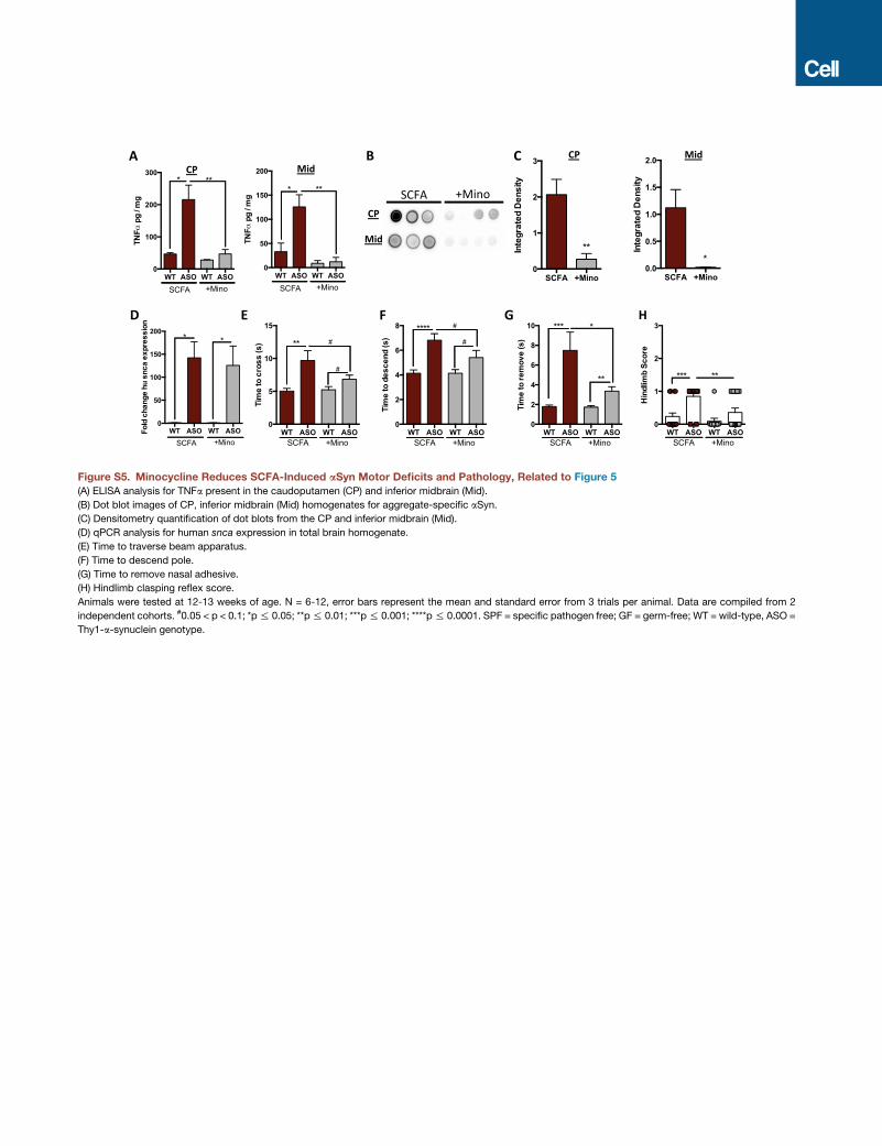
Figure S5. Minocycline Reduces SCFA-Induced aSyn Motor Deficits and Pathology, Related to Figure 5(A) ELISA analysis for TNFa present in the caudoputamen (CP) and inferior midbrain (Mid).
(B) Dot blot images of CP, inferior midbrain (Mid) homogenates for aggregate-specific aSyn.
(C) Densitometry quantification of dot blots from the CP and inferior midbrain (Mid).
(D) qPCR analysis for human snca expression in total brain homogenate.
(E) Time to traverse beam apparatus.
(F) Time to descend pole.
(G) Time to remove nasal adhesive.
(H) Hindlimb clasping reflex score.
Animals were tested at 12-13 weeks of age. N = 6-12, error bars represent the mean and standard error from 3 trials per animal. Data are compiled from 2
independent cohorts. #0.05 < p < 0.1; *p% 0.05; **p% 0.01; ***p% 0.001; ****p% 0.0001. SPF = specific pathogen free; GF = germ-free; WT = wild-type, ASO =
Thy1-a-synuclein genotype.

Figure S6. Microbial Metabolic Pathways Are Altered in Humanized Animals, Related to Figure 6
(A) Bray-Curtis distance comparisons between humanized groups.
(B and C) Bray-Curtis mean distance comparisons between (B) identical versus different donors, or (C) wild-type (WT) versus Thy1- a-synuclein (ASO) genotypes.
(D) PICRUSt analysis for specific KEGG families involved in SCFA production, light circles = healthy control derivedmicrobes, dark circles = PD derivedmicrobes.
(E) Fecal concentrations and relative abundances of acetate, propionate, and butyrate from humanized animals, normalized to soluble chemical oxygen demans.
Compiled from 6 independent donor pairs, HC = healthy controls; PD = Parkinson’s disease.
N = 3-6, over 3 time points post-colonization for KEGG analysis, N = 21-24 for SCFA abundances. Error bars represent the mean and standard error. *p% 0.05;
**p % 0.01; ***p % 0.001.
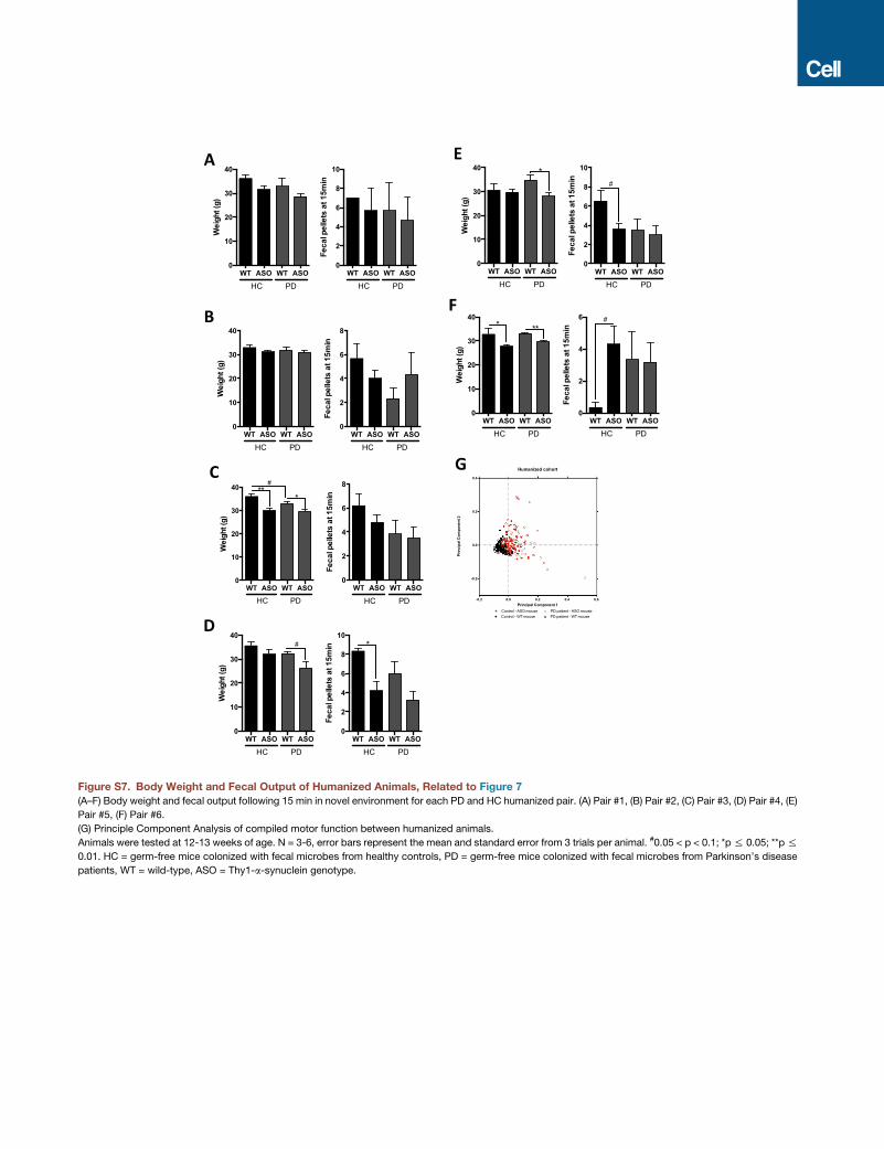
Figure S7. Body Weight and Fecal Output of Humanized Animals, Related to Figure 7
(A–F) Body weight and fecal output following 15 min in novel environment for each PD and HC humanized pair. (A) Pair #1, (B) Pair #2, (C) Pair #3, (D) Pair #4, (E)
Pair #5, (F) Pair #6.
(G) Principle Component Analysis of compiled motor function between humanized animals.
Animals were tested at 12-13 weeks of age. N = 3-6, error bars represent the mean and standard error from 3 trials per animal. #0.05 < p < 0.1; *p% 0.05; **p%
0.01. HC = germ-free mice colonized with fecal microbes from healthy controls, PD = germ-free mice colonized with fecal microbes from Parkinson’s disease
patients, WT = wild-type, ASO = Thy1-a-synuclein genotype.


![Gut microbiota and metabolite alterations …...the existence of a gut microbiota-bone axis [14–18], and the gut microbiota is a major regulator of bone mineral density (BMD) via](https://img.pdfslide.us/doc/110x75/5f0ecd4a7e708231d441023f/gut-microbiota-and-metabolite-alterations-the-existence-of-a-gut-microbiota-bone.jpg)