Embed Size (px)
Citation preview
501ISSN 1758-1907Diabetes Manag. (2014) 4(6), 501–513
part of
Diabetes Management
10.2217/DMT.14.47 © 2014 Future Medicine Ltd
DMTDiabetes Manag.
Diabetes Management1758-1907
1 7 5 8 - 1 9 15 F u t u r e Medicine LtdLondon, UK
1 0 . 2 2 1 7 /DMT.14.47
Review
Gut hormones and Type 2 diabetes mellitus
Falinska, Tan & Bloom
Gut hormones & Type 2 diabetes mellitus
Agnieszka M Falinska1, Tricia Tan1 & Stephen Bloom*,1
1Department of Investigative Medicine, Division of Diabetes, Endocrinology & Metabolism, Imperial College London, 6th Floor,
Commonwealth Building, London, W12 0HS, UK
*Author for correspondence: [email protected]
November2014November 2014
4
6
501
513
© 2014 FuTuRe MeDicine LTD
2014
Summary Type 2 diabetes mellitus (DM2) is a global threat to health with numbers of cases increasing exponentially. The pharmacological agents currently available for treatment of DM2 do not result in disease remission. Strikingly, bariatric surgery is successful in enforcing a prolonged remission of DM2 in a proportion of patients. The gut releases several hormones upon feeding, which affect hypothalamic, vagal and enteropancreatic pathways involved in the regulation of satiety and metabolism. One of the principal mechanisms by which bariatric surgery is capable of ameliorating DM2 is by means of increasing the secretion of key gut hormones such as glucagon-like peptide-1 (GLP-1), oxyntomodulin (OXM) and peptide YY (PYY). This review discusses available DM2 treatments based on GLP-1, as well as the potential therapeutic role of other gut hormones such as OXM, glucose-dependent insulinotropic polypeptide, ghrelin and PYY.
KeywordS • GLP-1 analogues • glucose metabolism • gut–brain axis • gut hormones • hypothalamus • Type 2 diabetes
Gut hormones & Type 2 diabetes Type 2 diabetes mellitus (DM2) is characterized by inappropriately high blood glucose concentra-tion resulting from insulin deficiency – the pancreas does not produce enough insulin to maintain a normal blood glucose level – and insulin resistance – the body is unable to use the insulin that is produced. DM2 is becoming a serious health problem, and the number of cases per year are increasing exponentially around the world [1]. It is becoming more prevalent among children and adolescents, a phenomenon that is linked to rising levels of obesity in these age groups [2].
DM2 is associated with secondary complications such as cardiovascular disease, stroke, chronic renal failure, blindness, neuropathy and leg ulcers. DM2 is strongly linked to obesity. People are being diagnosed with diabetes at ever younger ages and the risk of diabetic complications increases the earlier it is diagnosed, posing a substantial economic burden on health services around the world [1].
Practice points
● The gut–brain axis plays a significant role in glucose metabolism and appetite control.
● The improvement in glycemic control following bariatric surgery, such as Roux-en-Y gastric bypass, is likely to be driven by changes in gut hormone levels, thus exploiting those changes might help us to target future medical therapies for Type 2 diabetes mellitus.
● GLP-1 analogues improve glycemic control with associated weight loss but further studies are required to establish evidence of long-term benefit on cardiovascular risks.
● Combinations of gut hormone analogues have enhanced effects on glucose metabolism and suppression of food intake with minimized side effects.
Diabetes Manag. (2014) 4(6)502
review Falinska, Tan & Bloom
future science group
Lifestyle interventions, including dietary therapy, exercise and behavior modification, are often recommended as the foundation of DM2 management. However, the value of these interventions has been recently cast in doubt by the results of the Look AHEAD study, which showed that an intensive lifestyle intervention did not reduce the rate of cardiovascular events in overweight or obese adults with DM2 [3].
Conventional treatments for DM2 include metformin, sulphonylureas (SU), thiazolidin-ediones (TZD) and insulin. Apart from met-formin they all promote weight gain. Obesity and diabetes aggravate each other. Weight gain causes an increase in insulin resistance and can worsen co-morbidities such as osteoarthritis and obstructive sleep apnea. Moreover, weight gain makes patients more disinclined to persist with treatment, leading to poorer outcomes.
Consequently, it is vital to have effective and efficient treatments for DM2 that do not cause weight gain and successfully improve glycemic control. The phenomenon of diabetes remis-sion after gastric bypass surgery suggests that gut hormones have the important role in glucose control. This review will look at different gut hormones, their interactions with the nervous system and the potential for DM2 therapy.
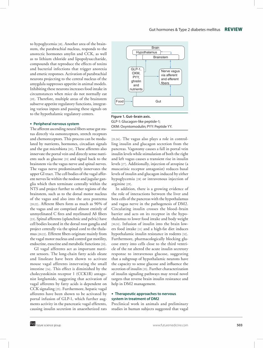
●● Gut–brain axisGut hormones, nutrients such as glucose and fatty acids, and the afferent and efferent auton-omous nervous system connect the gut and the brain. They inform the brain of the metabolic status of the gut (food intake, starvation and nutrient composition) through the activation of specific brain receptors. This then modulates complex brain pathways, generating efferent sig-nals via the central and peripheral autonomic nervous system to control the functions of organs (bowels, liver, gall bladder, pancreas), gut hormone secretion and tissue-specific hormone sensitivity (Figure 1) [4]. We will look at the role of brain, peripheral nervous system, nutrients and gut hormones in these complex interactions.
nervous systemThe brain receives and integrates signals com-ing from afferent neural pathways, gut peptides, direct nutrient sensing (glucose and fatty acids), and in response modulates vagal efferents (to control gut and metabolic function) and higher brain centers (to control the sensation of hunger and to trigger food-seeking behaviors).
●● Brain structuresThe key integrating brain centers for the infor-mation coming from the gut are the hypotha-lamic nuclei and brainstem. The blood–brain barrier is fenestrated at the median eminence in the hypothalamus and at the area postrema of the brainstem, and this allows access of hormones and nutrients to the brain [5,6].
The hypothalamus integrates majority of neu-ral, nutrient and hormonal signals coming from the gut. It modulates appetite in response to those signals via higher cortical centers (e.g., anterior cingulate cortex, amygdala, orbitofrontal cortex, nucleus accumbens, pre-frontal cortex) integrat-ing the sensory (smell, taste, sight) and hedonic (reward) aspects of food. Within the hypothala-mus, the arcuate nucleus (ARC) located in the mediobasal hypothalamus is one of the main nuclei regulating appetite [7]. The ARC, in turn, projects to the other parts of hypothalamus such as paraventricular nucleus (suppression of appe-tite), dorsomedial nucleus (circadian activity), lateral hypothalamus (hunger) and ventromedial nucleus (feeling of fullness), and ultimately to the limbic system and the cortex, which centrally coordinate food-seeking behavior [8]. The ARC contains neurons sensitive to both enteropancre-atic hormones (insulin, ghrelin, leptin, GLP-1) and nutrients (glucose, fatty acids) [8–10]. Both nutrients and hormones interact with each other. For example in rats receiving acute intracer-ebroventricular injection of insulin followed by intraperitoneal cholecystokinin (CCK)-8 imme-diately prior to the meal, the ability of CCK to reduce meal size was enhanced in the presence of elevated central insulin, suggesting they act syn-ergistically to reduce meal size [11,12]. Additionally, research from our laboratory suggested that ace-tate derived from the colonic fermentation of fatty acids crosses the blood–brain barrier and is taken up by the brain, especially the hypothalamus, resulting in appetite suppression [13].
The brainstem interacts with hypothalamic circuits and also itself plays a strategic role in the regulation of energy homeostasis [14]. Neural, nutrient and hormonal signals from the GI tract are sensed in the brainstem by mechanisms analogous to those seen in the hypothalamus [15–17]. One of the key structures is the nucleus of tractus solitarius (NTS). Glucose-sensing neurons in the NTS respond to low glucose by activating vagal motor nucleus causing nerve vagus firing and glucagon secretion, playing a crucial role in the counter-regulatory response
503
Figure 1. Gut–brain axis. GLP-1: Glucagon-like peptide-1; OXM: Oxyntomodulin; PYY: Peptide YY.
Brain
Gut
Hypothalamus
Brainstem
Food
GLP-1,OXM,PYY,
ghrelinand
nutrients
Nerve vagus via afferent and efferent fibers
Gut hormones & Type 2 diabetes mellitus review
future science group www.futuremedicine.com
to hypoglycemia [18]. Another area of the brain-stem, the parabrachial nucleus, responds to the anorectic hormones amylin and CCK, as well as to lithium chloride and lipopolysaccharide, compounds that reproduce the effects of toxins and bacterial infections that trigger anorexia and emetic responses. Activation of parabrachial neurons projecting to the central nucleus of the amygdala suppresses appetite in animal models. Inhibiting these neurons increases food intake in circumstances when mice do not normally eat [19]. Therefore, multiple areas of the brainstem subserve appetite regulatory functions, integrat-ing various inputs and passing these signals on to the hypothalamic regulatory centers.
●● Peripheral nervous systemThe afferent ascending neural fibers sense gut sta-tus directly via osmoreceptors, stretch receptors and chemoreceptors. This process can be modu-lated by nutrients, hormones, circadian signals and the gut microbiota [20]. These afferents also innervate the portal vein and directly sense nutri-ents such as glucose [21] and signal back to the brainstem via the vagus nerve and spinal nerves. The vagus nerve predominantly innervates the upper GI tract. The cell bodies of the vagal affer-ent nerves lie within the nodose and jugular gan-glia which then terminate centrally within the NTS and project further to other regions of the brainstem, such as to the dorsal motor nucleus of the vagus and also into the area postrema [20,22]. Afferent fibers form as much as 90% of the vagus and are comprised almost entirely of unmyelinated C fires and myelinated Aδ fibers [23]. Spinal afferents (splanchnic and pelvic) have cell bodies located in the dorsal root ganglia and project centrally via the spinal cord to the thala-mus [20,22]. Efferent fibers originate mainly from the vagal motor nucleus and control gut motility, endocrine, exocrine and metabolic functions [20].
GI vagal afferents act as important nutri-ent sensors. The long-chain fatty acids oleate and linoleate have been shown to activate mouse vagal afferents innervating the small intestine [24]. This effect is diminished by the cholecystokinin receptor 1 (CCK1R) antago-nist lorglumide, suggesting that activation of vagal afferents by fatty acids is dependent on CCK signaling [25]. Furthermore, hepatic vagal afferents have been shown to be activated by portal infusion of GLP-1, which further aug-ments activity in the pancreatic vagal efferents, causing insulin secretion in anaesthetized rats
[21,26]. The vagus also plays a role in control-ling insulin and glucagon secretion from the pancreas. Vagotomy causes a fall in portal vein insulin levels while stimulation of both the right and left vagus causes a transient rise in insulin levels [27]. Additionally, injection of atropine (a muscarinic receptor antagonist) reduces basal levels of insulin and glucagon induced by either hypoglycemia [28] or intravenous injection of arginine [29].
In addition, there is a growing evidence of the role of interactions between the liver and beta cells of the pancreas with the hypothalamus and vagus nerve in the pathogenesis of DM2. Circulating insulin crosses the blood–brain barrier and acts on its receptor in the hypo-thalamus to lower food intake and body weight [30,31]. Infusion of insulin into the brain low-ers food intake [11] and a high-fat diet induces hypothalamic insulin resistance in rodents [32]. Furthermore, pharmacologically blocking glu-cose entry into cells close to the third ventri-cle of the rat altered the acute insulin secretory response to intravenous glucose, suggesting that a subgroup of hypothalamic neurons have the capacity to sense glucose and influence the secretion of insulin [33]. Further characterization of insulin signaling pathways may reveal novel targets that reverse brain insulin resistance and help in DM2 management.
●● Therapeutic approaches to nervous system in treatment of DM2Preclinical work in animals and preliminary studies in human subjects suggested that vagal
Diabetes Manag. (2014) 4(6)504
review Falinska, Tan & Bloom
future science group
blockade was associated with enhanced satiety, decreases in food intake, and weight loss over prolonged follow-up periods. One of the pos-sible mechanisms suggests that CCK and lep-tin (as they both have receptors on the vagus nerve) could act synergistically to induce short-term inhibition of food intake and long-term reduction of body weight. This synergistic interaction between vagal CCK1R and leptin is mediated by the phosphorylation of signal trans-ducer and activator of transcription, which in turn, activates closure of K+ channels, leading to membrane depolarization and neuronal fir-ing (reviewed in [34]). The EMPOWER study, which used intermittent, bilateral blockade of both vagal nerves (VBLOC), failed to show a clinically relevant weight loss in obese individu-als [35]. More recently, the same group implanted VBLOC in twenty-eight obese subjects with DM2 showing significant weight reduction, reduction in HbA1c and mean arterial blood pressure [36]. These results suggest that the vagus nerve plays a role in the regulation of insulin secretion and appetite, and further studies are required to establish the therapeutic utility of vagal nerve modulation.
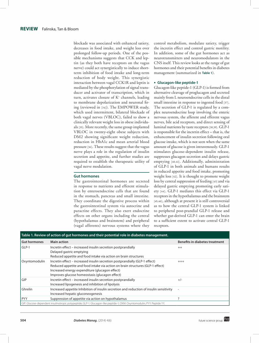
Gut hormonesThe gastrointestinal hormones are secreted in response to nutrients and efferent stimula-tion by enteroendocrine cells that are found in the stomach, pancreas and small intestine. They coordinate the digestive process within the gastrointestinal system via autocrine and paracrine effects. They also exert endocrine effects on other organs including the central (hypothalamus and brainstem) and peripheral (vagal afferents) nervous systems where they
control metabolism, modulate satiety, trigger the incretin effect and control gastric motility. In addition, some of the gut hormones act as neurotransmitters and neuromodulators in the CNS itself. This review looks at the range of gut hormones and their potential benefits in diabetes management (summarized in Table 1).
●● Glucagon-like peptide-1Glucagon-like peptide-1 (GLP-1) is formed from alternative cleavage of proglucagon and secreted mainly from L neuroendocrine cells in the distal small intestine in response to ingested food [37]. The secretion of GLP-1 is regulated by a com-plex neuroendocrine loop involving the enteric nervous system, the afferent and efferent vagus nerves, bile acid receptors, and direct sensing of luminal nutrients by taste receptors [38,39]. GLP-1 is responsible for the incretin effect – that is, the enhancement of insulin secretion following oral glucose intake, which is not seen when the same amount of glucose is given intravenously. GLP-1 stimulates glucose-dependent insulin release, suppresses glucagon secretion and delays gastric emptying [40,41]. Additionally, administration of GLP-1 in both animals and humans results in reduced appetite and food intake, promoting weight loss [42]. It is thought to promote weight loss by central suppression of feeding [43] and via delayed gastric emptying promoting early sati-ety [44]. GLP-1 mediates this effect via GLP-1 receptors in the hypothalamus and the brainstem [45,46], although at present it is still controversial as to how the central GLP-1 system is linked to peri pheral post-prandial GLP-1 release and whether gut-derived GLP-1 can enter the brain to a sufficient extent to activate central GLP-1 receptors.
Table 1. Review of action of gut hormones and their potential role in diabetes management.
Gut hormones Main action Benefits in diabetes treatment
GLP-1 Incretin effect – increased insulin secretion postprandially Delayed gastric emptying Reduced appetite and food intake via action on brain structures
++
Oxyntomodulin Incretin effect – increased insulin secretion postprandially (GLP-1 effect) Reduced appetite and food intake via action on brain structures (GLP-1 effect) Increased energy expenditure (glucagon effect) Improves glucose homeostasis (glucagon effect)
+++
GIP Incretin effect – increased insulin secretion postprandially Increased lipogenesis and inhibition of lipolysis
+/-
Ghrelin Increased appetite Inhibition of insulin secretion and reduction of insulin sensitivity Increased hepatic gluconeogenesis
-
PYY Suppression of appetite via action on hypothalamus ?GIP: Glucose-dependent insulinotropic polypeptide; GLP-1: Glucagon-like peptide-1; OXM: Oxyntomodulin; PYY: Peptide YY.
505
Gut hormones & Type 2 diabetes mellitus review
future science group www.futuremedicine.com
For these reasons GLP-1 action is beneficial in obese diabetic patients. Our group was one of the first ones to show that GLP-1 treatment (pre-meal injections) was able to improve glyce-mic control in patients with DM2 over 6 weeks [47]. GLP-1 has very short half-life (1.5–5 min) in plasma due to rapid degradation by dipepti-dyl peptidase-4 (DPP-4), which is a major limi-tation for its use in the clinical setting [41,48]. Degradation-resistant GLP-1 receptor agonists (incretin mimetics), and inhibitors of DPP-4 activity (incretin enhancers) are now used clinically for treatment of diabetes.
●● GLP-1 mimeticsGLP-1 analogues have been developed to pro-duce a longer half-life to allow for more sustained incretin effect and to make treatment practical for patients. The first analogue described in 1992 was exenatide (exendin-4, Amylin/Lilly, now BMS). It is a GLP-1 receptor agonist origi-nally isolated from the saliva of Heloderma sus-pectum, the Gila monster lizard. It shares a 53% homology with human GLP-1 and it is resistant to DPP-4 cleavage [49]. Recently a once-weekly formulation, exenatide LAR, was developed to allow for slow release over 7 days, and which performed better than exenatide twice daily in terms of HbA1c reduction with no increased risk of hypoglycemia and similar reductions in body weight [50].
The second GLP-1 analogue was liraglu-tide (Novo Nordisk). It is 97% homologous to native GLP-1 and its modification allows it to bind to albumin, increasing the half-life of liraglutide to 13 h [51]. Following exenatide and liraglutide’s commercial success, further com-panies are developing new GLP-1 analogues, for example, lixisenatide (Sanofi-Aventis) [52], albiglutide (GlaxoSmithKline) [53], dulaglutide (Eli Lilly) [54] and semaglutide (Novo Nordisk) [55]. Currently available GLP-1 analogues can be divided into a short-acting group (exenatide and lixisenatide) and longer acting group (lira-glutide, exenatide long-acting release, albiglutide and dulaglutide). It is postulated that shorter acting GLP-1 analogues reduce hyperglycaemia primarily by slowing gastric emptying, whereas longer acting GLP-1 analogues predominantly lower post-prandial glucose levels by increasing insulin and suppressing glucagon release [56].
A meta-analysis of GLP-1 analogues published in 2012 showed that the highest maintenance doses of the GLP-1 analogues were associated
with reduction in mean HbA1c of 1.1–1.6% [57]. One of the key advantages of GLP-1 analogue treatment is that patients also lose weight due to its appetite-suppressing effect. A recent meta-analysis including 25 trials involving exenatide twice daily, exenatide once weekly and liraglu-tide given for at least 20 weeks in both diabetics and non-diabetics found patients treated with GLP-1 analogues achieved a greater weight loss than control groups (weighted mean difference: 2.9 kg). Additionally, benefits were seen in terms of lowering systolic blood pressure, cholesterol and glycemia [58]. While rodent studies have found a favorable effect of GLP-1 analogues on β-cell mass, raising the possibility of a disease-modifying effect, it is too early to envisage the same will happen in humans, despite some positive studies [59].
As GLP-1-based therapies are still relatively new, safety concerns remain. The most common adverse event associated with GLP-1 receptor agonists is nausea and vomiting. It is common in the first few weeks of treatment and gradu-ally decreases with time [60]. Outside gastroin-testinal side effects, malignant thyroid C-cell carcinomas were observed in rodents following high-doses of liraglutide. It is unclear whether these risks apply to humans [61]. Furthermore, an association of long-term GLP-1 analogue therapy with an increased risk for pancreatitis is under debate. GLP-1 analogue therapy appears to be associated with hyperplasia of both the exocrine and endocrine pancreas, particularly alpha-cells [62]. A recent systematic review and meta-analysis suggested that the incidence of pancreatitis among patients using incretins is low and that the drugs do not increase the risk of pancreatitis [63].
●● DPP-4 inhibitorsGLP-1 enhancers work through inhibition of DPP-4, leading to a rise in endogenous GLP-1 levels, enhanced insulin release and suppression of glucagon secretion [64]. DPP-4 inhibitors are orally administered and currently there are five different drugs available on the market – sitag-liptin (2006), vildagliptin (2007), saxagliptin (2009), linagliptin (2011) and alogliptin (2013). Unlike GLP-1 agonists, they do not slow gastric emptying nor increase satiety, hence their effect on glycemia is less marked and they do not cause reductions in weight [64]. In a Cochrane review sitagliptin and vildagliptin were found to reduce HbA1c by 0.6–0.7% compared with placebo,
Diabetes Manag. (2014) 4(6)506
review Falinska, Tan & Bloom
future science group
with few adverse events and no weight gain [65]. A recent meta-analysis looking at DPP-4 inhibi-tors as a monotherapy and in combination with other oral glucose-lowering agents revealed a similar improvement in glycemic control as TZDs and SUs, but the key differences were that the gliptins are weight neutral and there are fewer reported cases of hypoglycemia [66]. Despite these salutary effects on glycemia, the recently published results of the SAVOR study in over 16,000 patients showed that saxagliptin did not affect the rate of ischemic events such as stroke or myocardial infarction [67].
●● OxyntomodulinOxyntomodulin (OXM) is a 37-amino acid peptide secreted from the intestine following nutrient ingestion, in a similar fashion to GLP-1 [68]. It originates from the same proglucagon precursor as GLP-1 [68]. OXM is a dual agonist of the GLP-1 receptor and the glucagon recep-tor combining the effects of both hormones [42,69,70]. OXM decreases food intake (GLP-1 action), increases energy expenditure (glucagon action) and enhances insulin secretion (GLP-1 action), thus representing a valuable candidate for possible therapeutic development [71].
Administration of OXM to high fat-fed mice improves glucose tolerance by enhancing glu-cose disposal and inhibiting endogenous glucose production [72]. One week of intraperitoneal OXM caused a reduction in food intake, weight gain and inhibited fasting plasma ghrelin secre-tion in rats [73]. Injections of OXM in humans cause significant weight loss in volunteers, both by reducing appetite and increasing energy expenditure [74].
Many companies and research institutes are working on OXM analogues resistant to pepti-dase degradation [75]. Pocai et al. described a dual GLP-1 and glucagon receptor agonist (DualAG), in essence a cholesterylated OXM. Studies in diet-induced obese mice revealed superior weight loss, lipid-lowering activity and antihyperglyce-mic efficacy compared with GLP-1 only recep-tor agonist [70]. Day et al. also described new PEGylated peptides with agonism at the glucagon and GLP-1 receptors. Administration of these peptides once per week normalized adiposity and glucose tolerance in diet-induced obese mice. Reduction of body weight was achieved by a loss of body fat resulting from decreased food intake and increased energy expenditure [76]. These preclinical studies indicate that when full GLP-1
agonism is augmented with a degree of glucagon receptor activation, there is a significant effect on glucose metabolism and weight, and such mul-tiple-acting peptides may hold promise as novel therapies for obesity–diabetes. Validating this approach, our group recently published results of a study in which glucagon and GLP-1 were coadministered to overweight or obese human volunteers. Resting energy expenditure rose sig-nificantly with glucagon alone or in combination with GLP-1. Glucagon infusion was accompa-nied by a rise in plasma glucose levels, but the addition of GLP-1 to glucagon rapidly reduced this excursion due to a synergistic insulinotropic effect. The data indicate that drugs with gluca-gon and GLP-1 agonist activity may represent a useful treatment for DM2, but clinical data with these drugs is needed to prove their efficacy [77]. Further studies are required to provide evidence of a translational effect in human studies.
●● Glucose-dependent insulinotropic polypeptideGlucose-dependent insulinotropic polypeptide (GIP) is a 42 amino acid peptide that is secreted in the duodenum and jejunum. It acts together with GLP-1 as an incretin to stimulate insu-lin release post-prandially. Additionally to the incretin effect, GIP has direct anabolic effects on adipose tissue by stimulating glucose import, fatty acid synthesis, lipogenesis and inhibiting lipolysis [78]. This action is not desirable as many diabetic patients are also obese and obesity contributes to insulin resistance.
A recent meta-analysis suggested that patients with DM2 have preserved GIP secre-tion in response to oral glucose and meal tests. Furthermore, obesity is associated with increased GIP responses while high age and HbA1c are associated with reduced GIP secretion [79]. Although the infusion of exogenous GIP into patients with DM2 worsens glucose tolerance [80], GIP analogues (developed similarly to GLP-1) have been shown to improve glucose homeostasis in obese mice [81]. Although the results are not encouraging, researchers attempted to include GIP in combination with other peptides. Bhat et al. designed a novel GIP-Oxm peptide incor-porating the actions of GIP, GLP-1 and gluca-gon in a single molecule. Acute administration of this compound to high fat fed mice resulted in a 45% reduction of plasma glucose and a 1.7-fold increase in insulin concentrations. Chronic once-daily administration for 15 days lowered body
507
Gut hormones & Type 2 diabetes mellitus review
future science group www.futuremedicine.com
weight (13% reduction), reduced plasma glucose (40% reduction) and increased plasma insulin [82]. Another group recently reported a novel peptide with potent co-agonism at both of the receptors for GLP-1 and GIP. This dual incretin agonist demonstrated superior efficacy compared with selective GLP-1 analogues in increasing insulin sensitivity and improving glycemic con-trol in multiple animal models including obese mice, diabetic rats and primates (cynomolgus macaques and humans) [83]. As the evidence of benefits of GIP agonists is conflicting, antago-nism of the GIP receptor was considered as GIP has direct anabolic effect on adipose tissue. Pro
3-
GIP, a GIP antagonist, has been shown to reduce weight and improve carbohydrate metabolism in diet-induced obese mice [84]. Further stud-ies are needed to evaluate the clinical potential of GIP agonism versus antagonism in humans. The current state of play would suggest that GIP is not a great candidate for diabetes therapy by itself, but may be more promising in combina-tion with other gut hormone therapies such as GLP-1-based therapies.
●● GhrelinGhrelin is a 28 amino acid octanoylated peptide that is secreted mainly in the stomach and pan-creas [85]. Ghrelin levels increase before meals and decrease afterwards, and it is known for its orexigenic (increasing food intake) proper-ties [86]. The ghrelin receptor, growth hormone secretagogue receptor (GHSR1a) is expressed in a wide variety of tissues, including the pituitary, hypothalamus, stomach, intestine, pancreas, thymus, gonads, thyroid and heart [87]. The diversity of ghrelin receptor locations suggests it has various biological functions.
Ghrelin affects glucose homeostasis by inhi-bition of insulin secretion [88], stimulation of the release of growth hormone and reduction of insulin sensitivity [89]. When mice were put on a calorie-restricted diet, their body fat dis-appeared, but blood glucose was maintained as long as the animals produced ghrelin. Calorie-restricted ghrelin knock-out mice fail to show the normal increase in growth hormone and become profoundly hypoglycemic when fasted [90], suggesting that ghrelin is needed for hepatic gluconeogenesis.
Our group showed that intravenous ghrelin enhances appetite and increases food intake in humans [91] and also causes hyperphagia and obesity in rats [92].
Since ghrelin has pro-diabetic actions, GHSR1a antagonism could be a promising strategy for the treatment of T2DM. Repeated administration of GHSR1a antagonists decreased body weight gain and improved glycemic control in obese mice [93]. Another study in wild-type rats using small-molecule GHSR1a antagonists also demonstrated improved glucose toler-ance [94]. Ghrelin-O-acyl transferase (GOAT) octanoylates ghrelin so it can act on its receptor GHSR1a, hence its inhibition may be an alterna-tive target [95]. Intraperitoneal administration of a GOAT inhibitor improved glucose tolerance and reduced weight gain in wild-type mice [96]. Taken together, these studies suggest that ghrelin plays an important role in the regulation of glu-cose homeostasis but as it acts on many organs, with both beneficial and detrimental effects, cau-tion must be taken to avoid side effects when developing therapies.
●● Peptide YYPeptide YY (PYY) is a satiety hormone released from the enteroendocrine L cells and its concen-tration in the circulation increases post-prandi-ally [97]. Full-length PYY
1–36 is quickly processed
by DPP-4 to PYY3–36
, which selectively binds to the neuropeptide Y subType 2 receptor and suppresses appetite, possibly by inhibiting the release of the appetite-stimulating neurotrans-mitter neuropeptide Y in the hypothalamus [98]. Intravenous infusion of PYY can reduce food intake in both lean and obese individu-als [97,99]. Although animal studies suggest that PYY can increase insulin sensitivity [98], in a study of healthy human volunteers PYY infu-sion caused increased thermogenesis, lipolysis, and increased postprandial insulin and glucose responses [100]. Therefore, the role of PYY in DM2 pathophysiology and potential treatment is yet to be clarified.
Lessons from bariatric surgeryThere is emerging a substantial amount of literature on the key role of bariatric surgery, particularly Roux-en-Y gastric bypass (RYGB), in the treatment of DM2. The effects can be visible within days and show improved glyce-mic control irrespective of weight loss. These observations have been reported since 1984, when the first study described 23 patients with DM2 using insulin who underwent bariatric surgery; 14 were able to discontinue its use, and in seven others the amount of insulin needed
Diabetes Manag. (2014) 4(6)508
review Falinska, Tan & Bloom
future science group
was substantially reduced [101]. More recently, Mingrone et al. observed severely obese patients with DM2 who underwent bariatric surgery. After 2 years they reported diabetes remission in 75% in the gastric bypass group, 95% in the biliopancreatic diversion group and no remis-sion in the medical therapy group. The HbA1c level (baseline 8.65 ± 1.45%) had decreased in all groups, but patients in the two surgical groups had the greatest degree of improvement (HbA1c 7.69 ± 0.57% in the medical therapy group, 6.35 ± 1.42% in the gastric bypass group, and 4.95 ± 0.49% in the biliopancreatic diver-sion group) [102]. Bariatric surgery has become a model to research the potential mechanisms contributing to resolution/improvement of DM2. Various hypotheses have been proposed for the mechanisms mediating the improve-ment and/or resolution of DM2 immediately post-bariatric surgery. These include changes in gut hormone levels (especially GLP-1, OXM and PYY) [103,104], alterations in bile acids metabo-lism [105], changes in gut microbiota [106] and changes in intestinal glucose metabolism [107]. Elevated levels of gut hormones such as PYY, OXM and GLP-1, both fasting and post-pran-dially, were shown to facilitate weight loss and improvements in glycemia via multiple mecha-nisms including: appetite suppression, increased energy expenditure, altered taste preferences, stimulation of insulin secretion and improve-ments in insulin sensitivity [103,108,109]. To study the significance of changes in gut hormone lev-els post-bariatric surgery in diabetic patients, Jørgensen et al. investigated postprandial glu-cose metabolism in obese patients with DM2 with concomitant infusion of GLP-1 receptor antagonist – exendin(9–39) or placebo. Nine patients were examined before, and 1 week and 3 months after RYGB. GLP-1 receptor blockade blunted postprandial β-cell responses more after than before RYGB, which in turn resulted in a greater impact of exendin(9–39) on glucose tolerance after the operation compared with preoperatively [104]. This suggests a major role of GLP-1 in maintaining glucose tolerance and insulin secretion in patients with DM2 after RYGB. RYGB is the most common form of gastric surgery and therefore the most studied procedure, but other types of surgery may have similar impacts. A comparison of RYGB with sleeve gastrectomy (SLG) in obese patients with DM2 showed that both procedures had a simi-lar impact on diabetes remission after 1 year,
as evidenced by β-cell glucose sensitivity tested in mixed-meal test and an increased GLP-1 response. In addition, other hormonal responses apart from PP and amylin were similar between the two groups [110]. Recently, Scholtz et al. used functional MRI to demonstrate that morbidly obese patients after RYGB show significantly lower responses to high-calorie food images in the reward system of the brain, compared with non-operated obese controls and obese patients after gastric banding. The RYGB group rated the high-calorie food images as less appealing and had lower activation in brain reward systems (amygdala and hippocampus). These changes were accompanied by higher levels of the ano-rexigenic plasma gut peptides – PYY and GLP-1, as well as bile acids – and lower levels of insulin during presentation of the images and/or meal intake [111].
The longitudinal Swedish Obese Subjects (SOS) study suggested both sustained remission and prevention of DM2 up to 15 years in severely obese patients after bariatric surgery compared with matched controls treated medically, but despite matching some baseline characteristics differed significantly between the groups (base-line body weight was greater and risk factors were more marked in the bariatric surgery arm than in controls) [112,113]. Schauer et al. recently pub-lished results of a 3-year study comparing obese patients with uncontrolled DM2 who either had intensive medical therapy plus bariatric surgery or medical therapy alone. By the end of 3-year period, 24–38% of patients who underwent sur-gery (SLG or RYGB) achieved HbA1c below 6% while only 5% of patients in the medical therapy group [114]. In summary, although bariatric sur-gery leads to greater body weight loss and higher remission rates of DM2 compared with medical treatment, the results are limited to 2–3 years of follow-up and based on a small number of studies and individuals [115]. There is a need for randomized controlled trials comparing the best medical and nutritional therapy with bariatric surgery to specifically treat DM2 and to assess its influence on long-term complications and cardiovascular outcomes. Additionally, the hor-monal changes after bariatric surgery can guide the development of gut hormone analogues as treatments for DM2.
novel technologiesMost recently endoscopy opened a novel path-way for less invasive methods to treat obesity
509
Gut hormones & Type 2 diabetes mellitus review
future science group www.futuremedicine.com
and T2DM. The duodenal–jejunal bypass liner (DJBL; EndoBarrier; GI Dynamics, MA, USA) is a 60-cm-long impermeable sleeve-like device placed endoscopically into the small intestine. It is supposed to mimic RYGB-related proxi-mal small intestinal exclusion, as ingested nutrients flow through the device lumen and do not contact the duodenal mucosa, keeping biliopancreatic secretions external to the device. A recent pilot nonrandomized study looked at glycemic control and gut hormone levels in 17 obese people with T2D who received a DJBL for 6 months [116]. At the end of the study par-ticipants lost weight and their glycemic control improved, as evidenced by significant reduction in HbA1c, fasting glucose levels and postpran-dial glucose response. Similar to post-RYGB, reduced glucagon and increased GLP-1 levels were documented, and this is likely due to the fact that nutrients are channeled straight to the jejunum where L cell density is higher. Although this study suggests potential hor-monal mechanisms for diabetes improvement, only few other small studies have been car-ried out until now with no large, multicenter, randomized controlled trials [117,118]. Further trials are essential to measure the benefits of the DJBL in patients with DM2 and also to examine its possible complications.
conclusionThe gut is a key endocrine organ secreting a vari-ety of hormones, which directly and indirectly affect brain. This gut–brain axis is crucial in the regulation of energy homeostasis and glucose metabolism. Bariatric surgery causes multiple changes in gut hormones, which are implicated in the weight loss and improvement in glyce-mic control of DM2 patients undergoing sur-gery. Some of these effects can be replicated by already available GLP-1 analogues, but it is clear that GLP-1 analogues alone are not capable of replicating all the improvements after bariatric
surgery. Currently multiple laboratories, includ-ing ours, are working on the development of gut hormone combinations aiming at synergistic action on appetite control and glucose metabo-lism to obtain benefits of a larger magnitude than obtainable with GLP-1 analogues. Further research is required to discover new treat-ments that will be more effective in reducing food intake without nausea, increasing energy expenditure and ameliorating the metabolic pathophysiology of DM2.
Future perspectiveDM2 and its management attracts a vast amount of research, both basic and clinical. Technological solutions such as nerve implants will not be widely available in the nearest future and pharmacological research carries the most prospects for the next 10 years. Evidence from bariatric surgery studies suggests it may be pos-sible to reset metabolism and put diabetes into remission. Gut hormones are vital in regulating energy homeostasis and their levels are affected by bariatric surgery. For these reasons, research into pharmacological agents that could mimic gut hormone changes and selectively reset energy metabolism is currently the most exciting pros-pect for significantly reducing the burden of DM2. As single agents are not able to capture all the benefits, all the effort is put into drug combinations aiming at synergistic action on appetite control and glucose metabolism.
Financial & competing interests disclosureThe authors have no relevant affiliations or financial involvement with any organization or entity with a finan-cial interest in or financial conflict with the subject matter or materials discussed in the manuscript. This includes employment, consultancies, honoraria, stock ownership or options, expert testimony, grants or patents received or pending, or royalties.
No writing assistance was utilized in the production of this manuscript.
ReferencesPapers of special note have been highlighted as: • of interest; •• of considerable interest
1 Wild S, Roglic G, Green A, Sicree R, King H. Global prevalence of diabetes: estimates for the year 2000 and projections for 2030. Diabetes Care 27(5), 1047–1053 (2004).
2 Group SFDIYS, Liese AD, D’agostino RB Jr. et al. The burden of diabetes mellitus among
US youth: prevalence estimates from the SEARCH for Diabetes in Youth Study. Pediatrics 118(4), 1510–1518 (2006).
3 Look ARG, Wing RR, Bolin P et al. Cardiovascular effects of intensive lifestyle intervention in Type 2 diabetes. N. Engl. J. Med. 369(2), 145–154 (2013).
4 Romijn JA, Corssmit EP, Havekes LM, Pijl H. Gut–brain axis. Curr. Opin. Clin. Nutr. Metabol. Care 11(4), 518–521 (2008).
5 Broadwell RD, Brightman MW. Entry of peroxidase into neurons of the central and peripheral nervous systems from extracerebral and cerebral blood. J. Compar. Neurol. 166(3), 257–283 (1976).
6 Peruzzo B, Pastor FE, Blazquez JL et al. A second look at the barriers of the medial basal hypothalamus. Exp. Brain Res. 132(1), 10–26 (2000).
Diabetes Manag. (2014) 4(6)510
review Falinska, Tan & Bloom
future science group
7 Konner AC, Klockener T, Bruning JC. Control of energy homeostasis by insulin and leptin: targeting the arcuate nucleus and beyond. Physiol. Behav. 97(5), 632–638 (2009).
8 Kalra SP, Dube MG, Pu S, Xu B, Horvath TL, Kalra PS. Interacting appetite-regulating pathways in the hypothalamic regulation of body weight. Endocr. Rev. 20(1), 68–100 (1999).
9 Lam TK, Pocai A, Gutierrez-Juarez R et al. Hypothalamic sensing of circulating fatty acids is required for glucose homeostasis. Nature Med. 11(3), 320–327 (2005).
10 Levin BE, Magnan C, Dunn-Meynell A, Le Foll C. Metabolic sensing and the brain: who, what, where, and how? Endocrinol. 152(7), 2552–2557 (2011).
11 Clegg DJ, Riedy CA, Smith KA, Benoit SC, Woods SC. Differential sensitivity to central leptin and insulin in male and female rats. Diabetes 52(3), 682–687 (2003).
12 Riedy CA, Chavez M, Figlewicz DP, Woods SC. Central insulin enhances sensitivity to cholecystokinin. Physiol. Behav. 58(4), 755–760 (1995).
13 Frost G, Sleeth ML, Sahuri-Arisoylu M et al. The short-chain fatty acid acetate reduces appetite via a central homeostatic mechanism. Nat. Commun. 5, 3611 (2014).
14 Schwartz GJ. Brainstem integrative function in the central nervous system control of food intake. Forum Nutr. 63, 141–151 (2010).
15 Blevins JE, Baskin DG. Hypothalamic–brainstem circuits controlling eating. Forum Nutr. 63, 133–140 (2010).
16 Grill HJ, Schwartz MW, Kaplan JM, Foxhall JS, Breininger J, Baskin DG. Evidence that the caudal brainstem is a target for the inhibitory effect of leptin on food intake. Endocrinol. 143(1), 239–246 (2002).
17 Lebrun B, Bariohay B, Moyse E, Jean A. Brain-derived neurotrophic factor (BDNF) and food intake regulation: a minireview. Auton. Neurosci. 126–127, 30–38 (2006).
18 Lamy CM, Sanno H, Labouebe G et al. Hypoglycemia-activated GLUT2 neurons of the nucleus tractus solitarius stimulate vagal activity and glucagon secretion. Cell Metabol. 19(3), 527–538 (2014).
19 Carter ME, Soden ME, Zweifel LS, Palmiter RD. Genetic identification of a neural circuit that suppresses appetite. Nature 503(7474), 111–114 (2013).
20 Berthoud HR, Blackshaw LA, Brookes SJ, Grundy D. Neuroanatomy of extrinsic afferents supplying the gastrointestinal tract. Neurogastroenterol. Motil. 16(Suppl. 1), 28–33 (2004).
21 Nakabayashi H, Nishizawa M, Nakagawa A, Takeda R, Niijima A. Vagal hepatopancreatic reflex effect evoked by intraportal appearance of tGLP-1. Am. J. Physiol. 271(5 Pt 1), E808–E813 (1996).
22 Hansen MB. Neurohumoral control of gastrointestinal motility. Physiol. Res. 52(1), 1–30 (2003).
23 Agostoni E, Chinnock JE, De Daly MB, Murray JG. Functional and histological studies of the vagus nerve and its branches to the heart, lungs and abdominal viscera in the cat. J. Physiol. 135(1), 182–205 (1957).
24 Webster WA, Beyak MJ. The long chain fatty acid oleate activates mouse intestinal afferent nerves in vitro. Can. J. Physiol. Pharmacol. 91(5), 375–379 (2013).
25 Cox JE, Kelm GR, Meller ST, Randich A. Suppression of food intake by GI fatty acid infusions: roles of celiac vagal afferents and cholecystokinin. Physiol. Behav. 82(1), 27–33 (2004).
26 Nishizawa M, Nakabayashi H, Uehara K, Nakagawa A, Uchida K, Koya D. Intraportal GLP-1 stimulates insulin secretion predominantly through the hepatoportal–pancreatic vagal reflex pathways. Am. J. Physiol. Endocrinol. Metabol. 305(3), E376–E387 (2013).
27 Frohman LA, Ezdinli EZ, Javid R. Effect of vagotomy and vagal stimulation on insulin secretion. Diabetes 16(7), 443–448 (1967).
28 Bloom SR, Edwards AV, Vaughan NJ. The role of the autonomic innervation in the control of glucagon release during hypoglycaemia in the calf. J. Physiol. 236(3), 611–623 (1974).
29 Bloom SR, Vaughan NJ, Russell RC. Vagal control of glucagon release in man. Lancet 2(7880), 546–549 (1974).
30 Schwartz MW, Woods SC, Porte D Jr, Seeley RJ, Baskin DG. Central nervous system control of food intake. Nature 404(6778), 661–671 (2000).
31 Niswender KD, Morrison CD, Clegg DJ et al. Insulin activation of phosphatidylinositol 3-kinase in the hypothalamic arcuate nucleus: a key mediator of insulin-induced anorexia. Diabetes 52(2), 227–231 (2003).
32 Clegg DJ, Gotoh K, Kemp C et al. Consumption of a high-fat diet induces central insulin resistance independent of adiposity. Physiol. Behav. 103(1), 10–16 (2011).
33 Osundiji MA, Lam DD, Shaw J et al. Brain glucose sensors play a significant role in the regulation of pancreatic glucose-stimulated insulin secretion. Diabetes 61(2), 321–328 (2012).
34 Owyang C, Heldsinger A. Vagal control of satiety and hormonal regulation of appetite. J. Neurogastroenterol. Motil. 17(4), 338–348 (2011).
35 Sarr MG, Billington CJ, Brancatisano R et al. The EMPOWER study: randomized, prospective, double-blind, multicenter trial of vagal blockade to induce weight loss in morbid obesity. Obes. Surg. 22(11), 1771–1782 (2012).
36 Shikora S, Toouli J, Herrera MF et al. Vagal blocking improves glycemic control and elevated blood pressure in obese subjects with Type 2 diabetes mellitus. J. Obes. 2013, 245683 (2013).
37 Herrmann C, Goke R, Richter G, Fehmann HC, Arnold R, Goke B. Glucagon-like peptide-1 and glucose-dependent insulin-releasing polypeptide plasma levels in response to nutrients. Digestion 56(2), 117–126 (1995).
38 Rocca AS, Brubaker PL. Role of the vagus nerve in mediating proximal nutrient-induced glucagon-like peptide-1 secretion. Endocrinol. 140(4), 1687–1694 (1999).
39 Chu ZL, Carroll C, Alfonso J et al. A role for intestinal endocrine cell-expressed G protein-coupled receptor 119 in glycemic control by enhancing glucagon-like peptide-1 and glucose-dependent insulinotropic peptide release. Endocrinol. 149(5), 2038–2047 (2008).
40 Kreymann B, Williams G, Ghatei MA, Bloom SR. Glucagon-like peptide-1 7–36: a physiological incretin in man. Lancet 2(8571), 1300–1304 (1987).
41 Drucker DJ. The biology of incretin hormones. Cell Metabol. 3(3), 153–165 (2006).
42 Chaudhri O, Small C, Bloom S. Gastrointestinal hormones regulating appetite. Philos. Trans. R. Soc. Lond. B. Biol. Sci. 361(1471), 1187–1209 (2006).
43 Turton MD, O’Shea D, Gunn I et al. A role for glucagon-like peptide-1 in the central regulation of feeding. Nature 379(6560), 69–72 (1996).
44 Naslund E, Bogefors J, Skogar S et al. GLP-1 slows solid gastric emptying and inhibits insulin, glucagon, and PYY release in humans. Am. J. Physiol. 277(3 Pt 2), R910–R916 (1999).
45 Parkinson JR, Chaudhri OB, Kuo YT et al. Differential patterns of neuronal activation in the brainstem and hypothalamus following peripheral injection of GLP-1, oxyntomodulin and lithium chloride in mice detected by manganese-enhanced magnetic resonance
511future science group www.futuremedicine.com
Gut hormones & Type 2 diabetes mellitus review
imaging (MEMRI). NeuroImage 44(3), 1022–1031 (2009).
46 Chaudhri OB, Wynne K, Bloom SR. Can gut hormones control appetite and prevent obesity? Diabetes Care 31(Suppl. 2), S284–S289 (2008).
47 Todd JF, Wilding JP, Edwards CM, Khan FA, Ghatei MA, Bloom SR. Glucagon-like peptide-1 (GLP-1): a trial of treatment in non-insulin-dependent diabetes mellitus. Eur. J. Clin. Invest. 27(6), 533–536 (1997).
48 Mentlein R, Gallwitz B, Schmidt WE. Dipeptidyl-peptidase IV hydrolyses gastric inhibitory polypeptide, glucagon-like peptide-1(7–36)amide, peptide histidine methionine and is responsible for their degradation in human serum. Eur. J. Biochem. 214(3), 829–835 (1993).
49 Eng J, Kleinman WA, Singh L, Singh G, Raufman JP. Isolation and characterization of exendin-4, an exendin-3 analogue, from Heloderma suspectum venom. Further evidence for an exendin receptor on dispersed acini from guinea pig pancreas. J. Biol. Chem. 267(11), 7402–7405 (1992).
50 Drucker DJ, Buse JB, Taylor K et al. Exenatide once weekly versus twice daily for the treatment of Type 2 diabetes: a randomised, open-label, non-inferiority study. Lancet 372(9645), 1240–1250 (2008).
51 Wajcberg E, Amarah A. Liraglutide in the management of Type 2 diabetes. Drug Des. Devel. Ther. 4, 279–290 (2010).
52 Rosenstock J, Raccah D, Koranyi L et al. Efficacy and safety of lixisenatide once daily versus exenatide twice daily in Type 2 diabetes inadequately controlled on metformin: a 24-week, randomized, open-label, active-controlled study (GetGoal-X). Diabetes Care 36(10), 2945–2951 (2013).
53 St Onge EL, Miller SA. Albiglutide: a new GLP-1 analog for the treatment of Type 2 diabetes. Expert Opin. Biol. Ther. 10(5), 801–806 (2010).
54 Jimenez-Solem E, Rasmussen MH, Christensen M, Knop FK. Dulaglutide, a long-acting GLP-1 analog fused with an Fc antibody fragment for the potential treatment of Type 2 diabetes. Curr. Opin. Mol. Ther. 12(6), 790–797 (2010).
55 Kapitza C LJ, During M et al. Safety, tolerability, pharmacokinetics (PK)/pharmacodynamics (PD) of single escalating doses of semaglutide, a unique once weekly GLP1 analogue, in healthy male subjects. EASD (2012).
56 Meier JJ. GLP-1 receptor agonists for individualized treatment of Type 2 diabetes mellitus. Nat. Rev. Endocrinol. 8(12), 728–742 (2012).
57 Aroda VR, Henry RR, Han J et al. Efficacy of GLP-1 receptor agonists and DPP-4 inhibitors: meta-analysis and systematic review. Clin. Ther. 34(6), 1247–1258 e1222 (2012).
58 Vilsboll T, Christensen M, Junker AE, Knop FK, Gluud LL. Effects of glucagon-like peptide-1 receptor agonists on weight loss: systematic review and meta-analyses of randomised controlled trials. BMJ 344, d7771 (2012).
59 Drucker DJ. Incretin-based therapy and the quest for sustained improvements in beta-cell health. Diabetes Care 34(9), 2133–2135 (2011).
60 Todd JF, Bloom SR. Incretins and other peptides in the treatment of diabetes. Diabet. Med. 24(3), 223–232 (2007).
61 Bjerre Knudsen L, Madsen LW, Andersen S et al. Glucagon-like Peptide-1 receptor agonists activate rodent thyroid C-cells causing calcitonin release and C-cell proliferation. Endocrinol. 151(4), 1473–1486 (2010).
62 Butler AE, Campbell-Thompson M, Gurlo T, Dawson DW, Atkinson M, Butler PC. Marked expansion of exocrine and endocrine pancreas with incretin therapy in humans with increased exocrine pancreas dysplasia and the potential for glucagon-producing neuroendocrine tumors. Diabetes 62(7), 2595–2604 (2013).
• ThispaperdemonstratesthatbothexocrineandendocrinepancreatichyperplasiaareassociatedwithGLP-1analoguetreatment,whichmayinturnpredisposetopancreatitisorevenpancreaticneuroendocrinetumors.
63 Li L, Shen J, Bala MM et al. Incretin treatment and risk of pancreatitis in patients with Type 2 diabetes mellitus: systematic review and meta-analysis of randomised and non-randomised studies. BMJ 348, g2366 (2014).
64 Drucker DJ. Dipeptidyl peptidase-4 inhibition and the treatment of Type 2 diabetes: preclinical biology and mechanisms of action. Diabetes Care 30(6), 1335–1343 (2007).
65 Richter B, Bandeira-Echtler E, Bergerhoff K, Lerch CL. Dipeptidyl peptidase-4 (DPP-4) inhibitors for Type 2 diabetes mellitus. Cochrane Database Syst. Rev. (2), CD006739 (2008).
66 Karagiannis T, Paschos P, Paletas K, Matthews DR, Tsapas A. Dipeptidyl peptidase-4 inhibitors for treatment of Type 2 diabetes mellitus in the Clin. setting: systematic review and meta-analysis. BMJ 344, e1369 (2012).
67 Scirica BM, Bhatt DL, Braunwald E et al. Saxagliptin and cardiovascular outcomes in patients with Type 2 diabetes mellitus. N. Engl. J. Med. 369(14), 1317–1326 (2013).
• Importantpaperconfirmingthatsaxagliptinimprovesglycemiccontrol,butshowingithasnoeffectontherateofischemicevents.Otherapproachesarenecessarytoreducecardiovascularriskinpatientswithdiabetes.
68 Cohen MA, Ellis SM, Le Roux CW et al. Oxyntomodulin suppresses appetite and reduces food intake in humans. J. Clin. Endocrinol. Metabol. 88(10), 4696–4701 (2003).
69 Baggio LL, Huang Q, Brown TJ, Drucker DJ. Oxyntomodulin and glucagon-like peptide-1 differentially regulate murine food intake and energy expenditure. Gastroenterology 127(2), 546–558 (2004).
70 Pocai A, Carrington PE, Adams JR et al. Glucagon-like peptide 1/glucagon receptor dual agonism reverses obesity in mice. Diabetes 58(10), 2258–2266 (2009).
71 Pocai A. Unraveling oxyntomodulin, GLP1’s enigmatic brother. J. Endocrinol. 215(3), 335–346 (2012).
72 Parlevliet ET, Heijboer AC, Schroder-Van Der Elst JP et al. Oxyntomodulin ameliorates glucose intolerance in mice fed a high-fat diet. Am. J. physiology. Endocrinol. Metabol. 294(1), E142–E147 (2008).
73 Dakin CL, Small CJ, Batterham RL et al. Peripheral oxyntomodulin reduces food intake and body weight gain in rats. Endocrinol. 145(6), 2687–2695 (2004).
74 Wynne K, Park AJ, Small CJ et al. Oxyntomodulin increases energy expenditure in addition to decreasing energy intake in overweight and obese humans: a randomised controlled trial. Int. J. Obes. 30(12), 1729–1736 (2006).
75 Druce MR, Minnion JS, Field BC et al. Investigation of structure-activity relationships of oxyntomodulin (Oxm) using Oxm analogs. Endocrinology 150(4), 1712–1722 (2009).
76 Day JW, Ottaway N, Patterson JT et al. A new glucagon and GLP-1 co-agonist eliminates obesity in rodents. Nat. Chem. Biol. 5(10), 749–757 (2009).
• Thedual-agonistpeptidespresentedinthepaperareshowntohavestrikingeffectsin
Diabetes Manag. (2014) 4(6)512
review Falinska, Tan & Bloom
future science group
improvingdiet-inducedobesityandhyperglycemiainanimalmodels.
77 Tan TM, Field BC, Mccullough KA et al. Coadministration of glucagon-like peptide-1 during glucagon infusion in humans results in increased energy expenditure and amelioration of hyperglycemia. Diabetes 62(4), 1131–1138 (2013).
78 Vella A, Rizza RA. Extrapancreatic effects of GIP and GLP-1. Hormone Metabol. Res. 36(11–12), 830–836 (2004).
79 Calanna S, Christensen M, Holst JJ et al. Secretion of glucose-dependent insulinotropic polypeptide in patients with Type 2 diabetes: systematic review and meta-analysis of clinical studies. Diabetes Care 36(10), 3346–3352 (2013).
80 Chia CW, Carlson OD, Kim W et al. Exogenous glucose-dependent insulinotropic polypeptide worsens post prandial hyperglycemia in Type 2 diabetes. Diabetes 58(6), 1342–1349 (2009).
81 Irwin N, Green BD, Mooney MH et al. A novel, long-acting agonist of glucose-dependent insulinotropic polypeptide suitable for once-daily administration in Type 2 diabetes. J. Pharmacol. Exp. Ther. 314(3), 1187–1194 (2005).
82 Bhat VK, Kerr BD, Flatt PR, Gault VA. A novel GIP-oxyntomodulin hybrid peptide acting through GIP, glucagon and GLP-1 receptors exhibits weight reducing and anti-diabetic properties. Biochem. Pharmacol. 85(11), 1655–1662 (2013).
83 Finan B, Ma T, Ottaway N et al. Unimolecular dual incretins maximize metabolic benefits in rodents, monkeys, and humans. Sci. Transl. Med. 5(209), 209ra151 (2013).
84 Mcclean PL, Irwin N, Cassidy RS, Holst JJ, Gault VA, Flatt PR. GIP receptor antagonism reverses obesity, insulin resistance, and associated metabolic disturbances induced in mice by prolonged consumption of high-fat diet. Am. J. Physiol. Endocrinol. Metabol. 293(6), E1746–E1755 (2007).
85 Kojima M, Hosoda H, Date Y, Nakazato M, Matsuo H, Kangawa K. Ghrelin is a growth-hormone-releasing acylated peptide from stomach. Nature 402(6762), 656–660 (1999).
86 Akamizu T, Murayama T, Teramukai S et al. Plasma ghrelin levels in healthy elderly volunteers: the levels of acylated ghrelin in elderly females correlate positively with serum IGF-I levels and bowel movement frequency and negatively with systolic blood pressure. J. Endocrinol. 188(2), 333–344 (2006).
87 Kirchner H, Heppner KM, Tschop MH. The role of ghrelin in the control of energy balance. Handbook Exp. Pharmacol. 209, 161–184 (2012).
88 Broglio F, Arvat E, Benso A et al. Ghrelin, a natural GH secretagogue produced by the stomach, induces hyperglycemia and reduces insulin secretion in humans. J. Clin. Endocrinol. Metabol. 86(10), 5083–5086 (2001).
89 Vestergaard ET, Gormsen LC, Jessen N et al. Ghrelin infusion in humans induces acute insulin resistance and lipolysis independent of growth hormone signaling. Diabetes 57(12), 3205–3210 (2008).
90 Li RL, Sherbet DP, Elsbernd BL, Goldstein JL, Brown MS, Zhao TJ. Profound hypoglycemia in starved, ghrelin-deficient mice is caused by decreased gluconeogenesis and reversed by lactate or fatty acids. J. Biol. Chem. 287(22), 17942–17950 (2012).
91 Wren AM, Seal LJ, Cohen MA et al. Ghrelin enhances appetite and increases food intake in humans. J. Clin. Endocrinol. Metabol. 86(12), 5992 (2001).
92 Wren AM, Small CJ, Abbott CR et al. Ghrelin causes hyperphagia and obesity in rats. Diabetes 50(11), 2540–2547 (2001).
93 Asakawa A, Inui A, Kaga T et al. Antagonism of ghrelin receptor reduces food intake and body weight gain in mice. Gut 52(7), 947–952 (2003).
94 Esler WP, Rudolph J, Claus TH et al. Small-molecule ghrelin receptor antagonists improve glucose tolerance, suppress appetite, and promote weight loss. Endocrinology 148(11), 5175–5185 (2007).
95 Yang J, Zhao TJ, Goldstein JL, Brown MS. Inhibition of ghrelin O-acyltransferase (GOAT) by octanoylated pentapeptides. Proc. Natl Acad. Sci. USA 105(31), 10750–10755 (2008).
96 Barnett BP, Hwang Y, Taylor MS et al. Glucose and weight control in mice with a designed ghrelin O-acyltransferase inhibitor. Science 330(6011), 1689–1692 (2010).
97 Batterham RL, Cowley MA, Small CJ et al. Gut hormone PYY(3–36) physiologically inhibits food intake. Nature 418(6898), 650–654 (2002).
98 Batterham RL, Bloom SR. The gut hormone peptide YY regulates appetite. Ann. NY Acad. Sci. 994, 162–168 (2003).
99 Batterham RL, Cohen MA, Ellis SM et al. Inhibition of food intake in obese subjects by peptide YY3–36. N. Engl. J. Med. 349(10), 941–948 (2003).
100 Sloth B, Holst JJ, Flint A, Gregersen NT, Astrup A. Effects of PYY1–36 and PYY3–36 on appetite, energy intake, energy expenditure, glucose and fat metabolism in obese and lean subjects. Am. J. Physiol. Endocrinol. Metabol. 292(4), E1062–E1068 (2007).
101 Herbst CA, Hughes TA, Gwynne JT, Buckwalter JA. Gastric bariatric operation in insulin-treated adults. Surg. 95(2), 209–214 (1984).
102 Mingrone G, Panunzi S, De Gaetano A et al. Bariatric surgery versus conventional medical therapy for Type 2 diabetes. N. Engl. J. Med. 366(17), 1577–1585 (2012).
103 Pournaras DJ, Osborne A, Hawkins SC et al. The gut hormone response following Roux-en-Y gastric bypass: cross-sectional and prospective study. Obes. Surg. 20(1), 56–60 (2010).
104 Jorgensen NB, Dirksen C, Bojsen-Moller KN et al. Exaggerated glucagon-like peptide 1 response is important for improved beta-cell function and glucose tolerance after Roux-en-Y gastric bypass in patients with Type 2 diabetes. Diabetes 62(9), 3044–3052 (2013).
105 Gerhard GS, Styer AM, Wood GC et al. A role for fibroblast growth factor 19 and bile acids in diabetes remission after Roux-en-Y gastric bypass. Diabetes Care 36(7), 1859–1864 (2013).
106 Liou AP, Paziuk M, Luevano JM Jr, Machineni S, Turnbaugh PJ, Kaplan LM. Conserved shifts in the gut microbiota due to gastric bypass reduce host weight and adiposity. Sci. Transl. Med. 5(178), 178ra141 (2013).
107 Saeidi N, Meoli L, Nestoridi E et al. Reprogramming of intestinal glucose metabolism and glycemic control in rats after gastric bypass. Science 341(6144), 406–410 (2013).
•• Theauthorssuggestedanovelmechanismofimprovedglycemiccontrolafterbariatricsurgery.
108 Bueter M, Ashrafian H, Le Roux CW. Mechanisms of weight loss after gastric bypass and gastric banding. Obes. Facts 2(5), 325–331 (2009).
109 Bueter M, Le Roux CW. Gastrointestinal hormones, energy balance and bariatric surgery. Int. J. Obes. 35(Suppl. 3), S35–S39 (2011).
110 Nannipieri M, Baldi S, Mari A et al. Roux-en-Y gastric bypass and sleeve gastrectomy: mechanisms of diabetes remission and role of gut hormones. J. Clin. Endocrinol. Metabol. 98(11), 4391–4399 (2013).
513future science group www.futuremedicine.com
Gut hormones & Type 2 diabetes mellitus review
111 Scholtz S, Miras AD, Chhina N et al. Obese patients after gastric bypass surgery have lower brain-hedonic responses to food than after gastric banding. Gut 63(6), 891–902 (2013).
112 Sjostrom L, Lindroos AK, Peltonen M et al. Lifestyle, diabetes, and cardiovascular risk factors 10 years after bariatric surgery. N. Engl. J. Med. 351(26), 2683–2693 (2004).
113 Carlsson LM, Peltonen M, Ahlin S et al. Bariatric surgery and prevention of Type 2 diabetes in Swedish obese subjects. N. Engl. J. Med. 367(8), 695–704 (2012).
• AuniquebutretrospectivestudylookingatpreventionofdiabetesType2andbariatricsurgery.Prospectiverandomizedtrialsareawaited.
114 Schauer PR, Bhatt DL, Kirwan JP et al. Bariatric surgery versus intensive medical therapy for diabetes – 3-year outcomes. N. Engl. J. Med. 370(21), 2002–2013 (2014).
•• Thewell-plannedprospectivestudyofobesepatientswithuncontrolledType2diabeteslookingat3-yearbenefitsofintensivemedicaltherapyplusbariatricsurgeryversusmedicaltherapyalone.
115 Gloy VL, Briel M, Bhatt DL et al. Bariatric surgery versus non-surgical treatment for obesity: a systematic review and meta-analysis of randomised controlled trials. BMJ 347, f5934 (2013).
116 De Jonge C, Rensen SS, Verdam FJ et al. Endoscopic duodenal–jejunal bypass liner
rapidly improves Type 2 diabetes. Obes. Surg. 23(9), 1354–1360 (2013).
117 Escalona A, Pimentel F, Sharp A et al. Weight loss and metabolic improvement in morbidly obese subjects implanted for 1 year with an endoscopic duodenal–jejunal bypass liner. Ann. Surg. 255(6), 1080–1085 (2012).
118 De Moura EG, Martins BC, Lopes GS et al. Metabolic improvements in obese Type 2 diabetes subjects implanted for 1 year with an endoscopically deployed duodenal-jejunal bypass liner. Diabetes Technol. Ther. 14(2), 183–189 (2012).