Embed Size (px)
Citation preview
http
://do
c.re
ro.c
h
GUP1 of Saccharomyces cerevisiae Encodes an O-AcyltransferaseInvolved in Remodeling of the GPI Anchor□D
Regine Bosson, Malika Jaquenoud, and Andreas Conzelmann
Department of Medicine, University of Fribourg, CH-1700 Fribourg, Switzerland
The anchors of mature glycosylphosphatidylinositol (GPI)-anchored proteins of Saccharomyces cerevisiae contain eitherceramide or diacylglycerol with a C26:0 fatty acid in the sn2 position. The primary GPI lipid added to newly synthesizedproteins in the ER consists of diacylglycerol with conventional C16 and C18 fatty acids. Here we show that GUP1 is essentialfor the synthesis of the C26:0-containing diacylglycerol anchors. Gup1p is an ER membrane protein with multiple membrane-spanning domains harboring a motif that is characteristic of membrane-bound O-acyl-transferases (MBOAT). Gup1� cellsmake normal amounts of GPI proteins but most mature GPI anchors contain lyso-phosphatidylinositol, and others possessphosphatidylinositol with conventional C16 and C18 fatty acids. The incorporation of the normal ceramides into the anchorsis also disturbed. As a consequence, the ER-to-Golgi transport of the GPI protein Gas1p is slow, and mature Gas1p is lost fromthe plasma membrane into the medium. Gup1� cells have fragile cell walls and a defect in bipolar bud site selection. GUP1function depends on the active site histidine of the MBOAT motif. GUP1 is highly conserved among fungi and protozoa andthe gup1� phenotype is partially corrected by GUP1 homologues of Aspergillus fumigatus and Trypanosoma cruzi.
INTRODUCTION
The biosynthesis of glycosylphosphatidylinositol (GPI)-an-chored proteins follows the same basic rules in all eu-karyotes, and all GPI anchors harbor a conserved carbohy-drate core structure linking a protein moiety to a lipidmoiety. In contrast, different organisms contain widely dif-fering kinds of lipid moieties (Kinoshita and Inoue, 2000;Ferguson et al., 2006). GPI lipid biosynthesis starts with theaddition of N-acetyl-glucosamine to phosphatidylinositol(PI) by PIG-A/GPI3 (Miyata et al., 1993). In many organisms,the spectrum of lipids found on mature GPI anchors is quitedifferent from the one displayed by the free PI. The situationin Saccharomyces cerevisiae is peculiar because two very dif-ferent types of lipid moieties can be found: ceramide (Cer)and diacylglycerol. Cer is found on the majority of anchors;it mainly consists of C18:0 phytosphingosine (PHS) and aC26:0 fatty acid (Fankhauser et al., 1993). On the other hand,Gas1p, a well-characterized GPI protein of yeast, is madewith a C26:0 fatty acid–containing, mild base-sensitive lipid.In both types of lipid moieties the C26:0 may be hydroxy-lated on C2. These lipids are introduced by remodelingreactions starting soon after the primary GPI lipid is addedto the nascent proteins in the lumen of the ER. During
remodeling the C16- and C18-containing diacylglycerol ofthe primary anchor is modified or replaced. Here we iden-tify GUP1 as a key remodelase. GUP1 and GUP2 were ini-tially identified in a screen for glycerol uptake–deficientmutants (Holst et al., 2000). A recent report however hasshown that in gup1� gup2� mutant yeast cells the glycerolH�/symport is still detectable (Neves et al., 2004). Otherreports have revealed other functions of Gup1p. GUP1 isinvolved in bipolar bud site selection (Ni and Snyder, 2001)and is implicated in vacuolar protein sorting (Bonangelino etal., 2002). Here we propose that GUP1 acts as an enzyme thatadds C26 fatty acids to the sn2 position of lyso-PI–contain-ing GPI anchors.
MATERIALS AND METHODS
Strains, Media, and MaterialsStrains with single deletions of nonessential genes were obtained fromEUROSCARF (http://web.uni-frankfurt.de/fb15/mikro/euroscarf/col_index.html), namely are1�, MAT� his3�1 leu2�0 lys2�0 ura3�0 are1::kanMX4; are2�,MAT� his3�1 leu2�0 lys2�0 ura3�0 are2::kanMX4; gup1�, MAT� his3�1 leu2�0lys2�0 ura3�0 gup1::kanMX4; gup2�, MAT� his3�1 leu2�0 lys2�0 ura3�0gup2::kanMX4; vps4�, MAT� his3�1 leu2�0 lys2�0 ura3�0 vps4::kanMX4; BY4742,MAT� his3�1 leu2�0 lys2�0 ura3�0. Several strains were obtained from StephenSturley, namely SCY63, MAT� ade2-1 trp1-1 ura3-1 can1-1 met14 are1::HIS3are2::LEU2; SCY1382, MATa ade2-1 trp1-1 his3-11,15 gup1::LEU2 gup2::URA3;SCY1414, are1::HIS3 are2::TRP1 gup1::LEU2 gup2::URA3; SCY325, MAT� ade2-1his3-11,15 leu2-3,112 trp1-1 ura3-1. The 5� strain are1::HIS3 are2::TRP1 gup1::LEU2gup2::URA3 yor175::kanMX4 was obtained from Nicolas Jacquier. Strains madefor this study were FBY8171, MATa ade2-101 ura3-52 pep4::LEU2 gup1::kanMX4;FBY8172, MAT� lys2 trp1 ura3 ubc6::HIS3 ubc7::LEU2 gup1::kanMX4; FBY8173,MAT� his3�200 ura3-52 lys2-801 trp1-1 doa4::LEU2 gup1::kanMX4; FBY8174,MAT� his3�200 leu2�1 ura3-52 cim5-1 gup1::kanMX4; FBY8175, MATa his3-115leu2-3,112 ura3 pre1-1 pre2-2 gup1::kanMX4. Strains were cultured at 24, 30, or 37°Cin YPD medium or in minimal media supplemented with glucose (SD) orgalactose (SG) and amino acids (aa; Sherman, 2002). Selection for integration ofKanMX4-containing deletion cassettes was performed on YPD plates containing200 �g/ml G418 (Calbiochem, San Diego, CA). Unless specified, chemicals werepurchased from Sigma (St. Louis, MO). Pepstatin was obtained from Alexis (SanDiego, CA), and protein A-Sepharose, octyl-Sepharose, and concanavalin A-Sepharose from Amersham Biosciences (Piscataway, NJ). Anti-Pma1p antibodywas a kind gift from Barbara Gaigg.
Address correspondence to: A. Conzelmann ([email protected]).
Abbreviations used: Cer, ceramide; CFW, calcofluor white; DHS,dihydrosphingosine; GPI, glycosylphosphatidylinositol; IPC, inosi-tolphosphoceramide; MIPC, mannosylated IPC; PHS, phytosphin-gosine; PI, phosphatidylinositol; TLC, thin-layer chromatography;wt, wild type.
1
file:///U|/docs/OA/articles/header_p1.txt
Published in "Molecular Biology of the Cell 17(6): 2636-2645, 2006" which should be cited to refer to this work.
file:///U|/docs/OA/articles/header_p1.txt22.09.2005 08:32:30
http
://do
c.re
ro.c
hStrain and Plasmid ConstructionTo delete GUP1 in different yeast strains a gup1::KanMX4 deletion cassettewas made by amplifying gup1::KanMX4 genomic region of the EUROSCARFdeletion strain using primers GUP1_F1 (5�-aatcatacaaaggcaaaaacaaa-3�) andGUP1_R1 (5�-taaaaatacatacatgatagcag-3�).
The expression vectors harboring GUP1 or GUP1H447A were obtained asfollows: the open reading frame of GUP1 was amplified by PCR using plasmidpBH2178 (kind gift from Morten Kielland-Brandt) as a template and usingprimers GUP1rec1sens (5�-gaattcgatatcaagcttatcgataccgatgtcgctgatcagcatcctgtc-tcc-3�) and GUP1rec2AS (5�-gacataactaattacatgactcgaggtcgactcagcattttaggtaaatt-ccg-3�), underlined sequences being homologous to the target vector pGREG505(Jansen et al., 2005). The PCR fragment was purified by a PCR purification kit(QIAGEN, Chatsworth, CA) and introduced into pGREG505 by cotransfectioninto yeast cells thus generating pGUP1 (Jansen et al., 2005). A point mutation tochange His 447 to Ala was introduced into GUP1 by PCR amplification of twofragments of GUP1 from plasmid pBH2178 using primers GUP1rec1sens (seeabove) and GUP1.HIS447ALA.AS (5�-gttcgatgtcagcccatatagctacg-3�), andGUP1.HIS447ALAsens (5�-cgtagctatatgggctgacatcgaac-3�) with GUP1rec2AS (seeabove), underlined nucleotides representing the His to Ala mutation. The twooverlapping fragments were fused by PCR using primers GUP1rec1sens andGUP1rec2AS (see above) and introduced into pGREG505 and pGREG535 byin vivo homologous recombination, yielding vectors pGUP1H447A andpHAGUP1H447A, respectively. A pGREG505 vector containing His 447 mu-tated to Asn (GUP1H447N) was obtained using the same strategy. Transfer ofGUP1 into pGREG535 yielded pHAGUP1. To generate pHAafGUP1 andpHAtcGUP1, the GUP1 ortholog of Aspergillus fumigatus was amplified froma cDNA preparation (kindly donated by Michel Monod, Lausanne, Switzer-land) using primers afGUP1-Rec1 (5�-gaattcgatatcaagcttatcgataccgatgacttcga-tcctttcctggttccgg-3� and afGUP1-Rec2 (5�-gacataactaattacatgactcgaggtcgac-tcaacacttcatcttgataccagcgcg-3�) and was similarly amplified from genomicDNA of Trypanosoma cruzi (prepared in the lab of Reto Brun (Basel, Switzer-land) using primers tcGUP1-Rec1 (5�-gaattcgatatcaagcttatcgataccgatgagtga-ggaaaaaaattgcgctaatatgc-3�) and tcGUP1-Rec2 (5�-gcgtgacataactaattacatgactcg-aggtcgacttaggcaccagcggaaattccgtatc-3�). The two PCR products were thenintroduced into pGREG535 as above. All inserts very verified by sequencingand corresponded to the published sequences over the entire reading frame.
Preparation of Radiolabeled GPI Protein Anchor PeptidesIsolation of the lipid moieties of GPI anchors was performed as described(Guillas et al., 2000), lipids were analyzed by TLC on 20 � 20-cm silica gel 60plates using solvent 1 (CHCl3/CH3OH/0.25% KCl, 55:45:10) or solvent 2(CHCl3/CH3OH/0.25% KCl, 55:45:5).
Lipid AnalysisLipids extracted from [3H]inositol–labeled cells were desalted by butanol/water partitioning and analyzed by TLC. Alkaline hydrolysis was performedusing 0.1 M NaOH in CHCl3/CH3OH/H2O (10:10:3) for 1 h at 37°C. Phos-pholipase A2 (PLA2) treatment of lipids was done in 25 mM Tris-HCl, pH 7.5,2 mM CaCl2, and 0.1% sodium deoxycholate for 2 h at 37°C.
Pulse-Chase AnalysisPulse-chase analysis was performed at 30°C as described (Gaigg et al., 2005).Immunoprecipitated proteins were solubilized by boiling 5 min in samplebuffer and analyzed by SDS-PAGE and visualized by fluorography or phos-phorimager in order to quantify by Quantity One software (Bio-Rad Labora-tories, Hercules, CA).
Raft Association AnalysisRaft association of Gas1p, CPY, and Pma1p was analyzed as described pre-viously (Bagnat et al., 2000) with some modifications. Cells were grown at30°C in rich medium. Twenty A600 of exponentially growing cells werecollected and lysed in TEPIN buffer (50 mM Tris-HCl, pH 7.4, 150 mM NaCl,5 mM EDTA, 1 mM phenylmethylsulfonyl fluoride, 2.5 �g/ml antipain, 2.5�g/ml chymostatin, 2.5 �g/ml leupeptin, and 2.5 �g/ml pepstatin) by vor-texing with glass beads. The lysate was centrifuged at 3000 rpm at 4°C for 5min to remove unbroken cells and debris. The cleared lysate was incubatedwith Triton X-100 added to 1% for 30 min on ice. After centrifugation at15,000 � g for 40 min at 4°C, the supernatant was collected and precipitatedwith 10% TCA. The 15,000 � g microsomal pellet and the TCA precipitatewere solubilized by boiling in reducing sample buffer for 5 min and analyzedby SDS-PAGE. Western blots were performed using antibodies against Gas1p,CPY, or Pma1p and revealed by chemiluminescence ECL kit.
RESULTS
The gup1� Mutant Is Deficient in GPI Anchor RemodelingTo find genes encoding remodelases, we analyzed the GPIanchor lipids of viable deletion strains lacking genes thatshow homology to known phospholipases, acyltrans-
ferases, or enzymes working on ceramides (Guillas et al.,2000; Conzelmann, 2005). To do so, deletion strains were la-
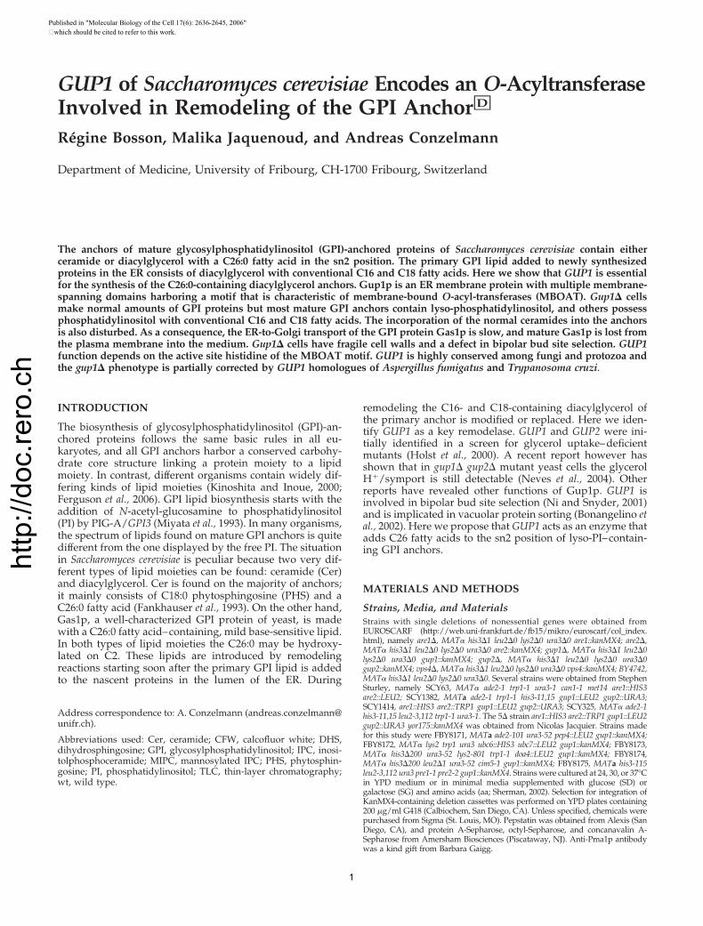
Figure 1. gup1� mutant is deficient in GPI anchor remodeling.(A–C) Cells of indicated genotype were labeled at 30°C with [3H]ino-sitol, and lipids were extracted and analyzed by TLC/fluorographyusing solvent 2, either directly (B) or after deacylation with NaOH(C). GPI anchors were isolated from delipidated proteins, and theirlipid moiety was released with nitrous acid and analyzed by TLC/fluorography (A) along with the free lipids of wt (SCY325) cells (wt,FL) using solvent 1. 2� are � are1� are2�; 2� gup � gup1� gup2�;4� � are1� are2� gup1� gup2�. pG1 is a remodeled form of PI,containing C26:0 in sn2. IPC/B contains PHS-C26:0; IPC/C containsPHS-C26:0-OH. (D) wt or gup1� cells were labeled at 24 or 37°Cwith [3H]inositol for 2 h. The delipidated labeled proteins wereanalyzed by SDS-PAGE/fluorography.
2
http
://do
c.re
ro.c
h
beled with [3H]inositol, proteins were extracted and exten-sively delipidated, enriched by concanavalin A-Sepharoseaffinity chromatography, and digested by protease to gen-erate [3H]inositol–labeled anchor peptides. The latter werefurther purified by octyl-Sepharose chromatography, and theirlipid moieties were liberated using nitrous acid deamination,which releases PI or inositolphosphoceramide (IPCs), depend-ing on the GPI anchor. Finally, these lipids were desalted andanalyzed by TLC. By this brute force approach, we foundgup1� having the phenotype shown in Figure 1A. As expected,the anchors of the wild-type (wt) strain (Figure 1A, lane 9)contained a remodeled PI (pG1) that migrated faster than thePI contained in the lipid extract (lane 1) because its sn2 fattyacid is not C18:1 as in the bulk of PI, but has been exchangedfor a more hydrophobic C26:0 (Sipos et al., 1997). wt anchorsalso contained the two ceramides: IPC/B (IPC containing PHSplus C26:0) and IPC/C (IPC containing PHS plus monohy-droxylated C26:0 [C26:0-OH]; Figure 1A, lane 9). In contrast,strains deleted for GUP1 were deficient in GPI anchor remod-eling (Figure 1A, lanes 4, 7, and 8). It appeared that gup1�strains were unable to attach pG1 or ceramides to the anchor.Their only anchor lipid seemed to be the primary, unremod-eled PI. Lipid extracts of gup1� cells contained normal amountsof IPC/C and MIPC, although they displayed a conspicuouslack of IPC/D (IPC containing PHS and C26:0-(OH)2; Figure1B), and this was a constant finding in several experiments. Wealso tested strains lacking GUP2, a homologue of GUP1 show-ing 53% of identities with GUP1, and strains lacking ergosterolacyltransferases ARE1 and ARE2, because all these genes alsocontain an MBOAT motif. As shown in Figure 1A, are1�, are2�,
and gup2� were able to correctly make pG1, IPC/B, andIPC/C. They seemed to accumulate more unremodeled PI thanwt, but unremodeled PI is occasionally also found in wt cells(unpublished data). Nevertheless, it can presently not be ruledout that Are1p and Gup2p play an auxiliary role to speed upthe remodeling reaction, but they are unable to perform re-modeling reactions in the absence of Gup1p. A fifth proteincontaining an MBOAT motif, encoded by YOR175c, has beenidentified. The single deletion yor175c� strain had normal GPIlipids, and the 5� strain (are1� are2� gup1� gup2� yor175c�)showed the same GPI anchor lipid pattern as the 4� strain(Supplementary Figure S1).
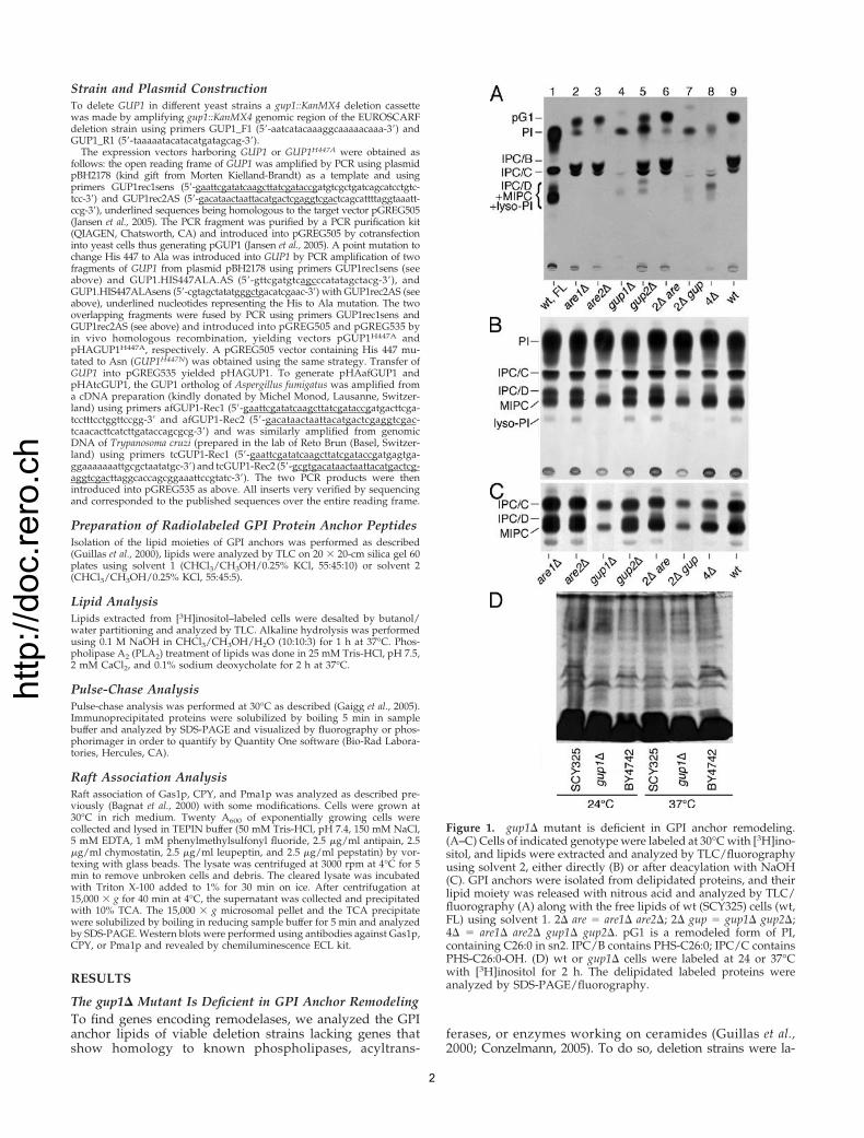
The gup1� Mutant Accumulates lyso-PIWhen proteins from [3H]inositol–labeled cells were ana-lyzed by SDS-PAGE/fluorography, it appeared that gup1�strains incorporated as much [3H]inositol into GPI proteinsas wt cells (Figure 1D). This result is in contrast to the lowamount of [3H]inositol–labeled anchor peptides one obtainsfrom gup1� cells (Figure 1A). However, it became apparentthat during the preparative octyl-Sepharose purification stepa significant amount of labeled peptides of gup1� wereeluted with 25% propanol, whereas the wt anchor peptideswere eluted only with 50% propanol, as previously reported(Figure 2A; Guillas et al., 2000). The fractions obtained with25% propanol were treated with HNO2, and liberated lipidswere separated by TLC. This revealed that gup1� cells con-tain massive amounts of anchor lipids that migrate less andend up in the zone of lyso-PI (Figure 2B, lanes 10–13). Forcomparison, we treated the lipid extracts of wt cells with
Figure 2. GPI anchor lipids of gup1� cellsare less hydrophobic than the ones of wt. (A)BY4742 wt and gup1� cells were labeled with[3H]inositol, and GPI proteins were purified,digested with pronase, and purified by chro-matography on octyl-Sepharose. The octyl-Sepharose column was washed with 5%,eluted with 25% and then 50% propanol inwater. Radioactivity in eluted fractions wasmeasured by scintillation counting. (B) Thefractions 1–9 of the gup1� derived peptides ofpanel A were subjected to nitrous acid treat-ment and the thus liberated lipid moietiesfrom indicated fractions were analyzed byTLC in solvent 1 (lanes 9–13). In parallel, thelipid extract of [3H]inositol–labeled wt cellswas treated with PLA2 and loaded onto thesame octyl-Sepharose column, and eluted frac-tions were analyzed by TLC as above (lanes2–7). Untreated lipid extract was run in lanes 1and 8. Note that anchor peptides eluting with25% propanol (lanes 10 and 11) are contami-nated by hydrophobic peptides, which slightlydistort the migration of lipids during TLC.
3
http
://do
c.re
ro.c
h
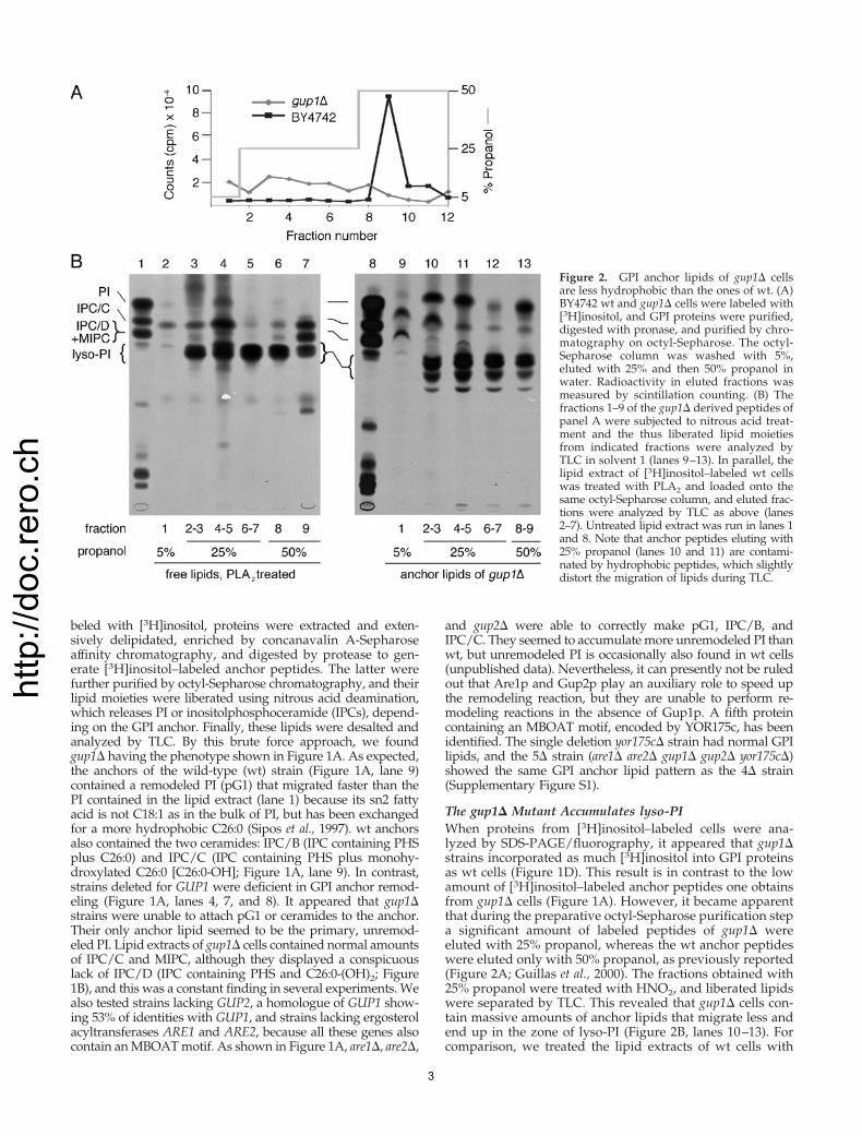
PLA2 to generate lyso-PI and fractioned the products onoctyl-Sepharose. Lyso-PI clearly was eluted with 25% ofpropanol as well (Figure 2B, lanes 3–5). However, only partof the gup1� anchor lipids comigrating with lyso-PI weresensitive to mild base (Figure 3, lanes 1 and 2). Indeed, inseveral experiments we observed base-resistant anchor lip-ids in gup1�, which were much more polar than the normal
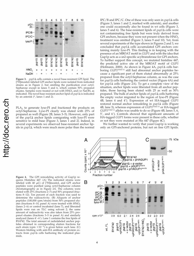
IPC/B and IPC/C. One of these was only seen in gup1� cells(Figure 3, lanes 1 and 2, marked with asterisk), and anotherone could occasionally also be found in wt cells (Figure 3,lanes 5 and 6). The base-resistant lipids of gup1� cells werenot contaminating free lipids but were truly derived fromGPI anchors, because they were not present when the HNO2treatment was omitted (Figure 3, lanes 9 and 10). Yet, fromseveral experiments of the type shown in Figures 2 and 3, weconcluded that gup1� cells accumulated GPI anchors con-taining mainly lyso-PI. This finding is in keeping with thepresence of an MBOAT motif in GUP1 and with the idea thatGup1p acts as a sn2-specific acyltransferase for GPI anchors.To further support this concept, we mutated histidine 447,the predicted active site of the MBOAT motif of GUP1(Hofmann, 2000). As shown in Figure 4A, gup1� cells har-boring GUP1H447A still had abnormal anchor peptides be-cause a significant part of them eluted abnormally at 25%propanol from the octyl-Sepharose column, as was the casefor gup1� cells harboring the control vector (Figure 4A) andfor gup1� cells (Figure 2A). To get a complete view of thesituation, anchor lipids were liberated from all anchor pep-tides, those having been eluted with 25 as well as 50%propanol. The bulk of anchor lipids of gup1� cells harboringthe empty vector migrated to the region of lyso-PI (Figure4B, lanes 4 and 4�). Expression of wt HA-tagged GUP1restored normal anchor remodeling in gup1� cells (Figure4B, lane 3), whereas expression of GUP1H447A or HA-taggedGUP1H447A alleles was unable to do so (Figure 4B, lanes 5, 6,5�, and 6�). Controls showed that significant amounts ofHA-tagged GUP1 forms were present in these cells, whetheror not they were mutated at His 447 (Figure 4C).
We further wanted to verify that yeast Gup1p is workingonly on GPI-anchored proteins, but not on free GPI lipids.
Figure 3. gup1� cells contain a novel base-resistant GPI lipid. The[3H]inositol–labeled GPI anchor lipids were isolated from indicatedstrains as in Figure 2, but omitting the purification over octyl-Sepharose except in lanes 5 and 6, which contain 50% propanoleluates. Samples were treated or not with HNO2 and/or NaOH, asindicated. The novel base resistant anchor lipid of gup1� is indicatedby an asterisk (*, lanes 1 and 2).
Figure 4. The GPI remodeling activity of Gup1p re-quires Histidine 447. (A) The indicated strains werelabeled with 40 �Ci of [3H]inositol, and GPI anchorpeptides were purified using octyl-Sepharose columnchromatography as in Figure 2A. The columns wereeluted with 25% (fractions 2–7) and 50% propanol (frac-tions 8–12). Ten percent of each fraction was used todetermine the radioactivity. (B) Aliquots of anchorpeptides (100,000 cpm/strain) from 50% propanol elu-ates (fractions 8–10, panel A) were treated with HNO2(lanes 2–6) or control incubated (lane 7), and liberatedlipids were run on TLC using solvent 2; the sameamount of radioactivity was also taken from 25% pro-panol eluates (fractions 3–5 in panel A) and similarlyanalyzed (lanes 4�–6�). Lane 1 contains the free lipids ofBY4742. The total amount of radiolabeled anchor pep-tides obtained in corresponding elution fractions foreach strain (cpm �10�5) is given below each lane. (C)Western blotting with anti-HA antibody of protein ex-tracts from gup1� cells harboring the indicated plas-mids.
4
http
://do
c.re
ro.c
h
For this we labeled yeast microsomes with UDP-[3H]Glc-NAc either in the presence or absence of C26-CoA, thepresumed substrate of Gup1p. Indeed, the profile of GPIlipids made by gup1� microsomes was the one of wt, and thesame was seen when reactions were done in the absence ofdivalent cations (Supplementary Figure S2).
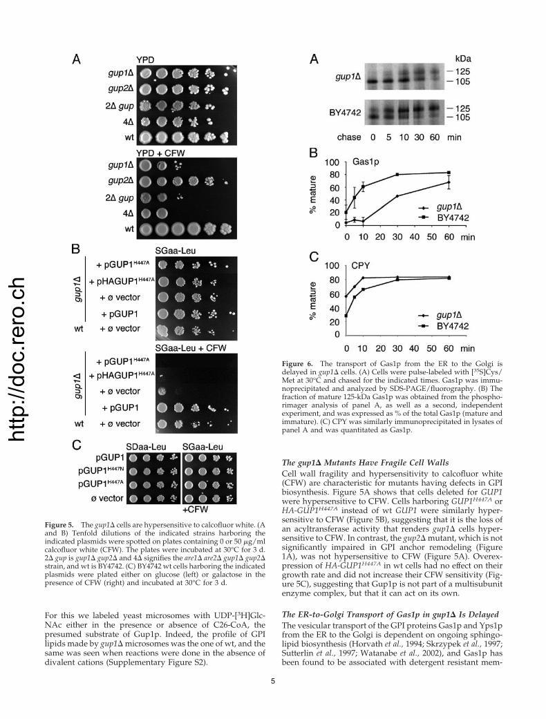
The gup1� Mutants Have Fragile Cell WallsCell wall fragility and hypersensitivity to calcofluor white(CFW) are characteristic for mutants having defects in GPIbiosynthesis. Figure 5A shows that cells deleted for GUP1were hypersensitive to CFW. Cells harboring GUP1H447A orHA-GUP1H447A instead of wt GUP1 were similarly hyper-sensitive to CFW (Figure 5B), suggesting that it is the loss ofan acyltransferase activity that renders gup1� cells hyper-sensitive to CFW. In contrast, the gup2� mutant, which is notsignificantly impaired in GPI anchor remodeling (Figure1A), was not hypersensitive to CFW (Figure 5A). Overex-pression of HA-GUP1H447A in wt cells had no effect on theirgrowth rate and did not increase their CFW sensitivity (Fig-ure 5C), suggesting that Gup1p is not part of a multisubunitenzyme complex, but that it can act on its own.
The ER-to-Golgi Transport of Gas1p in gup1� Is DelayedThe vesicular transport of the GPI proteins Gas1p and Yps1pfrom the ER to the Golgi is dependent on ongoing sphingo-lipid biosynthesis (Horvath et al., 1994; Skrzypek et al., 1997;Sutterlin et al., 1997; Watanabe et al., 2002), and Gas1p hasbeen found to be associated with detergent resistant mem-
Figure 5. The gup1� cells are hypersensitive to calcofluor white. (Aand B) Tenfold dilutions of the indicated strains harboring theindicated plasmids were spotted on plates containing 0 or 50 �g/mlcalcofluor white (CFW). The plates were incubated at 30°C for 3 d.2� gup is gup1� gup2� and 4� signifies the are1� are2� gup1� gup2�strain, and wt is BY4742. (C) BY4742 wt cells harboring the indicatedplasmids were plated either on glucose (left) or galactose in thepresence of CFW (right) and incubated at 30°C for 3 d.
Figure 6. The transport of Gas1p from the ER to the Golgi isdelayed in gup1� cells. (A) Cells were pulse-labeled with [35S]Cys/Met at 30°C and chased for the indicated times. Gas1p was immu-noprecipitated and analyzed by SDS-PAGE/fluorography. (B) Thefraction of mature 125-kDa Gas1p was obtained from the phospho-rimager analysis of panel A, as well as a second, independentexperiment, and was expressed as % of the total Gas1p (mature andimmature). (C) CPY was similarly immunoprecipitated in lysates ofpanel A and was quantitated as Gas1p.
5
http
://do
c.re
ro.c
h
brane fractions in the ER (Bagnat et al., 2000). From thesefindings, it was proposed that Gas1p has to partition intorafts in order to be incorporated into transport vesicles. Tostudy the transport of Gas1p, cells were pulsed with[35S]Cys/Met for 5 min and chased for 5, 10, 30, and 60 min.Gas1p was immunoprecipitated and analyzed by fluorogra-phy (Figure 6). In the wt strain the ratio between the ER form(105 kDa) and the Golgi form (125 kDa) is already �1:1 after5 min of chase, whereas in gup1� mutant, the ER form ispredominant up to 30 min of chase, indicating a delay inGas1p transport. Immunoprecipitation of CPY from thesame lysates showed that its maturation is not delayed ingup1� cells (Figure 6C).
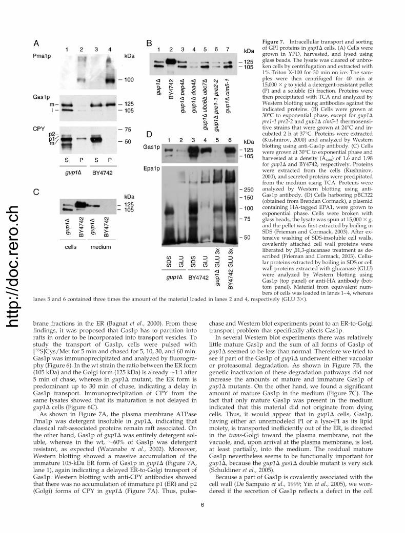
As shown in Figure 7A, the plasma membrane ATPasePma1p was detergent insoluble in gup1�, indicating thatclassical raft-associated proteins remain raft associated. Onthe other hand, Gas1p of gup1� was entirely detergent sol-uble, whereas in the wt, �60% of Gas1p was detergentresistant, as expected (Watanabe et al., 2002). Moreover,Western blotting showed a massive accumulation of theimmature 105-kDa ER form of Gas1p in gup1� (Figure 7A,lane 1), again indicating a delayed ER-to-Golgi transport ofGas1p. Western blotting with anti-CPY antibodies showedthat there was no accumulation of immature p1 (ER) and p2(Golgi) forms of CPY in gup1� (Figure 7A). Thus, pulse-
chase and Western blot experiments point to an ER-to-Golgitransport problem that specifically affects Gas1p.
In several Western blot experiments there was relativelylittle mature Gas1p and the sum of all forms of Gas1p ofgup1� seemed to be less than normal. Therefore we tried tosee if part of the Gas1p of gup1� underwent either vacuolaror proteasomal degradation. As shown in Figure 7B, thegenetic inactivation of these degradation pathways did notincrease the amounts of mature and immature Gas1p ofgup1� mutants. On the other hand, we found a significantamount of mature Gas1p in the medium (Figure 7C). Thefact that only mature Gas1p was present in the mediumindicated that this material did not originate from dyingcells. Thus, it would appear that in gup1� cells, Gas1p,having either an unremodeled PI or a lyso-PI as its lipidmoiety, is transported inefficiently out of the ER, is directedin the trans-Golgi toward the plasma membrane, not thevacuole, and, upon arrival at the plasma membrane, is lost,at least partially, into the medium. The residual matureGas1p nevertheless seems to be functionally important forgup1�, because the gup1� gas1� double mutant is very sick(Schuldiner et al., 2005).
Because a part of Gas1p is covalently associated with thecell wall (De Sampaio et al., 1999; Yin et al., 2005), we won-dered if the secretion of Gas1p reflects a defect in the cell
Figure 7. Intracellular transport and sortingof GPI proteins in gup1� cells. (A) Cells weregrown in YPD, harvested, and lysed usingglass beads. The lysate was cleared of unbro-ken cells by centrifugation and extracted with1% Triton X-100 for 30 min on ice. The sam-ples were then centrifuged for 40 min at15,000 � g to yield a detergent-resistant pellet(P) and a soluble (S) fraction. Proteins werethen precipitated with TCA and analyzed byWestern blotting using antibodies against theindicated proteins. (B) Cells were grown at30°C to exponential phase, except for gup1�pre1-1 pre2-2 and gup1� cim5-1 thermosensi-tive strains that were grown at 24°C and in-cubated 2 h at 37°C. Proteins were extracted(Kushnirov, 2000) and analyzed by Westernblotting using anti-Gas1p antibody. (C) Cellswere grown at 30°C to exponential phase andharvested at a density (A600) of 1.6 and 1.98for gup1� and BY4742, respectively. Proteinswere extracted from the cells (Kushnirov,2000), and secreted proteins were precipitatedfrom the medium using TCA. Proteins wereanalyzed by Western blotting using anti-Gas1p antibody. (D) Cells harboring pBC322(obtained from Brendan Cormack), a plasmidcontaining HA-tagged EPA1, were grown toexponential phase. Cells were broken withglass beads, the lysate was spun at 15,000 � g,and the pellet was first extracted by boiling inSDS (Frieman and Cormack, 2003). After ex-tensive washing of SDS-insoluble cell walls,covalently attached cell wall proteins wereliberated by �1,3-glucanase treatment as de-scribed (Frieman and Cormack, 2003). Cellu-lar proteins extracted by boiling in SDS or cellwall proteins extracted with glucanase (GLU)were analyzed by Western blotting usingGas1p (top panel) or anti-HA antibody (bot-tom panel). Material from equivalent num-bers of cells was loaded in lanes 1–4, whereas
lanes 5 and 6 contained three times the amount of the material loaded in lanes 2 and 4, respectively (GLU 3�).
6
http
://do
c.re
ro.c
h
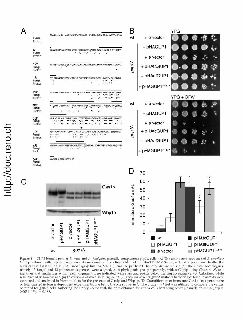
Figure 8. GUP1 homologues of T. cruzi and A. fumigatus partially complement gup1� cells. (A) The amino acid sequence of S. cerevisiaeGup1p is shown with its putative transmembrane domains (black lines, obtained with the TMHMM Server, v. 2.0 at http://www.cbs.dtu.dk/services/TMHMM/), the MBOAT motif (gray line, aa 271-510), and the predicted Histidine 447 active site (*). The closest homologues,namely 17 fungal and 13 protozoan sequences were aligned, each phylogentic group separately, with scGup1p using Clustalv W, andidentities and similarities within each alignment were indicated with stars and points below the Gup1p sequence. (B) Calcofluor whiteresistance of BY4742 wt and gup1� cells was assayed as in Figure 5B. (C) Proteins of wt or gup1� mutants harboring different plasmids wereextracted and analyzed in Western blots for the presence of Gas1p and Wbp1p. (D) Quantification of immature Gas1p (as a percentageof total Gas1p) in four independent experiments, one being the one shown in C. The Student’s t test was utilized to compare the valuesobtained for gup1� cells harboring the empty vector with the ones obtained for gup1� cells harboring other plasmids: *p � 0.40; **p �0.0034; ***p � 0.188.
7
http
://do
c.re
ro.c
h
wall integration of GPI proteins. This integration is probablyoperated by Dfg5p and/or Dcw1p, the enzymes proposed totransfer GPI proteins from GlcN-PI onto �1,6-glucans, thusincorporating them into the cell wall (Lu et al., 1995; Kollar et al.,1997; Kitagaki et al., 2002). To investigate this issue, we intro-duced into gup1� cells EPA1, an HA-tagged GPI protein that isdisplayed at the cell surface (Frieman and Cormack, 2003). Asshown in Figure 7D, the amount of EPA1 that could be liber-ated only by �-glucosidase treatment was comparable in wtand gup1� cells (lanes 2 vs. 4 and 5 vs. 6). On the other hand,the fraction of glucan-linked Gas1p was much higher in wtthan in gup1� cells (lanes 5 vs. 6). Thus, gup1� cells still incor-porate EPA1, but not Gas1p into the cell wall. It is conceivablethat in gup1� cells, Gas1p mainly contains a lyso-PI, whereasEPA1 contains some atypical base-resistant lipid anchor andthat this would be decisive. Further experiments are requiredto analyze the anchor lipids of these proteins and to see if theincorporation of GPI proteins into the cell wall is dependent ontheir anchor lipid moiety.
GUP1 Homologues from T. cruzi and A. fumigatusPartially Complement the Defect of gup1� CellsThe Blast link for GUP1 at NCBI lists homologues in allphyla except for archaea, but curiously, the closest homo-logues of GUP1 are all of fungal origin (scores between 1921and 887), the next closest homologues are from D. discoideumor protozoa (T. cruzi, T. brucei, L. major; scores from 765 to366), and the next closest homologues are all metazoan(scores from 254 to 181). This latter class also contains mam-malian homologues. The relatively strong conservation ofGUP1 among fungi and protozoa is also seen in the align-
ment shown in Figure 8A. Of the 79 residues conservedamong fungal GUP1 homologues, 66 were located in theMBOAT region. On the other hand, conventional alignmentprograms failed to correctly align the MBOAT motif andactive site histidine of metazoan homologues (Danio, Tetra-odon, Rattus, Canis, Xenopus, Mus, and Anopheles) with yeastGUP1. Expression of GUP1 homologues of T. cruzi and A.fumigatus significantly increased the CFW resistance ofgup1� cells (Figure 8B). As judged by Western blotting, theyfailed to reduce the abnormally high amounts of the imma-ture Gas1p form, but an increase of the mature form ofGas1p was noted in cells expressing afGUP1 (Figure 8C) sothat the ratio of immature/total Gas1p was significantlylower in afGUP1-complemented gup1� than in the emptyvector control (Figure 8D). Expression of tcGUP1 and af-GUP1 also significantly reduced the secretion of Gas1p (Sup-plementary Figure S3). The analysis of the anchor lipidsshowed that the expression of GUP1 homologues of T. cruziand A. fumigatus in gup1� cells reduced the fraction of lipidsin the region of lyso-PI but did not restore synthesis of pG1(unpublished data).
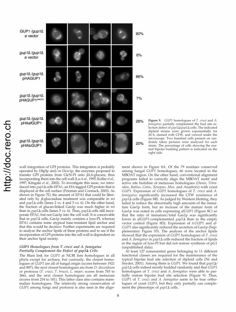
At least 127 nonessential genes belonging to 11 differentfunctional classes are required for the maintenance of thetypical bipolar bud site selection of diploid cells (Ni andSnyder, 2001). Among them is GUP1. We found that gup1�/gup1� cells indeed mostly budded randomly and that GUP1homologues of T. cruzi and A. fumigatus were able to par-tially restore bipolar bud site selection (Figure 9). Thus,GUP1 of T. cruzi and A. fumigatus seem to be true ortho-logues of yeast GUP1, but they only partially can comple-ment the phenotype of gup1� cells.
Figure 9. GUP1 homologues of T. cruzi and A.fumigatus partially complement the bud site se-lection defect of gup1�/gup1� cells. The indicateddiploid strains were grown exponentially for24 h, stained with CFW, and viewed under themicroscope. Two hundred cells present on ran-domly taken pictures were analyzed for eachstrain. The percentage of cells showing the nor-mal bipolar budding pattern is indicated on theright side.
8
http
://do
c.re
ro.c
hDISCUSSION
The lipids found on GPI anchors of different organisms arevery often not representative of the lipids present on the PI,on which the GPI structure is initially built. Lipid moietiesmay be changed either before or after the addition of the GPIto the nascent proteins. The best studied case for the formerscenario is the GPI remodeling of the blood form of T. brucei,where both of the primary fatty acids are exchanged formyristate through sequential enzymatic steps leading fromlipid A� via the lyso form � to A and furthermore to the finalA form, which is attached to proteins (Masterson et al., 1990).The remodeling occurring in S. cerevisiae is different in thatthe lipids are exchanged only once the GPI has been addedto the GPI protein. Yet, the Gup1p-mediated remodelingreaction leading from PI to pG1 may formally be similar tothe first reaction occurring in T. brucei, i.e., the replacementof a fatty acid in sn2 (Masterson et al., 1990).
Our present model for anchor lipid remodeling is that anunknown lipase first removes the acyl in sn2, that Gup1padds the C26:0 to generate pG1, and that the thus remodeledanchor then can be further remodeled through an exchangeof diacylglycerol for ceramide or phosphatidic acid for Cer-1-phosphate. This implies that the remodeling reaction op-erated by Gup1p greatly facilitates the introduction of PHS-C26:0 and PHS-C26:0-OH ceramides into the anchor, in thatGup1p produces the substrate for the still hypothetical re-modelases that introduce PHS-C26:0 and PHS-C26:0-OHceramides. (Gup1p-mediated remodeling may however notbe an absolute requirement for the introduction of PHS-C26:0,because in 1 of 8 experiments we still could detect significantamounts of IPC/B-type anchor lipids in gup1� cells.)
The only findings that are not predicted by this model arethe fact that gup1� cells contain reduced amounts of IPC/Dand that they still contain a minor base-resistant anchor lipidthat is not found in wt. Although these two phenomena maybe secondary to the absence of a normal remodeling process,we cannot at present exclude other models to explain theimportance of GUP1 for lipid remodeling. One such modelwould say that Gup1p may be involved in the ordinaryglycerophospholipid biosynthesis and act as a 1-acyl-glyc-erol-3-phosphate acyltransferase able to add not only C16and C18 but also C26:0 into sn2. Indeed such an sn2-specificacyltransferase may exist in yeast, because the only othergene known to perform the same reaction, SLC1, is notessential. If we thus assume that Gup1p acts as sn2 acyl-transferase to produce phosphatidic acid, the normal remod-eling of GPI anchors from PI to pG1 would have to be aphospholipase C (PLC) or D (PLD)-mediated exchange ofthe primary GPI anchor lipid for a C26:0-containing lipid,and the observed appearance of lyso-PI on the GPI anchorswould then have to be explained as a subsidiary hydrolyticreaction that only takes place when the cells cannot make theappropriate C26:0-containing lipid. Although this model isvaluable, it also would not explain the reduced amounts ofIPC/D in gup1� and several other findings render it lesslikely: 1) an exhaustive screen including all yeast strainsdeleted in genes having homology to PLC or PLD has notrevealed any mutant having a problem with anchor remod-eling (unpublished results). 2) The gup1� slc1� double mu-tant is perfectly viable (unpublished data). 3) Overexpres-sion of SLC1-1, a gain-of-function allele of SLC1 able totransfer C26:0 to sn2 (Nagiec et al., 1993) does not relieve thecalcofluor white hypersensitivity of gup1� (SupplementaryFigure S4A), even in the presence of low amounts of myri-ocin, which, by blocking the sphingolipid biosynthesis, de-viates C26-CoA toward other acyltransferases. Also, overex-
pression of SLC1-1 does not correct the remodeling defect ofgup1� cells (Supplementary Figure S4B). 4) Overexpressionof GUP1 cannot rescue the thermosensitivity of lcb1-100 cells(Supplementary Figure S5).
A further question raised by our findings concerns thenature of the base-resistant lipid attached to GPI anchors ingup1� cells (Figure 3, lipid marked by asterisk). This lipidmay consist of an unusual ceramide, a long-chain base, or alyso-alkyl-glycerol.
The gup1� null mutation shows a synthetic sick interac-tion with CHS3, CHS5, and CHS6, genes required for chitinsynthesis in a synthetic genetic array (SGA) analysis,whereas no such synthetic effect of chs� alleles with gup2�,are1�, are2�, or yor175w� were noted (Lesage et al., 2005).The specific role of Gup1p in GPI anchor remodeling ex-plains these synthetic sick interactions between the csh� andgup1� deletion mutations.
GPI anchoring seems to be important for the maintenanceof the typical bipolar budding pattern of diploid cells. Thismay not have been appreciated earlier, because most genesinvolved in GPI anchor biosynthesis or anchor processingare essential. However, GPI7, BST1, and GUP1 are nones-sential, GPI-related genes, and all of them are important forbud site selection. The same is true for GAS1 and CCW12encoding GPI proteins (Ni and Snyder, 2001). Moreover,bipolar bud site selection is deficient in four genes involvedin lipid biosynthesis, namely ELO2, ELO3, ERG3, and ERG4,the former two of which also affect GPI anchoring, becausethey affect biosynthesis of C26 (Ni and Snyder, 2001). OtherGPI proteins may not have been identified in the screen ofNi and Snyder because they are redundant.
A previous large-scale screen for CPY secretion showedthat GUP1 is involved in trafficking of CPY, Pep4p, andPho8p to the vacuole and that gup1� cells display vacuolarprotein sorting class C mutant vacuolar morphologies(Bonangelino et al., 2002). Abnormal secretion of CPY bygup1� was confirmed (Supplementary Figure S6), and our datasuggest that the defect in vacuolar sorting does not cause abnor-mal accumulation of the Golgi p2 from of CPY (Figure 7A).
It seems quite possible that the anchor remodeling of S.cerevisiae is paradigmatic for many other fungi and protozo-ans. Although the MBOAT motif occurs in a large variety ofacyl-CoA–dependent acyltransferases dedicated to lipidbiosynthesis in bacteria and eukaryotes, many fungi andprotozoan organisms contain GUP1 homologues having ahigh degree of homology to yeast GUP1 and containingsmall homology regions also outside the MBOAT motif(Figure 8A). Indeed, GUP1 homologues of T. cruzi and A.fumigatus could partially rescue the CFW hypersensitivity, theloss of Gas1p from the cell surface, and the bud site selectiondefect of gup1� cells (Figures 8 and 9 and SupplementaryFigure S3). HA-afGUP1 was more efficient than HA-tcGUP1in this respect (Figure 8), although HA-tcGUP1 could beexpressed to higher levels than HA-afGUP1 (unpublisheddata). T. cruzi contains both alkylacylglycerol- as well asceramide-based GPI anchors, whereas A. fumigatus only con-tains the latter type (Fontaine et al., 2003; Ferguson et al.,2006). The alkylacylglycerol anchors of T. cruzi contain C16and C18 fatty acids in sn2 (Ferguson et al., 2006), and we thuscan speculate that tcGUP1 expression in gup1� cells may notallow adding C26:0 acids in sn2 as does yeast GUP1, but thatit only may alter the proportions between lyso-PI and PI an-chors. Recent studies show that, similar to yeast, the first stepsof GPI biosynthesis in T. cruzi do not use ceramide as the lipidsupport, suggesting that ceramide is added by remodeling ofprotein-bound anchors also in T. cruzi (Bertello et al., 2004). Ourstudy raises the possibility that in both, T. cruzi and A.
9
http
://do
c.re
ro.c
hfumigatus the addition of the normal set of ceramides would bedependent on a foregoing remodeling by GUP1. GUP1 homo-logues are also found in other organisms that contain ceramideanchors such as certain plants, Paramecium, and Dictyostelium(Ferguson et al., 2006). Thus, a remodeling step catalyzed by aGUP1 homologue may be occurring in many organisms, evenif their sole anchor lipid is a ceramide.
Interestingly, a recent report shows that mammalian GPIanchors seem to be remodeled and undergo a deacylation–reacylation cycle in the Golgi, mediated by PGAP2, an enzymethat is completely unrelated to GUP1 (Tashima et al., 2006). Thissuggests that GPI lipid remodeling may be a common eventduring the transport of GPI proteins to the surface.
ACKNOWLEDGMENTS
We thank Stephen Sturley, Morten Kielland-Brandt, Marja Makarow, MichelMonod, Reto Brun, Roger Schneiter, and their lab members for strains, antibod-ies, cDNA libraries, DNA, and plasmids. We also acknowledge the excellenttechnical help provided by Christine Vionnet, Melanie Bapst, and Joel Robert.This work was supported by a grant from the Swiss National Science Foundation(31-67188.01).
REFERENCES
Bagnat, M., Keranen, S., Shevchenko, A., Shevchenko, A., and Simons, K.(2000). Lipid rafts function in biosynthetic delivery of proteins to the cellsurface in yeast. Proc. Natl. Acad. Sci. USA 97, 3254–3259.
Bertello, L. E., Alves, M. J., Colli, W., and de Lederkremer, R. M. (2004).Inositolphosphoceramide is not a substrate for the first steps in the biosyn-thesis of glycoinositolphospholipids in Trypanosoma cruzi. Mol. Biochem.Parasitol. 133, 71–80.
Bonangelino, C. J., Chavez, E. M., and Bonifacino, J. S. (2002). Genomic screenfor vacuolar protein sorting genes in Saccharomyces cerevisiae. Mol. Biol. Cell13, 2486–2501.
Conzelmann, A. (2005). Biosynthesis, remodeling and targeting of GPI pro-teins in the yeast Saccharomyces cerevisiae. In: Cell Biology and Dynamics ofYeast Lipids, ed. G. Daum, Trivandrum, India: Research Signpost, 133–159.
De Sampaio, G., Bourdineaud, J. P., and Lauquin, G. J. (1999). A constitutiverole for GPI anchors in Saccharomyces cerevisiae: cell wall targeting. Mol.Microbiol. 34, 247–256.
Fankhauser, C., Homans, S. W., Thomas-Oates, J. E., McConville, M. J.,Desponds, C., Conzelmann, A., and Ferguson, M. A. (1993). Structures of glycosyl-phosphatidylinositol membrane anchors from Saccharomyces cerevisiae. J. Biol.Chem. 268, 26365–26374.
Ferguson, M.A.J., Kinoshita, T., and Hart, G. W. (2006). Glycophospholipidanchors. In: Essential Glycobiology, 2nd ed., ed. Varki, A., Bertozzi, C.,Cummings, R., Etzler, M., Esko, J., Freeze, H., Hart, G., and Stanley, P. (inpress).
Fontaine, T., Magnin, T., Melhert, A., Lamont, D., Latge, J. P., and Ferguson,M. A. (2003). Structures of the glycosylphosphatidylinositol membrane anchorsfrom Aspergillus fumigatus membrane proteins. Glycobiology 13, 169–177.
Frieman, M. B., and Cormack, B. P. (2003). The omega-site sequence ofglycosylphosphatidylinositol-anchored proteins in Saccharomyces cerevisiaecan determine distribution between the membrane and the cell wall. Mol.Microbiol. 50, 883–896.
Gaigg, B., Timischl, B., Corbino, L., and Schneiter, R. (2005). Synthesis ofsphingolipids with very long chain fatty acids but not ergosterol is requiredfor routing of newly synthesized plasma membrane ATPase to the cell surfaceof yeast. J. Biol. Chem. 280, 22515–22522.
Guillas, I., Pfefferli, M., and Conzelmann, A. (2000). Analysis of ceramidespresent in glycosylphosphatidylinositol anchored proteins of Saccharomycescerevisiae. Methods Enzymol. 312, 506–515.
Hofmann, K. (2000). A superfamily of membrane-bound O-acyltransferaseswith implications for wnt signaling. Trends Biochem. Sci. 25, 111–112.
Holst, B., Lunde, C., Lages, F., Oliveira, R., Lucas, C., and Kielland-Brandt,M. C. (2000). GUP1 and its close homologue GUP2, encoding multimem-brane-spanning proteins involved in active glycerol uptake in Saccharomycescerevisiae. Mol. Microbiol. 37, 108–124.
Horvath, A., Sutterlin, C., Manning-Krieg, U., Movva, N. R., and Riezman, H.(1994). Ceramide synthesis enhances transport of GPI-anchored proteins tothe Golgi apparatus in yeast. EMBO J. 13, 3687–3695.
Imhof, I., Flury, I., Vionnet, C., Roubaty, C., Egger, D., and Conzelmann, A.(2004). Glycosylphosphatidylinositol (GPI) proteins of Saccharomyces cerevisiaecontain ethanolamine phosphate groups on the alpha1,4-linked mannose ofthe GPI anchor. J. Biol. Chem. 279, 19614–19627.
Jansen, G., Wu, C., Schade, B., Thomas, D. Y., and Whiteway, M. (2005).Drag&Drop cloning in yeast. Gene 344, 43–51.
Kinoshita, T., and Inoue, N. (2000). Dissecting and manipulating the pathwayfor glycosylphosphatidylinositol-anchor biosynthesis. Curr. Opin. Chem.Biol. 4, 632–638.
Kitagaki, H., Wu, H., Shimoi, H., and Ito, K. (2002). Two homologous genes,DCW1 (YKL046c) and DFG5, are essential for cell growth and encode glycosyl-phosphatidylinositol (GPI)-anchored membrane proteins required for cellwall biogenesis in Saccharomyces cerevisiae. Mol. Microbiol. 46, 1011–1022.
Kollar, R., Reinhold, B. B., Petrakova, E., Yeh, H. J., Ashwell, G., Drgonova, J.,Kapteyn, J. C., Klis, F. M., and Cabib, E. (1997). Architecture of the yeast cellwall. Beta(136)-glucan interconnects mannoprotein, beta(133)-glucan, andchitin. J. Biol. Chem. 272, 17762–17775.
Kushnirov, V. V. (2000). Rapid and reliable protein extraction from yeast.Yeast 16, 857–860.
Lesage, G., Shapiro, J., Specht, C. A., Sdicu, A. M., Menard, P., Hussein, S.,Tong, A. H., Boone, C., and Bussey, H. (2005). An interactional network ofgenes involved in chitin synthesis in Saccharomyces cerevisiae. BMC Genet. 6, 8.
Lu, C. F., Montijn, R. C., Brown, J. L., Klis, F., Kurjan, J., Bussey, H., and Lipke,P. N. (1995). Glycosyl phosphatidylinositol-dependent cross-linking of alpha-agglutinin and beta 1,6-glucan in the Saccharomyces cerevisiae cell wall. J. CellBiol. 128, 333–340.
Masterson, W. J., Raper, J., Doering, T. L., Hart, G. W., and Englund, P. T. (1990).Fatty acid remodeling: a novel reaction sequence in the biosynthesis of trypano-some glycosyl phosphatidylinositol membrane anchors. Cell 62, 73–80.
Miyata, T., Takeda, J., Iida, Y., Yamada, N., Inoue, N., Takahashi, M., Maeda,K., Kitani, T., and Kinoshita, T. (1993). The cloning of PIG-A, a component inthe early step of GPI-anchor biosynthesis. Science 259, 1318–1320.
Morita, Y. S., Acosta-Serrano, A., Buxbaum, L. U., and Englund, P. T. (2000).Glycosyl phosphatidylinositol myristoylation in African trypanosomes. Newintermediates in the pathway for fatty acid remodeling. J. Biol. Chem. 275,14147–14154.
Nagiec, M. M., Wells, G. B., Lester, R. L., and Dickson, R. C. (1993). Asuppressor gene that enables Saccharomyces cerevisiae to grow without makingsphingolipids encodes a protein that resembles an Escherichia coli fatty acyl-transferase. J. Biol. Chem. 268, 22156–22163.
Neves, L., Lages, F., and Lucas, C. (2004). New insights on glycerol transportin Saccharomyces cerevisiae. FEBS Lett. 565, 160–162.
Ni, L., and Snyder, M. (2001). A genomic study of the bipolar bud siteselection pattern in Saccharomyces cerevisiae. Mol. Biol. Cell 12, 2147–2170.
Schuldiner, M. et al. (2005). Exploration of the function and organization of theyeast early secretory pathway through an epistatic miniarray profile. Cell 123,507–519.
Sherman, F. (2002). Getting started with yeast. Methods Enzymol. 350, 3–41.
Sipos, G., Reggiori, F., Vionnet, C., and Conzelmann, A. (1997). Alternativelipid remodelling pathways for glycosylphosphatidylinositol membrane an-chors in Saccharomyces cerevisiae. EMBO J. 16, 3494–3505.
Skrzypek, M., Lester, R. L., and Dickson, R. C. (1997). Suppressor gene analysisreveals an essential role for sphingolipids in transport of glycosylphosphatidylinosi-tol-anchored proteins in Saccharomyces cerevisiae. J. Bacteriol. 179, 1513–1520.
Sutterlin, C., Doering, T. L., Schimmoller, F., Schroder, S., and Riezman, H.(1997). Specific requirements for the ER to Golgi transport of GPI-anchoredproteins in yeast. J. Cell Sci. 110, 2703–2714.
Tashima, Y., Taguchi, R., Murata, C., Ashida, H., Kinoshita, T., and Maeda, Y.(2006). PGAP2 is essential for correct processing and stable expression ofGPI-anchored proteins. Mol. Biol. Cell 17, 1410–1420.
Watanabe, R., Funato, K., Venkataraman, K., Futerman, A. H., and Riezman,H. (2002). Sphingolipids are required for the stable membrane association ofglycosylphosphatidylinositol-anchored proteins in yeast. J. Biol. Chem. 277,49538–49544.
Yin, Q. Y., de Groot, P. W., Dekker, H. L., de Jong, L., Klis, F. M., and deKoster, C. G. (2005). Comprehensive proteomic analysis of Saccharomycescerevisiae cell walls: identification of proteins covalently attached via glycosyl-phosphatidylinositol remnants or mild alkali-sensitive linkages. J. Biol. Chem.280, 20894–20901.
10