Embed Size (px)
Citation preview
Manchester Cancer
Guidelines for the management of renal cancer Approved by the urology pathway board September 2014 To be reviewed September 2016
Manchester Cancer
Guidelines for the management of renal cancer September 2014
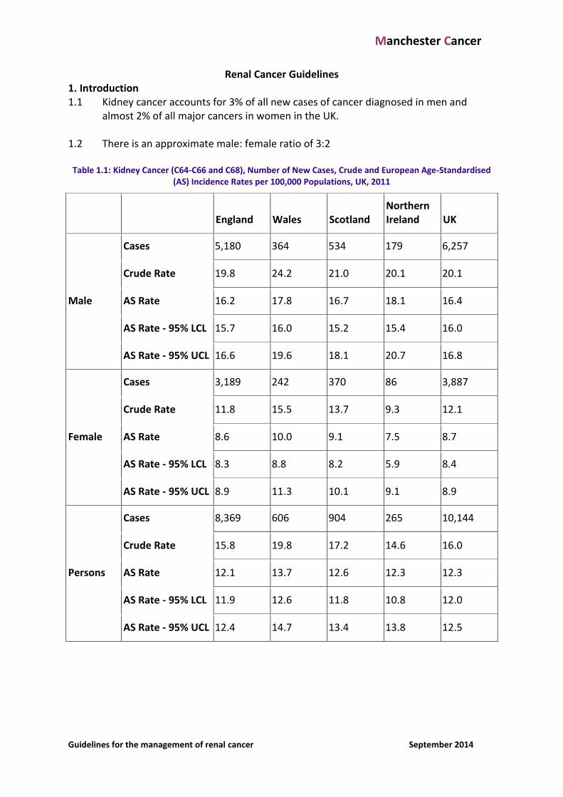
Renal Cancer Guidelines 1. Introduction 1.1 Kidney cancer accounts for 3% of all new cases of cancer diagnosed in men and
almost 2% of all major cancers in women in the UK. 1.2 There is an approximate male: female ratio of 3:2
Table 1.1: Kidney Cancer (C64-C66 and C68), Number of New Cases, Crude and European Age-Standardised (AS) Incidence Rates per 100,000 Populations, UK, 2011
England Wales Scotland Northern Ireland UK
Male
Cases 5,180 364 534 179 6,257
Crude Rate 19.8 24.2 21.0 20.1 20.1
AS Rate 16.2 17.8 16.7 18.1 16.4
AS Rate - 95% LCL 15.7 16.0 15.2 15.4 16.0
AS Rate - 95% UCL 16.6 19.6 18.1 20.7 16.8
Female
Cases 3,189 242 370 86 3,887
Crude Rate 11.8 15.5 13.7 9.3 12.1
AS Rate 8.6 10.0 9.1 7.5 8.7
AS Rate - 95% LCL 8.3 8.8 8.2 5.9 8.4
AS Rate - 95% UCL 8.9 11.3 10.1 9.1 8.9
Persons
Cases 8,369 606 904 265 10,144
Crude Rate 15.8 19.8 17.2 14.6 16.0
AS Rate 12.1 13.7 12.6 12.3 12.3
AS Rate - 95% LCL 11.9 12.6 11.8 10.8 12.0
AS Rate - 95% UCL 12.4 14.7 13.4 13.8 12.5
Manchester Cancer
Guidelines for the management of renal cancer September 2014
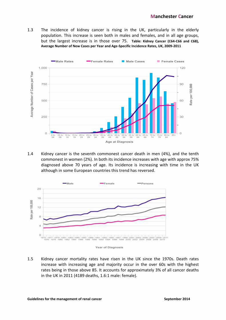
1.3 The incidence of kidney cancer is rising in the UK, particularly in the elderly population. This increase is seen both in males and females, and in all age groups, but the largest increase is in those over 75. Table: Kidney Cancer (C64-C66 and C68),
Average Number of New Cases per Year and Age-Specific Incidence Rates, UK, 2009-2011
1.4 Kidney cancer is the seventh commonest cancer death in men (4%), and the tenth
commonest in women (2%). In both its incidence increases with age with approx 75% diagnosed above 70 years of age. Its incidence is increasing with time in the UK although in some European countries this trend has reversed.
1.5 Kidney cancer mortality rates have risen in the UK since the 1970s. Death rates
increase with increasing age and majority occur in the over 60s with the highest rates being in those above 85. It accounts for approximately 3% of all cancer deaths in the UK in 2011 (4189 deaths, 1.6:1 male: female).
Manchester Cancer
Guidelines for the management of renal cancer September 2014
1.6 Renal cancer is associated with obesity and cigarette smoking. All patients encountered should therefore be encouraged and given help in smoking cessation and weight loss.
1.7 In the UK 86% of tumours are RCC and the remaining 12% are mainly TCC of the renal pelvis and ureter. There are five subgroups of RCCs: conventional (clear cell, also called non papillary), which account for 75-80% of RCC tumours; papillary (chromophilic) accounting for 10-15% and chromophobe, collecting duct carcinoma and unclassified renal cell carcinoma which together make up the remainder of RCC tumours.
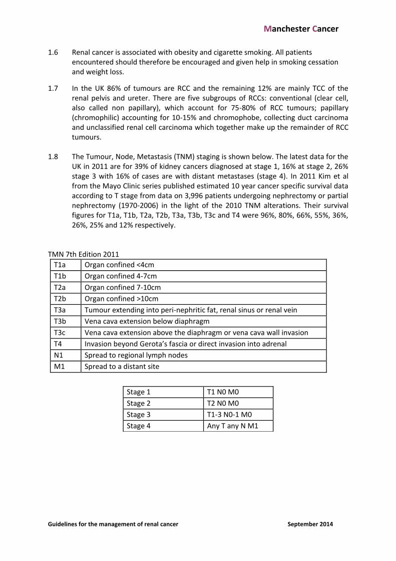
1.8 The Tumour, Node, Metastasis (TNM) staging is shown below. The latest data for the
UK in 2011 are for 39% of kidney cancers diagnosed at stage 1, 16% at stage 2, 26% stage 3 with 16% of cases are with distant metastases (stage 4). In 2011 Kim et al from the Mayo Clinic series published estimated 10 year cancer specific survival data according to T stage from data on 3,996 patients undergoing nephrectomy or partial nephrectomy (1970-2006) in the light of the 2010 TNM alterations. Their survival figures for T1a, T1b, T2a, T2b, T3a, T3b, T3c and T4 were 96%, 80%, 66%, 55%, 36%, 26%, 25% and 12% respectively.
TMN 7th Edition 2011
T1a Organ confined <4cm
T1b Organ confined 4-7cm
T2a Organ confined 7-10cm
T2b Organ confined >10cm
T3a Tumour extending into peri-nephritic fat, renal sinus or renal vein
T3b Vena cava extension below diaphragm
T3c Vena cava extension above the diaphragm or vena cava wall invasion
T4 Invasion beyond Gerota’s fascia or direct invasion into adrenal
N1 Spread to regional lymph nodes
M1 Spread to a distant site
Stage 1 T1 N0 M0
Stage 2 T2 N0 M0
Stage 3 T1-3 N0-1 M0
Stage 4 Any T any N M1
Manchester Cancer
Guidelines for the management of renal cancer September 2014
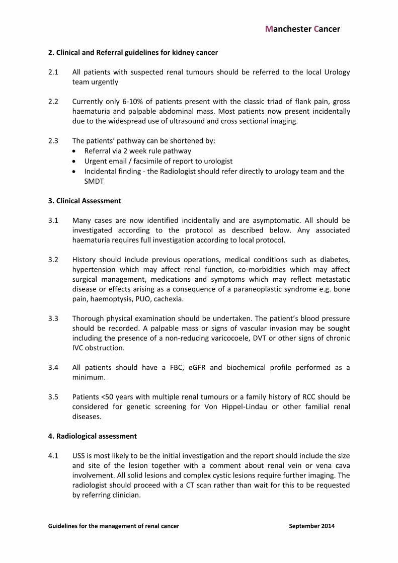
2. Clinical and Referral guidelines for kidney cancer 2.1 All patients with suspected renal tumours should be referred to the local Urology
team urgently 2.2 Currently only 6-10% of patients present with the classic triad of flank pain, gross
haematuria and palpable abdominal mass. Most patients now present incidentally due to the widespread use of ultrasound and cross sectional imaging.
2.3 The patients’ pathway can be shortened by:
Referral via 2 week rule pathway
Urgent email / facsimile of report to urologist
Incidental finding - the Radiologist should refer directly to urology team and the SMDT
3. Clinical Assessment 3.1 Many cases are now identified incidentally and are asymptomatic. All should be
investigated according to the protocol as described below. Any associated haematuria requires full investigation according to local protocol.
3.2 History should include previous operations, medical conditions such as diabetes,
hypertension which may affect renal function, co-morbidities which may affect surgical management, medications and symptoms which may reflect metastatic disease or effects arising as a consequence of a paraneoplastic syndrome e.g. bone pain, haemoptysis, PUO, cachexia.
3.3 Thorough physical examination should be undertaken. The patient’s blood pressure
should be recorded. A palpable mass or signs of vascular invasion may be sought including the presence of a non-reducing varicocoele, DVT or other signs of chronic IVC obstruction.
3.4 All patients should have a FBC, eGFR and biochemical profile performed as a
minimum. 3.5 Patients <50 years with multiple renal tumours or a family history of RCC should be
considered for genetic screening for Von Hippel-Lindau or other familial renal diseases.
4. Radiological assessment 4.1 USS is most likely to be the initial investigation and the report should include the size
and site of the lesion together with a comment about renal vein or vena cava involvement. All solid lesions and complex cystic lesions require further imaging. The radiologist should proceed with a CT scan rather than wait for this to be requested by referring clinician.
Manchester Cancer
Guidelines for the management of renal cancer September 2014
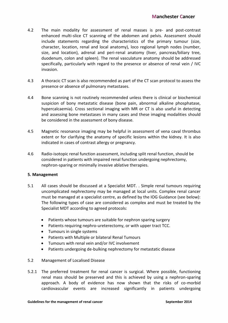
4.2 The main modality for assessment of renal masses is pre- and post-contrast enhanced multi-slice CT scanning of the abdomen and pelvis. Assessment should include statements regarding the characteristics of the primary tumour (size, character, location, renal and local anatomy), loco regional lymph nodes (number, size, and location), adrenal and peri-renal anatomy (liver, pancreas/biliary tree, duodenum, colon and spleen). The renal vasculature anatomy should be addressed specifically, particularly with regard to the presence or absence of renal vein / IVC invasion.
4.3 A thoracic CT scan is also recommended as part of the CT scan protocol to assess the
presence or absence of pulmonary metastases. 4.4 Bone scanning is not routinely recommended unless there is clinical or biochemical
suspicion of bony metastatic disease (bone pain, abnormal alkaline phosphatase, hypercalcaemia). Cross sectional imaging with MR or CT is also useful in detecting and assessing bone metastases in many cases and these imaging modalities should be considered in the assessment of bony disease.
4.5 Magnetic resonance imaging may be helpful in assessment of vena caval thrombus
extent or for clarifying the anatomy of specific lesions within the kidney. It is also indicated in cases of contrast allergy or pregnancy.
4.6 Radio-isotopic renal function assessment, including split renal function, should be
considered in patients with impaired renal function undergoing nephrectomy, nephron-sparing or minimally invasive ablative therapies.
5. Management 5.1 All cases should be discussed at a Specialist MDT. . Simple renal tumours requiring
uncomplicated nephrectomy may be managed at local units. Complex renal cancer must be managed at a specialist centre, as defined by the IOG Guidance (see below): The following types of case are considered as complex and must be treated by the Specialist MDT according to agreed protocols:
Patients whose tumours are suitable for nephron sparing surgery
Patients requiring nephro-ureterectomy, or with upper tract TCC.
Tumours in single systems
Patients with Multiple or bilateral Renal Tumours
Tumours with renal vein and/or IVC involvement
Patients undergoing de-bulking nephrectomy for metastatic disease 5.2 Management of Localised Disease 5.2.1 The preferred treatment for renal cancer is surgical. Where possible, functioning
renal mass should be preserved and this is achieved by using a nephron-sparing approach. A body of evidence has now shown that the risks of co-morbid cardiovascular events are increased significantly in patients undergoing
Manchester Cancer
Guidelines for the management of renal cancer September 2014
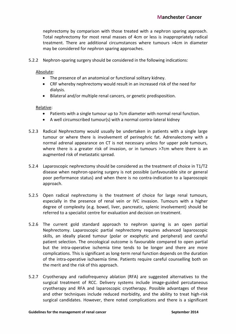
nephrectomy by comparison with those treated with a nephron sparing approach. Total nephrectomy for most renal masses of 4cm or less is inappropriately radical treatment. There are additional circumstances where tumours >4cm in diameter may be considered for nephron sparing approaches.
5.2.2 Nephron-sparing surgery should be considered in the following indications:
Absolute:
The presence of an anatomical or functional solitary kidney.
CRF whereby nephrectomy would result in an increased risk of the need for dialysis.
Bilateral and/or multiple renal cancers, or genetic predisposition.
Relative:
Patients with a single tumour up to 7cm diameter with normal renal function.
A well circumscribed tumour(s) with a normal contra-lateral kidney 5.2.3 Radical Nephrectomy would usually be undertaken in patients with a single large
tumour or where there is involvement of perinephric fat. Adrenalectomy with a normal adrenal appearance on CT is not necessary unless for upper pole tumours, where there is a greater risk of invasion, or in tumours >7cm where there is an augmented risk of metastatic spread.
5.2.4 Laparoscopic nephrectomy should be considered as the treatment of choice in T1/T2
disease when nephron-sparing surgery is not possible (unfavourable site or general poor performance status) and when there is no contra-indication to a laparoscopic approach.
5.2.5 Open radical nephrectomy is the treatment of choice for large renal tumours,
especially in the presence of renal vein or IVC invasion. Tumours with a higher degree of complexity (e.g. bowel, liver, pancreatic, splenic involvement) should be referred to a specialist centre for evaluation and decision on treatment.
5.2.6 The current gold standard approach to nephron sparing is an open partial
Nephrectomy. Laparoscopic partial nephrectomy requires advanced laparoscopic skills, an ideally placed tumour (polar or exophytic and peripheral) and careful patient selection. The oncological outcome is favourable compared to open partial but the intra-operative ischemia time tends to be longer and there are more complications. This is significant as long-term renal function depends on the duration of the intra-operative ischaemia time. Patients require careful counselling both on the merit and the risk of this approach.
5.2.7 Cryotherapy and radiofrequency ablation (RFA) are suggested alternatives to the
surgical treatment of RCC. Delivery systems include image-guided percutaneous cryotherapy and RFA and laparoscopic cryotherapy. Possible advantages of these and other techniques include reduced morbidity, and the ability to treat high-risk surgical candidates. However, there noted complications and there is a significant
Manchester Cancer
Guidelines for the management of renal cancer September 2014
treatment failure rate. Patients must be counselled fully about this prior to consideration of this therapeutic approach. Indications include: small, incidentally found lesions in elderly patients (although there is an increasing body of evidence to suggest that these can be managed by biopsy and active monitoring), patients with a genetic predisposition for developing multiple tumours, patients with bilateral tumours, patients with a solitary kidney at high risk of complete loss of renal function following surgical tumour resection. Contraindications include poor life expectancy of <1 year, multiple metastases, low possibility of successful treatment due to size (>3cm) or location of tumour (near the hilum/ ureter or in proximity to other vital structures e.g. bowel/ lung). Compared to RFA, cryoablation is more likely to be performed laparoscopically. The laparoscopic approach is more effective but has a higher complication rate. Repeat ablation is necessary more frequently following RFA. Local tumour progression is significantly higher with RFA. Cancer-specific survival rates for cryotherapy and RFA are poorer than survival rates for surgical procedures. Locally patients can be referred for cryotherapy at UHSM and, for RFA, to Central Manchester.
5.2.8 Patients with renal vein and / or IVC involvement should be referred to a team with
experience in the management of these complex cases. 5.2.9 Surveillance. In patients with small renal masses (<3cm), surveillance appears safe in
terms of risk of local tumour progression or an increased risk of metastatic disease. It is therefore an appropriate consideration especially in the elderly and those with multiple co-morbidities. Such patients with small renal masses should be counselled fully and monitored with intervention only if there is definitive evidence of progression. There is a body of evidence to suggest that there is a role for renal biopsy in these patients before instigating active monitoring.
5.2.10 Lymphadenectomy. During nephrectomy radical lymph node dissection does not
appear to improve long-term survival. For trial or staging purposes a limited lymph node dissection can be performed, especially if palpable or CT-detected.
5.2.11 Patients with pre-existing CKD and a potential requirement for post-surgical dialysis
should be referred to centres with co-existing nephrology services, with facilities for optimisation of renal function pre-operatively and for dialysis post-operatively when required.
5.2.12 Patients with severe co-morbidities precluding surgery and minimally invasive
treatment should be monitored and palliative treatment offered as appropriate; e.g. embolisation, palliative radiotherapy, supportive care.
5.2.13 Specialist urology nurse involvement should be available at an early stage and their
involvement recorded in the documentary record. MacMillan nurse support and/or palliative care team involvement may be indicated in specific patient journeys.
Manchester Cancer
Guidelines for the management of renal cancer September 2014
5.3 Metastatic disease 5.3.1 Debulking nephrectomy must be discussed at SMDT as an adjunct to systemic
therapy in patients with good performance status and low metastatic disease load. Consideration should be mandatory before advocating such surgery. This is to assess whether surgery is appropriate and/or feasible and to determine the most appropriate non-surgical intervention. Patient may then be referred back to the SMDT thereafter. Surgery of this type may be challenging and should only be performed by teams familiar with this type of therapeutic approach. Trial inclusion should be explored where possible.
5.3.2 Solitary metastases may be considered for local resection (metastasectomy).
Discussion at the SNMDT is required before definitive decisions relating to this are made. Patients who subsequently develop an apparent solitary metatasis after treatment for the primary should be consider for excision or localised radiotherapy.
5.3.3 Patients with advanced or metastatic disease should be considered for referral for
systemic treatment, either with immunotherapy or other targeted therapies such as sunitinib or Pazopanib.. Other therapies (e.g. mTOR inhibitors) will be considered in the secondline setting. All patients should be considered for entry into clinical trials where these exist. Systemic therapy for metastatic renal cancer is an area of rapid development and medical oncology input is of paramount importance in the management of these patients.
5.3.4 Palliative care team referral should be offered at an early stage for patient support
and symptom control as appropriate.
6. Surveillance following Surgery for RCC 6.1 Rationale for follow up. Follow up of patients with RCC after surgical and minimally
invasive treatment is recommended to detect local recurrence and distant metastases as early as possible to permit additional treatment when indicated and if possible. Such therapy includes; resection of pulmonary metastasis or local recurrences; radiotherapy, in the case of small and low number brain metastasis not suitable for excision or locally focused radiotherapy.
6.2 Patients should be stratified into risk groups for development of metastatic disease
following primary treatment (see pathological staging below) and a standardised follow-up protocol implemented such as the Mayo risk score should be considered for adoption (see below).
6.3 Patients developing metastases on surveillance should be discussed at an SMDT for
consideration regarding suitability for further non-surgical systemic therapy or surgical treatment, including metastasectomy. Palliative care team/ MacMillan referral may be appropriate.

Manchester Cancer
Guidelines for the management of renal cancer September 2014
6.4 Follow protocols should also seek to identify post-operative complication and monitor renal function. Improvement of primary care in renal monitoring and screening for diabetes and hypertension is recommended including referral on to nephrological services in the case of renal deterioration or significantly decreased GFR.
7. Outcomes 7.1 All units and surgeons undertaking renal surgery should be involved in an active
audit programme to review local outcomes and must submit data to on-going national renal cancer audit programmes (e.g. BAUS national database).
8. Pathology Reporting 8.1 Specimens shall be processed, and cases reported in accordance with, the Royal
College of Pathologists’ standards and datasets - Pathological Staging. 8.2 Histopathology reports for RCC should include the following as a minimum: Histological type
Furhmann Grade Pathological Stage using the most recent TNM system Tumour size
Presence or absence of necrosis Presence or absence of sarcomatoid changes In clear cell tumours, % of clear cell: granular cell ratio Information relating to renal vein / IVC invasion when this is present Specific statements about tumour margins in nephron sparing cases
These documents can be found at http://www.rcpath.org/Resources/RCPath/Migrated Resources/Documents/G/g099tpurologicaltissuepathwayfinalmay2010.pdf http://www.rcpath.org/publications-media/publications/datasets/adult-renal.htm
8.3 The risk of metastases can be calculated using the MAYO system [Leibovich BC, Blute ML, Zincke et al: Cancer 2003;97:1663-71] below:
Mayo scoring system:
Score pT Size grade necrosis nodes
0 1a < 10 1-2 No X / 0
1 >10 3 Yes
2 1b 1/2
3 2 4
4 3/4
Manchester Cancer
Guidelines for the management of renal cancer September 2014
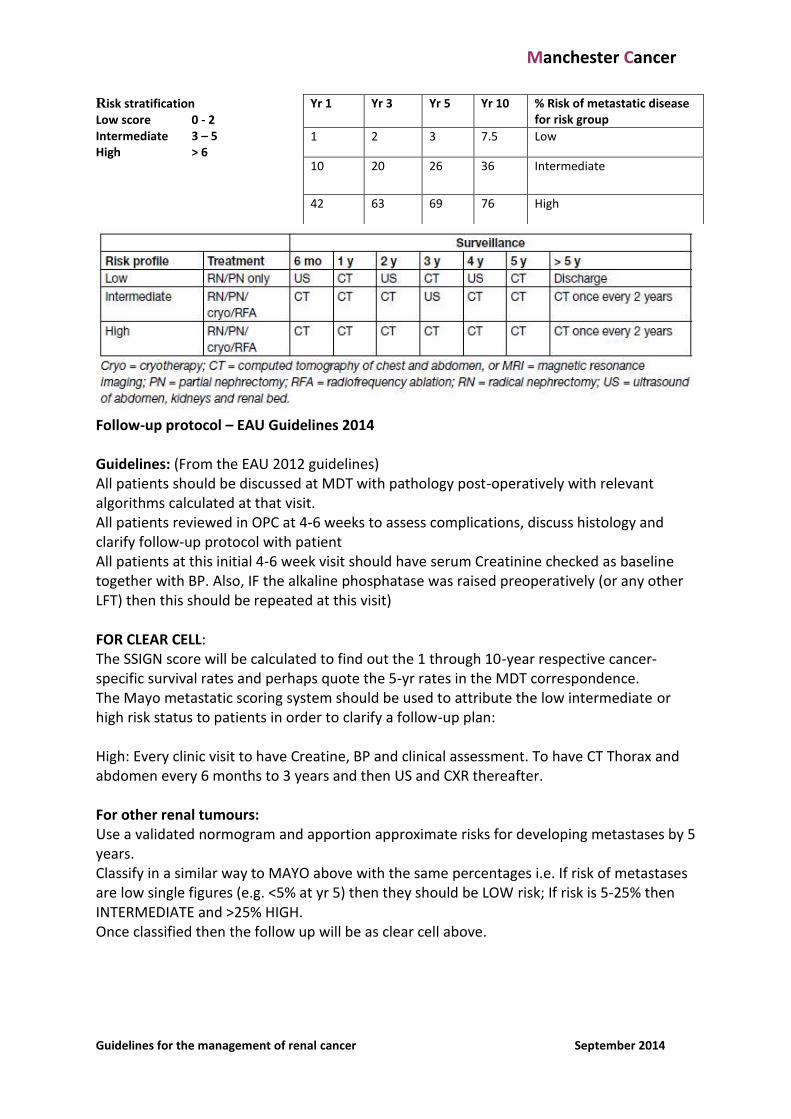
Risk stratification Low score 0 - 2 Intermediate 3 – 5 High > 6
Follow-up protocol – EAU Guidelines 2014 Guidelines: (From the EAU 2012 guidelines) All patients should be discussed at MDT with pathology post-operatively with relevant algorithms calculated at that visit. All patients reviewed in OPC at 4-6 weeks to assess complications, discuss histology and clarify follow-up protocol with patient All patients at this initial 4-6 week visit should have serum Creatinine checked as baseline together with BP. Also, IF the alkaline phosphatase was raised preoperatively (or any other LFT) then this should be repeated at this visit) FOR CLEAR CELL: The SSIGN score will be calculated to find out the 1 through 10-year respective cancer-specific survival rates and perhaps quote the 5-yr rates in the MDT correspondence. The Mayo metastatic scoring system should be used to attribute the low intermediate or high risk status to patients in order to clarify a follow-up plan: High: Every clinic visit to have Creatine, BP and clinical assessment. To have CT Thorax and abdomen every 6 months to 3 years and then US and CXR thereafter. For other renal tumours: Use a validated normogram and apportion approximate risks for developing metastases by 5 years. Classify in a similar way to MAYO above with the same percentages i.e. If risk of metastases are low single figures (e.g. <5% at yr 5) then they should be LOW risk; If risk is 5-25% then INTERMEDIATE and >25% HIGH. Once classified then the follow up will be as clear cell above.
Yr 1 Yr 3 Yr 5 Yr 10 % Risk of metastatic disease for risk group
1 2 3 7.5 Low
10 20 26 36 Intermediate
42 63 69 76 High