Embed Size (px)
Citation preview

Guidelines for the management and prevention of
Acute Kidney Injury (AKI)
1. The AKI 8 Care Bundle
2. The Critical Care Outreach Nurse team
3. Overview of AKI
4. Prevention of AKI
5. Detection of AKI
6. Essential Management of AKI
7. Complications of AKI
8. Referral to the renal team
9. Recovery from AKI and Follow-Up
Dr Russell Roberts and Dr John Stoves, with additional commentary from Dr Mansoor Ali
March 2019
Review March 2022
1. The AKI 8 Care Bundle Is your patient at risk of developing acute kidney injury (AKI), or has AKI already occurred? Your concern may have resulted from:
- an assessment of the patient’s current condition and/or
- an awareness that they have a chronic illness such as chronic kidney disease (CKD) that increases their susceptibility to AKI. The latter may have triggered a Sick Day AKI Alert when you logged on to SystmOne.
- a laboratory AKI staging report (see Section 3)
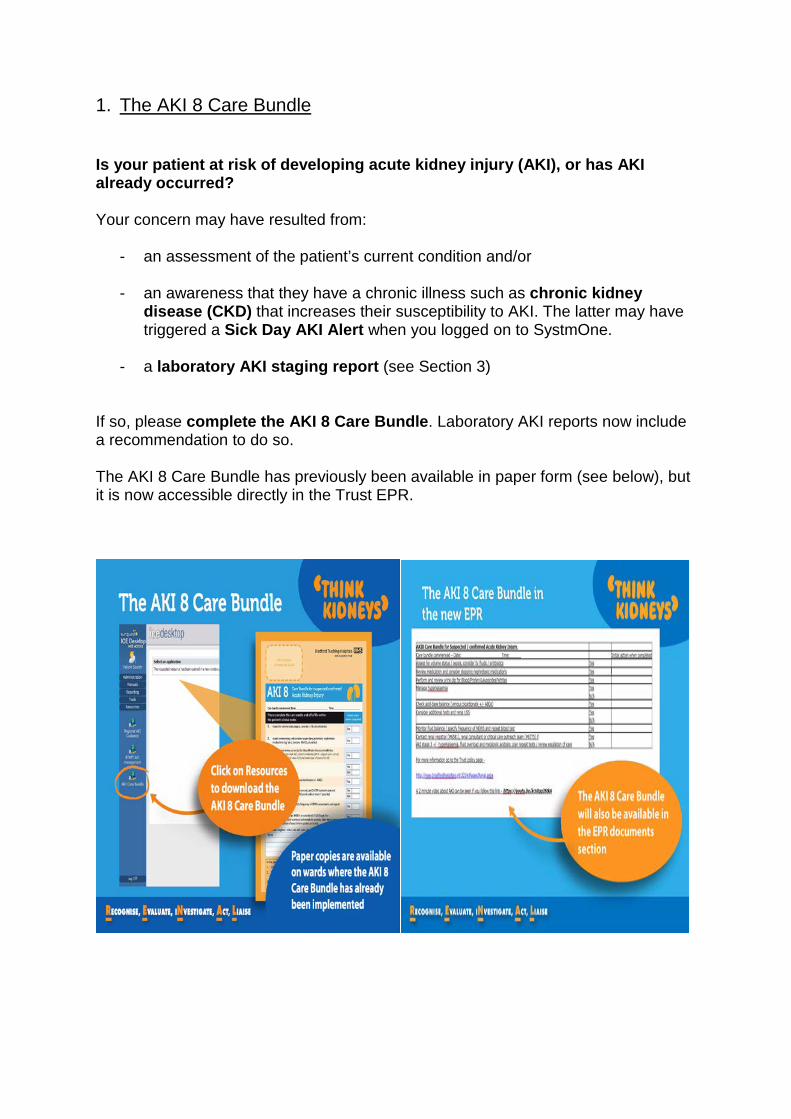
If so, please complete the AKI 8 Care Bundle. Laboratory AKI reports now include a recommendation to do so. The AKI 8 Care Bundle has previously been available in paper form (see below), but it is now accessible directly in the Trust EPR.

There are two ways to access the AKI 8 Care Bundle in the Trust EPR:
Option 1 Select the Pre-configured document AKI 8 Care Bundle BTHFT in Documentation
Within the document, add your initials to completed sections and then click ‘Save’.
If some sections are not yet ready for completion (eg urinalysis, pending a urine sample) an Addendum note can be added at a later stage.

Option 2 Use Clinician Workflow:
1. Navigate to Clinician Workflow and select the appropriate proforma (eg Clerking, Post Take, Progress)
2. In the appropriate section of Clinician Workflow, type ,AKI8 (with a comma first). From the menu that pops up double click on ,AKI8BTHFT.
3. The following then appears on the screen
Note that if you are assessing the patient in the ED, the same applies but type .AKI8 (with a full stop first) for the ED version and then double click on .AKI8BTHFT
As you can see, the AKI 8 Care Bundle comprises 8 elements that support early assessment and management, each element requiring confirmation that it has been reviewed.

2. The Critical Care Outreach Nurse team
We have recently introduced an AKI Nurse role within the Critical Care Outreach (CCOR) Team. As well the CCOR team providing better first line AKI management for acutely deteriorating patients, there have been opportunities for the team to promote AKI prevention through nurse-led education (for patients, fellow CCOR team members and MDT ward staff who care for patients with AKI or risk factors for developing AKI). The key role of the CCOR AKI nurse is illustrated in the schematic below. It complements a set of recent IT initiatives to improve AKI prevention, detection and early management. We have also introduced and refined a twice daily report of all new cases of AKI stages 2 and 3 within the Trust, which is collected by the CCOR AKI nurse and used as a prompt to ensure that all patients with AKI stages 2 and 3 have a completed AKI 8 Care Bundle and a linked AKI management plan.
3. Overview of AKI
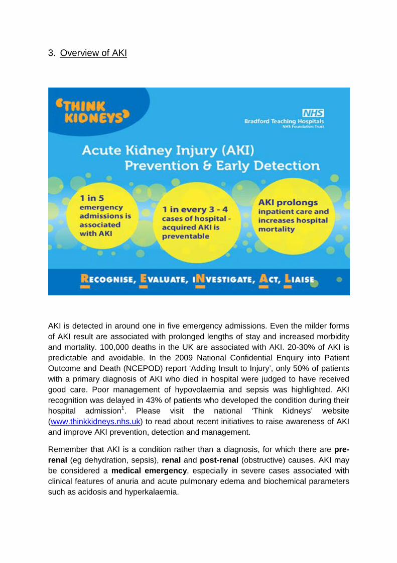
AKI is detected in around one in five emergency admissions. Even the milder forms of AKI result are associated with prolonged lengths of stay and increased morbidity and mortality. 100,000 deaths in the UK are associated with AKI. 20-30% of AKI is predictable and avoidable. In the 2009 National Confidential Enquiry into Patient Outcome and Death (NCEPOD) report ‘Adding Insult to Injury’, only 50% of patients with a primary diagnosis of AKI who died in hospital were judged to have received good care. Poor management of hypovolaemia and sepsis was highlighted. AKI recognition was delayed in 43% of patients who developed the condition during their hospital admission1. Please visit the national ‘Think Kidneys’ website (www.thinkkidneys.nhs.uk) to read about recent initiatives to raise awareness of AKI and improve AKI prevention, detection and management.
Remember that AKI is a condition rather than a diagnosis, for which there are pre-renal (eg dehydration, sepsis), renal and post-renal (obstructive) causes. AKI may be considered a medical emergency, especially in severe cases associated with clinical features of anuria and acute pulmonary edema and biochemical parameters such as acidosis and hyperkalaemia.
Definition of AKI
AKI is diagnosed when one of the following is present.
1. A rise in creatinine >26µmol/l from baseline in 48 hours or 2. A rise in creatinine to >1.5x baseline known or presumed to have occurred
within one week or 3. Urine output <0.5ml/kg/hr for >6 hours
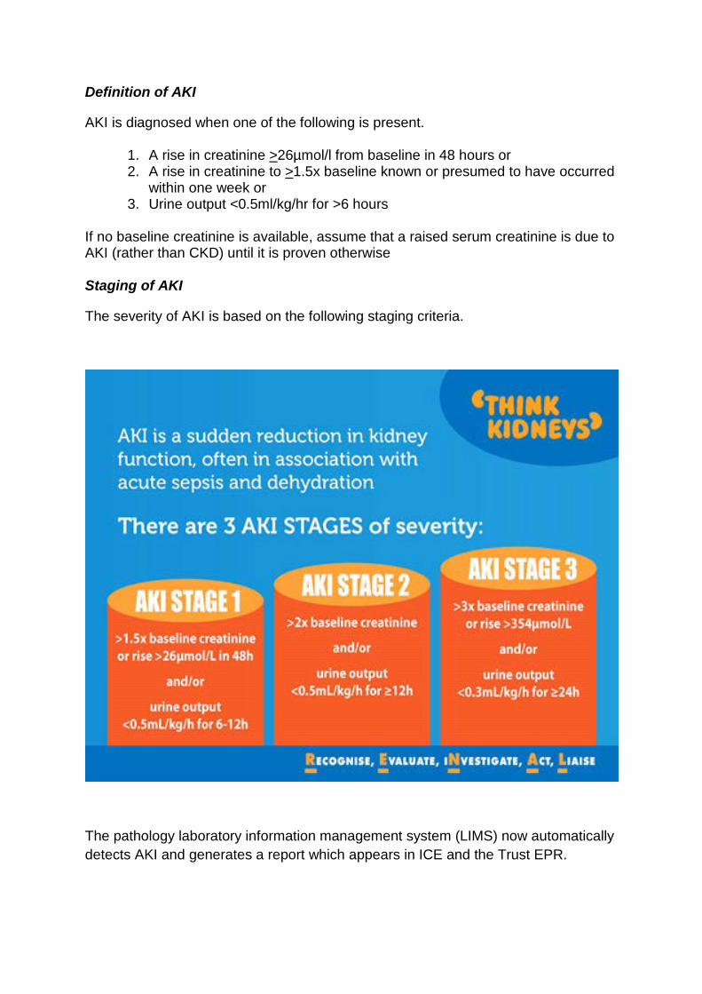
If no baseline creatinine is available, assume that a raised serum creatinine is due to AKI (rather than CKD) until it is proven otherwise Staging of AKI
The severity of AKI is based on the following staging criteria.
The pathology laboratory information management system (LIMS) now automatically detects AKI and generates a report which appears in ICE and the Trust EPR.
4. Prevention of AKI
Below is a more detailed guide to reducing the risk of AKI or AKI progression in emergency admissions mainly based on the recommendations from the 2009 NCEPOD report into the care of patients who died with a diagnosis of AKI.
A. Sick Day Advice This is summarised in the next section of the guideline2,3.
B. All medical and surgical emergency admissions should have their U&Es checked at the time of admission.
C. All medical and surgical emergency admissions should undergo a risk
assessment for AKI (Ref: NICE AKI Guidelines 2013) Risk factors for AKI: Chronic Kidney disease
Age >75 Sepsis Cardiac failure Liver disease Diabetes Drugs affecting renal function Hypovolaemia Peripheral vascular disease Previous h/o AKI
If present these MUST be documented in the medical record.
D. When one or more risk factors for AKI are present clinicians should undertake regular monitoring of U&Es Review pre-existing drugs that may affect renal function (eg NSAIDs, ACE/ARBs, diuretics) and generally withhold until there has been a senior review of the patient. If drugs that could affect renal function are to be prescribed this should be justified in the clinical record
E. NCEPOD recommend that all emergency admissions should have urinalysis
performed and recorded.
F. In addition to the above measures, cases of AKI will be avoided by adherence to good quality basic clinical care. Therefore all emergency admissions should have
• A documented plan for physiological observations based on the use of the NEWS algorithm
• Appropriate escalation of care according to the NEWS algorithm • Discussion with critical care services where appropriate and in
accordance with the NICE clinical guideline 50, acutely ill patients in hospital4.
G. Medication review Great care is needed when prescribing in patients at risk of AKI. It is often safer to withhold medication if you have any uncertainty and you must never simply transcribe a repeat prescription into the inpatient drug chart without careful consideration of each drug. The NPSA report ‘reducing harm from omitted and delayed medicines in hospital’ gives useful guidance on medicines that should not be delayed5.
Take special care and generally withhold the following ACE inhibitors (ramipril/lisinopril etc) and ARBs (losartan/irbesartan etc) NSAIDS Other antihypertensives Diuretics especially if potassium sparing (spironolactone, amiloride)
Metformin Gentamicin In addition a number of important medications can lead to cumulative toxicity in AKI and dose adjustments are required. Important examples are Penicillins Cephalosporins Vancomycin Opiates Gabapentin
Aciclovir Low molecular weight heparins (there is a BTHFT protocol for LMWH in renal disease on the renal unit intranet pages)
IF IN DOUBT ASK A PHARMACIST FOR ADVICE There is a guide to medicines optimisation in AKI available in the Resources section of ICE and on the Trust Intranet. Medicines Optimisation in AKI You can also access the on line version of the Renal Drug Handbook https://www.renaldrugdatabase.com/ Username: [email protected] Password: renalbri This gives straightforward prescribing advice for nearly 800 different drugs at different levels of renal function.
H. Risk of Contrast Nephropathy
Patients with risk factors for AKI will be at high risk of contrast induced nephropathy so you must think very carefully about radiological examinations involving contrast. The latest Trust guidelines can be accessed at http://nww.bradfordhospitals.int/departments/Radiology/Pages/ProtocolsGuidelinesandUsefulTools.aspx
Patients with risk factors for AKI or abnormal U&Es on admission must have their U&Es repeated at regular intervals throughout their admission and especially when significant clinical events occur. Patients transferring away from admitting units are especially vulnerable and arrangements must be made for follow up blood tests on handover to the receiving ward.

5. Detection of AKI
It may be possible to identify incipient AKI and so intervene before AKI has developed. The ACUTE risk factors for AKI shown on the right side of the figure below are potentially avoidable. It is also important to recognise the CHRONIC risk factors on the left side of the figure which increase the likelihood of a patient developing AKI.
To help with detecting patients who are most at risk of developing AKI, we have developed an Sick Day algorithm in System1 which advises healthcare staff in primary and secondary care to review medication (such as ACE inhibitors, Angiotensin II receptor blockers, diuretics, other antihypertensives and NSAIDs) and consider reversible elements such as dehydration and sepsis for patients with coded risk factors for AKI who present with a significant intercurrent illness.


AKI will be confirmed by blood tests that are taken when investigating a patient’s initial presentation or when there has been deterioration in his/ her clinical condition during the hospital admission period.
A national laboratory algorithm automatically detects a significant increase in a patient’s serum creatinine. An AKI alert message appears in ICE and EPR alongside the creatinine result. This gives the AKI stage and now also includes a recommendation to complete the AKI 8 Care Bundle (see below).


6. Essential Management of AKI The cornerstone of initial AKI management is completion of the tasks listed in the BTHT AKI8 Care Bundle. Subsequent management of AKI should be according to the NICE AKI Quality Standards document6.
A. Assessment of AKI Patients
1. History Must include an assessment and documentation of AKI risk factors (see below) Medication history Systemic symptoms eg fever, weight loss, joint pains, haemoptysis, nasal stuffiness Loin pain, haematuria or other urological features
2. Examination The most important part of the physical examination is an assessment of volume status: Pulse rate Blood pressure (look for a postural drop and compare BP with the patient’s usual as there may be relative hypotension) Skin turgor and capillary refill, JVP Oedema (pulmonary or peripheral-remember sacral) Fluid balance and weight charts Other features on examination Bladder/ prostate enlargement Signs of vasculitis- skin rash, arthropathy, uveitis Urinalysis is important- the absence of significant haematoproteinuria virtually excludes glomerulonephritis while the presence of leucocytes and nitrites suggests urinary tract infection (there is a prompt in the AKI8 Care Bundle to record this in the EPR)
3. Physiological observations Record a plan for these and speak to the nurses Fluid balance chart with daily input and output totals Daily weight Postural BP NEWS scoring Early nutritional assessment (MUST assessment)

4. Investigations Baseline investigations FBC and film, U&E (including bicarbonate), LFTs, bone profile including phosphate Urine culture Urine PCR if protein present on urinalysis Ultrasound Urgent USS if obstruction suspected
Within 24 hours in all cases (unless rapid recovery of renal function already in process)
CXR (to exclude pulmonary oedema and as part of sepsis screen) Blood cultures- if sepsis is suspected Other investigations to consider Creatinine kinase (if rhabdomyolysis suspected eg trauma, elderly patients after falls, long lies) CRP (?sepsis) Myeloma screen (clues age>50, calcium, back pain, high total protein) (Serum Free Light Chains plus Serum Electrophoresis in Bradford) Blood film, Lactate dehydrogenase (LDH) – Hemolytic uraemic syndrome (HUS)/ atypical HUS/ thrombotic microangiopathy (TMA) Renal Immunology tests (unlikely to be positive if no haematoproteinuria) Usual tests would be ANA, ANCA, anti-GBM, complement, immunoglobulins
If you are thinking of carrying out these tests you should be discussing the case with the renal team and you should always liaise with the lab to request that they be processed urgently (within 24 hours) if this is a case of AKI (telephone the immunology laboratory in Leeds 0113 3922587 and the BRI laboratory reception to request urgent transport)
Virology Hepatitis B/C, HIV screen (if emergency dialysis may be required)
B. Treatment of AKI TREAT THE UNDERLYING CAUSE Often but not always the cause is a combination of sepsis and relative hypovolaemia and will respond to resuscitation and antibiotics. Therefore the priority is
1. Identify and treat the underlying cause 2. Assess and manage the volume status
If you suspect sepsis or your patient has a NEWS score of 5 or more you must use the trust sepsis screening tool and complete the Sepsis 6 (BUFALO). http://nww.bradfordhospitals.int:2234/Pages/Acute%20Medicine%20Pathways.aspx and assess for the Systemic Inflammatory Response Syndrome (SIRS). There are

other useful resources available for acute management of suspected sepsis such as the Royal College of Physicians Acute care Toolkit for Sepsis7. If you suspect hypovolaemia: Fluid challenge with a stat bolus of 500ml crystalloid (0.9% NaCL is fine initially) and assess response (caution in elderly and cardiac patients, consider 250ml bolus) Response can be judged by Peripheral perfusion Pulse (falling if tachycardia) Blood pressure (postural) JVP Urine output REASSESS FOR PRESENCE OF PULMONARY OEDEMA If no response and no pulmonary oedema repeat 500ml bolus and discuss with senior member of team. If patient responding to fluid challenge continue intravenous fluids and discuss further management with a senior member of the team. Volume unresponsive AKI Oliguria (<0.3ml/kg/hr) despite adequate volume resuscitation means that the patient may be at risk of pulmonary oedema. Critical Care Team involvement and intensive monitoring would be appropriate in these circumstances.

7. Complications of AKI Hyperkalaemia
Metabolic acidosis
Pulmonary oedema
Uraemic syndrome Pericarditis or pleurisy Encephalopathy Neuropathy
A. Hyperkalaemia This is an emergency if ECG changes are present or if serum K >6.5. Needs cardiac monitoring. Remember that unless you remove the potassium from the body permanently (dialysis or restored kidney function) these measures are only temporary and may need to be repeated. The latest Trust guideline is available at http://nww.bradfordhospitals.int/policies-and-clinical-guidance/Clinical%20Guidelines/Trust%20Wide%20Clinical%20Guidelines/Guideline%20for%20Treatment%20of%20Hyperkalaemia%20in%20adults.pdf
1. Cardiac protection 10ml 10% calcium gluconate over 2-5minutes with cardiac monitoring. Onset (reversal of ECG changes) within 2-4 minutes, effect lasts for up to 1 hour but dose can be repeated at 5 minute intervals if ECG changes persist up to 4 times. This has no effect on serum potassium.
2. Increase movement of K+ into cells 10units of short acting insulin (actrapid) in 50ml of 50% dextrose via large bore cannula over 20minutes. Onset within 15-30 minutes and lasts for 4-6 hours. Risk of hypoglycaemia so monitor capillary blood glucose. 10-20mg salbutamol nebuliser. This does the same as insulin but less well tolerated as higher dose than normal of salbutamol is required to be effective. Correction of acidosis if present (venous bicarbonate <16). Consider 200-500ml of 1.26% or 1.4% sodium bicarbonate over 15-60 minutes if volume status permits. Risk of hypocalcaemia if acidosis corrected quickly. In a cardiac arrest situation attributed to hyperkalaemia consider 50ml 8.4% bicarbonate or 50-100ml 4.2% for rapid onset.
3. Remove potassium from the body If a urine output can be restored loop diuretics may increase potassium excretion but be aware that they can be nephrotoxic especially if hypovolaemia is present. Cation exchange resins (eg resonium) take up to 24 hours to be effective and are not relevant to the management of AKI.

B. Metabolic Acidosis
Management of metabolic acidosis in AKI remains an area of controversy and in general the best approaches to acidosis are either the restoration of renal function or dialysis. In the more stable patient not imminently requiring dialysis 1.26% or 1.4% sodium bicarbonate can be used as part of the volume replacement regime if there is acidosis but be aware that this will deliver a high sodium load and can worsen hypocalcaemia (check the Ca before using bicarbonate). Discuss this with a senior member of the team, if the pH is >7.2 there is very little evidence to support correction. A patient with severe acidosis and volume overload needs dialysis and a patient with pH < 7.15 needs discussion with critical care.
C. Pulmonary oedema Do not neglect basic measures, sit the patient up and use high flow oxygen. NIV may have a role. Sub-lingual GTN 2-5mg may be helpful and has a rapid onset. Intravenous GTN can be used titrating against systolic blood pressure Intravenous furosemide may be helpful and you need to use large doses (up to 160mg) in AKI but remember this is a treatment for the associated pulmonary oedema and diuretics have no role in treating or preventing the underlying AKI and can be harmful. Dialysis should not be delayed in the presence of pulmonary oedema.
D. Encephalopathy and uraemic pericarditis are indications for urgent dialysis

8. Referral to the Renal Team Not all AKI patients need to be referred to the renal team but in all cases you should use this guideline and you must always seek a more senior opinion if you are unsure of how to proceed with a case. See also the NICE AKI Quality Standard8. The following cases should be discussed with the renal team:
1. Suspicion for primary renal disease a. All AKI with ++blood and protein on dipstick b. Known myeloma c. Haemolysis features on blood film or bloody diarrheal illness with AKI d. Suspected rhabdomyolysis e. Suspicion for systemic vasculitis or connective tissue disease such as
lupus f. Haemoptysis- pulmonary haemorrhage g. Known CKD stage 4/5- previous nephrology follow-up h. AKI with no clear cause
2. AKI 3 at presentation or progression to AKI 3 despite supportive treatment 3. Refractory complications of AKI suggesting that dialysis is indicated
a. Hyperkalaemia b. Acidosis c. Pulmonary oedema d. Pericarditis e. Encephalopathy
Contacting the renal team in Bradford
1. Monday-Friday 9-5 During these hours you can usually contact the renal registrar using the on call mobile phone #6581 or via switchboard. If no registrar available then contact the on call consultant. Switchboard, ward 15 (3237) or the BRI dialysis unit (2143 or 3254) should all know who this is.
2. Overnight and at weekends contact the on call renal consultant. 3. If you require URGENT ADVICE it is ESSENTIAL to TELEPHONE the
RENAL ST or CONSULTANT

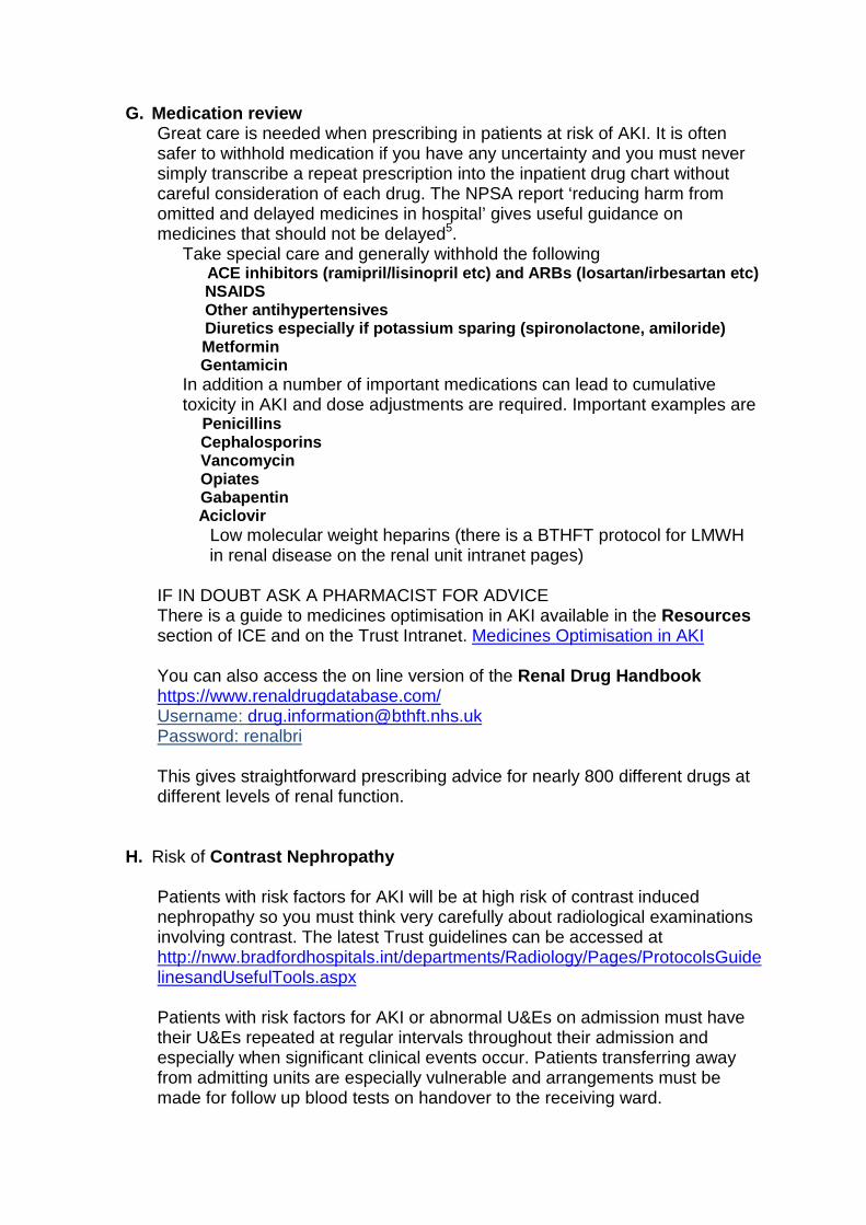
9. Recovery from AKI and Follow-Up The first signs of recovery from AKI may be an increase in urine output or a decrease in the daily rate of rise of serum creatinine which may then plateau and fall. The main issue to manage as patients recover from AKI is fluid balance as there will often be a polyuric phase when an inappropriately large volume of urine will be produced. Patients will often need intravenous fluid to maintain positive fluid balance and there is a risk of electrolyte disturbances and neurological sequelae. In addition dehydration is likely to halt the process of renal recovery. Follow up of AKI patients Patients who recover from AKI are at risk of developing chronic kidney disease (CKD). Renal function must be monitored up to the point of discharge from hospital and robust arrangements need to be made for follow up and monitoring post-discharge either in outpatients or via the GP. An AKI Virtual Clinic is currently being evaluated. In addition multiple medications have often been withdrawn during the acute episode and you need to give guidance to patient and GP about reintroduction of some or all of the medications if appropriate. If not the patient may end up with no medication or may collect an inappropriate repeat prescription that pre-dates the AKI episode. You need to describe all medication changes in the discharge summary together with any follow up arrangements or repeat blood tests. A senior doctor must be involved in the discharge plan for AKI patients. Clear discharge communication is essential and you must involve the patient in this. It is an NHS England requirement for AKI and the stage of AKI to be clearly mentioned in the discharge summary for all cases no matter which clinical area they are discharged from. To help with this, we have included a AKI prompt in the EPR discharge summary, as shown overleaf. It is good practice to inform the patient of their risk factors for AKI as they may then be able to play a role in reducing the risk of future episodes for example omitting ACE inhibitors in the presence of an intercurrent illness (sick day advice- see below). You should also make sure that all patients have a copy of the Bradford Hospitals AKI Patient Information leaflet which you can personalise with additional information on the back page regarding medication changes and follow up arrangements.
References
1. NCEPOD. Adding insult to injury – A review of care of patients who died in
hospital with a primary diagnosis of acute kidney injury (acute renal failure).
The National Confidential Enquiry into Patient Outcome and Death. 2009;
http://www.ncepod.org.uk/2009aki.htm (C)
2. Sick day rules in kidney disease. Drug and Therapeutics Bulletin, Vol 53. No4.
2015
3. Records Shared Nephrons Spared https://www.thinkkidneys.nhs.uk/kquip/wp-
content/uploads/sites/5/2016/11/Bradford-Teaching-Hospitals-NHS-FT-V3-
1.pdf
4. National Institute for Health and Clinical Excellence (2007) Acutely ill patients
in hospital. NICE clinical guideline 50. www.nice.org.uk
5. National Patient Safety Agency (2010) Reducing harm from omitted and
delayed medicines in hospital. NPSA/2010/RRR009.
www.nrls.npsa.nhs.uk/alerts/
6. National Institute for Health and Clinical Excellence (2014) Quality Standard
for Acute Kidney Injury https://www.nice.org.uk/guidance/qs76
7. https://www.rcplondon.ac.uk/resources/acute-care-toolkit-9-sepsis
8. Acute Kidney injury AKI quality standards (2014)
https://www.nice.org.uk/guidance/qs76/chapter/Quality-statement-5-
Discussion-with-a-nephrologist
Dr Russell Roberts and Dr John Stoves, with additional commentary from Dr Mansoor Ali
March 2019
Review March 2022
Additional links:
A 2-minute Powtoon AKI education video for the MDT https://www.youtube.com/watch?v=kcHtqq3StkA&rel=0