Embed Size (px)
Citation preview

1
GUIDELINE FOR THE DIAGNOSIS AND MANAGEMENT OF PATIENTS WITH
PERIPHERAL ARTERIAL DISEASE (PAD)
Title: Guidelines for the diagnosis and management of patients with
peripheral arterial disease (PAD) – resource notes Authors: Network Vascular Advisory Group (VAG) Contact Details: Professor G Stansby, VAG Chair E-mail [email protected]
Version History: Date: January
2014 Version: v1.0 Review Date: January 2016

2
ACKNOWLEDGMENT
Acknowledgement goes to Dr Jane Skinner, Consultant Community Cardiologist, Graeme Young, Advanced Podiatrist and Sue Long, Network Education and Training Lead for their contribution to these guidelines.

3
DIAGNOSIS AND MANAGEMENT OF PATIENTS WITH PERIPHERAL ARTERIAL DISEASE (PAD)
This guideline was originally developed in 2005 from the Newcastle, Northumberland and North Tyneside PAD guideline. The guideline was then adopted by the North of England Cardiovascular Network Vascular Advisory Group and subsequently updated in 2011. This document has now been updated in line with the NICE PAD clinical guideline published in August 20121.
In addition, in January 2014 NICE published PAD Quality Standard 522 which includes five quality statements covering the diagnosis and management of lower limb peripheral arterial disease, a list of these quality standards are included in Appendix 1.
Background:
Patients with PAD are at increased risk of coronary and cerebrovascular events, and more than half of patients with PAD have significant co-existing coronary disease. The commonest presentation of PAD is pain on walking termed intermittent claudication (IC). The management of IC can be divided into two strategies which are not mutually exclusive: reducing the patient’s cardiovascular risk and strategies designed to improve walking distances. All patients with suspected PAD should have an appropriate assessment and accurate diagnosis. Those with confirmed disease require appropriate management, including measures aimed at reducing cardiovascular risk, improving symptoms and making progression of disease less likely. Those with critical ischaemia (rest pain, necrosis or gangrene) are at high risk of progressing to amputation and need urgent/emergency referral for a vascular surgical opinion. Patients with PAD are at increased risk of having other forms of occlusive vascular disease (coronary heart disease, cerebro-vascular disease) and if they have symptoms of these require appropriate assessment and management. This network guideline is intended for all clinicians across the Northern Clinical Network involved in the diagnosis and management of patients with PAD. It does not include detailed recommendations for the management of patients seen in vascular surgical clinics, but does make recommendations to ensure care is effectively integrated between primary and secondary care.
This guideline makes recommendations for the diagnosis and management of PAD. The interventions should be offered to all people who are likely to benefit, irrespective of race, disability, gender, age, sexual orientation or religion. Information should be provided to patients in an accessible format and consideration should be given to mobility and communication issues, and being aware of sensitive and cultural issues.
1National Institute for Health and Clinical Excellence (2012) NICE clinical guideline 147. Lower limb peripheral arterial disease: diagnosis and management. NICE. http://guidance.nice.org.uk/CG147. Accessed January 2014. 2 National Institute for Health and Clinical Excellence (2014), Quality Standard 52, Peripheral Arterial Disease -
http://publications.nice.org.uk/peripheral-arterial-disease-qs52
4
HOW TO USE THE GUIDELINE
The guideline has two parts; a summary (Appendix 2) which can be folded to be double sided A5, easily laminated and kept readily available, and this set of supporting resource notes. The notes contain each of the summary statements with some background detail and, in some cases, expands the summary statement providing more detailed guidance. Clinicians should be familiar with these and use them to refer to for further clarification of management in individual patients as needed.
Where appropriate referral is made to other local guidelines and these are not duplicated here. Prescribers should review patients for any contra-indications before initiating drugs. The drugs are recommended assuming there are no contra-indications to treatment, and no contra-indications develop.
PATIENTS WITH SUSPECTED PAD Assess patients for the presence of PAD if they:
Have symptoms suggestive of PAD Or have diabetes, non-healing wounds on the leg Or are being considered for interventions to the leg or foot Or need to use compression hosiery
These should be asked routinely and are based on the Edinburgh Claudication Questionnaire:
Which leg (right, left or both)?
Does the pain only occur on walking? If no, it is less likely the patient has intermittent claudication (IC)
Does the pain go away on stopping? If yes, it is more likely the patient has IC
Is the pain worse going uphill or walking fast? If yes, it is more likely the patient has IC.
Does the patient have any associated numbness or weakness? If so, it might suggest spinal claudication. If so, enquire about back symptoms.
Where does the patient feel the pain? Pain from IC will be in a muscle group, most often
Ask about Symptoms:
Which leg?
Pain only on walking?
Pain resolves with rest?
Pain worse walking up hill quickly?
Any associated numbness / pain?
Location of pain? Symptoms / history other vascular disease.

5
the calf, sometimes the thigh or buttock.
Initial assessment will identify those with who need urgent/ emergency referral (urgent letter/fax, phone the on-call vascular team for advice, emergency admission, depending on severity). Those with acute ischaemia require emergency admission without delay, many patients with necrosis also require urgent admission (if in doubt discuss with the on call vascular team). Those with rest pain require urgent referral.
Peripheral pulses should be assessed and documented in all patients (see Appendix 3). However, the presence of pulses does not exclude the diagnosis of PAD.
Patients may have signs of other vascular disease, and occasionally of vascular risk factors (eg xanthelasmata, xanthomata). The presence of other vascular disease or the presence of multiple vascular risk factors makes it more likely the patient will have PAD.
PAD is an important diagnosis to make, is associated with an adverse prognosis, and patients require lifelong treatment and regular review. All patients with suspected PAD should have the diagnosis confirmed with measurement of the ankle brachial index (ABI), unless co-morbidity makes this inappropriate or the patient declines. It is anticipated that all GP practices will have access to an appropriately trained nurse competent in ABI assessment and the ABI should be taken by an appropriately trained individual and where there is doubt it should be re-checked3.
Patients who have been referred for assessment by the vascular surgeons will have ABI measurements as part of the hospital assessment.
If patients have mainly sensory symptoms, particularly if bilateral, spinal claudication is more likely (however, many patients with spinal claudication have co-existent vascular disease). Where doubt exists referral to the vascular surgeons for investigation as indicated.
ASSESSMENT OF ALL PATIENTS WITH CONFIRMED PAD
3 North of England Clinical Network. A Guide to the measurement of the ankle/brachial pressure index (ABI).
Examination:
Necrosis / critical ischaemia – refer
Peripheral pulses?
Signs other cardiovascular disease?
Check ABI (see ABI guide)
Leg ulcer? – refer to local guidelines
Ask about severity of symptoms; Distance able to walk, effect on quality of life, any threat to employment

6
Patients with confirmed PAD should be asked about their symptoms. It is important to establish the impact of patients symptoms on their quality of life as well as the distance a patient can walk before developing IC.
About 75% of patients will have other forms of vascular disease. This should be identified and appropriately managed.
Lifestyle risk factors should be identified, blood pressure checked and those with known diabetes identified and managed in line with local guidelines.
FBC; exclude anaemia, polycythaemia, thrombocythaemia
U&E; exclude chronic kidney disease (CKD). Patients are at risk of reno-vascular disease (if CKD, refer to local guidelines)
Glucose; can be a random sample, but those with a raised level will require a fasting sample taken to exclude diabetes or impaired fasting glycaemia
Lipids; in with local guidelines
LFTs; in line with local guidelines
TFTs; in line with local guidelines
MANAGEMENT OF PATIENTS WITH CONFIRMED PAD
Consistent Read coding in each practice will facilitate audit. Read codes for PAD are G73, Xa01v (systemone). Read code for ABI is Xalup.
All practices should have an accurate and regularly maintained disease register which is used to ensure patients are offered an annual review.
Check for symptoms/signs other cardiovascular disease
Identify lifestyle risk factors (smoking, exercise, diet, alcohol, BMI), blood pressure, known diabetes
FBC, U&E, glucose, lipids, LFT, TFT at baseline. Annual U&E, lipids, glucose. FBC if worsening symptoms or anaemia suspected
Ensure on disease register; Read codes G73, Xa01v
Arrange annual review

7
All patients should be offered oral and written information about their condition. This should include:
The causes of their symptoms and the severity of their disease
The risk of limb loss and cardiovascular events
The key modifiable risk factors including:
Smoking
Lipid modification
Control of diabetes
Weight and diet
Exercise As well as advice about risk factors they should be encouraged to set appropriate goals to address them. Those making changes to their lifestyle may need additional review for support and monitoring. Referral to other services may be appropriate e.g. stop smoking, weight management, exercise referral. Some patients may find that activities such as swimming, cycling allow them to achieve and maintain a higher level of fitness than walking, and programmes of supervised exercise have been shown to increase the distance that patients are able to walk before developing IC. Such a programme will typically involve 2 hours of supervised exercise a week for a 3-month period.
All patients should have lipids assessed and monitored in line with local guidelines. Patients with PAD have established atheromatous disease and should be managed as having occlusive vascular disease.
All patients should have blood pressure measured and hypertension managed in line with the local hypertension guidelines. Beta blockers are not specifically contra-indicated in PAD. Particular attention should be made to monitoring renal function in those treated with an ACE inhibitor in view of the increased risk of reno-vascular disease.
Lifestyle vascular risk factors; Smoking (stop smoking service referral if motivated to quit) Healthy eating, alcohol safe limits Regular physical activity (offer referral for supervised exercise) Weight reduction if appropriate
Lipids – refer to local guidelines
Hypertension – refer to local hypertension guidelines

8
In general patients with critical ischaemia should have further assessment of the PAD before high blood pressure is lowered. If high blood pressure is felt to need immediate treatment in such patients, this should be discussed with the vascular surgeons first.
Patients with established diabetes or those in whom the diagnosis is made during the baseline assessment should be managed in line with local guidelines. Foot care is particularly important.
All patients should take anti-platelet agents. In line with the NICE TAG 2104, clopidogrel is first line and aspirin second line5.
•
All patients with IC should have a therapeutic management plan which includes intensive lifestyle measures. A programme of regular physical activity should be discussed with the patient and goals set, with the aim of gradually increasing their claudication distance. Advice should be given that patients should aim for activity levels which lead to claudication pain, walking through the pain as far as possible (unlike patients with angina who are advised to stop / slow down if they develop angina). Supervised exercise programmes have been shown to increase the distance patients can walk before developing IC and have been shown to be superior to advice to exercise alone. Referral to an exercise programme should be offered to appropriate patients. Weight loss in those who are obese or overweight is also likely to improve symptoms. Stopping smoking will not improve symptoms of claudication, but will substantially reduce cardio-vascular risk, the chances of disease progression, and the chances of success from any revascularisastion procedure.
4 Available at http://www.nice.org.uk/nicemedia/live/13285/52030/52030.pdf
5 Surgery and invasive procedures in patients treated with anti-platelet agents. In general clopidogrel will be stopped for 10
days before and for the duration of an elective procedure, and aspirin is continued or substituted. In patients treated with
clopidogrel alone and who are more than 3 months following an acute event (eg acute ischaemic stroke or TIA), clopidogrel
can be temporarily withheld with minimal risk. However, in all patients treated with dual anti-platelet agents (aspirin in
combination with either clopidogrel or prasugrel) or if there is concern about the risks of withholding clopidogrel, any
proposed modification to the anti-platelet regime should be discussed in advance with the interventional cardiologist,
vascular surgeon or other clinician who initiated the treatment.
Optimal diabetes control – refer to local guidelines
Anti-platelet agents – clopidogrel first line
Therapeutic interventions; Regular physical activity – offer supervised exercise Specific drug therapy- only after full conservative treatment for 6 mths+, other therapeutic interventions not possible/declined Naftidrofuryl oxalate – only after at least 6 months of intensive conservative management
9
Specific drug therapy should only be considered in those with persistent significant limitation in activity which is leading to a reduced quality of life and or threatened employment. Patients should have had at least 6 months of intensive conservative management and failed to improve despite a supervised exercise programme and been considered for other therapeutic interventions (unless declined).
Following publication of the NICE TAG 2236, only naftidrofuryl oxalate is recommended. Cilostazol is no longer recommended In line with the NICE TAG 223, those already treated with cilostazol should have the option to continue treatment until they and their clinicians consider it appropriate to stop.
All patients treated with naftidrofuryl oxalate should be assessed 3 months after initiation and treatment only continued if there has been a clear improvement in symptoms.
Patients and their carers as appropriate should be provided with appropriate information about the nature of the disease, possible interventions and risk factor management. This should include the importance of appropriate foot care. Written information including a factfile on PAD has been developed by the British Heart Foundation (http://www.bhf.org.uk) and the Circulation Foundation (http://www.circulationfoundation.org.uk/).
Patients with definite or possible critical ischaemia need urgent/emergency specialist referral (urgent letter/fax, phone the on-call vascular team for advice, emergency admission, depending on severity). If there are signs of cellulitis, swabs should be taken and the patient treated with antibiotics at the same time as referral is considered. Staph. Aureus is a likely pathogen and flucloxacillin (at least 500mg qds) should be considered as first line (refer to the local primary care antibiotic guidelines). In patients with infected necrotic tissue or gangrene anaerobes are likely but such patients will usually require admission and should be discussed with the on call vascular team. Patients who are pyrexial and or have other signs of systemic upset should be admitted.
Patients with symptoms significantly impairing their Quality of Life, should be referred for vascular surgical assessment. Generally such patients should have had 3-6 months of intensive conservative management, including exercise. However, those with more severe symptoms or whose employment is threatened should be referred earlier. Patients with absent femoral pulses and more proximal disease are more likely to be suitable for
6 Available at http://guidance.nice.org.uk/TA223/Guidance/pdf/English
Identification of patients for specialist referral Critical ischaemia (rest pain, necrosis, gangrene) – urgent/emergency, Claudication significantly impairing Quality of Life, threatened employment, diagnostic doubt Leg ulcer
Patient / carer information and support, including appropriate foot care

10
revascularisation than those with more distal disease.
Patients without a secure diagnosis should be referred early.
Patients with a leg ulcer should be assessed and managed in line with local leg ulcer guidelines. Approximately 10% of ankle ulcers are due to a combination of venous and arterial disease.
For all patients who are referred, the following minimum information is helpful;
Brief summary of current symptoms. If critical ischaemia is present this should be emphasised.
Baseline assessment including, presence or absence of pulses, results of any baseline biochemical and haematological investigations and ABI measurement.
Previous / current management plan
Current medication
Important past medical history
Appendix 1
11

Appendix 2
12
Appendix 3
13
A Guide to the measurement of the
ankle/brachial pressure index (ABI).
14
Authors Graeme Young. Advanced Podiatrist, Community Resource Team, Campus for Aging and Vitality, Newcastle upon Tyne Hospitals NHS Foundation Trust. Sue Long. Education and Training Lead, North of England Cardiovascular Network Revised/updated (Gerry Stansby) 2014
Consultation Process Diabetic footcare network Tissue Viability Services Vascular Advisory Group

15
Contents
Section Title Page Number
1 Introduction
4
2 Evidence for practice
5
3 Key points for Doppler Use
6
4 Equipment
6
5 Indications
7
6 Contra-indications
7
7 Limitations to practice
7
8 Criteria for competence
8
9 Protocol and skills audit
9
10 Clinical incident reporting and management
9
11 References
10
12 Appendices Appendix 1 - Illustrations Appendix 2 – Report form
Appendix 3 – Competency form Appendix 4 – Case example
Appendix 5 – NECVN Guidelines for Peripheral Arterial Disease
11 13 14 20 21
16
1.0 Introduction • Doppler ultrasound is used to detect velocity and location of blood flow in both arteries and veins and is used to record a Resting Pressure Index (RPI) also known as Ankle Brachial Pressure Index (ABI). • Ankle brachial pressure index (ABI) compares the traditional arm systolic reading with the ankle systolic reading as an assessment of arterial blood flow to the feet. These recordings are expressed as a ratio. Doppler ultrasound is an essential part of the assessment of suspected peripheral arterial disease (PAD). • While an important element of the assessment process it should be used in conjunction with a holistic assessment of the patient. However, the ABI can help to inform the differential diagnosis, can be utilised to exclude arterial disease and to identify those patients with impaired arterial flow who should be referred to the vascular surgeon. Assessment should involve both legs regardless of whether only one limb is affected.

17
2.0 Evidence for practice
ABI is used during assessment of PAD as an objective aid to substantiate the presence or absence of significant disease1. It is essential that there is a consistent approach to undertaking the procedure and a period of supervision by an experienced practitioner is necessary for those new to performing ABI1, 2. The attached competencies outlining the procedure (Appendix 3) provide a framework for a standardised approach to ABI measurement3, 4, 5, while the reporting form (Appendix 2) supports consistent, clear calculation and recording of ABI measurement. Further information on the diagnosis and management of patients with Peripheral Arterial Disease can be found in Appendix 5.
18
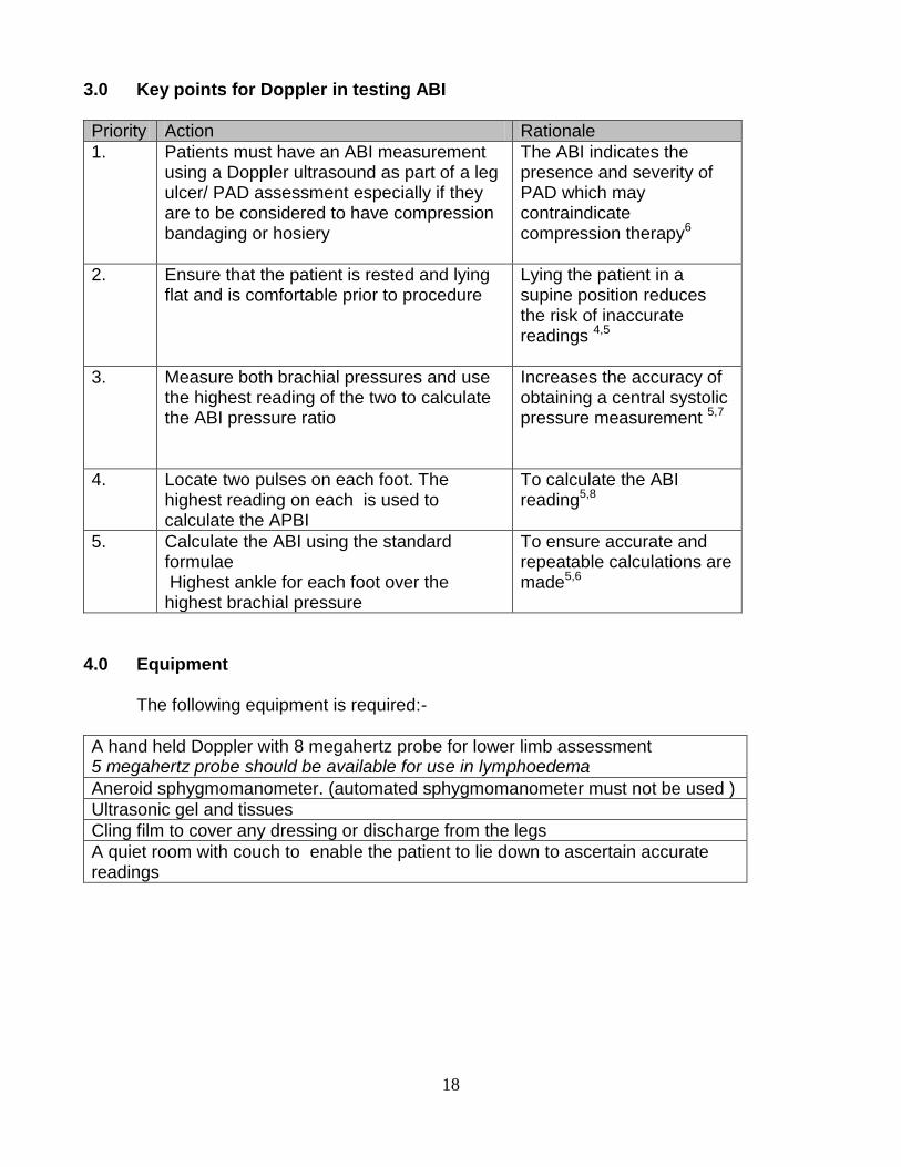
3.0 Key points for Doppler in testing ABI
Priority Action Rationale
1. Patients must have an ABI measurement using a Doppler ultrasound as part of a leg ulcer/ PAD assessment especially if they are to be considered to have compression bandaging or hosiery
The ABI indicates the presence and severity of PAD which may contraindicate compression therapy6
2. Ensure that the patient is rested and lying flat and is comfortable prior to procedure
Lying the patient in a supine position reduces the risk of inaccurate readings 4,5
3. Measure both brachial pressures and use the highest reading of the two to calculate the ABI pressure ratio
Increases the accuracy of obtaining a central systolic pressure measurement 5,7
4. Locate two pulses on each foot. The highest reading on each is used to calculate the APBI
To calculate the ABI reading5,8
5. Calculate the ABI using the standard formulae Highest ankle for each foot over the highest brachial pressure
To ensure accurate and repeatable calculations are made5,6
4.0 Equipment
The following equipment is required:-
A hand held Doppler with 8 megahertz probe for lower limb assessment 5 megahertz probe should be available for use in lymphoedema
Aneroid sphygmomanometer. (automated sphygmomanometer must not be used )
Ultrasonic gel and tissues
Cling film to cover any dressing or discharge from the legs
A quiet room with couch to enable the patient to lie down to ascertain accurate readings
19
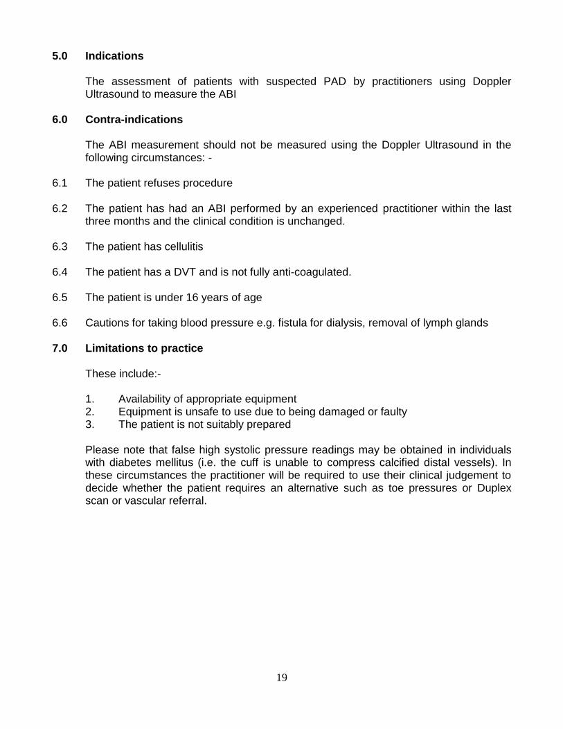
5.0 Indications
The assessment of patients with suspected PAD by practitioners using Doppler Ultrasound to measure the ABI
6.0 Contra-indications
The ABI measurement should not be measured using the Doppler Ultrasound in the following circumstances: -
6.1 The patient refuses procedure
6.2 The patient has had an ABI performed by an experienced practitioner within the last three months and the clinical condition is unchanged.
6.3 The patient has cellulitis
6.4 The patient has a DVT and is not fully anti-coagulated.
6.5 The patient is under 16 years of age
6.6 Cautions for taking blood pressure e.g. fistula for dialysis, removal of lymph glands 7.0 Limitations to practice
These include:-
1. Availability of appropriate equipment 2. Equipment is unsafe to use due to being damaged or faulty 3. The patient is not suitably prepared
Please note that false high systolic pressure readings may be obtained in individuals with diabetes mellitus (i.e. the cuff is unable to compress calcified distal vessels). In these circumstances the practitioner will be required to use their clinical judgement to decide whether the patient requires an alternative such as toe pressures or Duplex scan or vascular referral.

20
8.0 Criteria for competence 8.1 Registered practitioners undertaking ABI measurements will be required to complete a
competency framework or will have undertaken a recognised theoretical course and have evidence to demonstrate they have maintained their practical skills in the measurement of ABI’s.
8.2 The registered practitioner will have a working knowledge of guidelines for the
diagnosis and management of patients with PAD.
8.3 All registered practitioners undertaking this role are responsible for accessing an
appropriately experienced supervisor. Supervisors must be registered practitioners who have a theoretical knowledge and maintain practical skills in the management of PAD and/or leg ulceration.
8.4 The registered practitioner and their supervisor will determine the number of
supervised practices required to achieve competence. This will take into account the registered practitioner’s individual learning needs (appendix 3).
8.5 All registered practitioners undertaking the assessment of ABI will be required to
demonstrate competence in the relevant skills. Please refer to appendix 3. Evidence of competence must be provided and a copy kept in the registered practitioner’s individual file.
8.6 Competence must be maintained by regular clinical experience and supported by
updating of theoretical knowledge. Evidence of continuing professional development and maintenance of skill level will be required.
21
9.0 Skills audit In order to ensure delivery of quality care it is good practice to undertake an audit of compliance to guidelines and skills competencies on a regular basis. The audit may include:
Adherence to the protocol
Any untoward incidents or complaints
Number of competent registered practitioners
Any additional educational needs
10.0 Clinical incident reporting and management
Any untoward incidents and near misses must be reported and dealt with via the appropriate local pathway.

22
11.0 References
1. Scottish Intercollegiate Guidelines Network (SIGN) (2006). SIGN 89. Diagnosis and Management of Peripheral Arterial Disease.
2. Royal College of Nursing (2006) Management of Patients with Venous Leg Ulcers. RCN, London
3. Wirral Primary Care Trust (2008) Procedure for vascular assessment by Doppler ultrasound
4. Vowden K.R., Goulding V., Vowden P.,(1996) Hand-held Doppler assessment for
peripheral arterial disease. Journal of Wound Care Vol 5, No 6. Pp 14 -16 http://www.iocp.org.uk/sites/default/files/peripheral%20arterial%20disease_0.pdf accessed 6.5.11
5. National Institute for Health and Clinical Excellence (2012) NICE clinical guideline
147. Lower limb peripheral arterial disease: diagnosis and management. NICE. http://guidance.nice.org.uk/CG147. Accessed January 2014.
6. Vowden P. & Vowden K. (2001).Doppler assessment and ABPI: Interpretation in
the management of leg ulceration http://www.worldwidewounds.com/2001/march/Vowden/Doppler-assessment-and-ABPI.html accessed 23.5.11
7. Stubbing, N. J., Bailey, P. and Poole, M. (1997) Protocol for accurate assessment of ABPI in patients with leg ulcers. Journal of Wound Care 6 (9): 417-418.
23
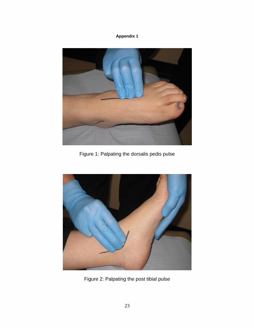
Appendix 1
Figure 1: Palpating the dorsalis pedis pulse
Figure 2: Palpating the post tibial pulse

24
Figure 3: Measuring ankle systolic pressure

25
Appendix 2
Clients Details NHS Number __________________________ Name __________________________
DOB __________________________
Address __________________________
__________________________
Tel __________________________
Indication __________________________ for referral
Interpretation of results
ABI normally 0.9- 1.3 at rest ABI 0.5- 0.89 indicates arterial disease ABI < 0.5 indicates severe arterial disease and requires referral to vascular ABI > 1.3 may indicate calcification of the arteries ABI > 0.8 is considered safe to apply compression therapy
Name of person completing ABI ______________________________ Date of ABI _________________ Contact Number_________________ Reason if unable to complete ABI ________________________________________________________________ ___________________________________________________________
Results RIGHT LEFT
Highest ankle pressure (for this leg)
Divided by
Highest brachial pressure (highest of 2 arm readings)
ABI Reading
REPORT FOR ABI MEASUREMENT
Right Brachial =
Left Brachial =
DP = PT=
DP= PT=

26
Appendix 3
Competency Title: ABI measurement for the assessment of PAD
ACTION
RATIONALE W A and S P
Score Score Score Score Score
Pre checks prior to test
1. Upon receipt of referral check clinical details and that there are no contraindications to the procedure
To ensure it is appropriate to perform the procedure
2. Check equipment is available and is working correctly i.e. no battery warning sign flashing and sphygmanometer works and appropriate cuff available.
To ensure the resources for the procedure are available. To meet the requirements of the Management of Medical Devices Policy and the Doppler machine competency statement
3. Select Doppler and appropriate probe
To use an 8MHz probe to detect peripheral vessels. In lymphoedema use a 5MHz probe.
4.
Select sphygmomanometer and appropriate size BP cuff for the patient’s limbs. The cuff must be 40% of the limb circumference at the mid-point
To use either a small, standard or obese cuff to ensure accurate measurement of BP
6.
Select appropriate ultrasound gel
To use a gel which will accurately transmit the sound without damaging the probe
27
ACTION
RATIONALE W A and S P
Score Score Score Score Score
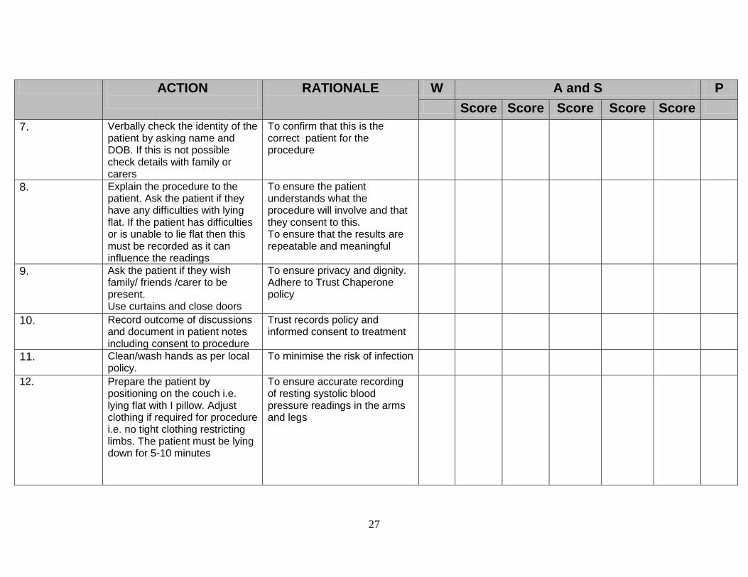
7. Verbally check the identity of the patient by asking name and DOB. If this is not possible check details with family or carers
To confirm that this is the correct patient for the procedure
8. Explain the procedure to the patient. Ask the patient if they have any difficulties with lying flat. If the patient has difficulties or is unable to lie flat then this must be recorded as it can influence the readings
To ensure the patient understands what the procedure will involve and that they consent to this. To ensure that the results are repeatable and meaningful
9. Ask the patient if they wish family/ friends /carer to be present. Use curtains and close doors
To ensure privacy and dignity. Adhere to Trust Chaperone policy
10. Record outcome of discussions and document in patient notes including consent to procedure
Trust records policy and informed consent to treatment
11. Clean/wash hands as per local policy.
To minimise the risk of infection
12. Prepare the patient by positioning on the couch i.e. lying flat with I pillow. Adjust clothing if required for procedure i.e. no tight clothing restricting limbs. The patient must be lying down for 5-10 minutes
To ensure accurate recording of resting systolic blood pressure readings in the arms and legs
28
ACTION
RATIONALE W A and S P
Score Score Score Score Score
Performing ABI procedure
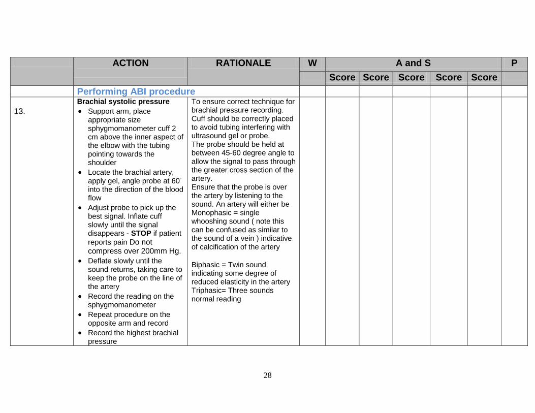
13.
Brachial systolic pressure
Support arm, place appropriate size sphygmomanometer cuff 2 cm above the inner aspect of the elbow with the tubing pointing towards the shoulder
Locate the brachial artery, apply gel, angle probe at 60◦ into the direction of the blood flow
Adjust probe to pick up the best signal. Inflate cuff slowly until the signal disappears - STOP if patient
reports pain Do not compress over 200mm Hg.
Deflate slowly until the sound returns, taking care to keep the probe on the line of the artery
Record the reading on the sphygmomanometer
Repeat procedure on the opposite arm and record
Record the highest brachial pressure
To ensure correct technique for brachial pressure recording. Cuff should be correctly placed to avoid tubing interfering with ultrasound gel or probe. The probe should be held at between 45-60 degree angle to allow the signal to pass through the greater cross section of the artery. Ensure that the probe is over the artery by listening to the sound. An artery will either be Monophasic = single whooshing sound ( note this can be confused as similar to the sound of a vein ) indicative of calcification of the artery Biphasic = Twin sound indicating some degree of reduced elasticity in the artery Triphasic= Three sounds normal reading

29
ACTION
RATIONALE W A and S P
Score Score Score Score Score
Remove gel from patient
Ankle systolic pressure. (See illustrations in Appendix 1)
14. If there are any wounds to the limb remove dressings and protect areas with cling film.
Locate the dorsalis pedis on the dorsum of the foot and posterior tibial pulse behind the medial malleolus. Apply appropriate size BP cuff just above the ankle with the tubing pointing towards the knee.
Apply gel to the pulse point on dorsum of the foot and angle probe at 60◦ to detect best signal
Inflate cuff until signal disappears STOP if pain
reported. Do not compress over 200mm Hg.
Deflate slowly until the signal reappears, taking care to keep to the line of the artery
Record this measurement next to dorsalis pedis pulse
To minimize cross infection To ensure correct technique for measurement of ankle systolic pressure
30
ACTION
RATIONALE W A and S P
Score Score Score Score Score
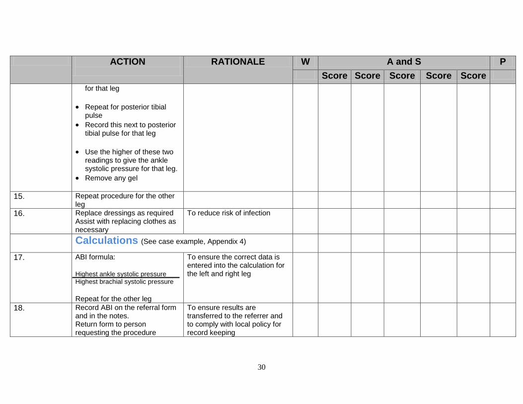
for that leg
Repeat for posterior tibial pulse
Record this next to posterior tibial pulse for that leg
Use the higher of these two readings to give the ankle systolic pressure for that leg.
Remove any gel
15. Repeat procedure for the other leg
16. Replace dressings as required Assist with replacing clothes as necessary
To reduce risk of infection
Calculations (See case example, Appendix 4)
17. ABI formula: Highest ankle systolic pressure Highest brachial systolic pressure
Repeat for the other leg
To ensure the correct data is entered into the calculation for the left and right leg
18.
Record ABI on the referral form and in the notes. Return form to person requesting the procedure
To ensure results are transferred to the referrer and to comply with local policy for record keeping

31
ACTION
RATIONALE W A and S P
Score Score Score Score Score
19. Discuss and agree subsequent actions with patient
The patient remains informed of the results and pathways
20. Clean the Doppler and probe Dispose of any dressings, wipes and tissues in a clinical waste bag Clean/wash hands as per local policy
To prevent cross infection and ensure adherence to infection control policy for decontamination of reusable instruments
DATE
SIGNATURE OF
CLINICIAN
SIGNATURE OF
ASSESSOR
Score as follows: 1 = NEEDS FURTHER PRACTICE
32
2 = SHOWS APTITUDE 3 = PROFICIENT
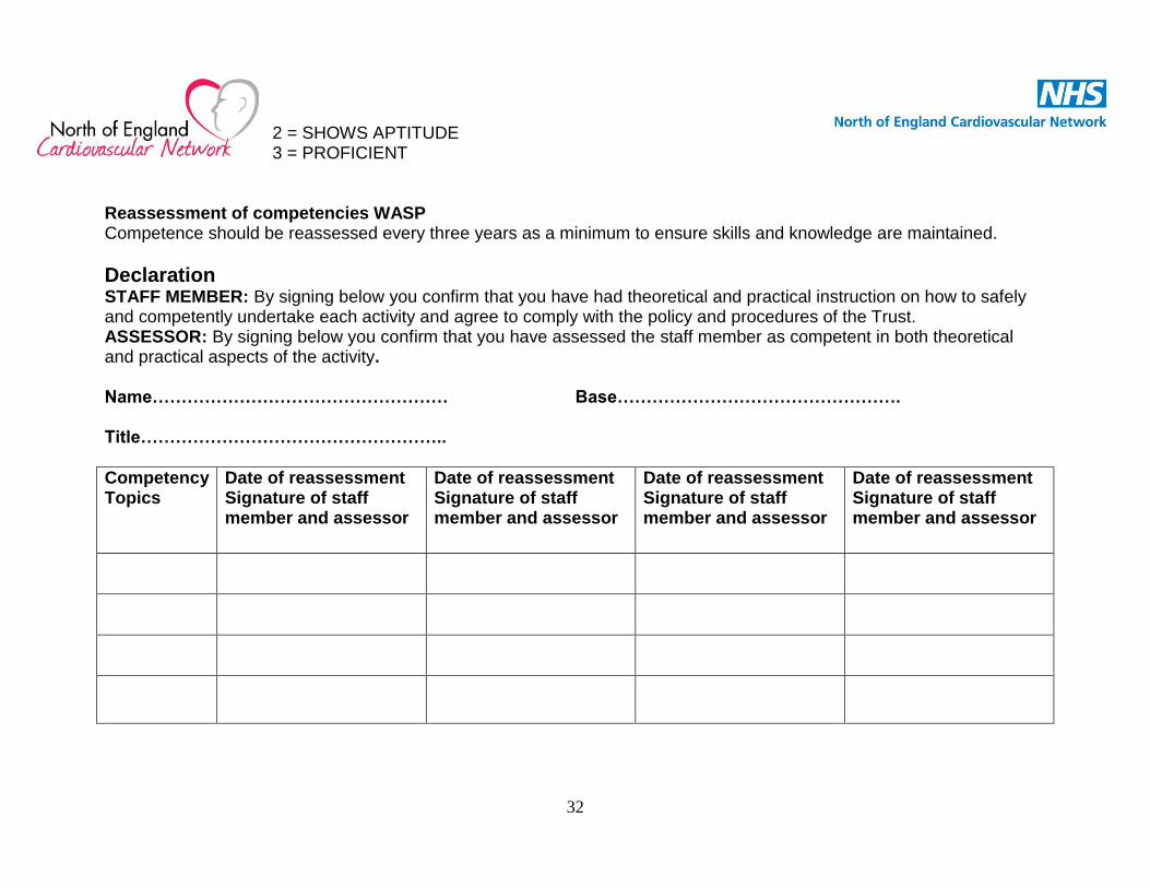
Reassessment of competencies WASP Competence should be reassessed every three years as a minimum to ensure skills and knowledge are maintained.
Declaration STAFF MEMBER: By signing below you confirm that you have had theoretical and practical instruction on how to safely and competently undertake each activity and agree to comply with the policy and procedures of the Trust. ASSESSOR: By signing below you confirm that you have assessed the staff member as competent in both theoretical and practical aspects of the activity. Name…………………………………………… Base…………………………………………. Title……………………………………………..
Competency Topics
Date of reassessment Signature of staff member and assessor
Date of reassessment Signature of staff member and assessor
Date of reassessment Signature of staff member and assessor
Date of reassessment Signature of staff member and assessor

33
34
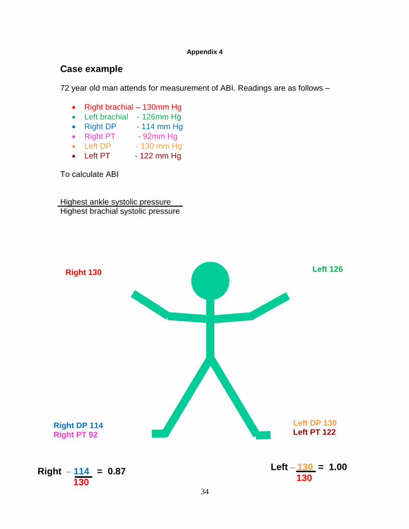
Appendix 4
Case example 72 year old man attends for measurement of ABI. Readings are as follows –
Right brachial – 130mm Hg
Left brachial - 126mm Hg
Right DP - 114 mm Hg
Right PT - 92mm Hg
Left DP - 130 mm Hg
Left PT - 122 mm Hg To calculate ABI Highest ankle systolic pressure Highest brachial systolic pressure
Right 130 Left 126
Right DP 114 Right PT 92
Left DP 130 Left PT 122
Right – 114 = 0.87 130
Left – 130 = 1.00 130