Embed Size (px)
Citation preview

CASE REPORT Open Access
Guided endodontic treatment of multipleteeth with dentin dysplasia: a case reportRalf Krug1* , Julian Volland1, Sebastian Reich1, Sebastian Soliman1, Thomas Connert2 and Gabriel Krastl1
Abstract
Background: To report the outcome of guided endodontic treatment (GET) of a case of dentin dysplasia with pulpcanal calcification (PCC) and apical periodontitis based on the use of a 3D-printed template designed by mergingcone-beam computed tomography (CBCT) and surface scan data.
Case presentation: A 12-year old female with radicular dentin dysplasia type I (DD-1) presented for endodontictreatment. Radiography revealed PCC in all teeth and apical radiolucency in seven teeth (12, 15, 26, 31, 32, 36 and46). Tooth 36 had the most acute symptoms and was thus treated first by conventional access cavity preparationand root canal detection. Despite meticulous technique, the distal and mesiolingual canals were perforated. Theperforations were immediately repaired with mineral trioxide aggregate, and the decision was made to switch toguided endodontic treatment for the remaining 6 teeth. CBCT and intraoral surface scans were acquired andmatched using coDiagnostix planning software (Dental Wings Inc.), the respective drill positions for root canallocation were determined, and templates were virtually designed and 3D-printed. The template was positioned onthe respective tooth, and a customized drill was used to penetrate the calcified part of the root canal and performminimally invasive access cavity preparation up to the apical region. All root canals were rapidly and successfullylocated with the templates. At 1-year follow-up, clear signs of apical healing were present in all treated teeth.
Conclusions: In patients with dentin dysplasia, conventional endodontic therapy is challenging. GET considerablyfacilitates the root canal treatment of teeth affected by dentin dysplasia.
Keywords: Guided endodontics, Pulp canal calcification, Dentin dysplasia, Root canal treatment, Template
BackgroundEndodontic treatment of calcified pulp systems is chal-lenging. Pulp canal calcification (PCC) is reported tooccur after various luxation injuries at rates of 15–40%[1, 2]. Chronic irritation (e.g. caries), cervical pulpotomy,or restorative therapies are known to promote theapposition of hard tissues within the root canal [3–5]. Itmay also occur after orthodontic treatment [6] or in eld-erly patients with a high rate of physiological appositionof dentin [7]. Specifically, dentin dysplasia (DD), a kind
of dentin malformation, also causes accelerated dentinalapposition [8, 9].DD, a rare disturbance of dentin formation (incidence:
1:100,000), is an autosomal dominant hereditary diseasecaused by a coding malfunction of the dentin sialopho-sphoprotein gene. The disorder is characterized by ap-parently normal enamel but atypical dentin formationand abnormal pulp morphology [10]. Two types of DDare distinguished based on clinical, radiological andhistological findings: type I (“radicular”) and type II(“coronal”), hereinafter referred to as DD-1 and DD-2[11]. In both DD-1 and DD-2, the crowns of primaryand permanent teeth mostly have a clinically and mor-phologically normal appearance and colour (in DD-2,however, the primary teeth are bluish amber-coloured).
© The Author(s). 2020 Open Access This article is licensed under a Creative Commons Attribution 4.0 International License,which permits use, sharing, adaptation, distribution and reproduction in any medium or format, as long as you giveappropriate credit to the original author(s) and the source, provide a link to the Creative Commons licence, and indicate ifchanges were made. The images or other third party material in this article are included in the article's Creative Commonslicence, unless indicated otherwise in a credit line to the material. If material is not included in the article's Creative Commonslicence and your intended use is not permitted by statutory regulation or exceeds the permitted use, you will need to obtainpermission directly from the copyright holder. To view a copy of this licence, visit http://creativecommons.org/licenses/by/4.0/.The Creative Commons Public Domain Dedication waiver (http://creativecommons.org/publicdomain/zero/1.0/) applies to thedata made available in this article, unless otherwise stated in a credit line to the data.
* Correspondence: [email protected] of Conservative Dentistry and Periodontology and Center ofDental Traumatology, University Hospital of Würzburg, Würzburg,Pleicherwall 2, 97070 Würzburg, GermanyFull list of author information is available at the end of the article
Krug et al. Head & Face Medicine (2020) 16:27 https://doi.org/10.1186/s13005-020-00240-4

The affected teeth may exhibit abnormal mobility, pre-mature exfoliation and, particularly in the case of DD-1,undeveloped or absent roots [12].Radiographically, the pulp spaces may be narrowed
and thus reduced in size or completely calcified. In DD-2, teeth in both dentitions often have thin roots but nor-mal root lengths. Common radiologic signs include earlypulp obliteration and thistle-tube shaped pulp chamberswith multiple pulp stones in the absence of periapical ra-diolucencies [13]. Besides aberrant dentin deposition inthe pulp chamber in both types (DD-1 and DD-2), mosttypically in DD-1 there is an increased incidence of peri-apical radiolucencies due to the infection of the rootcanal system [9, 13, 14]. To date, it is presumed that theseteeth are highly susceptible to bacterial invasion due to vari-ous factors, including the presence of atypical dentin for-mation within a highly irregular pulp chamber, the lack ofdentinal fluid, which induces enamel brittleness and, occa-sionally, micro cracks as a potential pathway for microor-ganisms, and aberrant dentin formation, which might affectthe formation and structure of the enamel as well [15]. Al-terations to the enamel, including micro cracks and in-creased dentin permeability in the case of DD-1, mayfacilitate bacterial invasion and lead to pulp necrosis andapical periodontitis. Such a crack was detected in a three-dimensional (3D) micro-computed tomographic study of amolar of a DD-1 patient, but the ability of this technologyto detect fine cracks seems to be limited [15].There is no available evidence on the occurrence of
apical periodontitis in teeth with DD, but several case re-ports suggest that these teeth may be at risk of develop-ing apical pathologies [10, 14–18].In the present case report, DD-1 was identified based
on its specific radiologic features, including the charac-teristic root morphology, obliteration of the pulp cham-bers and root canals, and the presence of severalperiapical lesions in sound teeth (Figs. 1, 3). Typically,the roots appear shortened, blunted, and partially mal-formed [14, 16, 19, 20].
Endodontic treatment of calcified pulp systems (due todental trauma or DD) is associated with a high risk ofcomplications, such as root perforation, extensive dentinalhard tissue loss, and missing the root canal [14, 17]. Thus,straight-line access close to or through the incisal edgewas emphasized as being a key to preventing technicalendodontic failures in anterior teeth with PCC [21].Guided endodontic treatment (GET) was recently intro-duced as a novel method of performing operator-independent guided straight-line access [22, 23]. Here, 3Ddata collected by cone-beam computed tomography(CBCT) were matched with surface scan data and used tofabricate a 3D drill guide. Once the drill paths were virtu-ally planned, templates could be fabricated to safely locatethe root canals. Other case reports and one case serieshave also shown the feasibility and reliability of GET incases with apical periodontitis as a late complication ofdental trauma [23–31]. To our knowledge, there is cur-rently only one guided endodontics case report, which de-scribes a promising 18-month outcome of a densinvaginatus malformation case treated, followed by end-odontic surgery and root-end filling with mineral trioxideaggregate [30].In DD cases in need of endodontic therapy, conven-
tional orthograde endodontic treatment was reported tobe potentially substitutable with periapical curettage andretrofilling, especially if the roots display normal lengthand development [32, 33]. In the case of DD-1, extractionof teeth with periapical radiolucencies might be favouredover endodontic therapy due to the above-mentionedtechnical challenges and anatomic limitations [34–36].To date, only a few case reports of endodontic treatment
attempts in patients with DD-1 have been published, andthey have partially sobering outcomes [14, 37–39]. This isthe first case report of a successful outcome of root canaltreatment of multiple teeth in a patient with DD-1. GETprevented technical complications and enabled toothretention in our patient.
Case presentationA 12-year old female with DD-1 was referred to ourdental clinic (Department of Conservative Dentistry andPeriodontology and Center of Dental Traumatology,University Hospital of Würzburg, Germany) for end-odontic treatment of multiple teeth. Due to the typicalclinical and radiologic appearance of the teeth, no fur-ther attempts were made to confirm the clinical diagno-sis of DD-1. The patient was not on any medication.Written informed consent for publication of their clin-ical details and clinical images was obtained from theparents of the patient. Clinically, the patient exhibitedacute pain on percussion of tooth 36 and was diagnosedwith symptomatic apical periodontitis. All of her teethwere unrestored and free of carious or non-carious loss
Fig. 1 Panoramic view of the dentition of a 12-year old female withDD-1: Note the obliterated pulp chambers and root canals ofreduced size in all teeth, and periapical radiolucencies in teeth 15,12, 26, 36, 32, 31, 46
Krug et al. Head & Face Medicine (2020) 16:27 Page 2 of 8
of dental hard tissue. They showed regular enamel(Fig. 2), but aberrant dentin formation and irregular pulpmorphology. Neither severe tooth mobility nor atypicaltooth positions were observed. Periapical radiographs re-vealed PCC in all teeth and apical radiolucencies inseven teeth: 15, 12, 26, 36, 32, 31 and 46 (Fig. 3). Thepulp spaces were reduced in size or completely calcified.Conventional endodontic treatment of tooth 36 was ini-tiated due to acute symptoms. While attempting to lo-cate the root canals, perforation of the distal andmesiolingual root canals occurred despite meticuloustreatment under a microscope. The perforations wereimmediately repaired with mineral trioxide aggregate(ProRoot® MTA, Dentsply Sirona). Conventional rootcanal treatment was performed in two sessions aftersuccessful root canal location in this tooth.Given the difficulty in locating the root canals, the op-
erator switched to GET for the remaining 6 asymptom-atic teeth. CBCT (3D Accuitomo 170, J. Morita Mfg.Corp.) and intra-oral surface scans (Cerec Omnicam,Dentsply Sirona GmbH) were acquired and matchedusing coDiagnostix planning software (Dental WingsInc.). After the drill position for root canal location wasdetermined (Fig. 4), a virtual template was designed. Thecorresponding surface-tessellation-language (STL) fileswere exported to a 3D printer (Form 2 Formlabs, Mater-ial: Dental SG Resin, Formlabs Inc., Somerville, MA,USA) for template fabrication. After inserting the drillsleeves (steco-system-technik GmbH), the template waspositioned on the respective teeth requiring endodontictreatment (Fig. 5). A customized drill (diameter = 1.0mm, steco-system-technik GmbH) was used to create aminimally invasive access to the calcified root canal inthe apical third of the root. The orifices of all root canalswere rapidly and successfully located. Endodontic ther-apy consisted of mechanical preparation using nickel-titanium rotary files (Mtwo®, VDW GmbH), sonic irri-gant activation was performed using Eddy® tips (VDWGmbH) and sodium hypochlorite (3%), followed bywarm vertical gutta-percha obturation with an epoxy
resin sealer (AH Plus®, Dentsply Sirona). Finally, the ac-cess cavities were restored with composite resin (Fig. 6).At the 1-year follow-up examination, the root-filledteeth were symptom-free. Radiography revealed signs ofapical lesion size reduction in teeth 36, 32, and 12. Fur-ther follow-ups are needed to clarify the ultimate end-odontic outcome. Complete healing of apicalperiodontitis was obtained in teeth 15, 26, 31, and 46(Fig. 7). Improvement of tooth mobility was observed inall endodontically treated teeth compared with the initialmobility.
DiscussionThis is the first case report of the use of GET to prepareminimally invasive access cavities for root canal location in apatient with DD-1. All of the patient’s teeth showed radio-graphic signs of PCC, and apical periodontitis was detectedin a total of seven teeth. Initially, endodontic therapy wasperformed on tooth 36 using the conventional approach,resulting in two perforations, whereas GET on the remainingaffected teeth led to a successful outcome. The 1-yearfollow-up examination revealed signs of healing of the le-sions of endodontic origin in all root canal treated teeth.Patients with DD-1 frequently have clinical signs of
apical periodontitis, spontaneous abscess formation ortooth hypermobility related to an unfavourable crown-to-root ratio. These conditions pose diagnostic chal-lenges, and the recommended treatment possibilities areunspecific and limited [40]. For management of suchaffected teeth with short roots, hypermobility, PCC, andperiapical lesions, it was recommended to focus on twoextreme options: either monitor without any treatmentor extract the tooth as the last resort [13, 18]. Endodon-tic instrumentation and obturation, an option in teethwithout extremely short roots [13], is often believed tobe unfeasible because of abnormal pulp canal morph-ology. Ramifications, the occurrence of pulp stones andvarious types of hard tissue formations within the rootcanal system are typical characteristics [18]. In aquestionnaire-based evaluation of one DD-1 case history,90.6% of a total of 64 dentists with different specialitiesand experience levels chose to monitor multiple periapi-cal radiolucencies in teeth with pulp necrosis instead ofperforming root canal treatment (3.1%) or extraction(6.2%) [41]. Likewise, in a case report of a 7-year-old fe-male DD-1 patient with a 6-year follow-up, the authorspreferred extracting permanent teeth due to extensivemobility. Extraction was suggested for teeth with pulpnecrosis and periapical abscess, and endodontic treat-ment was considered to be contraindicated in those withsevere calcification of the pulp [35]. Furthermore, end-odontic therapy might be limited in young children, asan early age of the patient is associated with poorcompliance in such difficult and lengthy treatment.
Fig. 2 Clinically, the enamel appears normal but there is atypicaldentin formation with abnormal pulp morphology
Krug et al. Head & Face Medicine (2020) 16:27 Page 3 of 8

Root canal location in calcified teeth with DD mightbe even more challenging than in those with calcificationafter trauma or caries. In teeth with DD-1, calcificationof the pulp space seems to follow a different yet un-known pathophysiological mechanism. The process ofcalcification occurs soon after or even prior to tooth
eruption [42], and it does not necessarily start in thecoronal part of the tooth in association with subsequentgradual narrowing of the root canals, as is often the caseafter luxation injuries. Instead, teeth with DD-1 seem tohave variable expressivity, ranging from total obliterationof the endodontic space to calcifications in specific
Fig. 3 Periapical radiolucencies were detected in teeth 15, 12, 46 by CBCT imaging (a, b, d) and in teeth 26, 36, 32, 31 by periapical radiography(c, e, f)
Fig. 4 The drill paths for tooth 26 (e.g. mesio- and disto-buccal) were virtually planned using coDiagnostix software (Dental Wings Inc.) (a-c)
Krug et al. Head & Face Medicine (2020) 16:27 Page 4 of 8

Fig. 5 Intraoral photograph showing the inserted templates and drill sleeves (steco-system-technik GmbH) for teeth 15 and 12 (a, b), for thepalatal root canal of tooth 26 (c), and for teeth 32 and 31 (d)
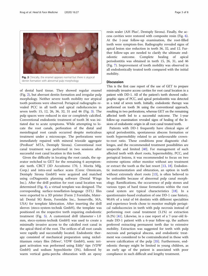
Fig. 6 The enamel has to be removed step-by-step, targeting each root canal. Staining of the tip of the bur with the template in place on thetooth crown makes it possible to locate the entry point of the bur within the dentin (a). Photo taken during endodontic treatment of tooth 26,with palatal, disto-buccal and two mesio-buccal root canals (b). Subsequently, obturation was carried out using gutta-percha and sealer (AHplus,Dentsply Sirona) (c), and the access cavity was filled with composite (d)
Krug et al. Head & Face Medicine (2020) 16:27 Page 5 of 8

regions in the apical or coronal part of the tooth [42].Thus, the presence of a narrow root canal in the apicalpart of the root, as reported for teeth with pulp canalcalcifications [43], might not apply for teeth with DD-1.Another aspect which makes root canal location more
difficult is dentin morphology. In patients with DD-1,the teeth have softer dentin that lacks a regular tubularmorphology and may contain irregular channels [44].Thus, regardless of the location of the root canal, fileswith cutting tips, which are frequently used to scout rootcanals and penetrate calcifications, might also penetratethe soft dentin and increase the risk of perforation.After the occurrence of two perforations during con-
ventional root canal treatment to manage a mandibularmolar with symptoms in the present case, it was consid-ered better to switch to GET for safe endodontic man-agement of the remaining teeth. The root canals in allteeth could be successfully located, prepared and ad-equately root-filled using GET approach. Nevertheless,the favourable results after 1 year, as reflected by clearsigns of periapical healing, do not guarantee long-termsuccess. While the success rates of primary root canaltreatment in teeth with preoperative apical periodontitisgenerally may reach 74% [45], little is known about thefate of root-canal-treated teeth in patients with DD-1.Three case reports describe successful conventionalendodontic management, partially completed with apicalsurgery [14, 37, 38]. Successful endodontic treatmentwith complete healing of periapical lesions in 16 teethwas solely observed in a 22-year old female with DD-1,who was monitored for up to 3 years [14].Most published cases had rather sobering outcomes
after different therapy approaches. This includes a switchto apical surgery after an unsuccessful endodontic
treatment attempt [39] or leaving teeth with periapical le-sions untreated [18, 35] or tooth extraction, particularly inteeth with short roots [10, 19, 20, 34, 36, 46]. The progno-sis of endodontically treated teeth with DD might be de-scribed as uncertain. It may be hypothesized that becausethe increased dentin permeability allows persistent bacter-ial penetration, the chances of complete healing of periapi-cal lesions may be limited, even in adequately root-filledteeth. Further, reversal of healing might be more likelythan in teeth without DD-1. In the present case reportedhere, a promising outcome of endodontic treatment wasobserved at 1-year follow-up. However, further follow-upsare needed to monitor the healing process and to identifynew lesions of endodontic origin which may occur inother teeth. In teeth with DD-1 there might be an in-creased risk for reinfection of the root canal space afterendodontic treatment due to the softer and more perme-able dentin and the presence of micro cracks [15]. Thus,all attempts should be made to optimally seal the rootcanal below the marginal bone with adhesive materials.From a technical perspective, the GET approach has
proven its worth in the present case, where multipleteeth were endodontically treated. GET is generally con-sidered to be clinically useful for facilitating the rootcanal treatment of anterior and posterior teeth withPCC and apical periodontitis. Various ex-vivo studyresults revealed that GET is a highly precise and time-saving technique of root canal location [47–49]. How-ever, in severely curved root canals, GET may fail to ne-gotiate the apical canal curvature. For such cases,apicectomy may be a viable alternative treatment ap-proach. Interestingly, a guided microsurgical techniquewas introduced to allow predefined osteotomy and rootresection [50].
Fig. 7 This 1-year follow-up radiograph showed size reduction of the apical lesions in endodontically treated teeth 12 (a), 36 (e), and 32 (d), andcomplete regression in 15 (a), 26 (b), 31 (d), and 46 (c)
Krug et al. Head & Face Medicine (2020) 16:27 Page 6 of 8
Prospectively, advanced guided endodontics based ona new computer-aided dynamic navigation technologymay soon be available for cases where multiple rootcanal treatments have to be performed in calcified teeth.However, further investigations are needed to confirmthe promising preliminary results of this novel techno-logy [51, 52].
ConclusionsConventional endodontic treatment of teeth affected bydentin dysplasia is challenging but can be facilitated byGET. As demonstrated in multiple teeth of the DD-1 pa-tient described in this case report, GET is a safe andclinically feasible technique that prevents root perfor-ation and enables tooth retention.
Abbreviations3D: Three-dimensional; AP: Apical periodontits; CBCT: Cone-beam computedtomography; DD: Dentin dysplasia; GET: Guided endodontic treatment;PCC: Pulp canal calcification; STL: Surface-tessellation-language
AcknowledgementsNot applicable.
Authors’ contributionsRK contributed to conception, design and interpretation, drafted andcritically revised the manuscript and performed the treatment. JV and SRcontributed to planning software management, conception and design. SScontributed to conception, design and interpretation, drafted and criticallyrevised the manuscript and performed calculations. TC contributed toconception, design and interpretation, drafted and critically revised themanuscript. GK contributed to conception, design and interpretation, draftedand critically revised the manuscript and supervised the work. All authorsgave final approval and agree to be accountable for all aspects of the work.
Authors’ informationRK has specialization in endodontology. The clinical performance was doneby RK. JV, SR, SS and TC are clinical dentists and researchers inendodontology. GK is chief researcher and head of the Department ofConservative Dentistry and Periodontology and Center of DentalTraumatology at the University of Würzburg.
FundingThis publication was supported by the Open Access Publication Fund of theUniversity of Wuerzburg.
Availability of data and materialsThe datasets used and/or analysed during the current case report areavailable from the corresponding author on reasonable request.
Ethics approval and consent to participateEthical approval is not applicable. The consent to participate is available, Thedocumentary evidence of consent can be supplied at any time if requested.
Consent for publicationWritten informed consents were obtained from the parents of the patient forpublication of the technical notes and images.
Competing interestsThe authors declare that they have no competing interests.
Author details1Department of Conservative Dentistry and Periodontology and Center ofDental Traumatology, University Hospital of Würzburg, Würzburg,Pleicherwall 2, 97070 Würzburg, Germany. 2Department of Periodontology,Endodontology and Cariology, University Centre for Dental Medicine,University of Basel, Mattenstraße 40, 4058 Basel, Switzerland.
Received: 18 June 2020 Accepted: 22 October 2020
References1. Nikoui M, Kenny DJ, Barrett EJ. Clinical outcomes for permanent incisor
luxations in a pediatric population. III. Lateral luxations. Dent Traumatol.2003;19(5):280–5.
2. Andreasen FM, Zhijie Y, Thomsen BL, Andersen PK. Occurrence of pulpcanal obliteration after luxation injuries in the permanent dentition. EndodDent Traumatol. 1987;3(3):103–15.
3. Fleig S, Attin T, Jungbluth H. Narrowing of the radicular pulp space incoronally restored teeth. Clin Oral Investig. 2017;21(4):1251–7.
4. Mass E, Zilberman U. Long-term radiologic pulp evaluation after partialpulpotomy in young permanent molars. Quintessence Int. 2011;42(7):547–54.
5. Sayegh FS, Reed AJ. Calcification in the dental pulp. Oral Surg Oral MedOral Pathol. 1968;25(6):873–82.
6. Delivanis HP, Sauer GJ. Incidence of canal calcification in the orthodonticpatient. Am J Orthod. 1982;82(1):58–61.
7. Carvalho TS, Lussi A. Age-related morphological, histological and functionalchanges in teeth. J Oral Rehabil. 2017;44(4):291–8.
8. MK OC, Duncan WK, Perkins TM. Dentin dysplasia: review of the literatureand a proposed subclassification based on radiographic findings. Oral SurgOral Med Oral Pathol. 1991;72(1):119–25.
9. Witkop CJ Jr. Manifestations of genetic diseases in the human pulp. OralSurg Oral Med Oral Pathol. 1971;32(2):278–316.
10. Khandelwal S, Gupta D, Likhyani L. A case of dentin dysplasia with fullmouth rehabilitation: a 3-year longitudinal study. Int J Clin Pediatr Dent.2014;7(2):119–24.
11. Shields ED, Bixler D. el-Kafrawy AM. A proposed classification for heritablehuman dentine defects with a description of a new entity. Arch Oral Biol.1973;18(4):543–53.
12. Chen D, Li X, Lu F, Wang Y, Xiong F, Li Q. Dentin dysplasia type I-A dentaldisease with genetic heterogeneity. Oral Dis. 2019;25(2):439–46.
13. American Academy of Pediatric D. Guideline on dental management ofheritable dental developmental anomalies. Pediatr Dent. 2013;35(5):E179–84.
14. Ravanshad S, Khayat A. Endodontic therapy on a dentition exhibitingmultiple periapical radiolucencies associated with dentinal dysplasia type 1.Austral Endod J. 2006;32(1):40–2.
15. Ranjitkar S, Yong R, Wu IC, Gully G, Farmer D, Watson I, et al. Dentinaldysplasia type 1: a 3D micro-computed tomographic study of enamel,dentine and root canal morphology. Austral Endod J. 2019;45(3):298–304.
16. Seow WK, Shusterman S. Spectrum of dentin dysplasia in a family: casereport and literature review. Pediatr Dent. 1994;16(6):437–42.
17. Ansari G, Reid JS. Dentinal dysplasia type I: review of the literature andreport of a family. ASDC J Dent Child. 1997;64(6):429–34.
18. Akhil Jose EJ, Palathingal P, Baby D, Thachil JM. Dentin dysplasia type I: arare case report. J Oral Maxillofac Pathol. 2019;23(2):309.
19. Malik S, Gupta S, Wadhwan V, Suhasini GP. Dentin dysplasia type I - a rareentity. J Oral Maxillofac Pathol. 2015;19(1):110.
20. Ozer L, Karasu H, Aras K, Tokman B, Ersoy E. Dentin dysplasia type I: reportof atypical cases in the permanent and mixed dentitions. Oral Surg OralMed Oral Pathol. 2004;98(1):85–90.
21. McCabe PS, Dummer PM. Pulp canal obliteration: an endodontic diagnosisand treatment challenge. Int Endod J. 2012;45(2):177–97.
22. Buchgreitz J, Buchgreitz M, Mortensen D, Bjorndal L. Guided access cavitypreparation using cone-beam computed tomography and optical surfacescans - an ex vivo study. Int Endod J. 2015;49(8):790.
23. Krastl G, Zehnder MS, Connert T, Weiger R, Kuhl S. Guided Endodontics: anovel treatment approach for teeth with pulp canal calcification and apicalpathology. Dent Traumatol. 2016;32(3):240–6.
24. Buchgreitz J, Buchgreitz M, Bjorndal L. Guided Endodontics modified fortreating molars by using an Intracoronal guide technique. J Endod. 2019;45(6):818–23.
25. Buchgreitz J, Buchgreitz M, Bjorndal L. Guided root canal preparation usingcone beam computed tomography and optical surface scans - anobservational study of pulp space obliteration and drill path depth in 50patients. Int Endod J. 2019;52(5):559–68.
26. Connert T, Zehnder MS, Amato M, Weiger R, Kuhl S, Krastl G. MicroguidedEndodontics: a method to achieve minimally invasive access cavitypreparation and root canal location in mandibular incisors using a novelcomputer-guided technique. Int Endod J. 2018;51(2):247–55.
Krug et al. Head & Face Medicine (2020) 16:27 Page 7 of 8

27. Fonseca Tavares WL, Diniz Viana AC, de Carvalho MV, Feitosa Henriques LC,Ribeiro Sobrinho AP. Guided endodontic access of calcified anterior teeth.J Endod. 2018;44(7):1195–9.
28. Lara-Mendes STO, Barbosa CFM, Santa-Rosa CC, Machado VC. Guidedendodontic access in maxillary molars using cone-beam computedtomography and computer-aided design/computer-aided manufacturingsystem: a case report. J Endod. 2018;44(5):875–9.
29. Tchorz JP, Wrbas KT, Hellwig E. Guided endodontic access of a calcifiedmandibular central incisor using a software-based three-dimensionaltreatment plan. Int Endod J. 2019;22(3):273–81.
30. Zubizarreta-Macho A, Ferreiroa A, Agustin-Panadero R, Rico-Romano C,Lobo-Galindo AB, Mena-Alvarez J. Endodontic re-treatment and restorativetreatment of a dens invaginatus type II through new technologies. J ClinExp Dent. 2019;11(6):e570–e6.
31. Maia LM, de Carvalho MV, da Silva N, Brito Junior M, da Silveira RR, MoreiraJunior G, et al. Case reports in maxillary posterior teeth by guidedendodontic access. J Endod. 2019;45(2):214–8.
32. Steidler NE, Radden BG, Reade PC. Dentinal dysplasia: a clinicopathologicalstudy of eight cases and review of the literature. Br J Oral Maxillofac Surg.1984;22(4):274–86.
33. Barron MJ, McDonnell ST, Mackie I, Dixon MJ. Hereditary dentine disorders:dentinogenesis imperfecta and dentine dysplasia. Orphanet J Rare Dis. 2008;3:31.
34. Toomarian L, Mashhadiabbas F, Mirkarimi M, Mehrdad L. Dentin dysplasiatype I: a case report and review of the literature. J Med Case Rep. 2010;4:1.
35. Ozer S, Ozden B, Otan Ozden F, Gunduz K. Dentinal dysplasia type I: a casereport with a 6-year followup. Case Rep Dent. 2013;2013:659084.
36. Shankly PE, Mackie IC, Sloan P. Dentinal dysplasia type I: report of a case. IntJ Paediatr Dent. 1999;9(1):37–42.
37. Rankow H, Miller AS. Dentin dysplasia: endodontic considerations andreport of involvement of three siblings. J Endod. 1984;10(8):384–6.
38. Tidwell E, Cummingham CJ. Dentinal dysplasia: endodontic treatment, withcase report. J Endod. 1979;5(12):372–6.
39. Coke JM, Del Rosso G, Remeikis N, Van Cura JE. Dentinal dysplasia, type I.report of a case with endodontic therapy. Oral Surg Oral Med Oral Pathol.1979;48(3):262–8.
40. Alhilou A, Beddis HP, Mighell AJ, Durey K. Dentin dysplasia: diagnosticchallenges. BMJ Case Rep. 2018;2018:1.
41. Rasaratnam L, Djemal S. Type-1 dentine dysplasia - diagnostic and clinicalchallenges in restorative management. Dent Update. 2017;44(3):174–6 8-80.
42. Kim JW, Simmer JP. Hereditary dentin defects. J Dent Res. 2007;86(5):392–9.43. de Cleen M. Obliteration of pulp canal space after concussion and
subluxation: endodontic considerations. Quintessence Int. 2002;33(9):661–9.44. Perl T, Farman AG. Radicular (type 1) dentin dysplasia. Oral Surg Oral Med
Oral Pathol. 1977;43(5):746–53.45. Friedman S, Abitbol S, Lawrence HP. Treatment outcome in endodontics:
the Toronto study. Phase 1: initial treatment. J Endod. 2003;29(12):787–93.46. Petrone JA, Noble ER. Dentin dysplasia type I: a clinical report. J Am Dent
Assoc. 1981;103(6):891–3.47. Buchgreitz J, Buchgreitz M, Mortensen D, Bjorndal L. Guided access cavity
preparation using cone-beam computed tomography and optical surfacescans - an ex vivo study. Int Endod J. 2016;49(8):790–5.
48. Zehnder MS, Connert T, Weiger R, Krastl G, Kuhl S. Guided endodontics:accuracy of a novel method for guided access cavity preparation and rootcanal location. Int Endod J. 2016;49(10):966–72.
49. Connert T, Zehnder MS, Weiger R, Kuhl S, Krastl G. MicroguidedEndodontics: accuracy of a miniaturized technique for apically extendedaccess cavity preparation in anterior teeth. J Endod. 2017;43(5):787–90.
50. Strbac GD, Schnappauf A, Giannis K, Moritz A, Ulm C. Guided modernendodontic surgery: a novel approach for guided osteotomy and rootresection. J Endod. 2017;43(3):496–501.
51. Chong BS, Dhesi M, Makdissi J. Computer-aided dynamic navigation: a novelmethod for guided endodontics. Quintessence Int. 2019;50(3):196–202.
52. Zubizarreta-Macho A, Munoz AP, Deglow ER, Agustin-Panadero R, AlvarezJM. Accuracy of Computer-Aided Dynamic Navigation Compared toComputer-Aided Static Procedure for Endodontic Access Cavities: An inVitro Study. J Clin Med. 2020;9(1):129.
Publisher’s NoteSpringer Nature remains neutral with regard to jurisdictional claims inpublished maps and institutional affiliations.
Krug et al. Head & Face Medicine (2020) 16:27 Page 8 of 8