Embed Size (px)
Citation preview
Guide for Eco-sensible
ELISA Development
Kem-En-Tec Diagnostics A/S www.kem-en-tec.com
Kem-En-Tec Diagnostics’ Guide for Eco-sensible ELISA
Development provides step by step information for
ELISA optimization in an environmentally sensible way.
It includes hints, tips and troubleshooting for the de-
velopment of assays based on chromogenic detection
of Alkaline Phosphatase (AP) and Horseradish Peroxi-
dase (HRP).
The Guide for Eco-sensible ELISA Development starts
with an overview of the different types of ELISA for-
mats. This is followed by sections describing the steps
and the reagents involved in ELISA development and
the critical factors that need to be addressed for each
step in order to develop a successfully optimized ELISA.
Eco-sensible properties of Kem-En-Tec Diagnostics’
products have green highlights.
1
Table of contents
TABLE OF CONTENTS ................................................................................................................................... 1
INTRODUCTION TO ELISA ............................................................................................................................ 2
1. ELISA DESIGN ...................................................................................................................................... 4
2. COATING ............................................................................................................................................ 9
3. WASHING ......................................................................................................................................... 10
4. BLOCKING AND STABILIZING ELISA PLATES ....................................................................................... 11
5. DILUTION OF SAMPLES, STANDARDS & CONTROLS .......................................................................... 14
6. DILUTION AND STABILIZATION OF THE CONJUGATE ......................................................................... 16
7. ENHANCED CONJUGATES: AMPLIFICATION SYSTEMS ....................................................................... 19
8. SUBSTRATES ..................................................................................................................................... 21
ELISA OPTIMIZATION ................................................................................................................................ 24
EXAMPLE OF ECO-SENSIBLE ASSAY OPTIMIZATION: SANDWICH ELISA ..................................................... 25
DEVELOP YOUR ASSAY STEP-BY-STEP WITH KEM-EN-TEC DIAGNOSTICS A/S ............................................ 26
Look for catalogue numbers in the end of this guide and ask for samples!
Abbreviations
Ab Antibody Abs Absorbance Ag Antigen AP Alkaline Phosphatase ELISA Enzyme Linked Immunosorbent Assay HAMA Human anti-mouse antibodies HRP Horseradish Peroxidase NSB Non Specific Binding OD Optical Density PBS Phosphate-Buffered Saline RT Room Temperature RTU Ready-To-Use SA Streptavidin SNR Signal-to-Noise Ratio TBS Tris-buffered Saline
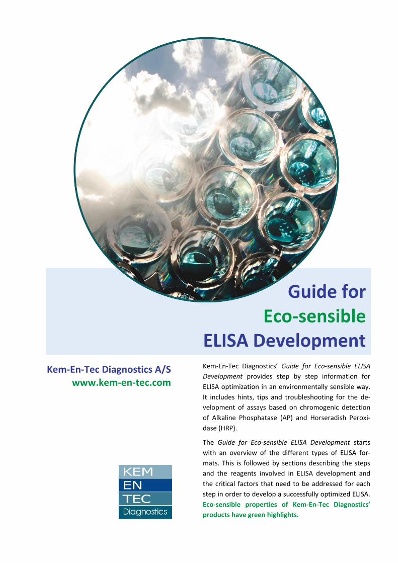
Legend for images
Antigens
Antibodies
Enzyme
Reacting enzyme
Substrate
Reacting substrate
Blocking agents
Biotin
Streptavidin
2
Introduction to ELISA
ELISAs are used to detect and quantitate peptides, proteins, antibodies and hormones in biologi-cal specimens. Antibodies (Ab) or antigens (Ag) are adsorbed to a solid surface and bind with high affinity their complementary reagents that are typically conjugated to enzymes. The enzymatic reaction triggered by appropriate substrates allows the quantitation of the analyte of interest.
ELISAs are useful tools because they allow for the rapid screening or quantitation of a large num-ber of samples. Other techniques have been developed, but ELISA remains popular because of the ease of performance of the assay, accuracy, and the low cost.
This guide is divided in eight sections that follow the traditional development of an ELISA.
1. ELISA Design This section introduces the most common ELISA formats, pre-senting their advantages and disadvantages. 2. Coating Suggestions for optimizing the coating of the solid phase are listed in this section. 3. Washing Wash out excess reagent to reduce background signal and in-crease the signal-to noise ratio (SNR). Guidelines for optimizing the washing buffer and the washing procedures are suggested. 4. Blocking and stabilizing ELISA plates The critical aspects to consider for preventing non-specific-binding (NSB) are discussed. Kem-En-Tec Diagnostics’ Synthetic Blocking Buffer and WellChampion are ideal reagents for block-ing and stabilizing ELISA plates. 5. Dilution of samples, standards & controls Specimens, standards and controls need to be diluted in specific buffers before adding them in the microwells. Kem-En-Tec Diag-nostics’ SamplePLUS2, Effect Diluents, and Protein-StabilPLUS are the ideal diluents for reducing cross reactions and interfer-ences. The use of Ab’s is also described in this section. 6. Dilution and stabilization of the conjugate Specific diluents are used for achieving the optimal concentra-tion of conjugated Ab’s or Ag’s to be added in the assay. Most of Kem-En-Tec Diagnostics’ diluents also have stabilizing properties to prevent loss of activity. The Alkaline Phosphatase (AP) and Horseradish Peroxidase (HRP) labels are also described. 7. Enhanced conjugates: amplification systems Kem-En-Tec Diagnostics has developed an amplification system based on the biotin-streptavidin binding, which enables 100 fold signal enhancement. 8. Substrates Adding the substrate is the last step in an ELISA. This guide fo-cuses on chromogenic substrates for AP and HRP. The broad range of Kem-En-Tec Diagnostics’ pNPP and TMB substrates is described in details for the highest level of optimization.
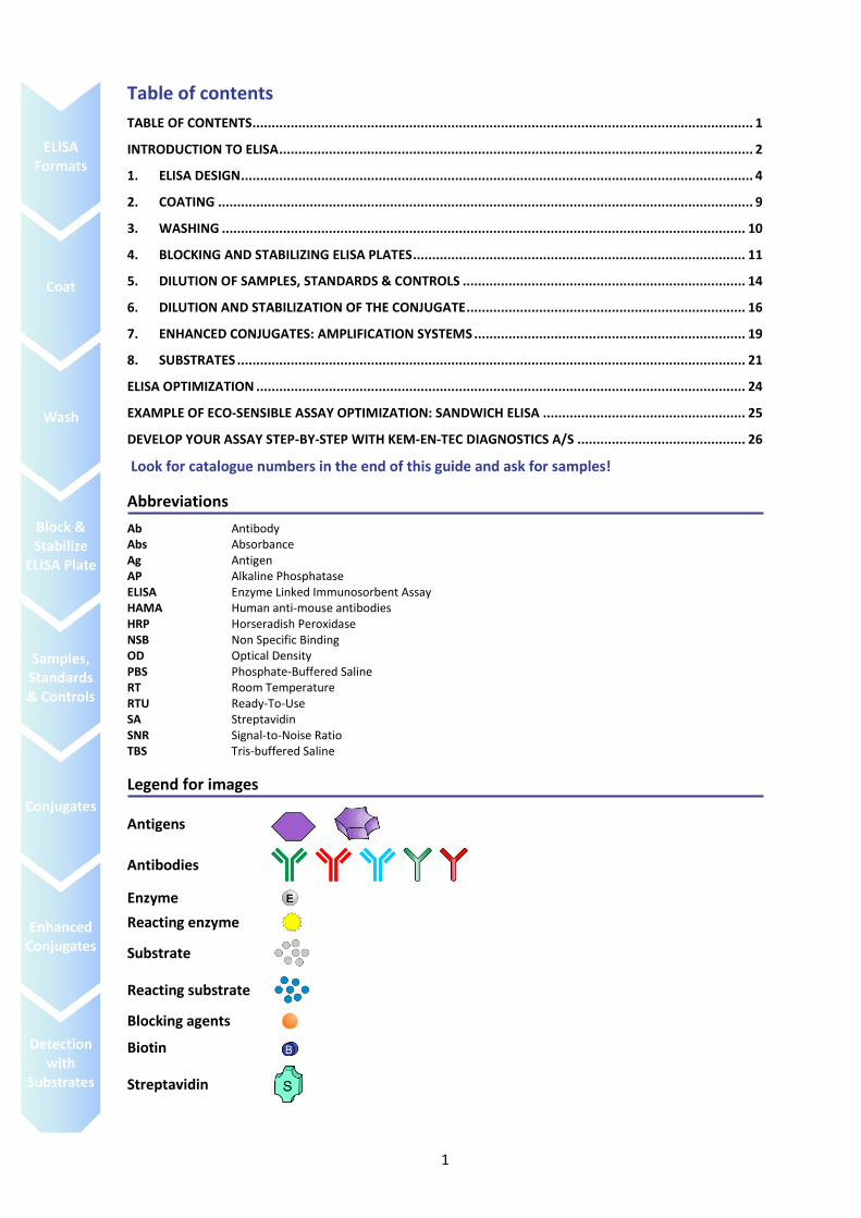
Figure 1. A schematic description of the most common type of ELISA, the sandwich ELISA.
3
Environmental considerations during assay development
Over the past decade, IVD manufacturers have been working towards more enviromentally con-scious and sustainable manufacturing. Much of this effort has been focused on reducing usage of facility water and energy use, reducing facility waste, increasing facility recycling, and sourcing recycled/recyclable product packaging. Eco-sensible options are now available for the develop-ment and production of diagnostics assays.
Many common components used in ELISAs contain biological and chemical hazards which were traditionally used to increase assay quality and to prolong assay shelf-life. Considering that the average automated immunoassay analyzer in a clinical lab can run 200 tests per hour while pro-cessing samples 24/7, these components can generate a considerable amount of hazardous waste. ELISAs have not traditionally been eco-sensible and several components in this type of assay are usually environmentally harmful:
Many components contain bovine serum albumin (BSA). This is a natural product from bo-vines and can carry disease. It is tightly regulated to avoid this issue. Kem-En-Tec Diagnostics totally avoids using BSA in its products. For instance Kem-En-tec Diagnostics has developed proprietary blockers which are more eco-sensible and block with high efficiency as well.
Reagents can also contain toxic preservatives such as mercury, azide and other toxic compo-nents which can be environmentally harmful. Kem-En-Tec Diagnostics avoids using any toxic chemicals as preservatives, but still maintains long product stability.
In order to maintain stability, substrates can be solvent based. Kem-En-Tec Diagnostics sub-strates are all aqueous based and contain no harmful organic solvent, but still maintain supe-rior activity and stability.
The acid used to stop the ELISA reaction can be hazardous (ex. greater than 0.5M H2SO4). Kem-En-Tec Diagnostics TMB is formulated to require a non-hazardous concentration of sul-phuric acid for stable stop stability, less than 0.3 M H2SO4.
During the execution of ELISAs, water is wasted in the washing steps. Kem-En-Tec’s Well Champion alleviates the need for 2 to 3 washing steps in the normal assay procedure allow-ing for less water wasting.
Kem-En-Tec Diagnostics’ eco-sensible options do not compromise assay stability or quality, and can reduce production costs. In some cases, non-hazardous alternatives can enhance perfor-mance, simplify assay production, and facilitate product transportation and distribution. The de-velopment and use of eco-sensible approaches enable individual IVD manufacturers to achieve more sustainable practices and allows other members of the IVD ecosystem (including the healthcare consumer) to improve their environmental sustainability as well, so that in the end, everyone benefits.
4
1. ELISA Design
ELISA procedures currently used for diagnostic testing are developed as combinations and modifi-cations of a number of basic assay formats.
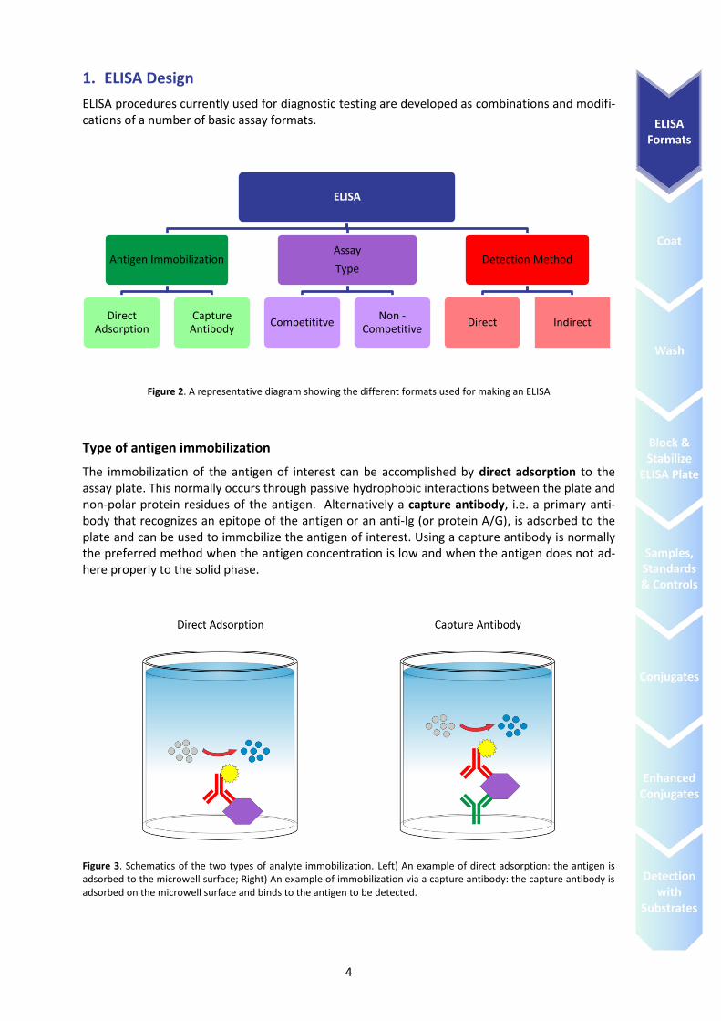
Figure 2. A representative diagram showing the different formats used for making an ELISA
Type of antigen immobilization
The immobilization of the antigen of interest can be accomplished by direct adsorption to the assay plate. This normally occurs through passive hydrophobic interactions between the plate and non-polar protein residues of the antigen. Alternatively a capture antibody, i.e. a primary anti-body that recognizes an epitope of the antigen or an anti-Ig (or protein A/G), is adsorbed to the plate and can be used to immobilize the antigen of interest. Using a capture antibody is normally the preferred method when the antigen concentration is low and when the antigen does not ad-here properly to the solid phase.
Figure 3. Schematics of the two types of analyte immobilization. Left) An example of direct adsorption: the antigen is adsorbed to the microwell surface; Right) An example of immobilization via a capture antibody: the capture antibody is adsorbed on the microwell surface and binds to the antigen to be detected.
ELISA
Antigen Immobilization
Direct Adsorption
Capture Antibody
Assay
Type
Competititve Non -
Competitive
Detection Method
Direct Indirect
5
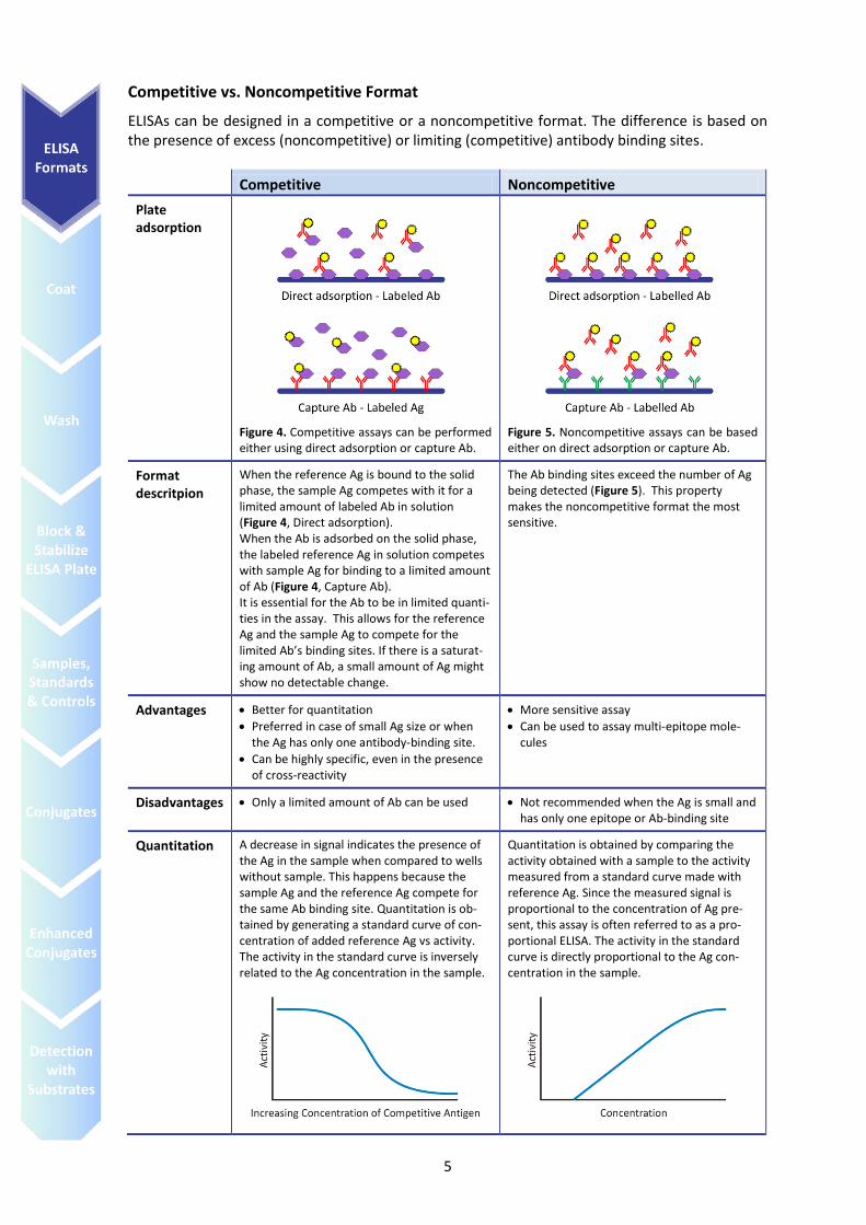
Competitive vs. Noncompetitive Format
ELISAs can be designed in a competitive or a noncompetitive format. The difference is based on the presence of excess (noncompetitive) or limiting (competitive) antibody binding sites.
Competitive Noncompetitive
Plate adsorption
Figure 4. Competitive assays can be performed either using direct adsorption or capture Ab.
Figure 5. Noncompetitive assays can be based either on direct adsorption or capture Ab.
Format descritpion
When the reference Ag is bound to the solid phase, the sample Ag competes with it for a limited amount of labeled Ab in solution (Figure 4, Direct adsorption). When the Ab is adsorbed on the solid phase, the labeled reference Ag in solution competes with sample Ag for binding to a limited amount of Ab (Figure 4, Capture Ab). It is essential for the Ab to be in limited quanti-ties in the assay. This allows for the reference Ag and the sample Ag to compete for the limited Ab’s binding sites. If there is a saturat-ing amount of Ab, a small amount of Ag might show no detectable change.
The Ab binding sites exceed the number of Ag being detected (Figure 5). This property makes the noncompetitive format the most sensitive.
Advantages Better for quantitation
Preferred in case of small Ag size or when the Ag has only one antibody-binding site.
Can be highly specific, even in the presence of cross-reactivity
More sensitive assay
Can be used to assay multi-epitope mole-cules
Disadvantages Only a limited amount of Ab can be used Not recommended when the Ag is small and has only one epitope or Ab-binding site
Quantitation A decrease in signal indicates the presence of the Ag in the sample when compared to wells without sample. This happens because the sample Ag and the reference Ag compete for the same Ab binding site. Quantitation is ob-tained by generating a standard curve of con-centration of added reference Ag vs activity. The activity in the standard curve is inversely related to the Ag concentration in the sample.
Quantitation is obtained by comparing the activity obtained with a sample to the activity measured from a standard curve made with reference Ag. Since the measured signal is proportional to the concentration of Ag pre-sent, this assay is often referred to as a pro-portional ELISA. The activity in the standard curve is directly proportional to the Ag con-centration in the sample.
6
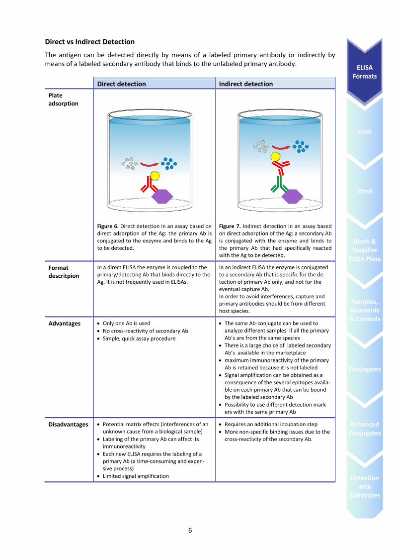
Direct vs Indirect Detection
The antigen can be detected directly by means of a labeled primary antibody or indirectly by means of a labeled secondary antibody that binds to the unlabeled primary antibody.
Direct detection Indirect detection
Plate adsorption
Figure 6. Direct detection in an assay based on direct adsorption of the Ag: the primary Ab is conjugated to the enzyme and binds to the Ag to be detected.
Figure 7. Indirect detection in an assay based on direct adsorption of the Ag: a secondary Ab is conjugated with the enzyme and binds to the primary Ab that had specifically reacted with the Ag to be detected.
Format descritpion
In a direct ELISA the enzyme is coupled to the primary/detecting Ab that binds directly to the Ag. It is not frequently used in ELISAs.
In an indirect ELISA the enzyme is conjugated to a secondary Ab that is specific for the de-tection of primary Ab only, and not for the eventual capture Ab. In order to avoid interferences, capture and primary antibodies should be from different host species.
Advantages Only one Ab is used
No cross-reactivity of secondary Ab
Simple, quick assay procedure
The same Ab-conjugate can be used to analyze different samples if all the primary Ab’s are from the same species
There is a large choice of labeled secondary Ab’s available in the marketplace
maximum immunoreactivity of the primary Ab is retained because it is not labeled
Signal amplification can be obtained as a consequence of the several epitopes availa-ble on each primary Ab that can be bound by the labeled secondary Ab
Possibility to use different detection mark-ers with the same primary Ab
Disadvantages Potential matrix effects (interferences of an unknown cause from a biological sample)
Labeling of the primary Ab can affect its immunoreactivity
Each new ELISA requires the labeling of a primary Ab (a time-consuming and expen-sive process)
Limited signal amplification
Requires an additional incubation step
More non-specific binding issues due to the cross-reactivity of the secondary Ab.
7
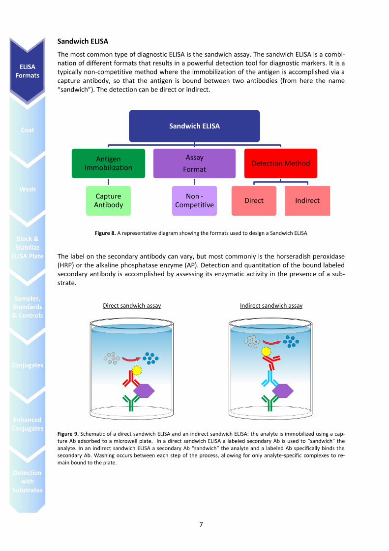
Sandwich ELISA
The most common type of diagnostic ELISA is the sandwich assay. The sandwich ELISA is a combi-nation of different formats that results in a powerful detection tool for diagnostic markers. It is a typically non-competitive method where the immobilization of the antigen is accomplished via a capture antibody, so that the antigen is bound between two antibodies (from here the name “sandwich”). The detection can be direct or indirect.
Figure 8. A representative diagram showing the formats used to design a Sandwich ELISA
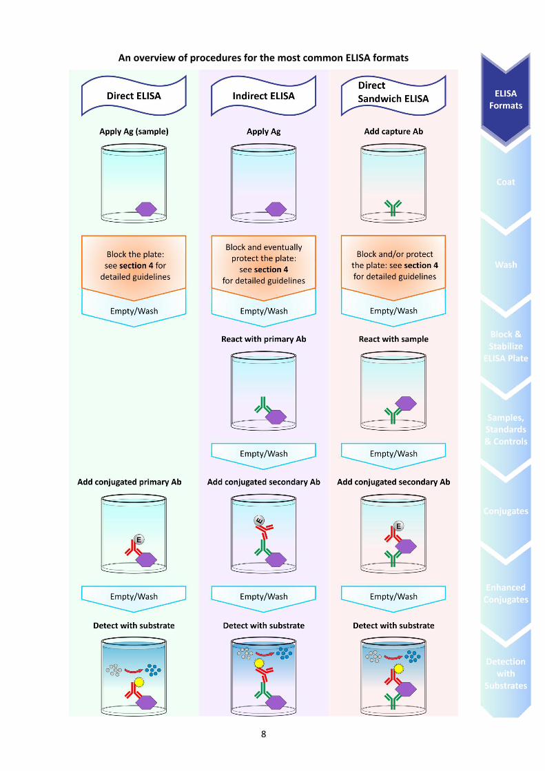
The label on the secondary antibody can vary, but most commonly is the horseradish peroxidase (HRP) or the alkaline phosphatase enzyme (AP). Detection and quantitation of the bound labeled secondary antibody is accomplished by assessing its enzymatic activity in the presence of a sub-strate.
Figure 9. Schematic of a direct sandwich ELISA and an indirect sandwich ELISA: the analyte is immobilized using a cap-ture Ab adsorbed to a microwell plate. In a direct sandwich ELISA a labeled secondary Ab is used to “sandwich” the analyte. In an indirect sandwich ELISA a secondary Ab “sandwich” the analyte and a labeled Ab specifically binds the secondary Ab. Washing occurs between each step of the process, allowing for only analyte-specific complexes to re-main bound to the plate.
Sandwich ELISA
Antigen Immobilization
Capture Antibody
Assay
Format
Non -Competitive
Detection Method
Direct Indirect
8
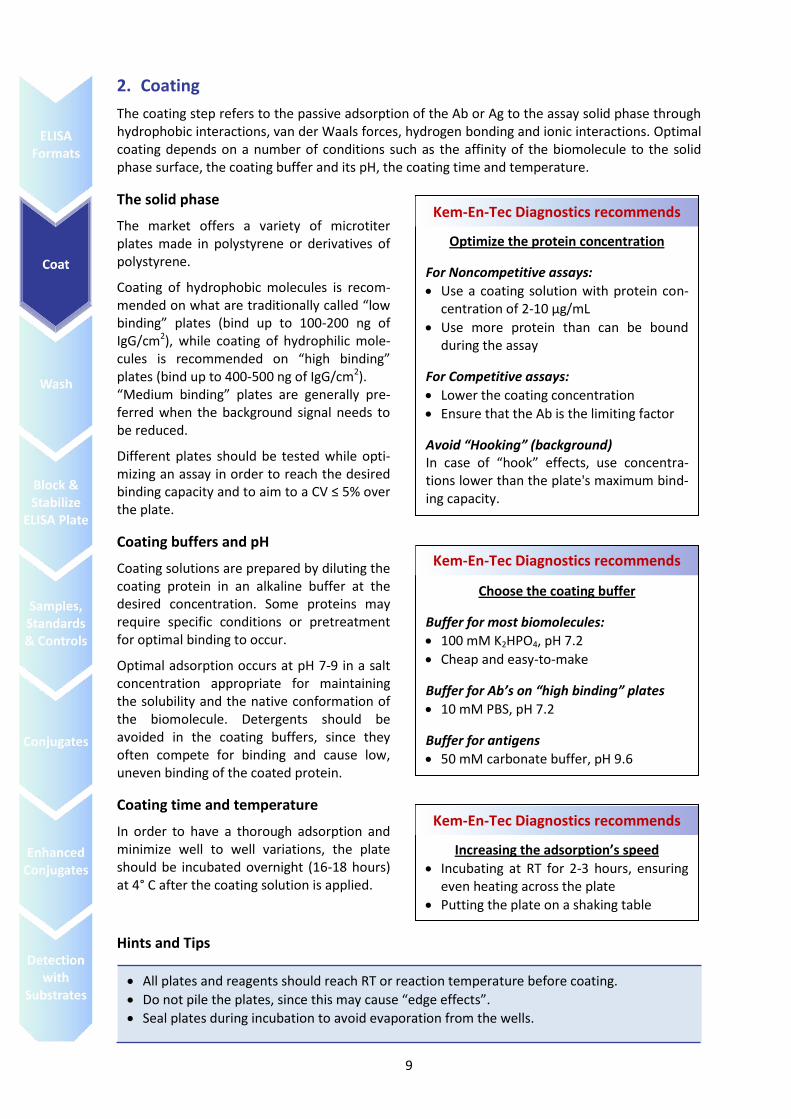
An overview of procedures for the most common ELISA formats
9
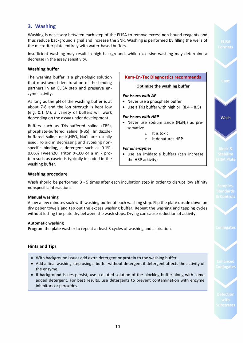
2. Coating
The coating step refers to the passive adsorption of the Ab or Ag to the assay solid phase through hydrophobic interactions, van der Waals forces, hydrogen bonding and ionic interactions. Optimal coating depends on a number of conditions such as the affinity of the biomolecule to the solid phase surface, the coating buffer and its pH, the coating time and temperature.
The solid phase
The market offers a variety of microtiter plates made in polystyrene or derivatives of polystyrene.
Coating of hydrophobic molecules is recom-mended on what are traditionally called “low binding” plates (bind up to 100-200 ng of IgG/cm2), while coating of hydrophilic mole-cules is recommended on “high binding” plates (bind up to 400-500 ng of IgG/cm2). “Medium binding” plates are generally pre-ferred when the background signal needs to be reduced.
Different plates should be tested while opti-mizing an assay in order to reach the desired binding capacity and to aim to a CV ≤ 5% over the plate.
Coating buffers and pH
Coating solutions are prepared by diluting the coating protein in an alkaline buffer at the desired concentration. Some proteins may require specific conditions or pretreatment for optimal binding to occur.
Optimal adsorption occurs at pH 7-9 in a salt concentration appropriate for maintaining the solubility and the native conformation of the biomolecule. Detergents should be avoided in the coating buffers, since they often compete for binding and cause low, uneven binding of the coated protein.
Coating time and temperature
In order to have a thorough adsorption and minimize well to well variations, the plate should be incubated overnight (16-18 hours) at 4° C after the coating solution is applied.
Hints and Tips
Optimize the protein concentration
For Noncompetitive assays:
Use a coating solution with protein con-centration of 2-10 μg/mL
Use more protein than can be bound during the assay
For Competitive assays:
Lower the coating concentration
Ensure that the Ab is the limiting factor
Avoid “Hooking” (background) In case of “hook” effects, use concentra-tions lower than the plate's maximum bind-ing capacity.
Kem-En-Tec Diagnostics recommends
All plates and reagents should reach RT or reaction temperature before coating.
Do not pile the plates, since this may cause “edge effects”.
Seal plates during incubation to avoid evaporation from the wells.
Choose the coating buffer
Buffer for most biomolecules:
100 mM K2HPO4, pH 7.2
Cheap and easy-to-make
Buffer for Ab’s on “high binding” plates
10 mM PBS, pH 7.2
Buffer for antigens
50 mM carbonate buffer, pH 9.6
Kem-En-Tec Diagnostics recommends
Increasing the adsorption’s speed
Incubating at RT for 2-3 hours, ensuring even heating across the plate
Putting the plate on a shaking table
Kem-En-Tec Diagnostics recommends
10
3. Washing
Washing is necessary between each step of the ELISA to remove excess non-bound reagents and thus reduce background signal and increase the SNR. Washing is performed by filling the wells of the microtiter plate entirely with water-based buffers.
Insufficient washing may result in high background, while excessive washing may determine a decrease in the assay sensitivity.
Washing buffer
The washing buffer is a physiologic solution that must avoid denaturation of the binding partners in an ELISA step and preserve en-zyme activity.
As long as the pH of the washing buffer is at about 7-8 and the ion strength is kept low (e.g. 0.1 M), a variety of buffers will work depending on the assay under development.
Buffers such as Tris-buffered saline (TBS), phosphate-buffered saline (PBS), Imidazole-buffered saline or K2HPO4-NaCl are usually used. To aid in decreasing and avoiding non-specific binding, a detergent such as 0.1%-0.05% Tween20, Triton X-100 or a milk pro-tein such as casein is typically included in the washing buffer.
Washing procedure
Wash should be performed 3 - 5 times after each incubation step in order to disrupt low affinity nonspecific interactions.
Manual washing Allow a few minutes soak with washing buffer at each washing step. Flip the plate upside down on dry paper towels and tap out the excess washing buffer. Repeat the washing and tapping cycles without letting the plate dry between the wash steps. Drying can cause reduction of activity.
Automatic washing Program the plate washer to repeat at least 3 cycles of washing and aspiration.
Hints and Tips
With background issues add extra detergent or protein to the washing buffer.
Add a final washing step using a buffer without detergent if detergent affects the activity of the enzyme.
If background issues persist, use a diluted solution of the blocking buffer along with some added detergent. For best results, use detergents to prevent contamination with enzyme inhibitors or peroxides.
Optimize the washing buffer
For issues with AP
Never use a phosphate buffer
Use a Tris buffer with high pH (8.4 – 8.5)
For issues with HRP
Never use sodium azide (NaN3) as pre-servative
o It is toxic o It denatures HRP
For all enzymes
Use an imidazole buffers (can increase the HRP activity)
Kem-En-Tec Diagnostics recommends
11
4. Blocking and stabilizing ELISA plates
Blocking
After coating, the unoccupied sites on the microplate’s wells surface must be blocked in order to reduce/prevent non-specific binding in the next steps of the ELISA. If blocking fails it will result in high background, low specificity and low sensitivity, i.e. low signal-to-noise ratio (SNR).
Blocking buffer The blocking buffer is a solution of compounds that passively adsorbs to unoccupied binding sites on the surface of the plate. The ideal blocking buffer improves the SNR by:
Blocking all potential binding sites of nonspecific interaction
Reducing background signal therefore improving the assay sensitivity
Not altering or obscuring the epitope for Ab binding
When preparing a blocking solution, optimization of the blocking step is required. Improper opti-mization may results in excessive background when the concentration of blocking agent is too low; or in enzyme inhibition and masking of the Ab-Ag interactions when the concentration of blocking agent is too high.
Categories of blocking agents Traditionally there are two categories of blocking agents: detergents and proteins.
Most of the detergents used as blockers are non-ionic compounds that disrupt non-specific pro-tein interactions. Nevertheless detergents may disrupts hydrophobic interactions, may leave hy-drophilic sites unblocked or may interfere with the enzyme activity. Moreover detergents can be washed away by washing buffers that contain detergents.
Proteins block both the hydrophilic and hydrophobic sites on the solid phase but enhance the risk of cross-reactions, tend to be inconsistent from batch-to-batch and unstable in solution. A com-monly used blocking agent is bovine serum albumin (BSA), which is relatively inexpensive, effi-cient and biochemically inactive. However, BSA is heavily regulated worldwide because of the risk of contamination by the prion responsible for the bovine spongiform encephalopathy.
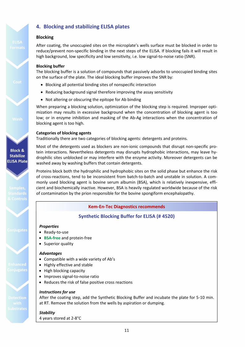
Synthetic Blocking Buffer for ELISA (# 4520)
Properties
Ready-to-use
BSA-free and protein-free
Superior quality
Advantages
Compatible with a wide variety of Ab’s
Highly effective and stable
High blocking capacity
Improves signal-to-noise ratio
Reduces the risk of false positive cross reactions
Instructions for use After the coating step, add the Synthetic Blocking Buffer and incubate the plate for 5-10 min. at RT. Remove the solution from the wells by aspiration or dumping.
Stability 4 years stored at 2-8°C
Kem-En-Tec Diagnostics recommends
12
Plate stabilization
When storage of coated plates is required, a stabilization buffer is necessary in order to prevent loss of activity of the adsorbed biomolecules. This procedure traditionally did require few addi-tional steps in the assay development.
Drying conditions should be optimized for a particular molecules being adsorbed before full scale processing begins. This can be done by coating, drying and storing a set of plates for at least 1 - 2 days and comparing the activity to freshly made plates using the same batch of coating reagent. Traditional stabilization methods require the protecting buffer to be easy to remove with a wash-ing step prior the use of the stored plate: uneasy removal can cause interference with the assay signal (background).
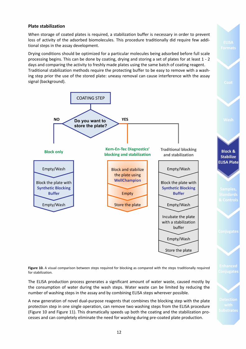
Figure 10. A visual comparison between steps required for blocking as compared with the steps traditionally required for stabilization.
The ELISA production process generates a significant amount of water waste, caused mostly by the consumption of water during the wash steps. Water waste can be limited by reducing the number of washing steps in the assay and by combining ELISA steps wherever possible.
A new generation of novel dual-purpose reagents that combines the blocking step with the plate protection step in one single operation, can remove two washing steps from the ELISA procedure (Figure 10 and Figure 11). This dramatically speeds up both the coating and the stabilization pro-cesses and can completely eliminate the need for washing during pre-coated plate production.
13
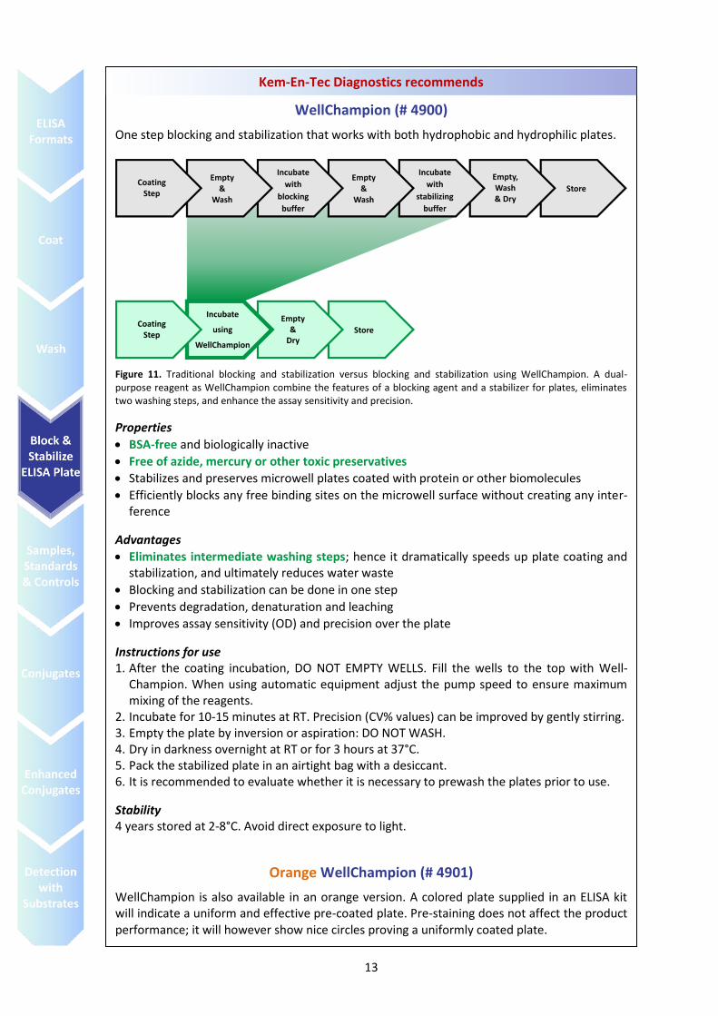
WellChampion (# 4900)
One step blocking and stabilization that works with both hydrophobic and hydrophilic plates.
Figure 11. Traditional blocking and stabilization versus blocking and stabilization using WellChampion. A dual-purpose reagent as WellChampion combine the features of a blocking agent and a stabilizer for plates, eliminates two washing steps, and enhance the assay sensitivity and precision.
Properties
BSA-free and biologically inactive
Free of azide, mercury or other toxic preservatives
Stabilizes and preserves microwell plates coated with protein or other biomolecules
Efficiently blocks any free binding sites on the microwell surface without creating any inter-ference
Advantages
Eliminates intermediate washing steps; hence it dramatically speeds up plate coating and stabilization, and ultimately reduces water waste
Blocking and stabilization can be done in one step
Prevents degradation, denaturation and leaching
Improves assay sensitivity (OD) and precision over the plate
Instructions for use 1. After the coating incubation, DO NOT EMPTY WELLS. Fill the wells to the top with Well-
Champion. When using automatic equipment adjust the pump speed to ensure maximum mixing of the reagents.
2. Incubate for 10-15 minutes at RT. Precision (CV% values) can be improved by gently stirring. 3. Empty the plate by inversion or aspiration: DO NOT WASH. 4. Dry in darkness overnight at RT or for 3 hours at 37°C. 5. Pack the stabilized plate in an airtight bag with a desiccant. 6. It is recommended to evaluate whether it is necessary to prewash the plates prior to use.
Stability 4 years stored at 2-8°C. Avoid direct exposure to light.
Orange WellChampion (# 4901)
WellChampion is also available in an orange version. A colored plate supplied in an ELISA kit will indicate a uniform and effective pre-coated plate. Pre-staining does not affect the product performance; it will however show nice circles proving a uniformly coated plate.
Kem-En-Tec Diagnostics recommends
14
5. Dilution of samples, standards & controls
Biological samples such as plasma, urine, saliva, serum, milk, cells and tissues extracts, contain the analyte to be quantified. The target antigen must also be present in known amounts in standard and control solutions.
Sample dilution
Samples tested in an immunoassay can require dilution for several reasons. Among others, these reasons include:
The Hook Effect can occur when an overwhelming amount of Ag is present in an assay and the sample reads as a false-negative. It is thought that the excess Ag binds to the receptor sites, preventing the conjugate antigen from being bound. Assaying a diluted serum will usually unmask this phenomenon and allow for an accurate measurement.
Complex sample matrices, such as serum and plasma, may contain interfering factors that af-fect the ability of the assay to accurately quantify the target analyte. Strong interferences are often caused by rheumatoid factors and HAMA’s. This matrix effect can cause high back-ground in the negative control or false negatives in the sample measurement. To reduce this effect the sample should be diluted, i.e. to show assay linearity.
The samples tested in an immunoassay might require dilution to be read within the dynamic range of the assay, i.e. to ensure that the signal response is within the standard curve.
It is important that buffers designed for sample dilution also reduces matrix effects and minimize nonspecific binding interactions in order to optimize the assay without affecting the correct bind-ing of the Ab or Ag.
Hints and Tips
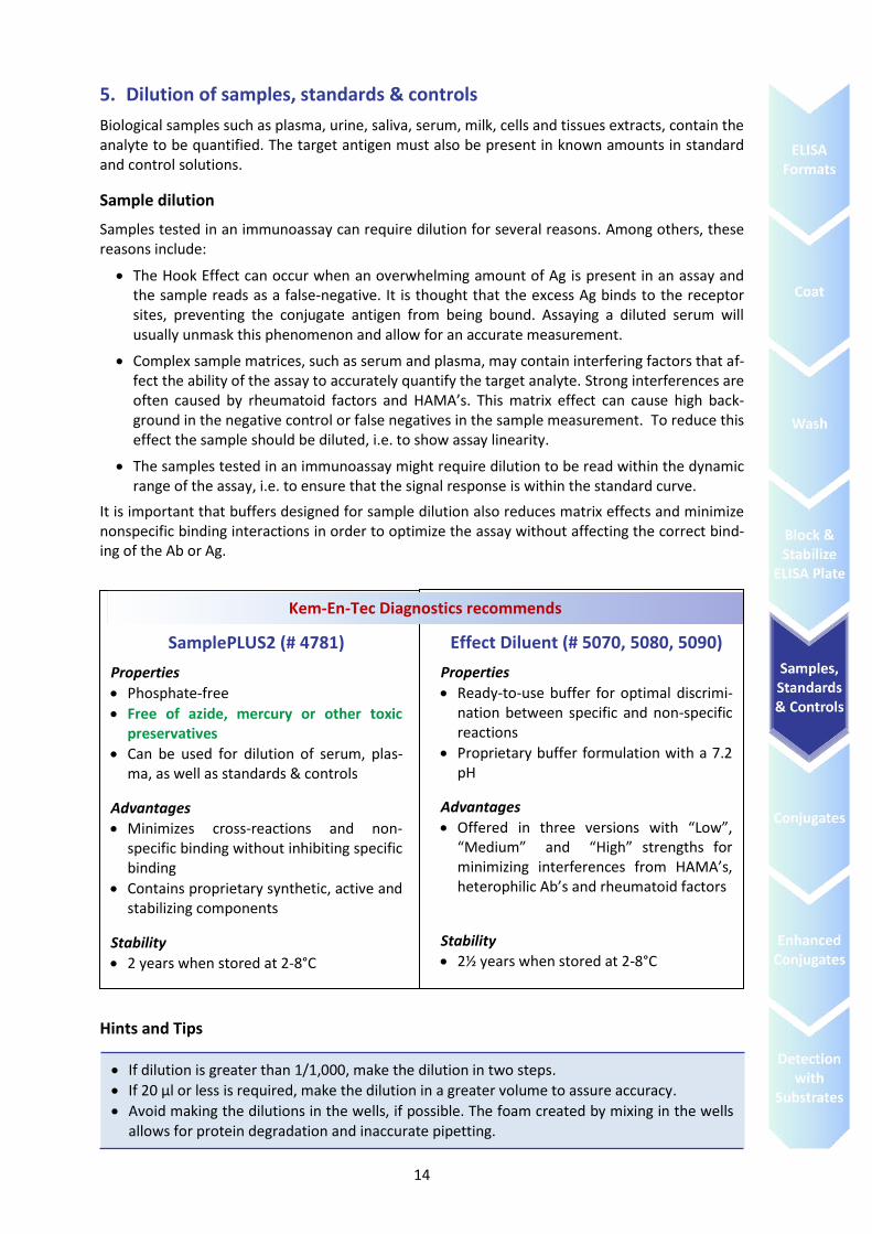
SamplePLUS2 (# 4781)
Properties
Phosphate-free
Free of azide, mercury or other toxic preservatives
Can be used for dilution of serum, plas-ma, as well as standards & controls
Advantages
Minimizes cross-reactions and non-specific binding without inhibiting specific binding
Contains proprietary synthetic, active and stabilizing components
Stability
2 years when stored at 2-8°C
Effect Diluent (# 5070, 5080, 5090)
Properties
Ready-to-use buffer for optimal discrimi-nation between specific and non-specific reactions
Proprietary buffer formulation with a 7.2 pH
Advantages
Offered in three versions with “Low”, “Medium” and “High” strengths for minimizing interferences from HAMA’s, heterophilic Ab’s and rheumatoid factors
Stability
2½ years when stored at 2-8°C
Kem-En-Tec Diagnostics recommends
If dilution is greater than 1/1,000, make the dilution in two steps.
If 20 µl or less is required, make the dilution in a greater volume to assure accuracy.
Avoid making the dilutions in the wells, if possible. The foam created by mixing in the wells allows for protein degradation and inaccurate pipetting.
15
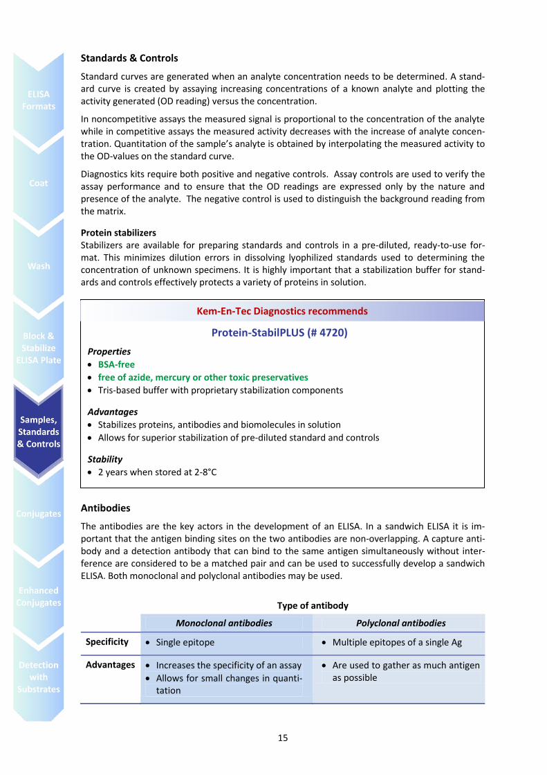
Standards & Controls
Standard curves are generated when an analyte concentration needs to be determined. A stand-ard curve is created by assaying increasing concentrations of a known analyte and plotting the activity generated (OD reading) versus the concentration.
In noncompetitive assays the measured signal is proportional to the concentration of the analyte while in competitive assays the measured activity decreases with the increase of analyte concen-tration. Quantitation of the sample’s analyte is obtained by interpolating the measured activity to the OD-values on the standard curve.
Diagnostics kits require both positive and negative controls. Assay controls are used to verify the assay performance and to ensure that the OD readings are expressed only by the nature and presence of the analyte. The negative control is used to distinguish the background reading from the matrix.
Protein stabilizers Stabilizers are available for preparing standards and controls in a pre-diluted, ready-to-use for-mat. This minimizes dilution errors in dissolving lyophilized standards used to determining the concentration of unknown specimens. It is highly important that a stabilization buffer for stand-ards and controls effectively protects a variety of proteins in solution.
Antibodies
The antibodies are the key actors in the development of an ELISA. In a sandwich ELISA it is im-portant that the antigen binding sites on the two antibodies are non-overlapping. A capture anti-body and a detection antibody that can bind to the same antigen simultaneously without inter-ference are considered to be a matched pair and can be used to successfully develop a sandwich ELISA. Both monoclonal and polyclonal antibodies may be used.
Type of antibody
Monoclonal antibodies Polyclonal antibodies
Specificity Single epitope Multiple epitopes of a single Ag
Advantages Increases the specificity of an assay
Allows for small changes in quanti-tation
Are used to gather as much antigen as possible
Protein-StabilPLUS (# 4720)
Properties
BSA-free
free of azide, mercury or other toxic preservatives
Tris-based buffer with proprietary stabilization components
Advantages
Stabilizes proteins, antibodies and biomolecules in solution
Allows for superior stabilization of pre-diluted standard and controls
Stability
2 years when stored at 2-8°C
Kem-En-Tec Diagnostics recommends
16
6. Dilution and stabilization of the conjugate
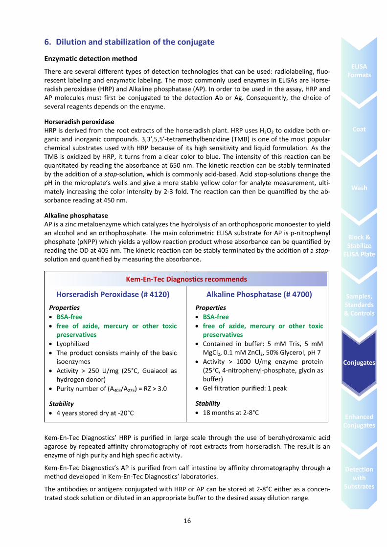
Enzymatic detection method
There are several different types of detection technologies that can be used: radiolabeling, fluo-rescent labeling and enzymatic labeling. The most commonly used enzymes in ELISAs are Horse-radish peroxidase (HRP) and Alkaline phosphatase (AP). In order to be used in the assay, HRP and AP molecules must first be conjugated to the detection Ab or Ag. Consequently, the choice of several reagents depends on the enzyme.
Horseradish peroxidase HRP is derived from the root extracts of the horseradish plant. HRP uses H2O2 to oxidize both or-ganic and inorganic compounds. 3,3′,5,5′-tetramethylbenzidine (TMB) is one of the most popular chemical substrates used with HRP because of its high sensitivity and liquid formulation. As the TMB is oxidized by HRP, it turns from a clear color to blue. The intensity of this reaction can be quantitated by reading the absorbance at 650 nm. The kinetic reaction can be stably terminated by the addition of a stop-solution, which is commonly acid-based. Acid stop-solutions change the pH in the microplate’s wells and give a more stable yellow color for analyte measurement, ulti-mately increasing the color intensity by 2-3 fold. The reaction can then be quantified by the ab-sorbance reading at 450 nm.
Alkaline phosphatase AP is a zinc metaloenzyme which catalyzes the hydrolysis of an orthophosporic monoester to yield an alcohol and an orthophosphate. The main colorimetric ELISA substrate for AP is p-nitrophenyl phosphate (pNPP) which yields a yellow reaction product whose absorbance can be quantified by reading the OD at 405 nm. The kinetic reaction can be stably terminated by the addition of a stop-solution and quantified by measuring the absorbance.
Kem-En-Tec Diagnostics’ HRP is purified in large scale through the use of benzhydroxamic acid agarose by repeated affinity chromatography of root extracts from horseradish. The result is an enzyme of high purity and high specific activity.
Kem-En-Tec Diagnostics’s AP is purified from calf intestine by affinity chromatography through a method developed in Kem-En-Tec Diagnostics’ laboratories.
The antibodies or antigens conjugated with HRP or AP can be stored at 2-8°C either as a concen-trated stock solution or diluted in an appropriate buffer to the desired assay dilution range.
Horseradish Peroxidase (# 4120)
Properties
BSA-free
free of azide, mercury or other toxic preservatives
Lyophilized
The product consists mainly of the basic isoenzymes
Activity > 250 U/mg (25°C, Guaiacol as hydrogen donor)
Purity number of (A403/A275) = RZ > 3.0
Stability
4 years stored dry at -20°C
Alkaline Phosphatase (# 4700)
Properties
BSA-free
free of azide, mercury or other toxic preservatives
Contained in buffer: 5 mM Tris, 5 mM MgCl2, 0.1 mM ZnCl2, 50% Glycerol, pH 7
Activity > 1000 U/mg enzyme protein (25°C, 4-nitrophenyl-phosphate, glycin as buffer)
Gel filtration purified: 1 peak
Stability
18 months at 2-8°C
Kem-En-Tec Diagnostics recommends
17
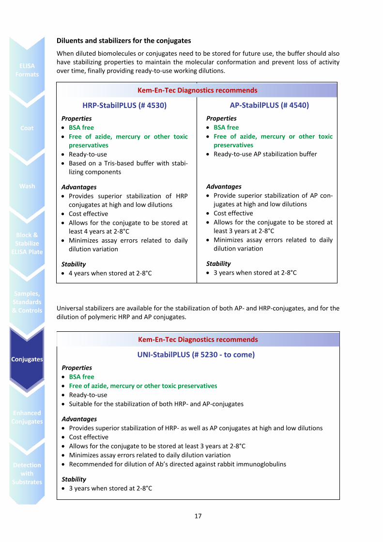
Diluents and stabilizers for the conjugates
When diluted biomolecules or conjugates need to be stored for future use, the buffer should also have stabilizing properties to maintain the molecular conformation and prevent loss of activity over time, finally providing ready-to-use working dilutions.
Universal stabilizers are available for the stabilization of both AP- and HRP-conjugates, and for the dilution of polymeric HRP and AP conjugates.
HRP-StabilPLUS (# 4530)
Properties
BSA free
Free of azide, mercury or other toxic preservatives
Ready-to-use
Based on a Tris-based buffer with stabi-lizing components
Advantages
Provides superior stabilization of HRP conjugates at high and low dilutions
Cost effective
Allows for the conjugate to be stored at least 4 years at 2-8°C
Minimizes assay errors related to daily dilution variation
Stability
4 years when stored at 2-8°C
AP-StabilPLUS (# 4540)
Properties
BSA free
Free of azide, mercury or other toxic preservatives
Ready-to-use AP stabilization buffer
Advantages
Provide superior stabilization of AP con-jugates at high and low dilutions
Cost effective
Allows for the conjugate to be stored at least 3 years at 2-8°C
Minimizes assay errors related to daily dilution variation
Stability
3 years when stored at 2-8°C
Kem-En-Tec Diagnostics recommends
UNI-StabilPLUS (# 5230 - to come)
Properties
BSA free
Free of azide, mercury or other toxic preservatives
Ready-to-use
Suitable for the stabilization of both HRP- and AP-conjugates
Advantages
Provides superior stabilization of HRP- as well as AP conjugates at high and low dilutions
Cost effective
Allows for the conjugate to be stored at least 3 years at 2-8°C
Minimizes assay errors related to daily dilution variation
Recommended for dilution of Ab’s directed against rabbit immunoglobulins
Stability
3 years when stored at 2-8°C
Kem-En-Tec Diagnostics recommends
18
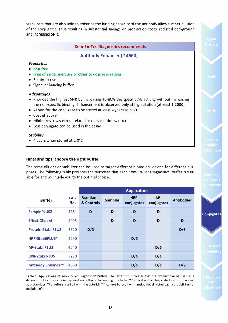
Stabilizers that are also able to enhance the binding capacity of the antibody allow further dilution of the conjugates, thus resulting in substantial savings on production costs, reduced background and increased SNR.
Hints and tips: choose the right buffer
The same diluent or stabilizer can be used to target different biomolecules and for different pur-poses. The following table presents the purposes that each Kem-En-Tec Diagnostics’ buffer is suit-able for and will guide you to the optimal choice.
Application
Buffer cat. No.
Standards & Controls
Samples HRP-
conjugates AP-
conjugates Antibodies
SamplePLUS2 4781 D D D D
Effect Diluent 5095 D D D D
Protein-StabilPLUS 4720 D/S D/S
HRP-StabilPLUS* 4530 D/S
AP-StabilPLUS 4540 D/S
UNI-StabilPLUS 5230 D/S D/S
Antibody Enhancer* 4660 D/S D/S D/S
Table 1. Applications of Kem-En-Tec Diagnostics’ buffers. The letter “D” indicates that the product can be used as a diluent for the corresponding application in the table heading; the letter “S” indicates that the product can also be used as a stabilizer. The buffers marked with the asterisk “*” cannot be used with antibodies directed against rabbit immu-noglobulin’s.
Antibody Enhancer (# 4660)
Properties
BSA free
Free of azide, mercury or other toxic preservatives
Ready-to-use
Signal enhancing buffer
Advantages
Provides the highest SNR by increasing 40-80% the specific Ab activity without increasing the non-specific binding. Enhancement is observed only at high dilution (at least 1:2000).
Allows for the conjugate to be stored at least 4 years at 2-8°C
Cost effective
Minimizes assay errors related to daily dilution variation
Less conjugate can be used in the assay
Stability
4 years when stored at 2-8°C
Kem-En-Tec Diagnostics recommends
19
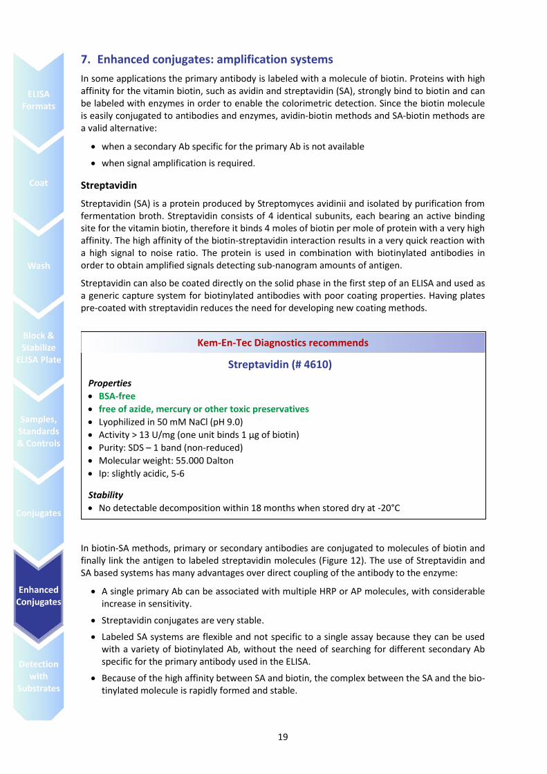
7. Enhanced conjugates: amplification systems
In some applications the primary antibody is labeled with a molecule of biotin. Proteins with high affinity for the vitamin biotin, such as avidin and streptavidin (SA), strongly bind to biotin and can be labeled with enzymes in order to enable the colorimetric detection. Since the biotin molecule is easily conjugated to antibodies and enzymes, avidin-biotin methods and SA-biotin methods are a valid alternative:
when a secondary Ab specific for the primary Ab is not available
when signal amplification is required.
Streptavidin
Streptavidin (SA) is a protein produced by Streptomyces avidinii and isolated by purification from fermentation broth. Streptavidin consists of 4 identical subunits, each bearing an active binding site for the vitamin biotin, therefore it binds 4 moles of biotin per mole of protein with a very high affinity. The high affinity of the biotin-streptavidin interaction results in a very quick reaction with a high signal to noise ratio. The protein is used in combination with biotinylated antibodies in order to obtain amplified signals detecting sub-nanogram amounts of antigen.
Streptavidin can also be coated directly on the solid phase in the first step of an ELISA and used as a generic capture system for biotinylated antibodies with poor coating properties. Having plates pre-coated with streptavidin reduces the need for developing new coating methods.
In biotin-SA methods, primary or secondary antibodies are conjugated to molecules of biotin and finally link the antigen to labeled streptavidin molecules (Figure 12). The use of Streptavidin and SA based systems has many advantages over direct coupling of the antibody to the enzyme:
A single primary Ab can be associated with multiple HRP or AP molecules, with considerable increase in sensitivity.
Streptavidin conjugates are very stable.
Labeled SA systems are flexible and not specific to a single assay because they can be used with a variety of biotinylated Ab, without the need of searching for different secondary Ab specific for the primary antibody used in the ELISA.
Because of the high affinity between SA and biotin, the complex between the SA and the bio-tinylated molecule is rapidly formed and stable.
Streptavidin (# 4610)
Properties
BSA-free
free of azide, mercury or other toxic preservatives
Lyophilized in 50 mM NaCl (pH 9.0)
Activity > 13 U/mg (one unit binds 1 μg of biotin)
Purity: SDS – 1 band (non-reduced)
Molecular weight: 55.000 Dalton
Ip: slightly acidic, 5-6
Stability
No detectable decomposition within 18 months when stored dry at -20°C
Stable over a wide range of pH and decompose only in the presence of SDS > 60°C
Kem-En-Tec Diagnostics recommends
20
Enhanced conjugates
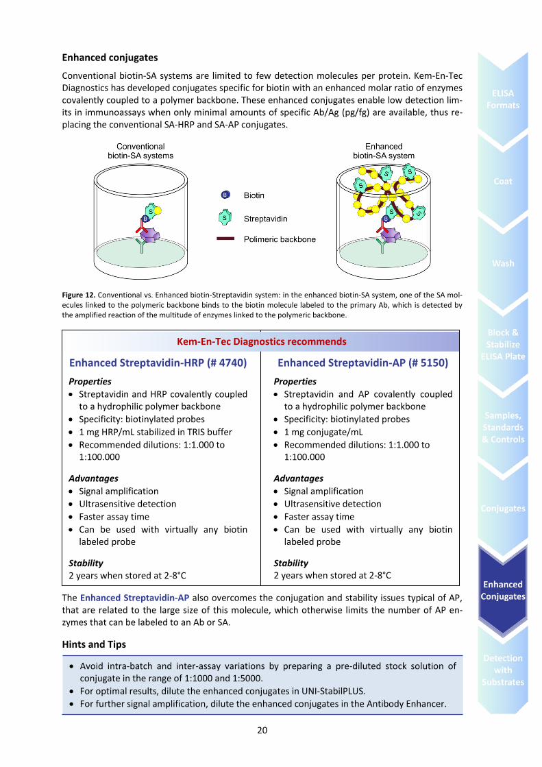
Conventional biotin-SA systems are limited to few detection molecules per protein. Kem-En-Tec Diagnostics has developed conjugates specific for biotin with an enhanced molar ratio of enzymes covalently coupled to a polymer backbone. These enhanced conjugates enable low detection lim-its in immunoassays when only minimal amounts of specific Ab/Ag (pg/fg) are available, thus re-placing the conventional SA-HRP and SA-AP conjugates.
Figure 12. Conventional vs. Enhanced biotin-Streptavidin system: in the enhanced biotin-SA system, one of the SA mol-ecules linked to the polymeric backbone binds to the biotin molecule labeled to the primary Ab, which is detected by the amplified reaction of the multitude of enzymes linked to the polymeric backbone.
The Enhanced Streptavidin-AP also overcomes the conjugation and stability issues typical of AP, that are related to the large size of this molecule, which otherwise limits the number of AP en-zymes that can be labeled to an Ab or SA.
Hints and Tips
Enhanced Streptavidin-HRP (# 4740)
Properties
Streptavidin and HRP covalently coupled to a hydrophilic polymer backbone
Specificity: biotinylated probes
1 mg HRP/mL stabilized in TRIS buffer
Recommended dilutions: 1:1.000 to 1:100.000
Advantages
Signal amplification
Ultrasensitive detection
Faster assay time
Can be used with virtually any biotin labeled probe
Stability 2 years when stored at 2-8°C
Enhanced Streptavidin-AP (# 5150)
Properties
Streptavidin and AP covalently coupled to a hydrophilic polymer backbone
Specificity: biotinylated probes
1 mg conjugate/mL
Recommended dilutions: 1:1.000 to 1:100.000
Advantages
Signal amplification
Ultrasensitive detection
Faster assay time
Can be used with virtually any biotin labeled probe
Stability 2 years when stored at 2-8°C
Kem-En-Tec Diagnostics recommends
Avoid intra-batch and inter-assay variations by preparing a pre-diluted stock solution of conjugate in the range of 1:1000 and 1:5000.
For optimal results, dilute the enhanced conjugates in UNI-StabilPLUS.
For further signal amplification, dilute the enhanced conjugates in the Antibody Enhancer.
21
8. Substrates
AP- and HRP-conjugates can be detected and quantitated using chromogenic substrates that re-sult in soluble, colored products when development takes place. The levels of the enzymatic reac-tion, and consequently the analyte’s concentration, are determined by monitoring the signal de-velopment at specific wavelengths. The most commonly used chromogenic ELISA substrates are 3,3',5,5'-Tetramethylbenzidine (TMB) for HRP and p-NitroPhenyl Phosphate (pNPP) for AP.
Substrate type Specificity Reaction
color Reaction read at
Stopped reaction
Stopped reaction read at
pNPP AP Yellow 405 nm Yellow 405 nm
TMB HRP Blue 620-655 nm Yellow 450 nm
p-NitroPhenyl Phosphate (pNPP)
Kem-En-Tec Diagnostics’s pNPP substrates are liquid buffer solutions containing stabilized pNPP. In the presence of AP, pNPP is hydrolyzed rapidly to p-nitrophenol and inorganic phosphate. The reaction results in a water-soluble yellow end product (p-nitrophenol) that absorbs light at 405 nm. The reaction can be stopped by adding an equal amount of 1 M sodium hydroxide (NaOH) solution and the absorbance read at 405 nm. The intensity of the color is directly proportional to the concentration of the reactant.
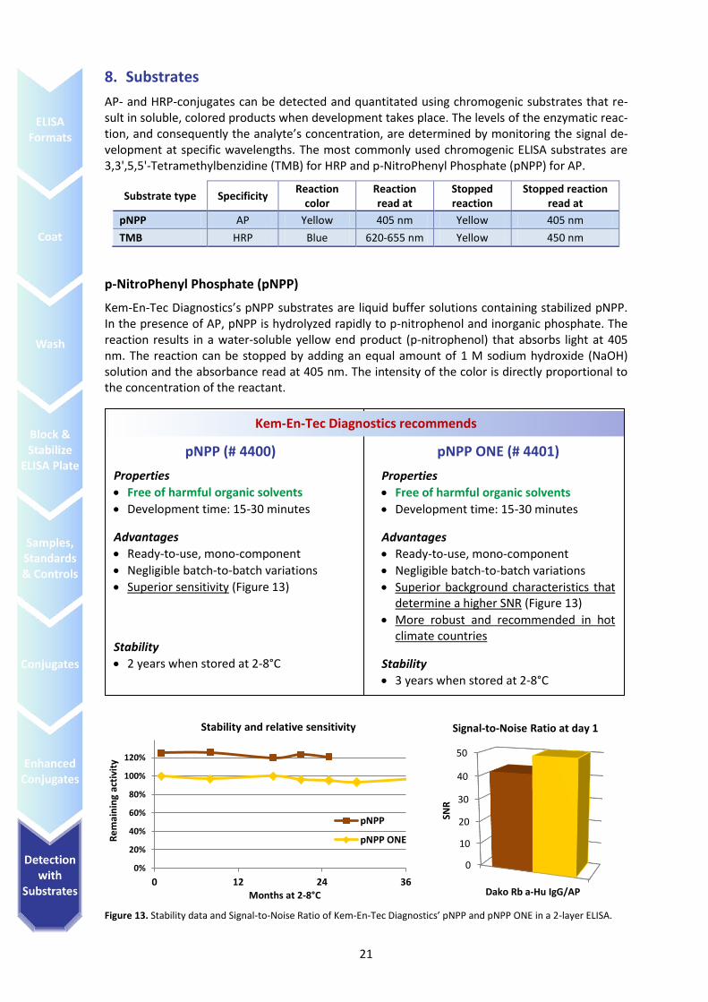
Figure 13. Stability data and Signal-to-Noise Ratio of Kem-En-Tec Diagnostics’ pNPP and pNPP ONE in a 2-layer ELISA.
0
10
20
30
40
50
Dako Rb a-Hu IgG/AP
SNR
Signal-to-Noise Ratio at day 1
0%
20%
40%
60%
80%
100%
120%
0 12 24 36
Re
mai
nin
g ac
tivi
ty
Months at 2-8°C
pNPP
pNPP ONE
Stability and relative sensitivity
pNPP (# 4400)
Properties
Free of harmful organic solvents
Development time: 15-30 minutes
Advantages
Ready-to-use, mono-component
Negligible batch-to-batch variations
Superior sensitivity (Figure 13)
Stability
2 years when stored at 2-8°C
pNPP ONE (# 4401)
Properties
Free of harmful organic solvents
Development time: 15-30 minutes
Advantages
Ready-to-use, mono-component
Negligible batch-to-batch variations
Superior background characteristics that determine a higher SNR (Figure 13)
More robust and recommended in hot climate countries
Stability
3 years when stored at 2-8°C
Kem-En-Tec Diagnostics recommends
22
Tetramethylbenzidine
Kem-En-Tec Diagnostics’ TMB solutions combine TMB, buffer and H2O2 for a ready-to-use, chro-mogenic system that yields a blue color whose absorbance is read at 620-655 nm. The reaction can be stopped with sulphuric acid resulting in a yellow color with an absorbance read at 450 nm and a 2-3 fold increase in sensitivity compared to the non-stopped absorbance reading. The stop-solutions recommended below guarantee an extended stop stability of the measured absorbance, with an OD decreases after 1 hour lower than 8%.
Prestained versions All TMB substrates are also available as unique color-coded products for traceable pipetting. The red indicator makes manual pipetting traceable, while in automatic analyzers, it offers a conven-ient tracking system for reagent supply. The color disappears after stopping and does not inter-fere with the assay.
TMB ONE (# 4380) and Prestained TMB ONE (# 4430)
Properties
Free of harmful organic solvents
Development time: 5-30 minutes
Stop solution 0.2 M H2SO4
Advantages
Ready-to-use
Mono-component
Negligible batch-to-batch variations
Extremely stable and robust
Stability
4 years when stored at 2-8°C
1 year when stored at RT
TMB PLUS2 (# 4395) and Prestained TMB PLUS2 (# 4445)
Properties
Free of harmful organic solvents
Development time: 5-30 minutes
Stop solution 0.2 M H2SO4
Advantages
Ready-to-use
Mono-component
Negligible batch-to-batch variations
Cost effective
Improved version of the TMB PLUS
Stability
4 years when stored at 2-8°C
1 year when stored at RT
Kem-En-Tec Diagnostics recommends
TMB X-tra (# 4800) and Prestained TMB X-tra (# 4810)
Properties
Free of harmful organic solvents
Development time: 5-30 minutes
Stop solution 0.2 M H2SO4
Advantages
Ready-to-use
Mono-component
Negligible batch-to-batch variations
High signal for a fast assay reaction
Resistant to raised temperatures
Stability
4 years when stored at 2-8°C
1 year when stored at RT
TMB SENS (# 4850) and Prestained TMB SENS (# 4860)
Properties
Free of harmful organic solvents
Development time: 5-15 minutes
Stop solution 0.3 M H2SO4
Advantages
Ready-to-use
Mono-component
Negligible batch-to-batch variations
Highly superior sensitivity
Fast assay development
Stability
2½ years when stored at 2-8°C
5 months when stored at RT
23
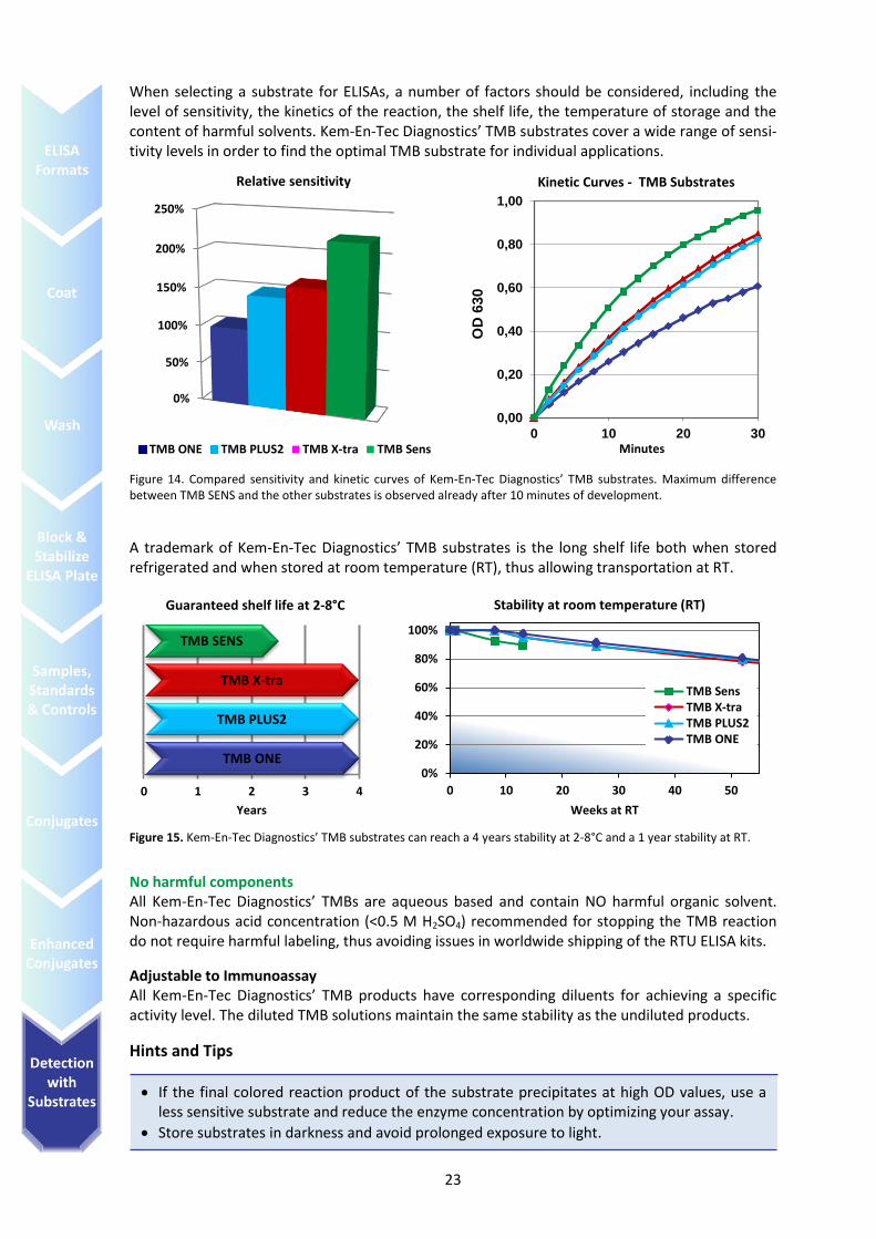
When selecting a substrate for ELISAs, a number of factors should be considered, including the level of sensitivity, the kinetics of the reaction, the shelf life, the temperature of storage and the content of harmful solvents. Kem-En-Tec Diagnostics’ TMB substrates cover a wide range of sensi-tivity levels in order to find the optimal TMB substrate for individual applications.
Figure 14. Compared sensitivity and kinetic curves of Kem-En-Tec Diagnostics’ TMB substrates. Maximum difference between TMB SENS and the other substrates is observed already after 10 minutes of development.
A trademark of Kem-En-Tec Diagnostics’ TMB substrates is the long shelf life both when stored refrigerated and when stored at room temperature (RT), thus allowing transportation at RT.
Figure 15. Kem-En-Tec Diagnostics’ TMB substrates can reach a 4 years stability at 2-8°C and a 1 year stability at RT.
No harmful components All Kem-En-Tec Diagnostics’ TMBs are aqueous based and contain NO harmful organic solvent. Non-hazardous acid concentration (<0.5 M H2SO4) recommended for stopping the TMB reaction do not require harmful labeling, thus avoiding issues in worldwide shipping of the RTU ELISA kits.
Adjustable to Immunoassay All Kem-En-Tec Diagnostics’ TMB products have corresponding diluents for achieving a specific activity level. The diluted TMB solutions maintain the same stability as the undiluted products.
Hints and Tips
0%
50%
100%
150%
200%
250%
Relative sensitivity
TMB ONE TMB PLUS2 TMB X-tra TMB Sens
0,00
0,20
0,40
0,60
0,80
1,00
0 10 20 30
OD
630
Minutes
Kinetic Curves - TMB Substrates
0 1 2 3 4
Years
Guaranteed shelf life at 2-8°C
TMB ONE
TMB PLUS2
TMB X-tra
TMB SENS
0%
20%
40%
60%
80%
100%
0 10 20 30 40 50
Weeks at RT
Stability at room temperature (RT)
TMB SensTMB X-traTMB PLUS2TMB ONE
If the final colored reaction product of the substrate precipitates at high OD values, use a less sensitive substrate and reduce the enzyme concentration by optimizing your assay.
Store substrates in darkness and avoid prolonged exposure to light.
24
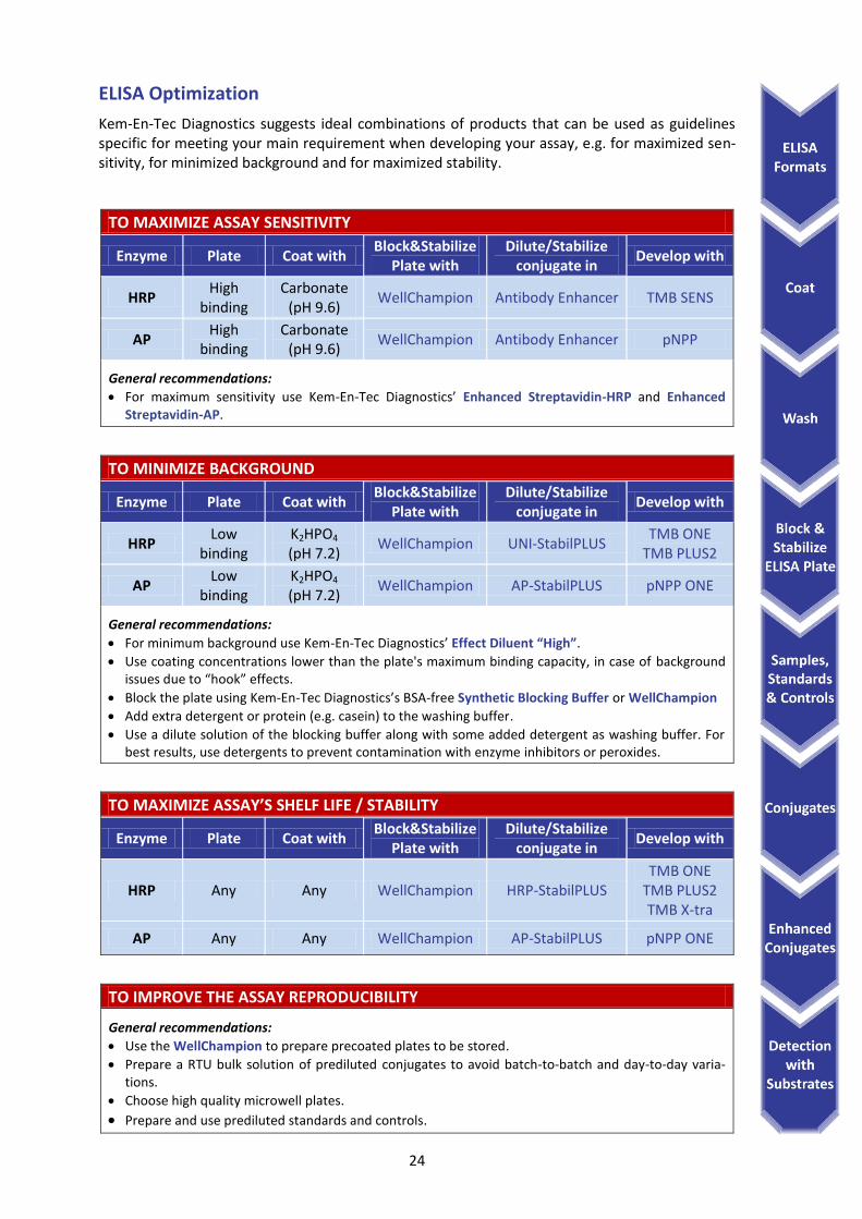
ELISA Optimization
Kem-En-Tec Diagnostics suggests ideal combinations of products that can be used as guidelines specific for meeting your main requirement when developing your assay, e.g. for maximized sen-sitivity, for minimized background and for maximized stability.
TO MAXIMIZE ASSAY SENSITIVITY
Enzyme Plate Coat with Block&Stabilize
Plate with Dilute/Stabilize
conjugate in Develop with
HRP High
binding Carbonate
(pH 9.6) WellChampion Antibody Enhancer TMB SENS
AP High
binding Carbonate
(pH 9.6) WellChampion Antibody Enhancer pNPP
General recommendations:
For maximum sensitivity use Kem-En-Tec Diagnostics’ Enhanced Streptavidin-HRP and Enhanced Streptavidin-AP.
TO MINIMIZE BACKGROUND
Enzyme Plate Coat with Block&Stabilize
Plate with Dilute/Stabilize
conjugate in Develop with
HRP Low
binding K2HPO4 (pH 7.2)
WellChampion UNI-StabilPLUS TMB ONE
TMB PLUS2
AP Low
binding K2HPO4 (pH 7.2)
WellChampion AP-StabilPLUS pNPP ONE
General recommendations:
For minimum background use Kem-En-Tec Diagnostics’ Effect Diluent “High”.
Use coating concentrations lower than the plate's maximum binding capacity, in case of background issues due to “hook” effects.
Block the plate using Kem-En-Tec Diagnostics’s BSA-free Synthetic Blocking Buffer or WellChampion
Add extra detergent or protein (e.g. casein) to the washing buffer.
Use a dilute solution of the blocking buffer along with some added detergent as washing buffer. For best results, use detergents to prevent contamination with enzyme inhibitors or peroxides.
TO MAXIMIZE ASSAY’S SHELF LIFE / STABILITY
Enzyme Plate Coat with Block&Stabilize
Plate with Dilute/Stabilize
conjugate in Develop with
HRP Any Any WellChampion HRP-StabilPLUS TMB ONE
TMB PLUS2 TMB X-tra
AP Any Any WellChampion AP-StabilPLUS pNPP ONE
TO IMPROVE THE ASSAY REPRODUCIBILITY
General recommendations:
Use the WellChampion to prepare precoated plates to be stored.
Prepare a RTU bulk solution of prediluted conjugates to avoid batch-to-batch and day-to-day varia-tions.
Choose high quality microwell plates.
Prepare and use prediluted standards and controls.
25
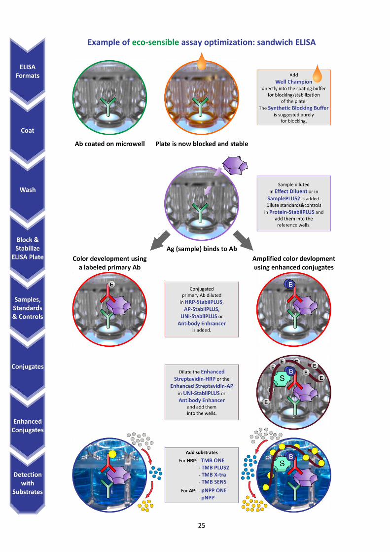
Example of eco-sensible assay optimization: sandwich ELISA
26
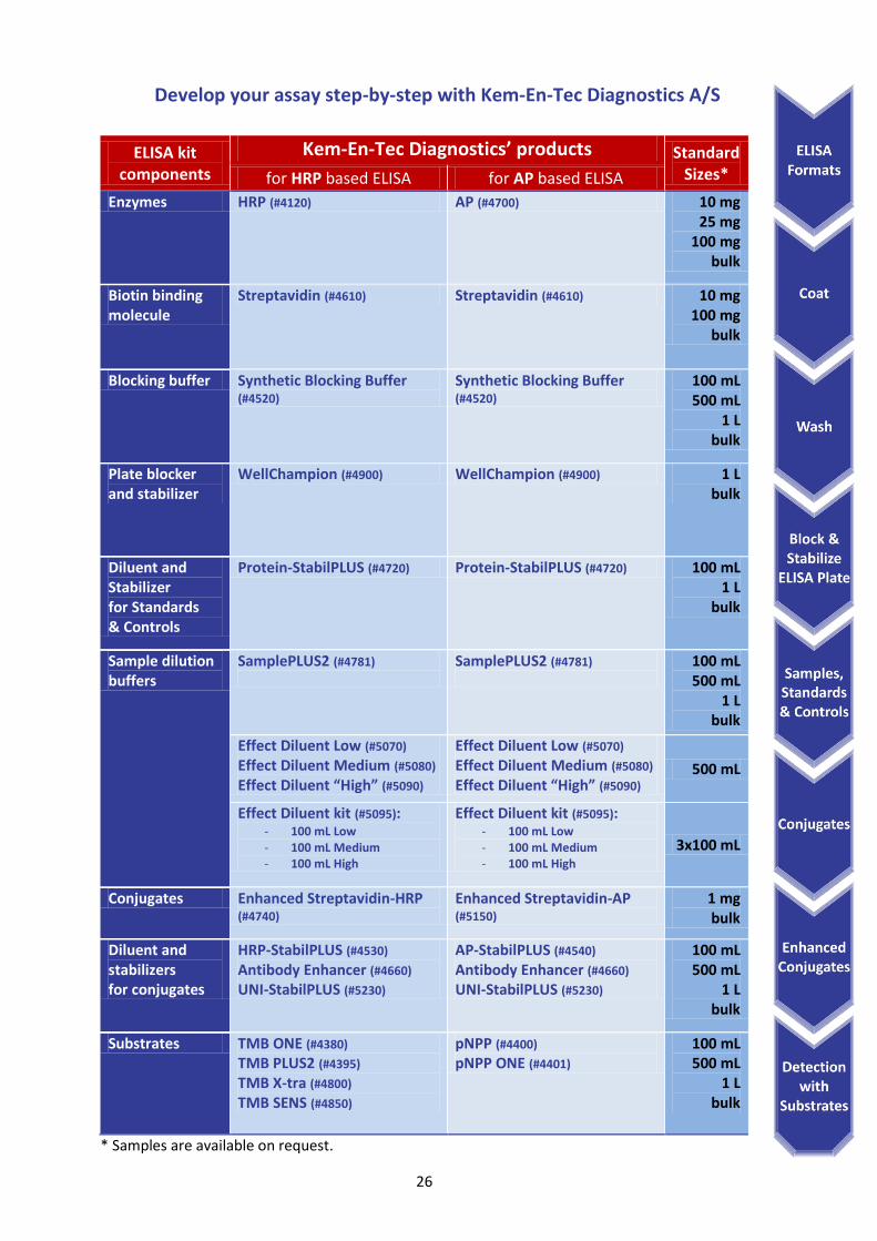
Develop your assay step-by-step with Kem-En-Tec Diagnostics A/S
ELISA kit components
Kem-En-Tec Diagnostics’ products Standard Sizes* for HRP based ELISA for AP based ELISA
Enzymes HRP (#4120) AP (#4700) 10 mg 25 mg
100 mg bulk
Biotin binding molecule
Streptavidin (#4610) Streptavidin (#4610) 10 mg 100 mg
bulk
Blocking buffer Synthetic Blocking Buffer (#4520)
Synthetic Blocking Buffer (#4520)
100 mL 500 mL
1 L bulk
Plate blocker and stabilizer
WellChampion (#4900) WellChampion (#4900) 1 L bulk
Diluent and Stabilizer for Standards & Controls
Protein-StabilPLUS (#4720) Protein-StabilPLUS (#4720) 100 mL 1 L
bulk
Sample dilution buffers
SamplePLUS2 (#4781)
SamplePLUS2 (#4781)
100 mL 500 mL
1 L bulk
Effect Diluent Low (#5070) Effect Diluent Medium (#5080)
Effect Diluent “High” (#5090)
Effect Diluent Low (#5070) Effect Diluent Medium (#5080)
Effect Diluent “High” (#5090) 500 mL
Effect Diluent kit (#5095): - 100 mL Low - 100 mL Medium - 100 mL High
Effect Diluent kit (#5095): - 100 mL Low - 100 mL Medium - 100 mL High
3x100 mL
Conjugates Enhanced Streptavidin-HRP (#4740)
Enhanced Streptavidin-AP (#5150)
1 mg bulk
Diluent and stabilizers for conjugates
HRP-StabilPLUS (#4530) Antibody Enhancer (#4660) UNI-StabilPLUS (#5230)
AP-StabilPLUS (#4540) Antibody Enhancer (#4660) UNI-StabilPLUS (#5230)
100 mL 500 mL
1 L bulk
Substrates TMB ONE (#4380) TMB PLUS2 (#4395) TMB X-tra (#4800) TMB SENS (#4850)
pNPP (#4400)
pNPP ONE (#4401) 100 mL 500 mL
1 L bulk
* Samples are available on request.

27
Bottling & Packaging
Kem-En-Tec Diagnostics can customize products as well as packaging in our state-of-the art facility to meet different dispensing needs (8 mL – 10 L). Our specialists can assist you with customized packaging and label design. Total batch volumes are up to 1200 L.
We monitor every step of the manufacturing process to ensure that our dispensed product have the same quality as our batch source. Automated liquid filling guarantees specific batch toleranc-es by precision weighing.
Manufacturing & Quality Assurance
Every lot released by Kem-En-Tec Diagnostics has undergone extensive Quality Control at every significant step, assuring our customers a high level of product validation. As an ongoing process we identify and manage critical process parameters.
Our ISO based QA System supports:
Change Control Notification
Batch Sample Storage
Document Control
Audit Availability
Batch Record Availability
Non-conforming Action Procedures Kem-En-Tec Diagnostics can handle our customer’s needs as they grow, ensuring:
Experience in process scale up and validation
Extensive refrigeration space
Filling lines that can accommodate large scale demands
Contact Kem-En-Tec Diagnostics A/S
To order or for Technical Support, please contact Kem-En-Tec Diagnostics A/S at:
Support: [email protected]
Sales: [email protected]
Orders: [email protected]
USA customers: [email protected]
For more information about Kem-En-Tec Diagnostics A/S, visit www.kem-en-tec.com or email us at [email protected].
Kem-En-Tec Diagnostics A/S Kuldyssen 10
DK-2630 Taastrup Denmark
Phone: +45 3927 1771 Fax: +45 3920 0178
www.kem-en-tec.com
US Contact:
83 Maple Avenue, Windsor, Hartford,
CT 06095, USA Phone: 860 298 0234
Fax 860 298 8586 [email protected]
©Copyright 2013, Kem-En-Tec Diagnostics A/S, All right reserved. ELISA GUIDE 13-02