Embed Size (px)
Citation preview
Effects of nitrogen starvation on two microalgae 275
Lat. Am. J. Aquat. Res., 44(2): 275-292, 2016
DOI: 10.3856/vol44-issue2-fulltext-9
Research Article
Growth, nutrient uptake and chemical composition of Chlorella sp.
and Nannochloropsis oculata under nitrogen starvation
Caroline R.P.S. Paes1, Gabrielle R. Faria
1, Natália A.B. Tinoco
1, Dominique J.F.A. Castro
1
Elisabete Barbarino1,2
& Sergio O. Lourenço1
1Universidade Federal Fluminense, Departamento de Biologia Marinha
CEP 24001-970, Niterói, RJ, Brazil 2Universidade Federal Fluminense, Programa de Pós-Graduação em Química, Niterói, RJ, Brazil
Corresponding author: Elisabete Barbarino ([email protected])
ABSTRACT. The production of microalgae biomass shows wide valuable uses, in the aquaculture,
biotechnology, and food science, among others. However, microalgae show fluctuations in their chemical profile generated mainly by the culture conditions. This study was designated to assess the effects of nitrogen starvation
on growth, nutrient uptake, and gross chemical composition of Chlorella sp. and Nannochloropsis oculata. The control experiments were performed with Conway culture medium in 13-day batch cultures, 12 h photoperiod,
and aeration. A second experimental condition was the addition of the nutrients except nitrogen, one week after the start of growth (experiments designed as N-). Cell yield were similar in the control and in the N- experiments
for both species. Cell biovolumes did not vary over growth in the control, but both microalgae exhibited larger cell biovolumes in N- experiments, probably as a consequence of the higher accumulation of storage substances.
Dissolved nitrogen was exhausted before the end of the experiments, but phosphorus was not totally consumed. Protein and total carotenoid did not vary from the exponential to the stationary growth phase of the control in
both species. For Chlorella sp., concentrations of lipid did not vary in the control either, but there was a significant increase of carbohydrate over growth. In the N- experiment, concentrations of all substances varied
throughout growth of Chlorella sp., except lipid. For N. oculata, all substances exhibited significant variations over growth, except protein and total carotenoid in the control. Protein and chlorophyll-a concentrations
decreased over growth in N- experiments for both species. In contrast, concentrations of carbohydrate increased throughout growth in N- experiments, especially in Chlorella sp. Nitrogen starvation caused accumulation of
carbohydrate, but increments of lipid were restricted to N. oculata. Both species showed a fast growth, but the small content of lipid in Chlorella sp. is unfavorable for its use as a food-species in a monospecific diet in
mariculture, and as a feedstock for biodiesel production. N. oculata is a lipid-rich species, and its lipid content can be successfully incremented through nitrogen starvation. This species is promising in uses that demand high
concentrations of lipid, such as the production of biodiesel.
Keywords: biodiesel, biomass, cultivation, lipid, mariculture, productivity.
Crecimiento, absorción de nutrientes y composición química de Chlorella sp.
y Nannochloropsis oculata bajo carencia de nitrógeno
RESUMEN. La producción de biomasa de microalgas presenta variados y valiosos usos en la acuicultura, biotecnología y ciencias de los alimentos, entre otros. Sin embargo, las microalgas muestran fluctuaciones en su
perfil químico causado principalmente por las condiciones de cultivo. En este estudio se evaluó los efectos de la carencia de nitrógeno sobre el crecimiento, absorción de nutrientes y composición química bruta de Chlorella
sp. y Nannochloropsis oculata. Los experimentos control se efectuaron en un medio de cultivo Conway durante 13 días, 12 h de fotoperiodo y aeración. Una segunda condición experimental fue la adición de nutrientes,
excepto nitrógeno, una semana después del inicio del crecimiento (denominados experimentos N-). Los
rendimientos celulares fueron similares en el control y en los experimentos N- en ambas especies. Los biovolúmenes celulares no variaron con el crecimiento en el control, pero ambas microalgas presentaron
mayores biovolúmenes celulares en los experimentos N-, probablemente como consecuencia de una mayor acumulación de sustancias almacenadas. El nitrógeno disuelto se agotó antes del término de los experimentos,
pero el fósforo no fue totalmente consumido. En ambas especies, las proteínas y carotenoides totales no variaron
____________________
Paper presented in the 5th Brazilian Congress of Marine Biology, 17-21 May 2015, Porto de Galinhas, Brazil.
276 Latin American Journal of Aquatic Research
desde la fase exponencial hasta la fase estacionaria de crecimiento del control. En Chlorella sp., las concentraciones de lípidos no variaron en el control, pero se determinó un aumento significativo de
carbohidratos durante el crecimiento. En los experimentos N-, las concentraciones de todas las sustancias variaron durante el crecimiento de Chlorella sp., a excepción de los lípidos. En N. oculata, todas las sustancias
presentaron variaciones significativas durante el crecimiento, excepto proteínas y carotenoides totales en el control. Las concentraciones de proteínas y clorofila-a disminuyeron durante el crecimiento en los experimentos
N- de ambas especies. En contraste, las concentraciones de carbohidratos aumentaron durante el crecimiento en los experimentos N-, especialmente en Chlorella sp. La carencia de nitrógeno causó una acumulación de
carbohidratos, pero el incremento de lípidos se restringió a N. oculata. Ambas especies presentaron un crecimiento rápido, pero el escaso contenido de lípidos en Chlorella sp. es desfavorable para su uso como
alimento en una dieta monoespecífica en acuicultura y como materia prima para la producción de biodiesel. N. oculata es una especie rica en lípidos y su contenido en lípidos puede ser incrementado exitosamente a través
de la carencia de nitrógeno. Esta especie es promisoria en usos que demanden altas concentraciones de lípidos, como la producción de biodiesel.
Palabras clave: biodiesel, biomasa, cultivo, lípidos, maricultura, productividad.
INTRODUCTION
Microalgae are the main components of the phytoplankton, and thus they are the most important primary producers of the majority of aquatic systems
(Falkowski & Raven, 2007). The term phytoplankton encompasses a heterogenous set of microscopic eukaryotic lineages, resulting in a broad diversity of not related groups such as green algae, diatoms, dinofla-gellates, and euglenoids, among others (Graham et al., 2009). Cyanobacteria are prokaryotic, but they are also
included in the phytoplankton due their ecological and evolutionary role as primary producers (Ratti et al., 2013). Marine phytoplankton is often categorized into groups based on taxonomic traits, abundance, role in biogeochemical fluxes, and/or primary production. While diatoms are considered the principal group
contributing to primary production and carbon export in coastal areas, dinoflagellates are important contributors to biomass in stratified or silica-limited areas, and cyanobacteria are the dominant group in offshore continental shelf and oceanic waters (Silva et al., 2009).
Many lineages of microalgae include fast-growing microorganisms with very high growth rates under optimum culture conditions. Fast-growing characteristics
combined with a huge chemical diversity open possible applications of microalgae biomass in many fields, such as aquaculture, biotechnology, and food science (Spolaore et al., 2006; Templeton & Laurens, 2015), for instance. It is expected an increase and a diversification of applications involving microalgae, as
a consequence of the ongoing search for more productive systems to supply the society with food, feedstocks, and high value biochemical products (Zeng et al., 2011; Lee-Chang et al., 2013). Despite microalgae biotechnology is still in its childhood, its development has been done in a context of sustainable
production using modern and efficient processes (Wijffels et al., 2013). Particularly in food science,
microalgae are useful to improve the nutritional content of conventional foods and hence to positively affect human health, due to their favorable chemical composition (Tokuşoglu & Ünal, 2003). In recent years, a growing attention on microalgae focuses on the
possible use of the biomass as a feedstock for biofuel production (Van Iersel & Flammini, 2010; Zeng et al., 2011; Mubarak et al., 2015).
Despite the wide scenarium of possible applications,
microalgae advantages and drawbacks should be
considered without excessive enthusiasm, but exclu-
sively with a scientific approach (Van Iersel &
Flammini, 2010). There are both technological and
biological issues to achieve a broader use of them.
Regarding the technological view, it is necessary to
develop more eficient systems to produce biomass and
valuable microalgae-based products (Wijffels &
Barbosa, 2010). On the other hand, biological issues
include the domestication of promising strains (Lim et
al., 2012), and the successful uses of mechanisms to
stimulate microalgae to grow and produce target
substances (Kaye et al., 2015). All possible
applications of microalgae are directly coupled to high
growth and a favourable chemical profile of the species
(Borges-Campos et al., 2010). Fluctuations in the
chemical profile of microalgae in cultures are a key
issue in their study and applications (Lourenço et al., 2002).
The chemical content of microalgae can vary with
culture age and with changes in culture conditions
(Carvalho et al., 2009). The effect of variation of
culture parameters on many microalgae species has
been studied in order to better understand their
physiology, as well as to answer specific and relevant
questions for mass culture (Grobbelaar, 2014). Data on
the chemical composition of microalgae may also vary widely due to differences of the methods of measure-
ment used (Barbarino & Lourenço, 2005), the physio-
logical state of the microalgae (Geider & La Roche,
Effects of nitrogen starvation on two microalgae 277
2002), as well as to the experimental conditions
applied, like temperature (Durmaz et al., 2009), light
intensity (Lourenço et al., 2008), and culture medium
(Huerlimann et al., 2010), especially in batch cultures.
Due to the interaction of the organisms with the culture
medium, a batch culture is under continuous chemical
change. These variations reflect on the cell metabolism
and consequently on their chemical composition
(Lourenço et al., 2002). Thus, the chemical compo-
sition of a given species may vary widely under
different growth conditions, and such changes may be
related to the growth phase of the culture (Costard et al., 2012). However, studies focusing sampling in
different growth phases are relatively scarce; most of
the papers report the chemical profile of given species
in a fixed momentum of the cultivation, ignoring the
continuing process of interaction between microalgae and the medium.
Nitrogen is added to most culture media in high
concentrations, and changes in N supply are known to
influence strongly both growth and chemical composition
of microalgae (Valenzuela-Espinoza et al., 1999;
Harrison & Berges, 2005). Studies of the effects of
nitrogen sources on the chemical composition in
different growth phases may give important information
on species metabolism (Fidalgo et al., 1995). On the
other hand, this knowledge is also important for both
aquaculture activities and biotechnological applications,
in which different culture media (with nutrients in
various chemical forms) may be used (Sepúlveda et al., 2015). Under many culture conditions, microalgae may
experience nitrogen starvation at least in part of their
growth, especially in the stationary growth phase of
batch cultures (Lourenço et al., 2004). Photosynthetic
activity and nitrogen metabolism are processes
integrally coupled, and a nitrogen limitation affects the
photosynthesis in microalgae by reducing the
efficiency of light collection due a decline in cell
pigment content (Geider et al., 1993). This physio-
logical stress may promote strong effects on the chemical
composition of a given microalga, such as demonstrated
by several authors (e.g., Silva et al., 2009; Jiang et al., 2011; Urreta et al., 2014). Production and accumulation
of protein, carbohydrate, lipid, and carotenoids are of
particular importance if the microalgae are cultivated
either to feed marine animals or to produce specific
valuable substances, for instance (Machado & Lourenço,
2008).
This paper aimed to assess growth, nutrient uptake,
and chemical composition of two marine microalgae,
the trebouxiophycean Chlorella sp. and the eustigma-tophycean Nannochloropsis oculata under standar-
dized culture conditions and under nitrogen starvation.
Growth under nitrogen starvation is thought to induce
protein and chlorophyll decreases, and increments in
carbohydrate and lipid productivity of microalgae, but
the intensity of these processes vary widely depending
on the species tested and experimental conditions. The
effects of the culture conditions on the two microalgae
were discussed in the context of possible use of the
microalgae as food-species in mariculture and as feedstocks for biofuel production.
MATERIALS AND METHODS
The microalgae tested
Two strains were used in this study: Chlorella sp. (division Chlorophyta, class Trebouxiophyceae, strain
CMEA BS04), and Nannochloropsis oculata (division
Heterokontophyta, class Eustigmatophyceae, strain
CMEA MO08). Both strains are available at Elizabeth
Aidar Microalgae Culture Collection, Fluminense
Federal University, Brazil (Lourenço & Vieira, 2004).
Culture conditions
Starter cultures of 150-250 mL in mid-exponential
growth phase were inoculated into 5 L of seawater,
previously autoclaved at 121°C for 60 min in 6-L
borosilicate flasks. The microalgae were cultured in two experimental conditions:
a) Seawater enriched with Conway nutrient solutions
(Walne, 1966) in its original concentrations and
continuously bubbled with filtered air at a rate of 2 L
min-1. This experimental condition is designed as “control”.
b) The same conditions described in item a, including a
new addition of Conway nutrient solutions without
nitrogen in the 7th day of growth. This procedure
promoted the enrichment of the culture medium with
the theoretical concentrations of all elements of the
Conway’s receipt (e.g., 128 μM NaH2PO4.H2O, 1.82
μM MnCl2.4H2O, 4.81 μM FeCl3.6H2O, etc.), except
NaNO3. The actual concentrations of the chemical
components available to the microalgae from the 7th
day of cultivation corresponded to the sum of the
enrichment done plus the residual concentrations of the
original substances that had not been taken up yet. This
experimental condition is designed as “N–”.
Each experiment was carried out in four culture
flasks (n = 4), exposed to 350 μmol photons m-2 s-1
measured with a Biospherical Instruments Quanta
Meter, model QLS100, provided from beneath by 40 W
fluorescent lamps (Sylvania daylight tubes), on a 12:12
h light:dark cycle. Mean temperatures in experiments were 21 ± 1°C and the salinity was 33.0. Growth was
estimated based on daily microscopic cell counting
with Neubauer chambers. Cultures were not buffered
278 Latin American Journal of Aquatic Research
and pH was determined daily, at the beginning (15-30
min after the start of the photoperiod), in the middle (6
h after the start of the photoperiod), and in the end of
the light period (11 h after the start of the photoperiod).
All samplings for cell counts occurred during the first
30 min of the light period. The initial cell densities of
cultures were 3.0x10⁴ cell mL-1 for Chlorella sp. and
2.5x10⁵ cell mL-1 for N. oculata. The experiments were
carried out for 13 days. Growth rates were calculated daily using the following equation:
lnNt - lnNt-1
∆t where r is the growth rate; ln is the logarithm of the
number of cells in a given day, in a standardized time
(beggining of the photoperiod), calculated using the
basis 2; Nt is the number of cells recorded in a given
day (t); Nt–1 is the number of cells 24 h before the
counting carried out in the time t (in days); t is the difference, in days, between the two cell countings.
The biovolumes of the cells were measured using
the equation provided by Hillebrand et al. (1999),
assuming a spherical shape for both microalgae. Mean
volumes were based on measurements of 30 cells in
each culture flask, giving four mean values used for
statistics (n = 4).
Sampling procedure
Each culture was sampled twice for chemical analysis
(on the 7 and 13th days of growth), corresponding to
late-exponential and stationary growth phases. Samples
of 1.6 to 2.3 L were concentrated by centrifugation at
8,000 x g for 9.0 min, at least once, using a Sigma
centrifuge, model σ-15, to obtain highly concentrated
pellets. Before the last centrifugation, cells were
washed with artificial seawater (Kester et al., 1967)
prepared without nitrogen, phosphorus and vitamins,
and adjusted to salinity of 10. All supernatants obtained
for each sample were combined and the number of cells
was determined (using Neubauer chambers) to quantify
possible cell losses. The pellets were frozen at -18°C
and then freeze-dried (using a Terroni-Fauvel, model
LB 1500 device), weighed and stored in desiccators
under vacuum and protected from light until the
chemical analyses. Samples to be analyzed for chloro-
phyll and carotenoid were obtained by filtering the
cultures under vacuum onto Whatman GF/F® glass
microfibre filters (0.7 μm nominal pore size). The
filtered samples were kept at -18°C in flasks containing
silica-gel until analysis. All sampling for chemical
analysis was done during the first 90 min of the light period.
Daily samples of the culture medium were taken to
evaluate the uptake of dissolved nutrients by the
microalgae. At the first 30 min of the photoperiod,
samples of 40 to 60 mL of the cultures from each flask
were collected. The samples were filtered in the same
manner as described above for photosynthetic pigments.
The filtered samples of culture media were kept at
-18°C in polyethylene flasks until analysis of dissolved
nutrients.
Chemical analysis
Total nitrogen and phosphorus were determined by peroxymonosulfuric acid digestion, using a Hach diges-
tor (Digesdhal®, Hach Co., model 23.130-20) (Hach et al., 1987). Calibration curves were prepared using NH4Cl and NaHPO4 as standards of nitrogen and
phosphorus, respectively. See Lourenço et al. (2005) for further details.
The Lowry et al. (1951) method was used to
evaluate protein in the samples, with bovine serum
albumin as a protein standard, following the extraction procedures proposed by Barbarino & Lourenço (2005).
Spectrophotometric determinations were done at 750 nm, 35 min after the start of the chemical reaction. Total
carbohydrate was extracted with 80% H2SO4, accor-
ding to Myklestad & Haug (1972). The carbohydrate concentration was determined spectrophotometrically
at 485 nm, 30 min after the start of the chemical reaction, by the phenol-sulfuric acid method (Dubois et al., 1956), using glucose as a standard. Total lipid was extracted following Folch et al. (1957), and determined
gravimetrically after solvent evaporation.
Chlorophyll-a and carotenoid were extracted in
90% acetone at 4°C for 20 h, after grinding the filters with the samples. Spectrophotometric determination of
chlorophyll-a was carried out as described by Jeffrey & Humphrey (1975), and the determination of total
carotenoid was carried out as described by Strickland
& Parsons (1968).
Determinations of ammonia + ammonium followed the procedure proposed by Aminot & Chaussepied
(1983), nitrite and nitrate analyses were performed following Strickland & Parsons (1968), and phosphate
was determined according to Grasshoff et al. (1983).
All nutrients were analyzed spectrophotometrically.
Statistical analysis
The results of growth and chemical composition were
compared using Student's t-test (Zar, 1996), adopting a level of significance = 0.05.
All experiments were performed twice in order to confirm the results. The chemical analyses were also done twice, using samples generated by all experiments carried out. In this paper, we show the results of only
one set of experiments.
r =
Effects of nitrogen starvation on two microalgae 279
RESULTS
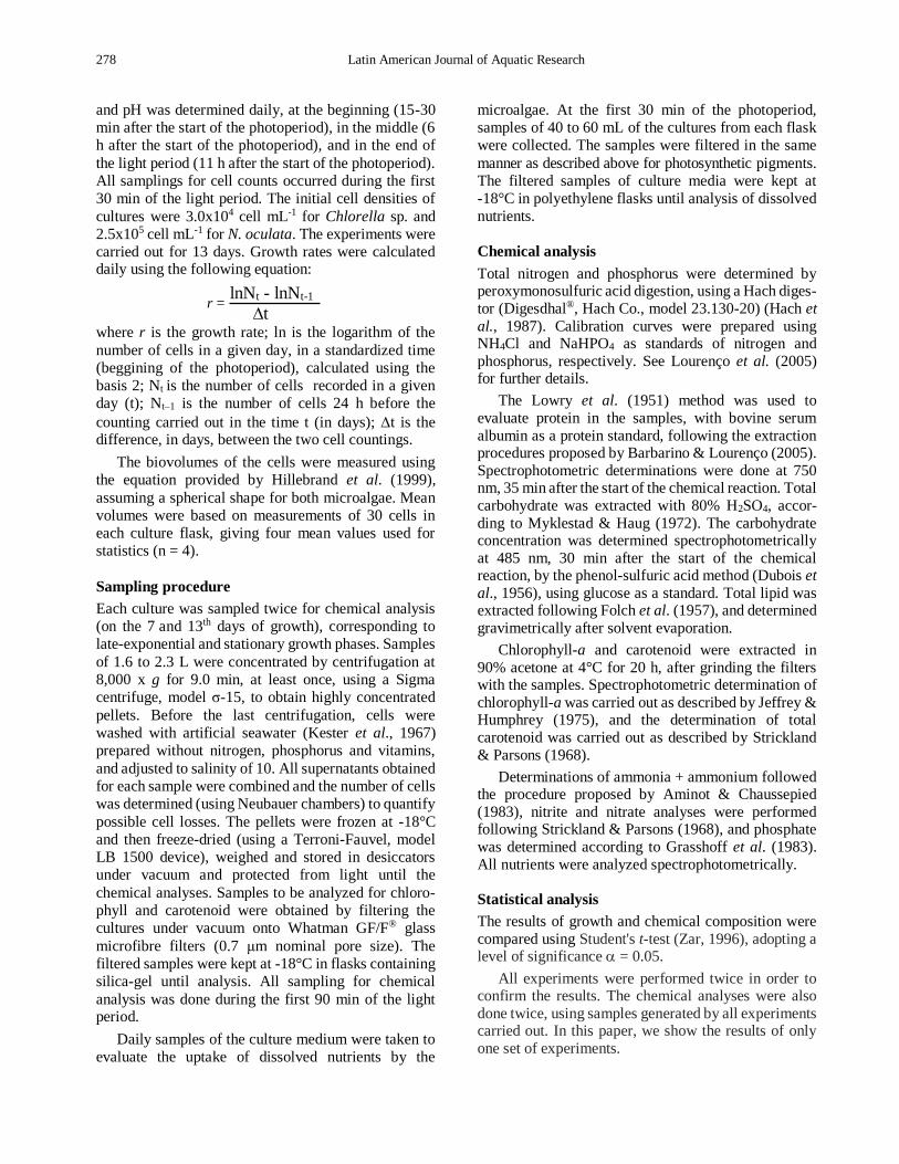
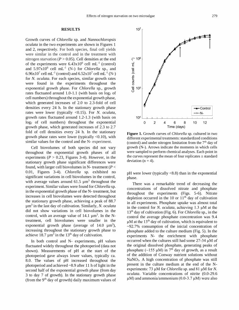
Growth curves of Chlorella sp. and Nannochloropsis
oculata in the two experiments are shown in Figures 1
and 2, respectively. For both species, final cell yields
were similar in the control and in the treatment with
nitrogen starvation (P > 0.05). Cell densities at the end
of the experiments were 6.43x10⁶ cell mL-1 (control)
and 5.97x10⁶ cell mL-1 (N-) for Chlorella sp., and
6.96x10⁷ cell mL-1 (control) and 6.52x10⁷ cell mL-1 (N-)
for N. oculata. For each species, similar growth rates
were found in the experiments throughout the
exponential growth phase. For Chlorella sp., growth
rates fluctuated around 1.0-1.1 (with basis on log2 of
cell numbers) throughout the exponential growth phase,
which generated increases of 2.0 to 2.3-fold of cell
densities every 24 h. In the stationary growth phase
rates were lower (typically <0.15). For N. oculata,
growth rates fluctuated around 1.2-1.3 (with basis on
log2 of cell numbers) throughout the exponential
growth phase, which generated increases of 2.3 to 2.7
fold of cell densities every 24 h. In the stationary
growth phase rates were lower (typically <0.10), with
similar values for the control and the N- experiment.
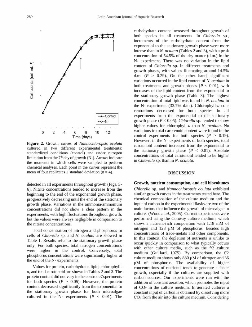
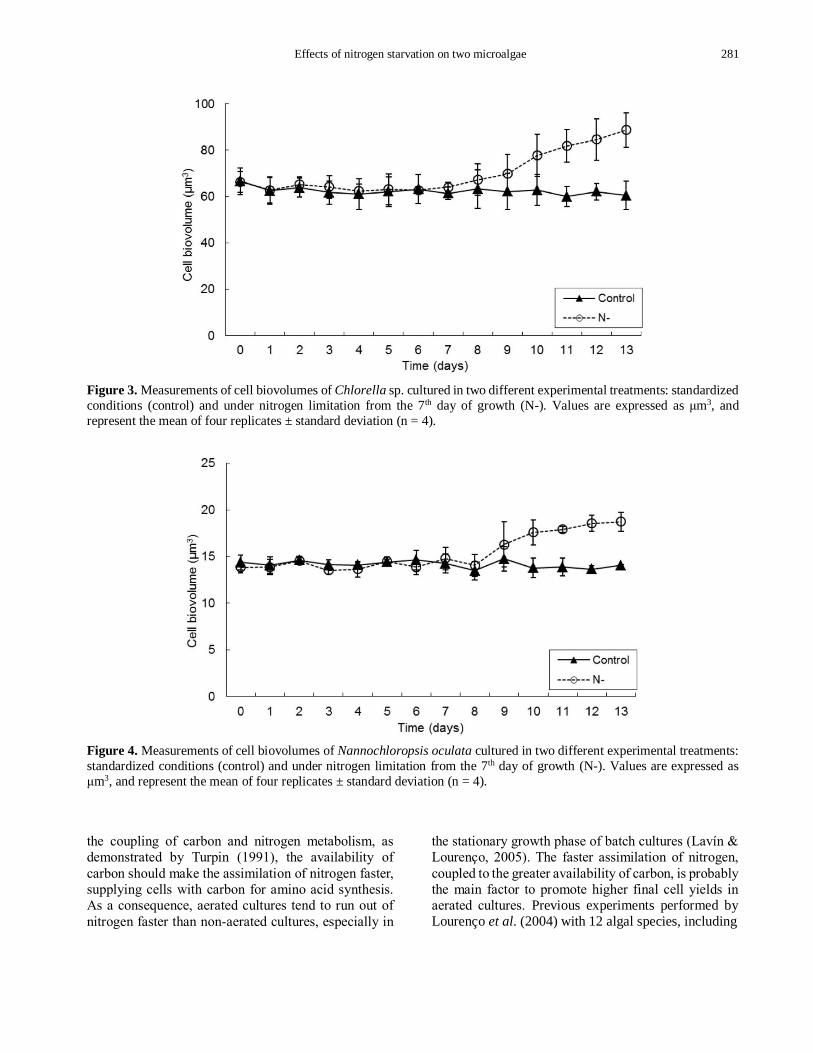
Cell biovolumes of both species did not vary
throughout the exponential growth phases of all
experiments (P > 0.23, Figures 3-4). However, in the
stationary growth phase significant differences were
found, with larger cell biovolumes in N- treatment (P <
0.01, Figures 3-4). Chlorella sp. exhibited no
significant variations in cell biovolumes in the control,
with average values around 61.5 µm3 throughout the
experiment. Similar values were found for Chlorella sp.
in the exponential growth phase of the N- treatment, but
increases in cell biovolumes were detected throughout
the stationary growth phase, achieving a peak of 88.7
µm3 in the last day of cultivation. Similarly, N. oculata
did not show variations in cell biovolumes in the
control, with an average value of 14.1 µm3. In the N-
treatment, cell biovolumes were smaller in the
exponential growth phase (average of 14.0 µm3),
increasing throughout the stationary growth phase to
achieve 18.7 µm3 in the 13th day of cultivation.
In both control and N- experiments, pH values
fluctuated widely throughout the photoperiod (data not
shown). Measurements of pH at the start of the
photoperiod gave always lower values, typically ca.
8.0. The values of pH increased throughout the
photoperiod and achieved ~8.9 after 11 h of light in the
second half of the exponential growth phase (from day
3 to day 7 of growth). In the stationary growth phase
(from the 9th day of growth) daily maximum values of
Figure 1. Growth curves of Chlorella sp. cultured in two
different experimental treatments: standardized conditions
(control) and under nitrogen limitation from the 7th day of
growth (N-). Arrows indicate the moments in which cells
were sampled to perform chemical analyses. Each point in
the curves represent the mean of four replicates ± standard
deviation (n = 4).
pH were lower (typically <8.8) than in the exponential phase.
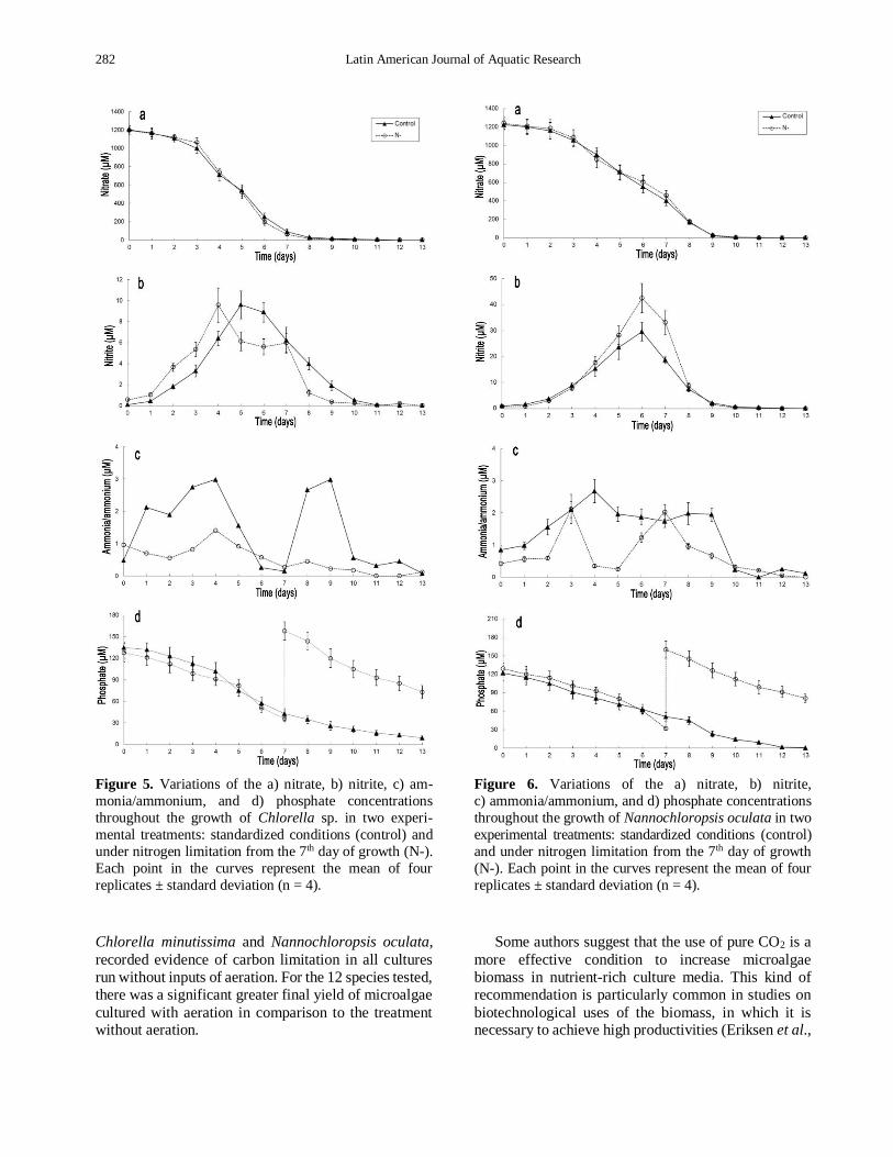
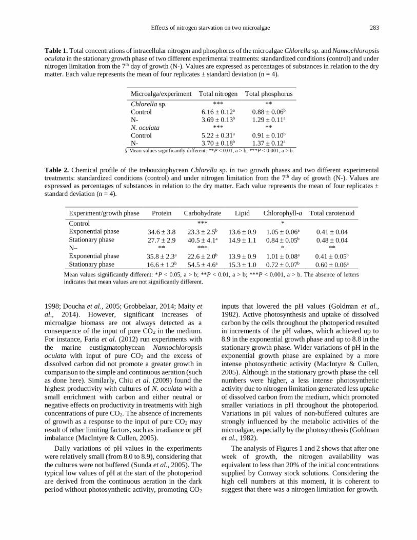
There was a remarkable trend of decreasing the
concentrations of dissolved nitrate and phosphate
throughout the experiments (Figs. 5-6). Nitrate
depletion occurred in the 10 or 11th day of cultivation
in all experiments. Phosphate uptake was almost total
in the control for N. oculata, achieving 1.3 μM at the
13th day of cultivation (Fig. 6). For Chlorella sp., in the
control the average phosphate concentration was 9.4
μM at the 13th day of cultivation, which is equivalent to
~92.7% consumption of the inicial concentration of
phosphate added to the culture medium (Fig. 5). In the
experiments N- the enrichment with phosphate
occurred when the cultures still had some 27-34 μM of
the original dissolved phosphate, generating peaks of
phosphate (~155 μM) in 7th day of growth, as a result
of the addition of Conway nutrient solutions without
NaNO3. A high concentration of phosphate was still
present in the culture medium at the end of the N-
experiments: 73 μM for Chlorella sp. and 81 μM for N. oculata. Variable concentrations of nitrite (0.0-29.6 μM) and ammonia/ammonium (0.0-3.7 μM) were also
280 Latin American Journal of Aquatic Research
Figure 2. Growth curves of Nannochloropsis oculata
cultured in two different experimental treatments:
standardized conditions (control) and under nitrogen
limitation from the 7th day of growth (N-). Arrows indicate
the moments in which cells were sampled to perform
chemical analyses. Each point in the curves represent the
mean of four replicates ± standard deviation (n = 4).
detected in all experiments throughout growth (Figs. 5-
6). Nitrite concentrations tended to increase from the
beginning to the end of the exponential growth phase,
progressively decreasing until the end of the stationary
growth phase. Variations in the ammonia/ammonium
concentrations did not show a clear pattern in the
experiments, with high fluctuations throughout growth,
but the values were always negligible in comparison to the nitrate concentrations.
Total concentration of nitrogen and phosphorus in
cells of Chlorella sp. and N. oculata are showed in
Table 1. Results refer to the stationary growth phase
only. For both species, total nitrogen concentrations
were higher in the control. Conversely, total
phosphorus concentrations were significantly higher at the end of the N- experiments.
Values for protein, carbohydrate, lipid, chlorophyll-
a, and total carotenoid are shown in Tables 2 and 3. The
protein content did not vary in the control e7xperiments
for both species (P > 0.05). However, the protein content decreased significantly from the exponential to
the stationary growth phase for both microalgae
cultured in the N- experiments (P < 0.01). The
carbohydrate content increased throughout growth of
both species in all treatments. In Chlorella sp.,
increments of the carbohydrate content from the
exponential to the stationary growth phase were more
intense than in N. oculata (Tables 2 and 3), with a peak
concentration of 54.5% of the dry matter (d.m.) in the
N- experiment. There was no variation in the lipid
content of Chlorella sp. in different treatments and
growth phases, with values fluctuating around 14.5%
d.m. (P > 0.29). On the other hand, significant
variations occurred in the lipid content of N. oculata in
both treatments and growth phases (P < 0.01), with
increases of the lipid content from the exponential to
the stationary growth phase (Table 3). The highest
concentration of total lipid was found in N. oculata in
the N- experiment (33.7% d.m.). Chlorophyll-a con-
centrations decreased for both species in all
experiments from the exponential to the stationary
growth phase (P < 0.05). Chlorella sp. tended to show
higher values for chlorophyll-a than N. oculata. No
variations in total carotenoid content were found in the
control experiments for both species (P > 0.19).
However, in the N- experiments of both species, total
carotenoid contend increased from the exponential to
the stationary growth phase (P < 0.01). Absolute
concentrations of total carotenoid tended to be higher
in Chlorella sp. than in N. oculata.
DISCUSSION
Growth, nutrient consumption, and cell biovolumes
Chlorella sp. and Nannochloropsis oculata exhibited
similar growth curves in the treatments tested here. The
chemical composition of the culture medium and the input of carbon in the experimental flasks are two of the
main factors that influence the growth of microalgae in cultures (Wood et al., 2005). Current experiments were
performed using the Conway culture medium, which shows a nutrient-rich composition with 1.18 mM of
nitrogen and 128 µM of phosphorus, besides high
concentrations of trace-metals and other components. In this context, the depletion of nutrients is unlike to
occur quickly in comparison to what typically occurs with other culture media, such as the f/2 culture
medium (Guillard, 1975). By comparison, the f/2
culture medium shows only 880 µM of nitrogen and 36 µM of phosphorus. The availability of higher
concentrations of nutrients tends to generate a faster growth, especially if the cultures are supplied with
carbon sources. Our experiments were run with the addition of constant aeration, which promotes the input of CO2 in the culture medium. In aerated cultures a
constant input of carbon is obtained by dissolving more CO2 from the air into the culture medium. Considering
Effects of nitrogen starvation on two microalgae 281
Figure 3. Measurements of cell biovolumes of Chlorella sp. cultured in two different experimental treatments: standardized
conditions (control) and under nitrogen limitation from the 7th day of growth (N-). Values are expressed as μm3, and
represent the mean of four replicates ± standard deviation (n = 4).
Figure 4. Measurements of cell biovolumes of Nannochloropsis oculata cultured in two different experimental treatments:
standardized conditions (control) and under nitrogen limitation from the 7th day of growth (N-). Values are expressed as
μm3, and represent the mean of four replicates ± standard deviation (n = 4).
the coupling of carbon and nitrogen metabolism, as
demonstrated by Turpin (1991), the availability of
carbon should make the assimilation of nitrogen faster,
supplying cells with carbon for amino acid synthesis.
As a consequence, aerated cultures tend to run out of
nitrogen faster than non-aerated cultures, especially in
the stationary growth phase of batch cultures (Lavín &
Lourenço, 2005). The faster assimilation of nitrogen,
coupled to the greater availability of carbon, is probably
the main factor to promote higher final cell yields in
aerated cultures. Previous experiments performed by Lourenço et al. (2004) with 12 algal species, including
282 Latin American Journal of Aquatic Research
Figure 5. Variations of the a) nitrate, b) nitrite, c) am-
monia/ammonium, and d) phosphate concentrations
throughout the growth of Chlorella sp. in two experi-
mental treatments: standardized conditions (control) and
under nitrogen limitation from the 7th day of growth (N-).
Each point in the curves represent the mean of four
replicates ± standard deviation (n = 4).
Chlorella minutissima and Nannochloropsis oculata,
recorded evidence of carbon limitation in all cultures
run without inputs of aeration. For the 12 species tested,
there was a significant greater final yield of microalgae
cultured with aeration in comparison to the treatment without aeration.
Figure 6. Variations of the a) nitrate, b) nitrite,
c) ammonia/ammonium, and d) phosphate concentrations
throughout the growth of Nannochloropsis oculata in two
experimental treatments: standardized conditions (control)
and under nitrogen limitation from the 7th day of growth
(N-). Each point in the curves represent the mean of four
replicates ± standard deviation (n = 4).
Some authors suggest that the use of pure CO2 is a
more effective condition to increase microalgae
biomass in nutrient-rich culture media. This kind of
recommendation is particularly common in studies on
biotechnological uses of the biomass, in which it is necessary to achieve high productivities (Eriksen et al.,
Effects of nitrogen starvation on two microalgae 283
Table 1. Total concentrations of intracellular nitrogen and phosphorus of the microalgae Chlorella sp. and Nannochloropsis
oculata in the stationary growth phase of two different experimental treatments: standardized conditions (control) and under nitrogen limitation from the 7th day of growth (N-). Values are expressed as percentages of substances in relation to the dry
matter. Each value represents the mean of four replicates ± standard deviation (n = 4).
Microalga/experiment Total nitrogen Total phosphorus
Chlorella sp. *** **
Control 6.16 ± 0.12a 0.88 ± 0.06b
N- 3.69 ± 0.13b 1.29 ± 0.11a
N. oculata *** **
Control 5.22 ± 0.31a 0.91 ± 0.10b N- 3.70 ± 0.18b 1.37 ± 0.12a
§ Mean values significantly different: **P < 0.01, a > b; ***P < 0.001, a > b.
Table 2. Chemical profile of the trebouxiophycean Chlorella sp. in two growth phases and two different experimental
treatments: standardized conditions (control) and under nitrogen limitation from the 7th day of growth (N-). Values are
expressed as percentages of substances in relation to the dry matter. Each value represents the mean of four replicates ±
standard deviation (n = 4).
Experiment/growth phase Protein Carbohydrate Lipid Chlorophyll-a Total carotenoid
Control *** *
Exponential phase 34.6 3.8 23.3 2.5b 13.6 0.9 1.05 0.06a 0.41 0.04 Stationary phase 27.7 2.9 40.5 4.1a 14.9 1.1 0.84 0.05b 0.48 0.04 N– ** *** * **
Exponential phase 35.8 2.3a 22.6 2.0b 13.9 0.9 1.01 0.08a 0.41 0.05b Stationary phase 16.6 1.2b 54.5 4.6a 15.3 1.0 0.72 0.07b 0.60 0.06a
Mean values significantly different: *P < 0.05, a > b; **P < 0.01, a > b; ***P < 0.001, a > b. The absence of letters
indicates that mean values are not significantly different.
1998; Doucha et al., 2005; Grobbelaar, 2014; Maity et al., 2014). However, significant increases of
microalgae biomass are not always detected as a
consequence of the input of pure CO2 in the medium.
For instance, Faria et al. (2012) run experiments with
the marine eustigmatophycean Nannochloropsis oculata with input of pure CO2 and the excess of
dissolved carbon did not promote a greater growth in
comparison to the simple and continuous aeration (such
as done here). Similarly, Chiu et al. (2009) found the
highest productivity with cultures of N. oculata with a
small enrichment with carbon and either neutral or
negative effects on productivity in treatments with high
concentrations of pure CO2. The absence of increments
of growth as a response to the input of pure CO2 may
result of other limiting factors, such as irradiance or pH imbalance (MacIntyre & Cullen, 2005).
Daily variations of pH values in the experiments
were relatively small (from 8.0 to 8.9), considering that
the cultures were not buffered (Sunda et al., 2005). The typical low values of pH at the start of the photoperiod
are derived from the continuous aeration in the dark
period without photosynthetic activity, promoting CO2
inputs that lowered the pH values (Goldman et al., 1982). Active photosynthesis and uptake of dissolved
carbon by the cells throughout the photoperiod resulted
in increments of the pH values, which achieved up to
8.9 in the exponential growth phase and up to 8.8 in the
stationary growth phase. Wider variations of pH in the
exponential growth phase are explained by a more
intense photosynthetic activity (MacIntyre & Cullen,
2005). Although in the stationary growth phase the cell
numbers were higher, a less intense photosynthetic
activity due to nitrogen limitation generated less uptake
of dissolved carbon from the medium, which promoted
smaller variations in pH throughout the photoperiod.
Variations in pH values of non-buffered cultures are
strongly influenced by the metabolic activities of the
microalgae, especially by the photosynthesis (Goldman et al., 1982).
The analysis of Figures 1 and 2 shows that after one
week of growth, the nitrogen availability was
equivalent to less than 20% of the initial concentrations supplied by Conway stock solutions. Considering the
high cell numbers at this moment, it is coherent to suggest that there was a nitrogen limitation for growth.
284 Latin American Journal of Aquatic Research
Table 3. Chemical profile of the eustigmatophycean Nannochloropsis oculata in two growth phases and two different
experimental treatments: standardized conditions (control) and under nitrogen limitation from the 7th day of growth (N-). Values are expressed as percentages of substances in relation to the dry matter. Each value represents the mean of four
replicates ± standard deviation (n = 4).
Experiment/growth phase Protein Carbohydrate Lipid Chlorophyll-a Total carotenoid
Control ** ** *
Exponential phase 30.8 2.9 23.3 2.0b 18.5 1.5b 0.65 0.04a 0.25 0.03
Stationary phase 26.1 1.7 29.3 2.5a 26.3 1.4a 0.53 0.03b 0.32 0.04
N– ** ** *** ** **
Exponential phase 29.1 1.9a 24.1 1.7b 17.8 1.5b 0.64 0.02a 0.24 0.03b
Stationary phase 17.9 1.0b 29.0 2.9a 33.7 2.8a 0.46 0.04b 0.33 0.02a
Mean values significantly different: *P < 0.05, a > b; **P < 0.01, a > b; ***P < 0.001, a > b. The absence of
letters indicates that mean values are not significantly different.
Thus, the start of the stationary growth phase, some two
or three days later, probably was promoted by the
insufficient availability of nitrogen to the microalgae
(Eriksen et al., 2007). A second factor that might have
contributed to the start of the stationary growth phase
was light availability. The excess of cells possibly
produced a self-shading in the cultures and a presumed
effect of photoacclimation by the microalgae (Johnsen
& Sakshaug, 2007).
Despite the low concentration of nitrogen in the
culture medium after seven days of growth, cells
continued to divide in relatively high rates (growth
rates fluctuated between 0.30 and 0.15) in the transition
between the exponential and the stationary growth
phases. This may be due the use of internal pools of
inorganic nitrogen, which sometimes achieve high
concentrations (Lavín & Lourenço, 2005). The
capability of accumulating inorganic nitrogen is a key
factor to keep phytoplankton alive under fluctuations in
nitrogen supply in natural environments (Dortch,
1982). In cultures, microalgae maintain the trend of
taking up high concentrations of dissolved inorganic
nitrogen supplied by the culture medium. The
accumulation of high concentrations of inorganic pools
of nitrogen (such as nitrate, nitrite and ammonia/
ammonium) in the exponential phase reflects the rapid
nitrogen uptake in the first days of growth, when no
factor is limiting (Lavín & Lourenço, 2005). Thus, the
consumption of intracellular pools of inorganic
nitrogen can promote growth of microalgae even under
scarce concentrations of nitrogen in the culture
medium. This could also explain the lack of differences
in final cell yield when the control and the N– treatment of both species are compared.
The assimilatory process of nitrogen may be limited
by the activity of enzymes, and the accumulation of
high concentrations of intracellular inorganic nitrogen
may also be a consequence of this condition. Marine
microalgae may accumulate large transitory pools of
inorganic nitrogen when the rates of uptake are higher
than growth rates. The accumulation of pools of
inorganic nitrogen, such as NO3-, may be a conse-
quence of differences in the rates of the previous step
in the assimilatory process (Berges, 1997). Imbalance
between up take and assimilation of nitrogen was
detected in our experiments. High concentrations of
nitrite might have been built up, and excretion of this
ion may have occured preventing its toxic effects, as
demonstrated by Lourenço et al. (1997) in nitrogen-rich
cultures of the prasinophycean Tetraselmis gracilis.
Lomas & Glibert (2000) also reported the release of
nitrite in nitrogen-sufficient cultures of some diatoms
and flagellates as a response to rapid increases in
irradiance. Concentrations of nitrite in the culture
medium in experiments with Chlorella sp. increased
from almost zero to ~8.0-9.0 M after four days of
exponential growth, and to ~30.0-40.0 M in cultures
of N. oculata. A similar trend was found by Aidar et al. (1991) in experiments run with the diatom Phaeo-dactylum tricornutum. This behaviour results from the
reduction of nitrate to nitrite by nitrate reductase
activity, not followed by further reduction to ammonia by nitrite reductase (Berges, 1997).
Phosphorus has not limited the growth of the
microalgae in the present study. The phosphate
concentrations declined throughout all experiments, but
in none of them it was depleted. Phosphorus is an
integral part of nucleic acids and biological mem-
branes, and it acts as a carrier substrate of chemical
energy (ATP) in the cytoplasm (La Roche et al., 1993).
The excess of phosphorus in N- experiments could
prevent any constraint by energy, considering that ATP-dependent physiological processes would not be
limited by phosphorus. In addition, the lack of nitrogen
would stimulate the cells to synthesize more
carbohydrate and/or lipid (e.g., Rodolfi et al., 2009; Li
Effects of nitrogen starvation on two microalgae 285
et al., 2011). However, our data suggest that even in the
control the microalgae had sufficient phosphorus to
synthesize all cell constituints and run ATP-dependent
physiological processes. Dean et al. (2008) demons-
trated that cultures of the chlorophycean Chlamydo-monas reinhardtii run with high concentrations of
phosphorus promoted increments if intracelular P
quota, as well as more accumulation of both
carbohydrate and lipid. Other studies claim that the
synthesis of storage products is favoured under
phosphorus limitation. Studying the trebouxiophycean
Chlorella vulgaris, Chia et al. (2013) concluded that
the accumulation of carbohydrate and lipid was greater
under phosphorus limitation. Similarly, Sigee et al. (2007) detected more production of carbohydrate and
lipid in P-limitated cultures of the chlorophycean
Scenedesmus subspicatus. Our experimental design
does not allow us to evaluate if the extremely high
availability of phosphorus in the N- experiments had
any stimulatory effect on the production of particular substances, such as carbohydrate and lipid.
The dynamics of growth in algal and cyanobacterial
cultures is also influenced by changes in cell
biovolumes. According to Borges-Campos et al. (2010)
microalgal species with small cell volumes tend to have
higher growth rates in comparison to large-sized cells.
This interpretation seems to be coherent here,
explaining well the higher growth rate of N. oculata, a
small-sized species, in comparison to Chlorella sp. On
the other hand, cell biovolumes did not vary in the
control throughout growth, but increments in
biovolumes were found only in N- experiments for both
species. This trend seems to be related to the
progressive reduction of nitrogen concentrations in the
culture medium and the ability to accumulate energy
reserves (carbohydrate and/or lipid) by the test-species.
The accumulation of high concentrations of
carbohydrate and/or lipid may contribute to enlarge
cells (Dean et al., 2010; Traller & Hildebrand, 2013),
since reserves accumulate in the cytoplasm in most of
the algal lineages (Graham et al., 2009). Our results
indicate that Chlorella sp. showed at the 13th day of
cultivation cell biovolumes ca. 32% larger in N-
experiments than in the control. For N. oculata, cell
biovolumes in the 13th day of cultivation were ca. 25%
larger in N– experiments than in the control. The
accumulation of carbohydrate and lipid was remarkable
for Chlorella sp. and N. oculata, respectively, throu-
ghout time in N– experiments (see discussion on gross
chemical composition of the microalgae). In addition,
in the stationary growth phase the lower growth rates reflect in slower cell divisions, which contribute to
increase of frequence of more cells with larger cell volumes (Carfagna et al., 2015).
Gross chemical composition
Protein content of the two microalgae exhibited diferent
trends in the experiments. The lack of variation of
protein content over growth in the control indicates that
the exhaustion of nitrogen in the culture medium,
recorded from the 10th day of growth, had no detectable
effect on the protein content of the microalgae. A
possible explanation for this trend results from the
consumption of internal pools of inorganic nitrogen,
presumably created due the luxuriant uptake of nitrate
(Lourenço et al., 1998). Large pools of nitrate, nitrite
and ammonia/ammonium are accumulated in cell
vacuoles during the exponential growth of batch
cultures run with sufficience in nitrate (Lomas &
Glibert 2000; Lavín & Lourenço, 2005). The
consumption of the inorganic pools of nitrogen occurs
when dissolved nitrogen declines in the culture
medium, and this makes both growth and production of
protein viable even with scarce concentrations of
nitrogen in the culture medium. This scenarium suggest
an “apparent” starvation in the control, based on
measurements of nitrogenaceous nutrients in the
culture medium. The actual nitrogen starvation would
be reached only when the internal pools of nitrogen
were consumed by the microalgae, normally later in the
stationary growth phase. On the other hand, a
significant decrease in protein concentrations were
recorded from the exponential to the stationary growth
phase of N- experiments for both species. This trend
suggests that the addition of nutrient solutions (without
nitrogen) of the Conway culture medium created an
imbalance that affected the production of protein. The
high availability of phosphorus, trace metals and other
components might have stimulated the synthesis of
non-nitrogenaceous substances, such as ATP and
carotenoids. The low availability of nitrogen in the
stationary growth phase of N- experiments lead to a low
protein content, which is in accordance with many other
studies (e.g., Young & Beardall, 2003; Setta et al., 2014). Our data on total nitrogen corroborate these
interpretations, considering that most of the intra-
cellular nitrogen of marine microalgae is contained in
protein (Lourenço et al., 2004). In N- experiments total
N was significantly lower than in the control in the
stationary growth phase for both species, which points
to a physiological stress caused by nitrogen starvation (Young & Beardall, 2003).
Our results for protein content (>30% d.m.) in the
control (and the exponential growth phase of the N-
experiments) were slightly higher than the values
reported by Renaud et al. (1999) for 18 microalgae cultured in Australia, which typically ranged from 24 to
29% d.m. This may be a result of different culture
conditions and also the efficient protocol of protein
286 Latin American Journal of Aquatic Research
extraction used by us, previously tested with different
macro- and microalgae (Barbarino & Lourenço, 2005).
Our results for protein content are similar to those
presented by Brown (1991) for some species of green
algae and Nannochloropsis oculata, one of the species
studied by us. Our results of protein content in the
stationary growth phase are typically lower than those
recorded in the literature for microalgae cutured in
sufficiency of nutrients (e.g., Lourenço et al., 2002;
Natrah et al., 2007; González-López et al., 2010), but
they were similar to data of studies run with nitrogen
starvation (e.g., Pelah et al., 2004; Silva et al., 2009; Tunçay et al., 2013).
The accumulation of carbohydrate reserves
occurred in both species in all experiments in the
stationary growth phase. In all comparisons throughout
time, concentrations of carbohydrate were higher in
older cultures. However, the accumulation of carbohy-
drate seemed to be more intense in Chlorella sp., in
comparison to N. oculata. For Chlorella sp., the
concentrations of carbohydrate roughly doubled from
the exponential to the stationary growth phases in the
control, and a ~2.5-fold increase occurred in N-
experiment for the same microalga (Table 2). The
increment of carbohydrate in N. oculata throughout
growth was smaller than in Chlorella sp., achieving ca.
31% in the two experiments. These differences are
related to taxonomic traits. Green algae have
carbohydrate as the main product to store energy,
especially as starch. Eustigmatophycean algae also
accumulate carbohydrate as reserves, but they produce
great amounts of lipid too (Graham et al., 2009). This
trend is verified in members of other classes of the algal
lineage Heterokontophyta (= Ochrophyta), such as the
chrysophyceans, the bolidophyceans, and the pinguio-
phyceans, among others (Reviers, 2006). Reinforcing
the interpretations on the energy reserves produced by
different algal lineages, there was no variation in the
total lipid produced by the trebouxiophycean Chlorella
sp. in the two experiments. Nevertheless, the eustigma-
tophycean N. oculata exhibited a ~42% increase in lipid
content over growth in the control, and a ~2.1-fold
increase in lipid content over growth in the N- experiment (Table 3).
Concentrations of carbohydrate in the exponential
growth phase of all experiments were similar to the
results published by Lourenço et al. (2002) for
Chlorella minutissima and N. oculata, as well as
Machado & Lourenço (2008) for green microalgae. In
the stationaty growth phase of both control and N-
experiments our results (>31% d.m. for N. oculata; > 40% d.m. for Chlorella sp.) are higher than most of the
available data in the literature (e.g., Brown, 1991;
Renaud et al., 1999), but they were similar to results
found in studies run with nitrogen starvation (e.g., Silva et al., 2009; Ikaran et al., 2015).
Concentrations of lipid in Chlorella sp. were low,
always lower than 15.5% d.m., even in the N-
experiment. These results are similar to those reported
by different authors for green algae (e.g., Machado &
Lourenço, 2008; Li et al., 2008). However, this species
did not respond to nitrogen starvation with increases in
total lipid. In many other studies, authors were able to
detect significant increments of the lipid content of
green microalgae under nitrogen starvation (e.g., Tang
et al., 2011; Karemore et al., 2013; Urreta et al., 2014).
As Chlorella sp. faild to produce more lipid under
nitrogen starvation, this strain may be unsuitable in
applications that need high concentrations of lipid, such
as biodiesel production. On the other hand, the
production of lipid by N. oculata was influenced by the
nitrogen availability such as other eukaryotic micro-
algae (Chen et al., 2011; Suali & Sarbatly, 2012).
Particularly in the N- experiment, N. oculata almost
doubled its lipid content over growth, in a similar trend
reported by other studies run with nitrogen starvation
(e.g., Rodolfi et al., 2009; Jiang et al., 2011; Kaye et al., 2015). Our results confirm N. oculata as a suitable
species to be used in applications that need high
concentrations of lipid, such as nutrition of some marine animals (Costard et al., 2012).
In general, increments in storage substances
throughout growth of both species seems to be related
to the progressively lower availability of N in the
culture medium. This general trend is widely
documented in the literature (e.g., Geider & La Roche,
2002; Silva et al., 2009; Praveenkumar et al., 2012).
Production of protein is favoured during periods of
nitrogen sufficiency, with limited carbohydrate and lipid
synthesis; conversely, during periods of reduced nitrogen
availability, carbohydrate and/or lipid accumulate and
protein production drops (Guo et al., 2013). On the other
hand, the increase of cell volumes over growth
probably results mainly of the intense accumulation of
non-nitrogenaceous substances, such as carbohydrate and lipid, as discussed in the previous section.
Concentrations of chlorophyll dropped from the
exponential to the stationary growth phase of both
microalgae in all experiments in a similar way. In the
control, the concentrations of chlorophyll decreased ca.
20.0% over growth of Chlorella sp. and ca. 18.5% in
N. oculata. In N- experiments, the concentrations of
chlorophyll dropped ca. 28.5% over growth of
Chlorella sp. and ca. 28.0% in N. oculata. These trends
are also connected to the availability of nitrogen. Chlorophyll-a is a nitrogenaceous substance, with
6.28% N in its molecular mass (Lourenço et al., 1998).
Growth under nitrogen limitation generates variations
Effects of nitrogen starvation on two microalgae 287
of chlorophyll content similar to those described for
protein: decrease of the percentages throughout time
(Bellefeuille et al., 2014). Absolute percentages of
chlorophyll-a were bigger in Chlorella sp. (0.72-1.05%
d.w.) than in N. oculata (0.46-0.65% d.w.), which is in
accordance with taxonomic traits. Chlorophytes are
probably the richest algal group in chlorophyll-a, which
may achieve more than 1.5% d.w. in some species
(Machado & Lourenço, 2008). Most of the algal groups
typically show less than 0.7% d.w. of chlorophyll-a
when grown in nutrient sufficiency and even lower
concentrations under nitrogen starvation (Lourenço et al., 2004).
Both microalgae showed similar trends of variation
of total carotenoid in the experiments. The lack of
variation in the control is regarded as a consequence of
sufficiency in nutrients to synthesize carotenoids.
These substances do not contain nitrogen and their
synthesis is not inhibited by the depletion of nitrogen,
such as chlorophyll. As the cultures experienced
nitrogen limitation for only two or three days in the
control (see discussion of the previos section), this
probably did not reflect in significant increments in
total carotenoid. On the other hand, in N- experiments
there was an increase in total carotenoid over growth.
This trend is supported by studies run with the
chlorophytes Dunaliella salina (Lamers et al., 2012)
and Chlorella zofingiensis (Mulders et al., 2014) and
with the haptophyte Isochrysis galbana (Roopnarain et al., 2014), among many others. Increments in total
carotenoid are possibly a consequence of the need of
more photosynthesis atennae to support growth of the
species. Chlorophyll-a is the main photosynthetic
pigment, but under nitrogen starvation it is not possible
to produce additional chlorophyll molecules for new
daughter-cells. Microalgae can compensate the
decrease in chlorophyll producing more carotenoids
(Young & Beardall, 2003).
Both microalgae tested here showed a significant
increment of total carotenoid over growth in N-
experiments. Despite this trend points for potential uses
of these microalgae to produce carotenoids, it is
necessary to identify their individual carotenoids in
order to evaluate if they produce any substance of high
value. Commercial applications of carotenoids currently
known involve some specific valuable substances, such
as astaxathin, β-carotene, and zeaxanthin, for instance (Spolaore et al., 2006).
Mariculture uses and potential for biofuel production
Our study provided data on growth, nutrient uptake,
and gross chemical compositon of two microalgae in
two experimental conditions. Current results confirm
that the two microalgae tested present variable
chemical composition and their gross chemical profile
can be changed depending on the experimental
conditions. We used here a two-step cultivation,
creating a physiological condition of nitrogen
starvation after the cultures achieve high cell densities.
This strategy combines the need of high biomass and
the induction of the synthesis of substances of interest.
A growth under nitrogen limitation since the beginning
of the cultures would not provide enough biomass for possible applications.
The high concentrations of lipid showed by N.
oculata are suitable for the use of this microalga as a
food-species in mariculture and it is also favorable for
the potential production of biodiesel. Cultivation under
nitrogen starvation provided significant increments in
total lipid, which is important to feed animals with a
high demand for fatty food, such as larvae of oysters
(Martínez-Fernández et al., 2006). Under nitrogen
starvation, N. oculata exhibited low concenrations of
protein, which may be unsuitable for some animal
species. In theory, this possible defficiency in protein
can be compensated if N. oculata is offered in diets
combined with a protein-rich microalga of similar size
and shape. On the other hand, a possible future use of
N. oculata as a candidate feedstock to produce biodiesel
is strengthened by the current results, considering that
its chemical composition could be successfully changed to produce more lipid.
The new strain tested here, Chlorella sp., exhibited
a good productivity of biomass, but its low
concentration of lipid may limit its use both to feed
some marine animals and as well as to convert its
biomass in biodiesel. The high concentration of protein
presented by Chlorella sp. may be suitable to feed
marine animals with high demand for protein (Brown
et al., 1997). Trials to assess the digestibility of its cell
wall are needed to better evaluate its use in mariculture.
Currently there are many research groups creating new
photobioreactors that can be more efficient to produce
microalgae biomass and even specific substances, such
as lipids (Rodolfi et al., 2009). However, even with the
expected progress to produce microalgae biomass it is
unlike that low-lipid strains can be selected as
feedstocks for biodiesel production, considering the
tremendous technological challenges of this enterprise
(Davis et al., 2011). The high content of carbohydrate
of Chlorella sp. may be potentially useful to produce
bioethanol, which technical and economic viabilities still have to be demonstrated.
In the next step of our research, the fatty acid profile
of the microalgae tested here will be evaluated in different experimental conditions, which will be important to better interpret their possible uses.
288 Latin American Journal of Aquatic Research
ACKNOWLEDGEMENTS
The authors thank to Brazil’s National Council for
Scientific and Technological Development (CNPq) and
Foundation for Research Support of the State of Rio de
Janeiro, Brazil (FAPERJ) for the finantial support of
this study. The authors thank CNPq for their research
fellowships. Authors thank Dr. Renato Crespo Pereira
and Aguinaldo Nepomuceno Marques Júnior for the use of laboratory facilities.
REFERENCES
Aidar, E., R. Ehrlich, C.S. Asano & T.C.S. Sigaud. 1991.
Variação da composição bioquímica de Phaeodac-
tylum tricornutum (Bohlin), em cultivos estanques. Bolm. Inst. Oceanogr. São Paulo, 39(2): 131-139.
Aminot, A. & M. Chaussepied. 1983. Manuel des analyses
chimiques en milieu marin. CNEXO, Brest, 395 pp.
Barbarino, E. & S.O. Lourenço. 2005. An evaluation of
methods for extraction and quantification of protein
from marine macro- and microalgae. J. Appl. Phycol.,
17: 447-460.
Bellefeuille, S.D., S. Dorion, J. Rivoal & D. Morse. 2014.
The dinoflagellate Lingulodinium polyedrum responds
to N depletion by a polarized deposition of starch and
lipid bodies. PLoS ONE, 9(11): e111067.
Berges, J.A. 1997. Algal nitrate reductases. Eur. J. Phycol. 32(1): 3-8.
Borges-Campos, V., E. Barbarino & S.O. Lourenço. 2010.
Crescimento e composição química de dez espécies de
microalgas marinhas em cultivos estanques. Cienc.
Rural, 40(2): 339-347.
Brown, M.R. 1991. The amino-acid and sugar composi-
tion of 16 species of microalgae used in mariculture. J.
Exp. Mar. Biol. Ecol., 145: 79-99.
Brown, M.R., S.W. Jeffrey, J.K. Volkman & G.A.
Dunstan. 1997. Nutritional properties of microalgae
for mariculture. Aquaculture, 151(1-4): 315-331.
Carfagna, S., G. Salbitani, C. Bottone, A. De Marco & V.
Vona. 2015. Cross-effects of nitrogen and sulphur
starvation in Chlorella sorokiniana 211/8K. Nat.
Resour., 6: 221-229.
Carvalho, A.P., C.M. Monteiro & F.X. Malcata. 2009.
Simultaneous effect of irradiance and temperature on biochemical composition of the microalga Pavlova
lutheri. J. Appl. Phycol., 21(5): 543-552.
Chen, M., H. Tang, H. Ma, T.C. Holland, K.Y.S. Ng &
S.T. Salley. 2011. Effect of nutrients on growth and
lipid accumulation in the green algae Dunaliella
tertiolecta. Bioresource Technol., 102: 1629-1655.
Chia, M.A., A.T. Lombardi & M.G.G. Melão 2013.
Calorific values of Chlorella vulgaris (Trebouxio-
phyceae) as a function of different phosphorus
concentrations. Phycological Res., 61(4): 286-291.
Chiu, S.-Y., C.-Y. Kao, M.-T. Tsai, S.-C. Ong, C.-H. Chen
& C.-S. Lin. 2009. Lipid acumulation and CO2
utilization of Nannochloropsis oculata in response to
CO2 aeration. Bioresource Technol., 100: 833-838.
Costard, G.S., R.R. Machado, E. Barbarino, R.C. Martino
& S.O. Lourenço. 2012. Chemical composition of five
marine microalgae that occur on the Brazilian coast.
Int. J. Fish. Aquacult., 4(9): 191-201.
Davis, R., A. Aden & P.T. Pienkos. 2011. Techno-
economic analysis of autotrophic microalgae for fuel
production. Appl. Energy, 88(10): 3524-3531.
Dean, A.P., J.M. Nicholson & D.C. Sigee. 2008. Impact
of phosphorus quota and growth phase on carbon
allocation in Chlamydomonas reinhardtii: an FTIR
microspectroscopy study. Eur. J. Phycol., 43(4): 345-
354.
Dean, A.P., D.C. Sigee, B. Estrada & J.K. Pittman. 2010.
Using FTIR spectroscopy for rapid determination of
lipid accumulation in response to nitrogen limitation in
freshwater microalgae. Bioresource Technol., 101:
4499-4507.
Dortch, Q. 1982. Effect of growth conditions on
accumulation of internal nitrate, ammonium, amino
acids, and protein in three marine diatoms. J. Exp. Mar.
Biol. Ecol., 61: 243-264.
Doucha, J., F. Straka & K. Lívanský. 2005. Utilization of
flue gas for cultivation of microalgae (Chlorella sp.) in
an outdoor open thin-layer photobioreactor. J. Appl.
Phycol., 17(5): 403-412.
Dubois, M., K.A. Gilles, J.K. Hamilton, P.A. Rebers & F.
Smith. 1956. Colorimetric method for the
determination of sugars and related substances. Anal.
Chem., 18: 350-356.
Durmaz, Y., M. Donato, M. Monteiro, L. Gouveia, M.L.
Nunes, T. Gama Pereira, S. Gökpmar, N.M. Bandarra.
2009. Effect of temperature on α-tocopherol, fatty acid
profile, and pigments of Diacronema vlkianum
(Haptophyceae). Aquacult. Int. 17: 391-399.
Eriksen, N.T., B.R. Poulsen & J.J. Lønsmann Iversen.
1998. Dual sparging laboratory-scale photobioreactor
for continuous production of microalgae. J. Appl.
Phycol., 10(4): 377-382.
Eriksen, N.T., F. Riisgård, W.S. Gunther & J.J.L. Iversen.
2007. On-line estimation of O2 production, CO2
uptake, and growth kinetics of microalgal cultures in a
gas-tight photobioreactor. J. Appl. Phycol., 19(2): 161-
174.
Falkowski, P.G. & J.A. Raven. 2007. Aquatic photosyn-
thesis. Princeton University Press, Princeton, 484 pp.
Effects of nitrogen starvation on two microalgae 289
Faria, G.R., C.R.P.S. Paes, D.J.F.A. Castro, N.A.B.
Tinoco, E. Barbarino & S.O. Lourenço. 2012. Effects
of the availability of CO2 on growth, nutrient uptake,
and chemical composition of the marine microalgae
Chlorella sp. and Nannochloropsis oculata, two
potentially useful strains for biofuel production. Int.
Res. J. Biotechnol., 3(5): 65-75.
Fidalgo, J.P., A. Cid, J. Abalde & C. Herrero. 1995.
Culture of the marine diatom Phaeodactylum
tricornutum with different nitrogen sources: growth,
nutrient conversion and biochemical composition.
Cah. Biol. Mar., 36: 165-173.
Folch, J., M. Lees & G.M. Sloane-Stanley. 1957. A simple
method for the isolation and purification of total lipid
from animal tissues. J. Biol. Chem., 226: 497-509.
Geider, R., J. La Roche, R. Greene & M. Olaizola. 1993.
Response of the photosynthetic apparatus of Phaeo-
dactylum tricornutum (Bacillariophyceae) to nitrate,
phosphate, or iron starvation. J. Phycol. 29: 755-66.
Geider, R. & J. La Roche. 2002. Redfield revisited:
variability of C:N:P in marine microalgae and its
biochemical basis. Eur. J. Phycol., 37(1): 1-17.
Goldman, J.C., Y. Azov, C.B. Riley & M.R. Dennett.
1982. The effect of pH in intensive microalgal
cultures. I. Biomass regulation. J. Exp. Mar. Biol. Ecol., 57: 1-13.
González López, C.V., M.C. Cerón García, F.G. Acién
Fernández, C. Segovia Bustos, Y. Chisti & J.M.
Fernández-Sevilla. 2010. Protein measurements of
microalgal and cyanobacterial biomass. Bioresource Technol., 101(19): 7587-7591.
Graham, L.E., J.E. Graham & L.W. Wilcox. 2009. Algae.
Benjamin Cummings (Pearson), San Francisco, 720
pp.
Grasshoff, K., M. Ehrhardt & K. Kremling. 1983. Methods
of seawater analysis. Verlag Chemie, Weinheim, 419
pp.
Grobbelaar, J.U. 2014. Microalgal biomass production:
challenges and realities. Photosynth. Res., 106(1-2):
135-144.
Guillard, R.R.L. 1975. Culture of phytoplankton for
feeding marine invertebrates. In: W.L. Smith & M.H. Chanley (eds.). Culture of marine invertebrete
animals. Plenum Publishing Corporation, New York,
pp. 29-60.
Guo, H., M. Daroch, L. Liu, G. Qiu, S. Geng & G. Wang.
2013. Biochemical features and bioethanol production of microalgae from coastal waters of Pearl River Delta.
Bioresource Technol., 127: 422-428.
Hach, C.C., B.K. Bowden, A.B. Kopelove & S.V.
Brayton. 1987. More powerful peroxide Kjeldahl
digestion method. J. Assoc. Off. Anal. Chem., 70(5):
783-787.
Harrison, P.J. & J.A. Berges. 2005. Marine culture media.
In: R.A. Andersen (ed.). Algal culturing techniques. Academic Press, San Diego, pp. 21-33.
Hillebrand, H., C.-D. Dürselen, D. Kirschtel, U. Pollingher
& T. Zohary. 1999. Biovolume calculation for pelagic
and benthic microalgae. J. Phycol., 35(2): 403-424.
Huerlimann, R., R. de Nys & K. Heimann. 2010. Growth,
lipid content, productivity, and fatty acid composition
of tropical microalgae for scale-up production.
Biotechnol. Bioeng., 107(2): 245-257.
Ikaran, Z., S. Suárez-Alvarez, I. Urreta & S. Castañón.
2015. The effect of nitrogen limitation on the
physiology and metabolism of Chlorella vulgaris var
L3. Algal Res., 10: 134-144.
Jeffrey, S.W. & G.F. Humphrey. 1975. New spectropho-
tometric equations for determining chlorophylls a, b,
c1 and c2 in higher plants, algae and natural phyto-
plankton. Biochem. Physiol. Pflantz., 167: 191-194.
Jiang, L., S. Luo, X. Fan, Z. Yang & R. Guo. 2011. Biomass and lipid production of marine microalgae
using municipal wastewater and high concentration of
CO2. Appl. Energ., 88: 3336-3341.
Johnsen, G. & E. Sakshaug. 2007. Biooptical charac-
teristics of PSII and PSI in 33 species (13 pigment
groups) of marine phytoplankton, and the relevance for pulse-amplitude-modulated and fast-repetition-rate
fluorometry. J. Phycol., 43(6): 1236-1251.
Karemore, A., R. Pal & R. Sen. 2013. Strategic
enhancement of algal biomass and lipid in Chloroco-
ccum infusionum as bioenergy feedstock. Algal Res.,
2(2): 113-121.
Kaye, Y., O. Grundman, S. Leu, A. Zarka, B. Zorin, S.
Didi-Cohen, I. Khozin-Goldberg & S. Boussiba. 2015.
Metabolic engineering toward enhanced LC-PUFA
biosynthesis in Nannochloropsis oceanica: Overex-
pression of endogenous Δ12 desaturase driven by
stress-inducible promoter leads to enhanced deposition of polyunsaturated fatty acids in TAG. Algal Res., 11:
387-398.
Kester, D.R., I.W. Duedall, D.N. Connors & R.M.
Pytkowicz. 1967. Preparation of artificial seawater.
Limnol. Oceanogr., 12(1): 176-179.
Lamers, P.P., M. Janssen, R.C.H. De Vos, R.J. Bino &
R.H. Wijffels. 2012. Carotenoid and fatty acid
metabolism in nitrogen-starved Dunaliella salina, a
unicellular green microalga. J. Biotechnol., 162(1):
21-27.
La Roche, J., R.J. Geider, L.M. Graziano, H. Murray & K.
Lewis. 1993. Induction of specific proteins in
eukaryotic algae grown under iron-, phosphorus-, or
nitrogen-deficient conditions. J. Phycol., 29: 767-777.
Lavín, P.L. & S.O. Lourenço. 2005. An evaluation of the
accumulation of intracellular inorganic nitrogen pools
290 Latin American Journal of Aquatic Research
by marine microalgae in batch cultures. Braz. J.
Oceanogr., 53(1/2): 55-68.
Lee-Chang, K.J., P.D. Nichols & S.I. Blackburn. 2013.
More than biofuels-potential uses of microalgae as
sources of high-value lipids. Lipid Technol., 25(9):
199-203.
Li, Y., M. Horsman, B. Wang, N. Wu & C. Lan. 2008.
Effects of nitrogen sources on cell growth and lipid
accumulation of green alga Neochloris oleoabundans.
Appl. Microbiol. Biotechnol., 81(4): 629-636.
Li, Z.S., H.L. Yuan, J.S. Yang & B.Z. Li. 2011.
Optimization of the biomass production of oil algae
Chlorella minutissima UTEX2341. Bioresource
Technol., 102(19): 9128-9134.
Lim, D.K.Y., S. Garg, M. Timmins, E.S.B. Zhang, S.R.
Thomas-Hall, H. Schuhmann, Y. Li & P.M. Schenk.
2012. Isolation and evaluation of oil-producing
microalgae from subtropical coastal and brackish
waters. PLoS ONE, 7(7): e40751.
Lomas, M.W. & P.M. Glibert. 2000. Comparisons of
nitrate uptake, storage, and reduction in marine
diatoms and dinoflagellates. J. Phycol., 36: 903-913.
Lourenço, S.O. & A.A.H. Vieira. 2004. Culture collec-
tions of microalgae in Brazil: progress and constraints.
Nova Hedwigia, 79(1-2): 149-173.
Lourenço, S.O., E. Barbarino, U.M. Lanfer-Marquez & E.
Aidar. 1998. Distribution of intracelluar nitrogen in
marine microalgae: basis for the calculation of specific
nitrogen-to-protein conversion factors. J. Phycol.,
34(5): 798-811.
Lourenço, S.O., E. Barbarino, A. Nascimento & R.
Paranhos. 2005. Seasonal variations in tissue nitrogen
and phosphorus of eight macroalgae from a tropical
hypersaline coastal environment. Cryptogamie, 26(4):
355-371.
Lourenço, S.O., U.M. Lanfer-Marquez, J. Mancini-Filho,
E. Barbarino & E. Aidar. 1997. Changes in bioche-
mical profile of Tetraselmis gracilis. I. Comparison of
two culture media. Aquaculture, 148(2/3): 153-168.
Lourenço, S.O., E. Barbarino, J. Mancini-Filho, K.P.
Schinke & E. Aidar. 2002. Effects of different nitrogen
sources on growth and biochemical profile of ten
marine microalgae under batch cultures: an evaluation
for aquaculture. Phycologia, 41(2): 158-168.
Lourenço, S.O., E. Barbarino, P.L. Lavín, U.M.L.
Marquez & E. Aidar. 2004. Distribution of
intracellular nitrogen in marine microalgae.
Calculation of new nitrogen-to-protein conversion
factors. Eur. J. Phycol., 39(1): 17-32.
Lourenço, S.O., E. Barbarino, M.G. Bispo, D.A. Borges,
C. Coelho-Gomes, P.L. Lavín & F. Santos. 2008.
Effects of light intensity on growth, inorganic nitrogen
storage, and gross chemical composition of four
marine microalgae in batch cultures. Museu Nacional,
Série Livros, 30: 203-214.
Lowry, O.H., N.J. Rosebrough, A.L. Farr & R.L. Randall.
1951. Protein measurement with Folin phenol reagent.
J. Biol. Chem., 193: 265-275.
Machado, R.R. & S.O. Lourenço. 2008. Propriedades
nutricionais de microalgas usadas como alimento de
moluscos bivalves: uma revisão. Museu Nacional,
Série Livros, 30: 281-304.
MacIntyre, H.L. & J.J. Cullen. 2005. Using cultures to
investigate the physiological ecology of microalgae.
In: R.A. Andersen (ed.). Algal culturing techniques.
Academic Press, San Diego, pp. 287-326.
Maity, J.P., J. Bundschuh, C.-Y. Chen & P. Bhattacharya. 2014. Microalgae for third generation biofuel
production, mitigation of greenhouse gas emissions
and wastewater treatment: present and future
perspectives - a mini review. Energy, 78: 104-113.
Martínez-Fernández, E., H. Acosta-Salmón & P.C. Southgate. 2006. The nutritional value of seven
species of tropical microalgae for black-lip pearl
oyster (Pinctada margaritifera, L.) larvae. Aqua-
culture, 257(1-4): 491-503.
Myklestad, S. & A. Haug. 1972. Production of
carbohydrates by the marine diatom Chaetoceros affinis var. willei (Gran) Hustedt. I. Efect of the
concentration of nutrients in the culture medium. J.
Exp. Mar. Biol. Ecol., 9: 125-136.
Mubarak, M., A. Shaija & T.V. Suchithra. 2015. A review
on the extraction of lipid from microalgae for biodiesel
production. Algal Res., 7: 117-123.
Mulders, K.J.M., J.H. Janssen, D.E. Martens, R.H.
Wijffels & P.P. Lamers. 2014. Effect of biomass
concentration on secondary carotenoids and triacyl-
glycerol (TAG) accumulation in nitrogen-depleted
Chlorella zofingiensis. Algal Res., 6: 8-16.
Natrah, F.M.I., F.M. Yusoff, M. Shariff, F. Abas & N.S.
Mariana. 2007. Screening of Malaysian indigenous
microalgae for antioxidant properties and nutritional
value. J. Appl. Phycol., 19(6): 711-718.
Pelah, D., I. Marton, W. Wang, O. Shoseyov, A. Altman
& E. Cohen. 2004. Accumulation and protection
activity of protease-resistant heat-stable proteins in
Haematococcus pluvialis during high light and
nitrogen starvation. J. Appl. Phycol., 16(2): 153-156.
Praveenkumar, R., K. Shameera, G. Mahalakshmi, M.A.
Akbarsha & N. Thajuddin. 2012. Influence of nutrient
deprivations on lipid accumulation in a dominant
indigenous microalga Chlorella sp., BUM11008:
evaluation for biodiesel production. Biomass Bio-
energ., 37: 60-66.
Renaud, S.M., L.-V. Thinh & D.L. Parry. 1999. The gross
chemical composition and fatty acid composition of 18
Effects of nitrogen starvation on two microalgae 291
species of tropical Australian microalgae for possible
use in mariculture. Aquaculture, 170(2): 147-159.
Ratti, S., A.H. Knoll & M. Giordano. 2013. Grazers and
phytoplankton growth in the oceans: an experimental
and evolutionary perspective. PLoS ONE, 8(11):
e77349.
Reviers, B. 2006. Biologia e filogenia das algas. Artmed,
Porto Alegre, 280 pp.
Rodolfi, L., G.C. Zitelli, N. Bassi, G. Padovani, N. Biondi,
G. Bonini & M.R. Tredici. 2009. Microalgae for oil:
strain selection, induction of lipid synthesis and
outdoor mass cultivation in a low-cost photobio-
reactor. Biotechnol. Bioeng., 102: 100-112.
Roopnarain, A., V.M. Gray & S. Sym. 2014. Influence of nitrogen stress on Isochrysis galbana strain U4, a
candidate for biodiesel production. Phycol. Res.,
62(4): 237-249.
Sepúlveda, C., F.G. Acién, C. Gómez, N. Jiménez-Ruíz,
C. Riquelme & E. Molina-Grima. 2015. Utilization of centrate for the production of the marine microalgae
Nannochloropsis gaditana. Algal Res., 9: 107-116.
Setta, B.R.S., E. Barbarino, F.B. Passos & S.O. Lourenço.
2014. An assessment of the usefulness of the
cyanobacterium Synechococcus subsalsus as a source
of biomass for biofuel production. Lat. Am. J. Aquat. Res., 42(2): 364-375.
Sigee, D.C., F., Bahrami, B. Estrada, R.E. Webster & A.P.
Dean. 2007. The influence of phosphorus availability
on carbon allocation and P quota in Scenedesmus
subspicatus: a synchrotron-based FTIR analysis.
Phycologia, 46(5): 583-592.
Silva, A.F., S.O. Lourenço & R.M. Chaloub. 2009. Effects
of nitrogen starvation on the photosynthetic
physiology of a tropical marine microalga Rhodo-
monas sp. (Cryptophyceae). Aquacult. Bot., 91(4):
291-297.
Spolaore, P., C. Joannis-Cassan, E. Duran & A. Isambert.
2006. Commercial applications of microalgae. J.
Biosci. Bioeng., 101(2): 87-96.
Strickland, J.D.H. & T.R. Parsons. 1968. A practical
handbook of seawater analysis. Bull. Fish. Res. Bd.
Can., 167: 1-311.
Suali, E. & R. Sarbatly. 2012. Conversion of microalgae
to biofuel. Renew. Sust. Energ. Rev., 16(6): 4316-
4342.
Sunda, W.G., N.M. Price & F.M.M. Morel. 2005. Trace
metal ion buffers and their use in culture studies. In:
R.A. Andersen (ed.). Algal culturing techniques. Academic Press, San Diego, pp. 35-63.
Tang, H., M. Chen, M.E.D. Garcia, N. Abunasser, K.Y.S.
Ng & S.O. Salley. 2011. Culture of microalgae
Chlorella minutissima for biodiesel feedstock
production. Biotechnol. Bioeng., 108(10): 2280-2287.
Templeton, D.W. & L.M.L. Laurens. 2015. Nitrogen-to-
protein conversion factors revisited for applications of
microalgal biomass conversion to food, feed and fuel.
Algal Res., 11: 359-367.
Tokuşoglu, Ö. & M.K. Ünal. 2003. Biomass nutrient
profiles of three microalgae: Spirulina platensis,
Chlorella vulgaris, and Isochrisis galbana. J. Food
Sci., 68(4): 1144-1148.
Traller, J.C. & M. Hildebrand. 2013. High throughput
imaging to the diatom Cyclotella cryptica
demonstrates substantial cell-to-cell variability in the
rate and extent of triacylglycerol accumulation. Algal
Res., 2(3): 244-252.
Tunçay, H., J. Findinier, T. Duchêne, V. Cogez, C.
Cousin, G. Peltier, S.G. Ball & D. Dauvillée. 2013. A
forward genetic approach in Chlamydomonas rein-
hardtii as a strategy for exploring starch catabolism.
PLoS ONE, 8(9): e74763.
Turpin, D.H. 1991. Effects of inorganic N availability on
algal photosynthesis and carbon metabolism. J.
Phycol., 27(1): 14-20.
Urreta, I., Z. Ikaran, I. Janices, E. Ibañez, M. Castro-
Puyana, S. Castañón & S. Suárez-Alvarez. 2014.
Revalorization of Neochloris oleoabundans biomass
as source of biodiesel by concurrent production of
lipids and carotenoids. Algal Res., 5: 16-22.
Valenzuela-Espinoza, E., R. Millán-Núñez & F. Núñez-
Cebrero. 1999. Biomass production and nutrient
uptake by Isochrysis aff. galbana (Clone T-ISO)
cultured with a low cost alternative to the f/2 medium.
Aquacult. Eng., 20: 135-147.
Van Iersel, S. & A. Flammini. 2010. Algae-based biofuels:
applications and Co-Products. FAO, Rome, 117 pp.
Walne, P.R. 1966. Experiments in the large scale culture
of the larvae of Ostrea edulis. Fish. Invest. Lond. Ser.,
(2)25: 1-53.
Wijffels, R.H. & M.J. Barbosa. 2010. An outlook on
microalgal biofuels. Science, 329: 796-799.
Wijffels, R.H., O. Kruse & K.J. Hellingwerf. 2013.
Potential of industrial biotechnology with cyanobac-
teria and eukaryotic microalgae. Curr. Opin. Biotech.,
24: 405-413.
Wood, A.M., R.C. Everroad & L.M. Wingard. 2005.
Measuring growh rates in microalgal cultures. In: R.A.
Andersen (ed.). Algal culturing techniques. Academic
Press, San Diego, pp. 269-285.
Young, E.B. & J. Beardall. 2003. Photosynthetic function
in Dunaliella tertiolecta (Chlorophyta) during a
nitrogen starvation and recovery cycle. J. Phycol.,
39(5): 897-905.
Zar, J.H. 1996. Biostatistical analysis. Prentice Hall,
Englewood Cliffs, 662 pp.
292 Latin American Journal of Aquatic Research
Zeng, X., M.K. Danquah, X.D. Chen & Y. Lu. 2011.
Microalgae bioengineering: from CO2 fixation to
biofuel production. Renew. Sust. Energ. Rev., 15(6):
3252-3260.
Received: 11 November 2015; Accepted: 2 March 2016