Embed Size (px)
Citation preview

(CANCER RKSF.ARCH 49. 7124-71.11. December 15. 1989]
Growth and Differentiation of a Human T-Cell Leukemia Cell Line, CCRF-CEM,Grafted in Mice1
Peter J. Houghton,2 Joseph Mirro, Jr.,3 Rakesh M. Goorha, Susana C. Raimondi, Arnold Fridland, and
Janet A. HoughtonDepartments nf Biochemical anil Clinical Pharmacology ¡P.J. II.. A. F., J. A. H.J, Laboratories for Developmental Therapeutics ¡P.J. H., J. A. H.I. Department ofllemaloloxy/Oncolog)- (J. M./. Department of I irology and Molecular Biology [R. M. fi.I. and Department of Laboratory Medicine and Pathology IS. C. R.I. St. Jude( hildren 's Research Hospital, and the Department of Pediatrics /J. M.I, The t 'nirersity of Tennessee, Memphis, Tennessee 38101
ABSTRACT
The growth of human CCRF-CEM T-cell lymphoblastic leukemia wasstudied in mice immune deprived by different techniques, and in CD-nu/nu atli>mie mice. Female CBA/CaJ mice were immune deprived by infantthymectomy, priming with l-/î-i>-arabinofuranosylcytosine(200 mg/kg)48 h prior to total body irradiation (925 cGy) designated Oara-Cy; orafter thymectomy the mice received 925 cGy total body irradiation withmarrow reconstitution (4 x HI"nucleated cells), designated «•»HM. Onlyin mice immune deprived by «->IIAI. subsequently given a single dose ofcyclophosphamide(100 mg/kg) 18-24 h before transplantation of CCRF-CEM, was there progressive reproducible engraftment and tumor growth.For mice immune deprived in this manner the tumor engraftment ratewas 100 and 80% of tumors achieved >1 cm ' within 46 days. In immune-
deprived CBA/CaJ mice, but not CD-nu/nu athymic mice, tumor transplanted to the s.c. site metastasized to paraaortic and axillary nodes.Metastatic spread to lymph nodes was confirmed by immunophenotypingand by karyotyping.
In contrast to the CCRF-CEM cells in culture, which expressedcytoplasmic CD3 (T3) but not surface CD3, both s.c. and metastaticCCRF-CEM cells xenografted in mice expressed surface CD3. TheCCRF-CEM line was exposed to phorbol-12-myristate 13-acetate invitro to mimic the apparent differentiation which occurred in the xenografted cells, and a similar expression of surface CD3 after treatmentwas seen. This surface expression of CD3 was accompanied by productionof inKN \ for the T-cell receptor a chain and surface expression of theT-cell receptor. Identical T-cell receptor ßand 7 chain gene rearrangements were found for the CCRF-CEM line in vitro and thexenografted cells in vivo, demonstrating that only one clone was presentand that differences in immunophenotyping were not the result of clonalselection. These results suggest that host (mouse) hematopoictic factorscould affect human leukemic cell differentiation.
be propagated only in newborn mice (5), suggesting a possibleinfluence of host immunity. Further immune deprivation ofathymic mice enhances successful heterotransplantation of human leukemic cells (6), using established cell lines (1, 7).Transplantation to the intracranial site in athymic mice increases the ability to transplant human lymphomas (8). Thereare two reports of successful engraftment of human T-cellleukemia (1, 7), using the MOLT-4 line, and in both instancesfurther immune deprivation of athymic mice was necessary- Ithas been postulated that T-cell leukemias may be sensitive tonatural killer cells (9), and that the decrease in natural killeractivity that occurs after irradiation (10) might explain theincreased tumor growth (7).
Despite the increased rate of engraftment, few data are available concerning the uniformity of growth of individual tumors,which would allow such tumors to have utility as preclinicalmodels. The T-cell leukemia, CCRF-CEM, has been used extensively for studies of drug metabolism and cytotoxicity invitro, hence we were interested in developing a reproduciblemodel which would allow extension of these experiments invivo. CCRF-CEM cells have been grown as xenografts inathymic nude mice (11), although a systematic study of growthcharacteristics has not been reported. We have xenograftedCCRF-CEM cells into immune-deprived mice and have documented that the resulting tumors are human by karyotyping,immunophenotyping, and genotyping (using T-cell receptorgenes). We report the growth, metastatic characteristics, andthe apparent ability of these human cells to differentiate inimmune-deprived mice.
INTRODUCTION
Despite successful transplantation of many tumor types, heterotransplantation of human hematopoietic malignancies hasbeen difficult (1, 2). For acute myeloblastic leukemia, the lackof growth may be a result of terminal differentiation (3, 4).Clutterbuck et al. (4) reported that 17 of 19 fresh acute myeloblastic leukemia samples transplanted into mice resulted information of palpable tumors, but 16 of these tumors regressed.These authors suggested that tumor regression was a consequence of cell differentiation. Machado et al. (5) have demonstrated that the human myeloid cell lines KG-1, KG-la, andHL60 may also undergo some changes in mice that are consistent with differentiation. The KG-la cell line, however, could
Received 2/9/89: revised 8/28/89; accepted 9/18/89.The costs of publication of this article were defrayed in part by the payment
of page charges. This article must therefore be hereby marked advertisement inaccordance with 18 I'.S.C. Section 1734 solely to indicate this fact.
'Supported in part by Grants CA-23099. CA-43237, CA-20180 and CA-21765 (Cancer Center Support (¡rant)from the National Cancer Institute and bythe American Lebanese Syrian Associated Charities (ALSAC).
!To whom requests for reprints should be addressed, at Department ofBiochemical and Clinical Pharmacology. St. Jude Children's Research Hospital.
P.O. Box 318. Memphis. TN .18101.*Recipient of a Clinical Oncology Career Development Award from the
American Cancer Society.
MATERIALS AND METHODS
CCRF-CEM Cell Line. The human T-lymphoblastic leukemiaCCRF-CEM was originally established by Foley et al. (12), and wasobtained from American Type Culture Collection (Rockville, MD).Cells were maintained in suspension culture in RPMI-1640 supplemented with glutamine and 10% fetal calf serum. For inoculation intomice, CCRF-CEM cells were suspended at 1 x 10*/ml in serum-free
medium and 100 n( were inoculated s.c. into each flank. Cells wereinoculated into mice 7-14 days after total body irradiation.
Immune Deprivation. Female CBA/CaJ mice, 3-4 weeks old. werethymectomized (13). Subsequently they received 925 cGy total bodyirradiation by using a "7Cs source. Mice either received priming withl-fi-i>-arabinofuranosylcytosine, 48 h prior to radiation (designatedWara-C7>,or received 4 x IO6nucleated bone marrow cells within 6 hof irradiation (designated A-yBM). A third protocol was used wheremice were immune deprived by ff-y-BMand in addition received CPA4(100 mg/kg) 18-24 h prior to tumor transplantation (designated OyBM+ CPA). For serial transplantation, CCRF-CEM xenografts were excised aseptically, cut into pieces approximately 3 mm', and transplanted
to the s.c. space of recipient mice, within 2 weeks of irradiation. Allprocedures utilized a class B biological safety cabinet. Mortality asso-
4The abbreviations used are: CPA. cyelophosphamide: MAb. monoclonalantibody: TPA. phorbol-12-myristate 13-acetate; DMSO. dimethyl sulfoxide;TCR. T-cell receptor.
7124
on May 23, 2018. © 1989 American Association for Cancer Research. cancerres.aacrjournals.org Downloaded from

GROWTH AND DIFFERENTIATION OF CTRF-CEM CELLS
dated with immune deprivation was <4% for each protocol. Six-week-old female CD-nu/nu mice were purchased from Charles River BreedingLaboratories (Wilmington, MA). Mice were housed in filter top cagesin humidity and temperature-controlled conditions. Experiments were
in accordance with institutional guidelines, using protocols reviewedand approved by an internal review committee.
Xenograft Tumor. Tumor growth was determined by measurementof two perpendicular diameters at 7-day intervals (Maxcal digital caliperinterfaced with a microcomputer). Tumor volume was calculated fromthe formula V = r(6(ff, where d is the mean diameter ( 13).
Xenograft s.c. tumors and grossly involved lymph nodes were removed by sterile technique after the mice had undergone cervicaldislocation. The lymph nodes and the primary tumor were grosslyexcised from normal surrounding mouse tissue, placed in RMPI-I640medium, and minced by using scalpel and forceps. A single cell suspension was made by passing the cells through a 21-gauge needle. Viablecells were subsequently separated by using Ficoll-Hypaque (1.077g/ml).
For controls, normal mouse lymphocytes were obtained after cervicaldislocation from mice that were not inoculated with CCRF-CEM cells.Lymphocytes from peripheral blood were isolated by using Ficoll-Hypaque (1.077 g/ml), while normal lymphocytes from lymph nodeswere isolated in exactly the manner described above for the metastatictumor.
Morphological and Cytochemical Studies. Microscopic sections ofxenografted tumor and metastatic lymph nodes were made by fixingtissue in 10% buffered formaldehyde and routine processing. Sectionswere stained with hematoxylin-eosin and examined by light microscopy.
Cells in suspension were stained by using standard techniques includingWright-Gicmsa, periodic acid-Schiff reagent, myeloperoxidase, Sudanblack B, and «-naphthyl butyrate esterase. The CCRF-CEM cell line,xenograft tumor, metastatic tumor, and normal mouse lymphocytes(from blood and lymph nodes) were examined by light microscopy afterstaining.
Surface Immunophenotyping. All cells were incubated in heat-inactivated pooled human AB serum to prevent nonspecific Fc binding. Theexpression of all cell surface antigens was detected by a direct immu-nofluorescence assay. Monoclonal antibodies from clusters of differentiation (CD) groups defined by the International Workshops onLeukocyte Differentiation Antigens were used for phenotyping (14).Antibodies for CD7 (Leu-9), CD2 (Leu-5b), CDS (T8), CD3 (T3), CD4(T4), and the T-cell receptor (WT-31 ) were used to assess the expressionof T-cell surface antigens; CD 10 (J5, CALLA), CD 19 (B4), and CD22(Leu-16) were used to detect B-cell-associated surface antigens. Mye-loid-associated antigens were also assessed by using antibodies CD 13(MY7) and CD33 (MY9). All antibodies were directly conjugated tofluorochromes and were purchased commercially (Leu series fromBecton Dickenson, Mountain View, CA, and all other MAbs fromCoulter Immunology, I link-ali. FL). Antibody usage conformed tosupplier's suggestions. In all experiments, mouse immunoglobulins
directly conjugated to the appropriate fluorochrome were used tocontrol for nonspecific binding (negative control) and nonspecific reactivity was subtracted from the histogram.
After fluorescent staining, cells were analyzed with a Coulter EPICS753 flow cytometer (Coulter Immunology). For MAbs conjugated tofiuorescein, the fluorescence was detected with a 525-nm narrow bandpass niter, with dead cells excluded from analysis, based on propidiumiodide staining. Phycoerythrin fluorescence was detected with a 575-nm narrow band pass filter.
Cytoplasmic Immunophenotyping. Cryopreserved leukemic cells weretested for cytoplasmic antigens by an alkaline phosphatase stainingmethod (15). Cytocentrifuge cells (2 x 10⁵/slide) were fixed in coldacetone (4°Cfor 3 min) and were then stained with Leu-4 (CD3) or
isotype-matched myeloma immunoglobulin as the negative control(25"C for 45 min). Sequentially, with washing in phosphate-buffered
saline between steps, an affinity-purified biotinylated horse antimouseIgG (Vector Laboratories, Burlingame, CA) was added (25°Cfor 30
min), followed by avidin DH:biotinylated alkaline phosphatase H complex (Vector Laboratories) (25°Cfor 45 min) and finally, an alkalinephosphatase substrate (Vector Laboratories) (25°Cfor 20 min).
TPA Induction of Differentiation in CCRF-CEM Cells. TPA (SigmaChemical Co., St. Louis, MO) was dissolved in DMSO. Exponentiallygrowing cell cultures were treated with TPA at a final concentration of16nM (10ng/ml) for 5 or 7 days. CCRF-CEM cultures with an identicalconcentration of DMSO (0.1%), were used for controls in all immu-nophenotyping studies and in the analysis of T-cell receptor geneexpression. DMSO at this concentration did not affect cell growth ordifferentiation.
Cytogenetic Analysis. The cytogenetic analysis was performed by themethod of Williams et al. (16) that includes treatment with trypsin andstaining with Wright stain to obtain G banding. The chromosomalabnormalities are described as recommended by the international System for Cytogenetic Nomenclature.
Molecular Genetic Analysis. High-molecular-weight DNA was extracted from the cell line and s.c. xenograft tumor by the method ofWigler et al. ( 17). DNA was quantified by using a fluorometric assay(Hoefer Scientific Instruments, San Francisco, CA). It was then digested with BamHl (Boehringer Mannheim. Indianapolis, IN), £coRI(Bethesda Research Laboratory, Gaithersburg, MD) or llind\\\ (Be-thesda Research Laboratory) restriction cndonucleases (3 to 10 units//jg of DNA), electrophoresed through 1% agarose gels (FMC Bioprod-ucts. Rockland, MD); and transferred to nitrocellulose (Schleicher &Schuell, Keene, NH). Blots were then probed with oligolabcled DNAprobes for the genes encoding the fi and y chains of the TCR. Theprobe for the ßchain of the TCR was pB400, which contains about 0.4kilobasc from the Cft region and hybridizes equally well to the Cfi, andCft (18). The y chain probe PJ-yl contains 0.7 kilobasc from thejoining region of the 7-TCR gene (19). After hybridization, the filterswere washed (52°C)in O.I x standard saline citrate/0.1% sodium
dodecyl sulfate and then exposed to Kodak XAR film (Kodak, Rochester, NY).
Cytoplasmic RNA (10-20 >jg)was sized-fractionated by electropho-resis through 1% agarose gel containing 2.2 M formaldehyde, followedby linkage to Nytran (Schleicher & Schuell) by UV light. The «chaingene of the complementary DNA clone containing an insert encodingthe human T-cell receptor has been described (20). The insert wasoligolabeled to specific activity of 1 to 2 x 10" cpm/^g DNA (21). Total
cellular RNA was prepared by a guanidium/cesium chloride procedure(22). RNA was denatured in 50% formamide/2.2 M formaldehyde,electrophoresed in 1% agarose, and linked to Nytran by UV light. Afterhybridization for 24 h, the membrane was washed twice in 0.1 M salinecitrate solution at 50°Cfor 15 min. The membranes were exposed toKodak XAR film (-70°C, 24-48 h) with an intensifying screen.
RESULTS
Preliminary experiments with CCRF-CEM showed that consistent growth of s.c. tumors was rare. This was further examined by using mice immune deprived by infant thymectomyfollowed by priming with Hara-Cy. bone marrow reconstitutionalone (fl-yBM), 07BM conditioning with CPA (fl-yBM + CPA)prior to CCRF-CEM transplant, or in CD athymic nude mice.Results, presented in Table 1, have been analyzed for frequencyof palpable tumors, the proportion that attained 1 cm', and the
number of tumors that regressed having once become palpable.For mice prepared by using flara-C-y, 84% of impluntations gaverise to palpable tumors, but only 47% reached 1 cm' or greater.
In this model 50% of tumors regressed. Growth patterns fortumors growing in opposite flanks of the same mice are presented in Fig. \A. Clearly, growth was dependent upon thehost, as bilateral tumors grew in a similar manner, i.e., progressive growth of each tumor, or regression of both. Whilesuch data did not eliminate the possibility of host-induceddifferentiation, it suggested that variable growth in the (lara-Cymice may be caused by host-mediated rejection. An alternativemodel in which mice were reconstituted with syngeneic bonemarrow (W-yBM)was evaluated (Table 1). In these mice there
was a higher frequency of palpable tumors, with 75% reaching7125
on May 23, 2018. © 1989 American Association for Cancer Research. cancerres.aacrjournals.org Downloaded from
GROWTH AND DIFFERENTIATION OF CCRF-CEM CELLS
Table I Heterotransplantation of CCRF-CEM lymphoblastic leukemia 10
Passage1345561011131415Host0ara-C-y°flara-C-y«ara-C-y»ara-C-y»ara-C-yCD-nu/nuSyBM'HyBMHyBM
+CP\äOyBM
+CPAOyBM+ CPANo.
ofimplant
sites1412141412201212142814No.
of palpable tumors(%)12101481216(80)1012(94)1428(100')14No.
oftumors10102
(47)*451510(75)121022
(79')12No.
of regressed tumors(%)2511486(38)8(53)206(13)1
"Infant thymectomy, 1-ii-D-arabinofuranosylcytosine (200 mg/kg). 925 cGy
total body irradiation.* Numbers in parentheses, mean for experiments using a given immune-
deprivation technique.' Infant thymectomy. 925 cGy total body irradiation, 4x10' nucleated bone
marrow i ells.d As for (f). plus CPA (100 mg/kg) l day prior to transplant.' Comparisons between »ara-C-yand HyBM + CPA. /><0.05.
10
o
o
0.120 40 20 40 60 20 40 60 80
DaysFig. I. Growth of CCRF-CEM cells as xenografts in immune-deprived mice
or athymic CD-nu/nu mice. Tumor fragments were transplanted bilaterally intoeach flank of recipient mice. Growth of tumor was determined by measurementof two perpendicular diameters at 7-day intervals. Open and closed symbolsrepresent growth of tumors in the same host. A, Passage 5 xenografts in CBA/CaJ mice prepared by »ara-C7; ß.Passage 5 xenografts in CBA/CaJ miceprepared by flyBM. C, Passage 6 xenografts in athymic female CD-nu/nu mice.Data are from representative experiments.
1 cm1. However, many of these tumors (in two studies) re
gressed. Again, tumor growth was dependent upon the individual host, as determined by the similar growth pattern betweenbilateral tumors (Fig. IB). Similar growth patterns were observed when CCRF-CEM tumors were transplanted in CDathymic nude mice (Fig. 1C). In these mice 75% of tumorsgrew to 1 cm' but approximately 37% of palpable tumors
regressed.As 07BM appeared to be superior to 0ara-C7 with respect to
frequency of tumor takes (P —¿�0.05) and the proportion oftumors reaching 1 cm' or greater (P = 0.048), this model waschosen for further development. Mice immune-deprived 0-yBMwere given a single nontoxic dose of CPA (100 mg/kg; 0.55 x5% lethal dose level) 18 to 24 h prior to transplantation ofCCRF-CEM. Results from 3 separate experiments are shownin Table 1. All implantations gave rise to palpable tumors with78.5% reaching 1 cm' within 46 days of transplantation (Table
1). Growth of tumors in mice prepared by flyBM + CPA areshown in Fig. 2. The time for tumors to double in volume,during exponential growth was 9.1 ±1.2, 9.2 ±1.5, and 8.4 ±1.2 (SE) in 3 separate studies.
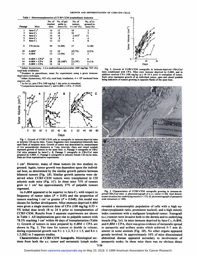
Characteristics of CCRF-CEM Xenografts. Histológica! sections from both the s.c. tumor and metastatic lymph nodes
O
0120
Days40
Fig. 2. Growth of CCRF-CEM xenografts in immune-deprived CBA/CaJmice conditioned with CPA. Mice were immune deprived by fl-yBM. and inaddition received CPA (100 mg/kg i.p.) 18-24 h prior to transplant of tumor.Each cune represents growth of an individual tumor, open and closed symbolsbeing indicative of tumors growing in opposite flanks of the same host.
Fig. 3. Characteristics of CCRF-CEM xenografts growing in immune-deprived CBA/CaJ mice. A, photomicrograph of a s.c. tumor (x 60). Inset demonstrates invasion into underlying muscle (x 175). fi. photomicrograph of paraaorticnode metastasis (x 300).
revealed a monomorphic population of cells with a high nu-clearcytoplasmic ratio, prominent nucleoli, and a high mitoticindex consistent with a malignant lymphoid tumor. Xenograft(s.c.) tumors were invasive both to the dermis and to underlyingmuscle (Fig. 3/1). In mice immune deprived by Wara-C-y,llyBM,
and flyBM + CPA, there was gross evidence of metastalic spreadto paraaortic and axillary nodes which achieved 5-7 mm diameter in some animals (Fig. 3Ä).No other organs appearedgrossly involved. In approximately 10% of mice disseminatedabdominal disease appeared secondary to involvement ofparaaortic nodes. In these mice there was no obvious direct
7126
on May 23, 2018. © 1989 American Association for Cancer Research. cancerres.aacrjournals.org Downloaded from
GROWTH AND DIFFERENTIATION OF CCRF-CEM CELLS
extension of tumor from the s.c. site. Involvement of lymphnodes was not found in CD-nu/nu mice at autopsy.
The morphology and cytochemical characteristics of theCCRF-CEM cell line (from both early and late passage) andxenograft tumor were identical. Cultured and xenografted cellshad the same lymphoid appearance with a large nuclear/cyto-
plasmic ratio, opened nuclear chromatin pattern with nucleoli.There were cytoplasmic vacuoles present in both the cell lineand the xenograft tumor. The cytochemical staining patternwas identical (Table 2), except for a slight decrease in theintensity of the periodic acid-Schiff staining in the xenografttumor cells.
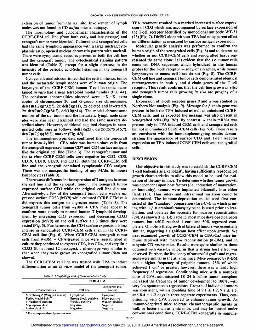
Cytogenetic analysis confirmed that the cells in the s.c. tumorand the metastatic lymph nodes were of human origin. Thekaryotype of the CCRF-CEM human T-cell leukemia maintained in vitro had a near tetraploid modal number (Fig. 4A).The consistent abnormalities observed were: —¿�X,—X,extracopies of chromosome 20 and G-group size chromosome,der(l)t(l;?)(p?32;?), 2x del(8)(pll), 2x deleted and inverted 9,2x der(9)t(9;?)(p24;?), del(10)(pl4). The modal chromosomenumber of the s.c. tumor and the metastatic lymph node samples were also near tetraploid and had the same markers described above. However, additional acquired changes in xenografted cells were as follows: del(3)(q25), der(5)t(5;?)(pl5;?),der(7)t(7;?)(q36;?), marker (Fig. 4Ä).
The immunophenotypic data confirmed that the xenografttumor from 0-yBM + CPA mice was human since cells from
the xenograft expressed human CD7 and CD4 surface antigenslike the original cell line (Table 3). The xenograft tumor andthe in vitro CCRF-CEM cells were negative for CD2, CDS,CD19, CD10, CD20, and CD13. Both the CCRF-CEM cellline and the xenograft contained cytoplasmic CD3 antigen.There was no nonspecific binding of any MAbs to mouselymphocytes (Table 3).
There was a difference in the expression of 2 antigens betweenthe cell line and the xenograft tumor. The xenograft tumorexpressed surface CD3 while the original cell line did not.Alternatively, a few of the xenograft tumor cells weakly expressed surface CD33 (MY9) while cultured CCRF-CEM cells
did express this antigen to a greater extent (Table 3). Thexenograft tumor cells from 0-yBM + CPA mice appear toconform more closely to normal human T-lymphoid development by increasing CD3 expression and decreasing CD33expression (MY9) on the cell surface all 3 times they weretested (Fig. 5). Furthermore, the CD4 surface expression is lessintense in xenografted CCRF-CEM cells than in the CCRF-CEM cell line (Fig. 6). When CCRF-CEM xenograft tumorcells from 07BM + CPA-treated mice were reestablished inculture they continued to express CD3, less CD4, and very littleCD33 (for at least 12 passages), a phenotype very similar tothat when they were grown as xenografted tumor (data notshown).
The CCRF-CEM cell line was treated with TPA to inducedifferentiation as an in vitro model of the xenograft tumor.
Table 2 Morphology and cytochemical reactivity
CCRF-CEMCharacteristicsMorphology"
(\Vrigh t-Giemsa)Periodic acid-Schiffrt-Naphthyl butyratcMyeloperoxidascSudan black BCell
lineLymphoid
Strong block positiveWeakly positiveNegativeNegativeXenograft
(s.c.tumor)LymphoidBlock
positiveWeakly positive
NegativeNegativea
For complete description see text.
TPA treatment resulted in a marked increased surface expression of CD3 which was accompanied by surface expression ofthe T-cell receptor identified by monoclonal antibody WT-31(23) (Fig. 7). DMSO alone without TPA had no apparent effecton differentiation as measured by surface antigen expression.
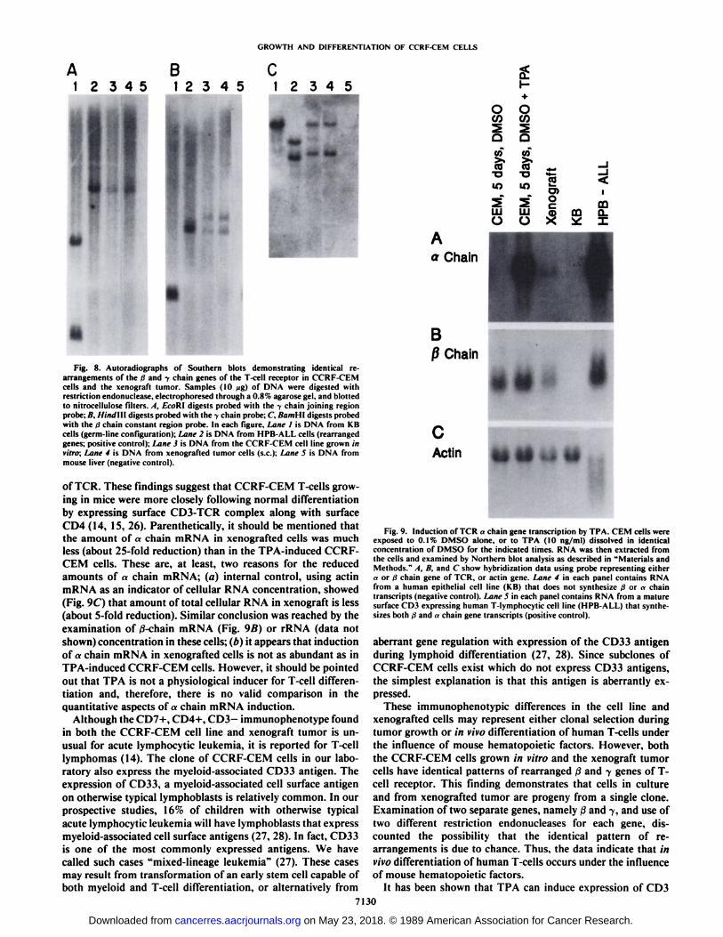
Molecular genetic analysis was performed to confirm thehuman origin of the xenografted cells (Fig. 8) and to determinewhether or not CCRF-CEM cells and xenografted tissue represented the same clone. It is evident that the s.c. tumor cellscontained DNA sequences which hybridized to the humanprobes for the T-cell receptor 7- and /3-chain genes, while mouselymphocytes or mouse cell lines do not (Fig. 8). The CCRF-CEM cell line and xenograft tumor cells demonstrated identicalrearrangements in both 7 and ßchain genes of the T-cellreceptor. This result confirms that the cell line grown in vitroand xenograft tumor cells growing in vivo are progeny of asingle clone.
Expression of T-cell receptor genes ßand a was studied byNorthern blot analysis (Fig. 9). Message for ßchain gene waspresent in both the TPA-induced as well as uninduced CCRF-CEM cells, and as expected the message was also present inxenografted cells (Fig. 9B). By contrast, «chain mRNA waspresent only in TPA-induced CEM cells and xenografted cellsbut not in uninduced CCRF-CEM cells (Fig. 9A). These resultsare consistent with the immunophenotyping results demonstrating the appearance of surface CD3 and T-cell receptorexpression on TPA-induced CCRF-CEM cells and xenograftedcells.
DISCUSSION
Our objective in this study was to establish the CCRF-CEMT-cell leukemia as a xenograft, having sufficiently reproduciblegrowth characteristics to allow this model to be used for evaluation of therapy in mice. To determine whether tumor growthwas dependent upon host factors (i.e., induction of maturation,or immunity), tumors were implanted bilaterally into eitherflank (13). Thus inter- and intraanimal variation could bedetermined. The immune-deprivation model used first consisted of the "standard" preparation (oara-C7), in which prim
ing with l-ß-D-arabinofuranosylcytosine is used prior to irradiation, and obviates the necessity for marrow reconstitution(24). As shown (Fig. \A\ Table 1), most mice developed palpabletumors, but <50% reached 1 cm3, and 50% regressed com
pletely. Of note is that growth of bilateral tumors was essentiallysimilar, suggesting a significant host effect upon growth. Wenext examined growth of CCRF-CEM in CBA/CaJ mice immune deprived with marrow reconstitution (fii-yBM), and inathymic CD-nu/nu mice. Results were quite similar to thoseobtained with 0ara-C7 mice, in that a strong host effect wasobserved. Further, the frequency of successful grafts and regressions were similar in the athymic mice. Mice prepared by ItyBMhad a higher frequency of palpable tumors, 75% of whichachieved 1 cm3 or greater; however, there was a fairly high
frequency of regression. Conditioning mice with a nontoxicdose of CPA, administered 18-24 h before tumor transplantincreased the frequency of tumor development to 100%, withvery few spontaneous regressions. Growth of individual tumorswas consistent, with a doubling time of 9.1 ±1.2, 9.2 ±1.5,and 8.4 ±1.2 days in three separate experiments. Thus, conditioning with CPA appeared to enhance tumor growth. Asimmune-deprived mice tolerate chemotherapeutic agents aswell, or better than athymic mice, and may be housed underconventional conditions, CCRF-CEM xenografts in immune-
7127
on May 23, 2018. © 1989 American Association for Cancer Research. cancerres.aacrjournals.org Downloaded from

SSES Jill2
A
lui SAgg *ji«»x*»<»9 10 11 12
C
13 14 15 16 17 18
D E t t
til ti»* ** * **
20 21 22
F G
lile aiti, laid IMI23 45
A B
v>«ty-mi*¿ss«»»*9 10 11 12
C
t t»O*» »ta. «»» »M««
13 14 15 16 17 18
t t
19 20 mar 1 21 22F G
Fig. 4. A. karyotype of CCRF-CEM human T-cell leukemia maintained in vitro demonstrating that the cells were of human origin and had a near tetraploid modalnumber of chromosomes with the deleted and inverted chromosome 9. B, the karyotype of the CCRF-CEM heterografted cells were identical in the s.c. tumor andmetastatic lymph node. The modal chromosomal number was also near tetraploid with the same markers as the cell line but with additional acquired changes (seetext for details).
7128
on May 23, 2018. © 1989 American Association for Cancer Research. cancerres.aacrjournals.org Downloaded from

GROWTH AND DIFFERENTIATION OF CCRF-CEM CELLS
Table 3 Immunophenolype results"
Clusler group(CD)CD7
CD2CD3CD4CD8CD19
CDIOCD20CD33CD
13Cytoplasm
CD3CCRF-CEMEarly
passage97+++
100+++-69
+IOO++lineLale
passage95
+++
100+++:88
+100++Early
passage80
+40
+95++--NDXenograftLale
passage59
+65
+97++--100+++Metastatic
lymphnodeND*50
+85++NDNDN
nNDND
NDNDMouse
controlsLymph
node LymphocytesND
NDND
NDNDNDND
NDND-
a For all positive results given as percentage of positive cells, intensity of staining is given: +. weak; ++, moderate; +++, intense; —¿�,negative.* ND, not done.
CD3Control
CD3Control
Cell Line
CD33Control
Xenograft Tumor
CD33Control
Cell Line Xenograft TumorFig. 5. Fluorescence histograms; (A and C) CCRF-CEM cells grown in vitro
and used to establish xcnografts. (B and /)) CCRF-CEM cells from xenografts.Thin lines demonstrate results for control immunoglobulins. while heavy linesdemonstrate results for test antibodies; A and B demonstrate increased CD3expression by xenografted tumor, while C and /) demonstrate the decreasedexpression of CD33 by xenografted tumor.
—¿�CDS
Control
—¿�CD3
Control
NoTPA
—¿�anti-TCR
Control
TPA Treated
—¿�anti-TCR
Control
No TPA TPA TreatedFig. 7. Fluorescence histograms of CCRF-CEM cells grown in the absence of
TPA (DMSO control). A and C or in the presence of TPA (16 n\t); B and D.Thin lines demonstrate the results for control isotype matched immunoglobulins,while heavy lines demonstrate results of test antibodies. Treatment with TPAresults in a marked increased expression of surface CD3 (A and K) and a markedincreased expression of surface TCR ((' and D).
—¿�CD4- Control
—¿�CD4- Control
Cell Line
—¿�CD4—¿�Control
Xenograft Tumor
—¿�CD4- Control
Metastatic Xenograft(lymph node)
Mouse Lymphocytes
Fig. 6. Fluorescence histograms of CCRF-CEM cells (A) grown in vitro'. (lì)Xenograft; (C) xenograft-metastatic lymph node. /) is normal mouse lymphocytesfor comparison. Thin lint's demonstrate the results for control immunoglobulins.while heavy lines demonstrate the results for CD4 antibody. Xenografted tumorcells express less CD4 antigen (B and C) than the CCRF-CEM cell line grown invitro (A). Normal mouse lymphocytes do not express the human CD4 antigen(D).
deprived mice prepared by ttyBM + CPA appear to be a suitablemodel for therapeutic studies.
As a xenograft growing s.c. in immune-deprived CBA/CaJmice, CCRF-CEM was highly invasive, both to the dermis andto underlying muscle. In each immune-deprived model (flara-
Oy, 6yBM, 8yßM+ CPA), >90% of mice with tumor >1 cm3
had involvement of paraaortic and axillary nodes without directextension of the s.c. tumor into the abdominal cavity. In approximately 10% of mice, massive abdominal disease appearedsecondary to involvement of paraaortic nodes. The consistentand reproducible metastatic behavior of this tumor is thus ofinterest. Using the T-cell leukemia MOLT 4 or CCRF-CEM,neither Ziegler et al. (7), Ohsugi et al. (1), or Weil-Hillman etal. (11) were able to detect metastatic spread in athymic nudemice. Of note is that we did not observe nodal métastaseswhenCCRF-CEM was transplanted in CD-nu/nu athymic mice.
Spread of tumor to both paraaortic and axillary lymph nodeswas confirmed by karyotype analysis and by immunophenotyp-ing, in addition to standard morphological techniques. Cyto-genetic analysis demonstrated that the tumor cells observed inboth tissues were derived from the original CCRF-CEM cellline (25).
Of note were differences in the immunophenotype betweenCCRF-CEM cells grown in mice (s.c. and métastases)comparedto the original cell line, which suggests that differentiation invivo of leukemic human T-cells may occur under the influenceof mouse hematopoietic factors. Specifically, xenograftedCCRF-CEM cells expressed CD3 antigen on the cell surface,whereas in culture only cytoplasmic CD3 was detected. Similarly, Northern blot analysis showed that xenografted but notcultured CCRF-CEM cells synthesized mRNA for «-chaingene
7129
on May 23, 2018. © 1989 American Association for Cancer Research. cancerres.aacrjournals.org Downloaded from
GROWTH AND DIFFERENTIATION OF CCRF-CEM CELLS
12345
, :
Fig. 8. Autoradiographs of Soulhern blots demonstrating identical rearrangements of the d and y chain genes of the T-cell receptor in CCRF-CEMcells and the xenograft tumor. Samples (10 wg) of DNA were digested withrestriction endonuclease, electrophorescd through a 0.8% agarosc gel, and blottedto nitrocellulose filters. A, EcoRl digests probed with the y chain joining regionprobe; B. IlinJ\\\ digests probed with the y chain probe: C. BamHt digests probedwith the >1chain constant region probe. In each figure. Lane I is DNA from KBcells (germ-line configuration): Lane 2 is DNA from HPB-ALL cells (rearrangedgenes: positive control): Lane 3 is DNA from the CCRF-CEM cell line grown invitro; Lane 4 is DNA from xenografted tumor cells (s.c.); Lane 5 is DNA frommouse liver (negative control).
of TCR. These findings suggest that CCRF-CEM T-cells growing in mice were more closely following normal differentiationby expressing surface CD3-TCR complex along with surface
CD4 (14, 15, 26). Parenthetically, it should be mentioned thatthe amount of «chain mRNA in xenografted cells was muchless (about 25-fold reduction) than in the TPA-induced CCRF-CEM cells. These are, at least, two reasons for the reducedamounts of «chain mRNA; (a) internal control, using actinmRNA as an indicator of cellular RNA concentration, showed(Fig. 9C) that amount of total cellular RNA in xenograft is less(about 5-fold reduction). Similar conclusion was reached by theexamination of ß-chainmRNA (Fig. 9ß)or rRNA (data notshown) concentration in these cells; (b) it appears that inductionof a chain mRNA in xenografted cells is not as abundant as inTPA-induced CCRF-CEM cells. However, it should be pointedout that TPA is not a physiological inducer for T-cell differentiation and, therefore, there is no valid comparison in thequantitative aspects of«chain mRNA induction.
Although the CD7+, CD4+. CD3—immunophenotype foundin both the CCRF-CEM cell line and xenograft tumor is unusual for acute lymphocytic leukemia, it is reported for T-celllymphomas (14). The clone of CCRF-CEM cells in our laboratory also express the myeloid-associated CD33 antigen. Theexpression of CD33, a myeloid-associated cell surface antigenon otherwise typical lymphoblasts is relatively common. In ourprospective studies, 16% of children with otherwise typicalacute lymphocytic leukemia will have lymphoblasts that expressmyeloid-associated cell surface antigens (27, 28). In fact, CD33is one of the most commonly expressed antigens. We havecalled such cases "mixed-lineage leukemia" (27). These cases
may result from transformation of an early stem cell capable ofboth myeloid and T-cell differentiation, or alternatively from
Fig. 9. Induction of TCR «chain gene transcription by TPA. CEM cells wereexposed lo 0.1% DMSO alone, or to TPA (10 ng/ml) dissolved in identicalconcentration of DMSO for the indicated times. RNA was then extracted fromthe cells and examined by Northern blot analysis as described in "Materials andMethods." A, B, and C show hybridization data using probe representing either
a or d chain gene of TCR, or actin gene. Lane 4 in each panel contains RNAfrom a human epithelial cell line (KB) that does not synthesize d or «chaintranscripts (negative control). Lane 5 in each panel contains RNA from a maturesurface CD3 expressing human T-lymphocytic cell line (HPB-AL1.) that synthesizes both ii and »chain gene transcripts (positive control).
aberrant gene regulation with expression of the CD33 antigenduring lymphoid differentiation (27, 28). Since subclones ofCCRF-CEM cells exist which do not express CD33 antigens,the simplest explanation is that this antigen is aberrantly expressed.
These immunophenotypic differences in the cell line andxenografted cells may represent either clonal selection duringtumor growth or in vivo differentiation of human T-cells underthe influence of mouse hematopoietic factors. However, boththe CCRF-CEM cells grown in vitro and the xenograft tumorcells have identical patterns of rearranged /J and -ygenes of T-
cell receptor. This finding demonstrates that cells in cultureand from xenografted tumor are progeny from a single clone.Examination of two separate genes, namely /i and y, and use oftwo different restriction endonucleases for each gene, discounted the possibility that the identical pattern of rearrangements is due to chance. Thus, the data indicate that invivo differentiation of human T-cells occurs under the influenceof mouse hematopoietic factors.
It has been shown that TPA can induce expression of CD37130
on May 23, 2018. © 1989 American Association for Cancer Research. cancerres.aacrjournals.org Downloaded from

GROWTH AND DIFFERENTIATION OF CCRF-CEM CELLS
polypeptides with or without the T-cell receptor on the surfaceof T-cell lines (29, 30). Carrel et al. (29) reported that CCRF-CEM cells could express surface CD3 without surface TCR,and suggested that separate regulatory mechanisms may existfor CD3 surface expression and TCR. Their studies also suggested that expression of the TCR complex is not controlled bytranscription of the TCR «chain, which may be increased afterTPA treatment in CCRF-CEM subclones that do not expressTCR on the cell surface. Some sublines of CCRF-CEM cells,however, do express CD3 polypeptides accompanied by the T-cell receptor when treated with TPA (30), which is consistentwith our results. Our clone of CCRF-CEM cells demonstrateda marked increase in surface CD3 and TCR expression aftertreatment with TPA (Fig. 7). Furthermore, cells also demonstrated abundant appearance of mRNA for the «chain of theTCR after TPA exposure. These results do not eliminate thepossibility that separate regulatory mechanisms exist for surfaceexpression of CD3, the TCR complex, and «-TCRtranscription(29), but are in accordance with our (15) and other investigators(30) results which show that a chain transcription is usuallyaccompanied by TCR surface expression.
In summary, we have established conditions for reproducibleand metastatic growth of human T-cell leukemia in mice. Underthese conditions, >90% of host animals develop distant lymphnode métastases,and conditions for growth appear to stimulatematuration of those cells to give a more normally differentiatedT-cell phenotype. The model described may be of value forstudy of T-cell differentiation, and as an in vivo model forpreclinical pharmacological studies of T-cell leukemia.
ACKNOWLEDGMENTS
We wish to thank L. Woodruff, P. Cheshire, M. Odiorne, and R.Tirey for technical assistance; Dr. R. Ashmun for assistance with flowcytometry studies; Dr. J. Seidman for the gift of the 7-TCR complementary DNA probe and Dr. M. J. Owen for the pB400 probe; and R.Dodge for statistical analysis.
REFERENCES
1. Ohsugi. Y.. Gershwin, M. E., Owens, R. B.. and Nelson-Rees. W. A.Tumorigenicity of human malignant lymphoblasts: comparative study withunmanipulatcd nude mice, antilymphocyte serum-treated nude mice and x-irradiated nude mice. J. Nati. Cancer Inst., 65: 715-718, 1980.
2. Franks. C. R., Bishop. D., Balkwill. F. R.. Oliver, R. T. D., and Spector, W.G. Growth of acute myeloid leukemia as discrete subcutaneous tumors inimmune-deprived mice. Br. J. Cancer, 35:697-700. 1977.
3. Palu. G., Selby. P.. Powles. R.. and Alexander, P. Spontaneous regression ofhuman aeute myeloid leukaemia xenografts and phenotypic evidence formaturation. Br. J. Cancer. 40: 731-735, 1979.
4. Clutlcrbuck, R. D.. Hills. C. A., Hoey, P.. Alexander. P.. Powles, R. L., andMillar. J. L. Studies on the development of human acute myeloid leukaemiaxenografts in immune-deprived mice: comparison with cells in short-termculture. Leuk. Res.. 9: 1511-1518, 1985.
5. Machado, E. A., Gerard, D. A.. Lozzio, C. B., Lozzio. B. B.. Mitchell. J. R.,and Golde. D. W. Proliferation and differentiation of human myeloid leukemia cells in immunodeficient mice: electron microscopy and cytochemistry.Blood. 63: 1015-1022, 1984.
6. Nara. N.. and Miyamoto, T. Direct and serial transplantation of human acutemyeloid leukemia into nude mice. Br. J. Cancer. 45: 778-782, 1982.
7. Ziegler. H. W.. Frizzerà .G., and Bach. F. H. Successful transplantation of ahuman leukemia cell line into nude mice: conditions optimizing graft ac
ceptance. J. Nail. Cancer Inst.. 68: 15-18, 1982.8. Epstein. A. L.. Herman. M. M., Kim, H., Dorfman, R. F.. and Kaplan. II.
S. Biology of human malignant lymphomas. III. Intracranial hcterotransplan-tation in nude, athymic mice. Cancer (Phila.), 37: 2158-2176. 1976.
9. Ono, A., Amos, D. B., and Koren, H. S. Selective cellular natural killingagainst leukemic T cells and thymus. Nature (Lond.). 266: 546-547, 1977.
10. Cudkowicz, G., and Hochman, P. S. Do natural killer cells engage inregulated reactions against self to ensure homeostasis? Immunol. Rev., 44:13-41. 1979.
11. Weil-Hillman. G.. Uckun, F. M.. Manske, J. M., and Vallera, D. A. Combined immunochemotherapy of human solid tumors in nude mice. CancerRes.. 47: 579-585. 1987.
12. Foley, G. E., Lazarus. H., Farber, S., Uzman. B. G.. Boone. B. A., andMcCarthy, R. E. Continuous culture of human lymphoblasts from peripheralblood of a child with acute leukemia. Cancer (Phila.). IS: 522-529. 1965.
13. Houghton, J. A., and Taylor. D. M. Growth characteristics of humancolorectal tumours during serial passage in immune-deprived mice. Br. J.Cancer, 37: 213-223. 1978.
14. Foon. K. A., and Todd, R. F., III. Immunologie classification of leukemiaand lymphoma (review). Blood. 68: 1-31, 1986.
15. Mirro, J.. Jr.. Kitchingman. G.. Behm, F. G.. Murphy. S. B.. and Goorha.R. M. T cell differentiation stages identified by molecular and immunologieanalysis of the T cell receptor complex in childhood lymphoblastic leukemia.Blood, 69:908-912, 1987.
16. Williams, D. L.. Harris. A.. Williams, K. J.. Brosius, M. J.. and Lemonds,W. A direct bone marrow chromosome technique for acute lymphoblasticleukemia. Cancer Genet. Cytogenet., 13: 239-257, 1984.
17. Wigler, M., Sweet, R., Sim, G. K., Wold. B.. Pcllicer, A.. Lacy. E.. Maniatis,T.. Silverstein, S.. and Axel, R. Transformation of mammalian cells withgenes from procaryotes and eucaryotes. Cell. 16: 777-785, 1979.
18. Collins, M. K. L.. Kissonerghis. A. M.. Dunne. M. J.. Watson. C. J.. Rigby,P. W. J., and Owen, M. J. Transcripts from an aberrantly rearranged humanT-cell receptor (i-chain gene. EMBOJ.,4: 1211-1215. 1985.
19. Quertermous. T.. Murre, C., Dialynas. D.. Duby. A. D.. Strominger. J. L.,Waldman. T. A., and Seidman. J. G. Human T-eell 7-ehain genes: organization, diversity, and rearrangement. Science (Wash. DC). 231: 252-255,1986.
20. Yoshikai. Y.. Clark. S. P.. Taylor. S., Sohn, U.. Wilson. B. !.. Minden. M.D.. and Mak. T. W. Organization and sequences of the variable, joining andconstant region genes of human T-cell receptor «-chain.Nature (Lond.), 316:837, 1985.
21. Feinberg, A. P.. and Vogelstein. B. A technique for radiolabeling DNArestriction endonuelease fragment to high specific activity. Anal. Biochem..132:6, 1983.
22. Maniatis. T., Fritsch, E. F., and Sambrook, J. Extraction, purification, andanalysis of mRNA from cukaryotic cells. In: T. Maniatis. E. F. Fritsch, andJ. Sambrook (eds.). Molecular Cloning (A Laboratory Manual), p. 196. ColdSpring Harbor, NY: Cold Spring Harbor Laboratory, 1982.
23. Spits. H.. Borst. J., Tax, W.. Capel. P. J. A., Terhorst, C, and De Vries, J.E. Characteristics of a monoclonal antibody (WT-31) that recognizes acommon epitope on the human T cell for antigen. J. Immunol.. 135: 1922-1928. 1985.
24. Steel. G. G., Courtenay, V. D., and Rostom, A. Y. Improved immuno-suppression techniques for the xenografting of human tumors. Br. J. Cancer,57:224-230. 1978.
25. Moore. D. E.. W'erse. K.. Zawydewski. R.. and Thompson. E. B. Thekaryotype of the glucocorticoid-sensitive, lymphoblastic human T-cell lineCCRF-CEM shows a unique deleted and inverted chromosome 9. CancerGenet. Cytogenet.. 14: 89-94. 1985.
26. Reinherz, E. L., Kung, P. C.. Goldstein, G., Levey, R. H., and Schlossman,S. F. Discrete stages of human intrathymic differentiation: analysis of normalthymocytes and leukemia lymphoblasts of T-cell lineage. Proc. Nati. Acad.Sci. USA, 77: 1588-1592. 1980.
27. Mirro. J.. Zipf. T. F., Pui. C-H., Kitchingman, G.. Williams. D.. Melvin, S.,Murphy, S. B.. and Stass. S. Acute mixed lineage leukemia: clinicopathologiccorrelation and prognostic significance. Blood. 66: 1115-1123, 1985.
28. Mirro, J., and Kitchingman, G. The morphology, cytochemistry, molecularcharacteristics and clinical significance of acute mixed lineage leukemia. In:S. Scott (ed.). Leukemia Cytochemistry, pp. 155-179. Chichester. England:Ellis Horwood Ltd., 1989.
29. Carrel. S.. Mach, J-P.. Micschcr, G.. Salvi, S., Giuffrc. L.. Schreyer. M.. andIsler. P. Phorbol 12-myristate 13-acelate induces surface expression of T3on human immature T cell lines with and without concomitant expressionof the T cell antigen receptor complex. Eur. J. Immunol.. 17: 1079. 1987.
30. Shackelford. D. A.. Smith. A. V.. and Trowbridge. I. S. Changes in geneexpression induced by a phorbol diester: expression of IL-2 receptor. T3 andTcell antigen receptor. J. Immunol.. 183: 613-619, 1987.
7131
on May 23, 2018. © 1989 American Association for Cancer Research. cancerres.aacrjournals.org Downloaded from

1989;49:7124-7131. Cancer Res Peter J. Houghton, Joseph Mirro, Jr., Rakesh M. Goorha, et al. Line, CCRF-CEM, Grafted in MiceGrowth and Differentiation of a Human T-Cell Leukemia Cell
Updated version
http://cancerres.aacrjournals.org/content/49/24_Part_1/7124
Access the most recent version of this article at:
E-mail alerts related to this article or journal.Sign up to receive free email-alerts
Subscriptions
Reprints and
To order reprints of this article or to subscribe to the journal, contact the AACR Publications
Permissions
Rightslink site. Click on "Request Permissions" which will take you to the Copyright Clearance Center's (CCC)
.http://cancerres.aacrjournals.org/content/49/24_Part_1/7124To request permission to re-use all or part of this article, use this link
on May 23, 2018. © 1989 American Association for Cancer Research. cancerres.aacrjournals.org Downloaded from