Embed Size (px)
Citation preview
8/12/2019 Groch Et Al 2012_Skeletal Abnormalities in Humpback Whales
http://slidepdf.com/reader/full/groch-et-al-2012skeletal-abnormalities-in-humpback-whales 1/14
DISEASES OF AQUATIC ORGANISMS
Dis Aquat Org
Vol. 101: 145–158, 2012
doi: 10.3354/dao02518Published November 8
INTRODUCTION
Every winter, humpback whales Megaptera no-
vaeangliae of the southwestern Atlantic Ocean pop-
ulation migrate from feeding grounds in Antarctic
waters to tropical waters on the Brazilian coast to
breed and calve. They concentrate mainly in the
Abrolhos Bank (16° 40’−19°30’S, 37° 25’−39°45’W),
an enlargement of the continental shelf (Andriolo et
al. 2006). A total of 9330 whales was estimated for the
Brazilian coast in 2008 (Wedekin et al. 2010). Strand-
ings commonly occur; from 2002 until 2011, therewere 153 single stranding cases along the Abrolhos
Bank seashore and adjacent waters. In 91% of these
cases, the whale died at sea and was washed ashore
(Instituto Baleia Jubarte [IBJ] unpubl. data).
Full necropsy and histopathological analysis of soft
tissues are often impaired by decomposition state and
necessary logistics related to the size of the animal.
Analysis of skeletal changes, which are relatively re-
sistant to decomposition, may provide valuable infor-
mation on the life history and pathological conditions
© Inter-Research 2012 · www.int-res.com*Email: [email protected]
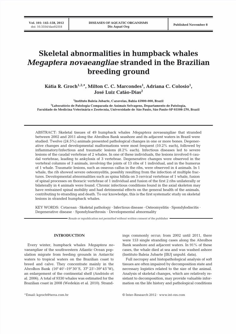
Skeletal abnormalities in humpback whalesMegaptera novaeangliae stranded in the Brazilian
breeding ground
Kátia R. Groch1,2,*, Milton C. C. Marcondes1, Adriana C. Colosio1,
José Luiz Catão-Dias2
1Instituto Baleia Jubarte, Caravelas, Bahia 45900-000, Brazil2Laboratório de Patologia Comparada de Animais Selvagens, Departamento de Patologia,
Faculdade de Medicina Veterinária e Zootecnia, Universidade de São Paulo, São Paulo-SP 05508-270, Brazil
ABSTRACT: Skeletal tissues of 49 humpback whales Megaptera novaeangliae that stranded
between 2002 and 2011 along the Abrolhos Bank seashore and its adjacent waters in Brazil were
studied. Twelve (24.5%) animals presented pathological changes in one or more bones. Degener-
ative changes and developmental malformations were most frequent (10.2% each), followed by
inflammatory/infectious and traumatic lesions (8.2% each). Infectious diseases led to severelesions of the caudal vertebrae of 2 whales. In one of these individuals, the lesions involved 6 cau-
dal vertebrae, leading to ankylosis of 3 vertebrae. Degenerative changes were observed in the
vertebral columns of 3 animals, involving the joints of 13 ribs of 1 individual, and in the humerus
of 1 whale. Traumatic lesions, such as osseous callus in the ribs, were observed in 4 animals. In 1
whale, the rib showed severe osteomyelitis, possibly resulting from the infection of multiple frac-
tures. Developmental abnormalities such as spina bifida on 3 cervical vertebrae of 1 whale, fusionof spinal processes on thoracic vertebrae of 1 individual and fusion of the first 2 ribs unilaterally orbilaterally in 4 animals were found. Chronic infectious conditions found in the axial skeleton may
have restrained spinal mobility and had detrimental effects on the general health of the animals,
contributing to stranding and death. To our knowledge, this is the first systematic study on skeletal
lesions in stranded humpback whales.
KEY WORDS: Cetacean · Skeletal pathology · Infectious disease · Osteomyelitis · Spondylodiscitis ·
Degenerative disease · Spondyloarthrosis · Developmental abnormality
Resale or republication not permitted without written consent of the publisher

8/12/2019 Groch Et Al 2012_Skeletal Abnormalities in Humpback Whales
http://slidepdf.com/reader/full/groch-et-al-2012skeletal-abnormalities-in-humpback-whales 2/14
Dis Aquat Org 101: 145–158, 2012
of the animal. However, information on skeletal ab-
normalities in humpback whales is scarce: 5 single
cases have been reported (Stede 1994, Paterson &
Van Dyck 1996, Kompanje 1999, Félix et al. 2007,
Hellier et al. 2011). Stede (1994) reported osteope-
riostitis ossificans hypertrophicans in the caudal ver-tebra of a humpback whale calf from Germany. Pater-
son & Van Dyck (1996) described a humpback whale
fetus from Australia with injuries consistent with
trauma during parturition, such as bilaterally frac-
tured ribs and superficial focally extensive cystic os-
seous lesions associated with periosteal new bone
formation in the supra-orbital region. Hellier et al.
(2011) reported the presence of a remodeled lesion
ventrally in the maxilla, possibly related to trauma to
the baleen plates and/or adjacent soft tissue in a
13.5-m-long humpback whale. The animal also pre-
sented bony bridging in the left aspect of the neuralarches of 2 cervical vertebrae, and multiple localized
areas of reactive bone growth in the vertebral column
(Hellier et al. 2011). Spondyloarthritis was suspected
to occur in caudal vertebrae of a specimen from Den-
mark (Kompanje 1999). Severe infectious spondy-
litis affected 7 lumbar and 4 caudal vertebrae of a
7.25-m-long humpback whale from Ecuador (Félix et
al. 2007). The goal of the present study was to investi-
gate the occurrence of skeletal pathologic processes
in humpback whales, as well as to evaluate their
potential to compromise health and contribute to the
stranding event or death.
MATERIALS AND METHODS
The total coastal area covered consists of approxi-
mately 500 km between the municipalities of Bel-
monte, State of Bahia (15° 44’ S, 38° 53’ W), and Santa
Cruz, State of Espírito Santo (20°00’S, 40° 09’W),
Brazil, including the coastline of Abrolhos Bank and
Royal Charlotte Bank.
One hundred and fifty-three humpback whales
were found stranded from 2002 until 2011 (IBJ un-
publ. data). For each animal, a specimen number wasissued and information regarding date and location of
stranding, body length and sex were recorded. Bones
from 49 animals that stranded alive and subsequently
died, or were found dead, were examined in situ.
Some examinations were performed opportunistically
when the team was helping section carcasses to facil-
itate removal. Bones were cleaned during dissection
and the remaining soft tissue was removed through
maceration in water and/or washing and drying. A
rib with extensive bone outgrowth and remodeling
(Specimen 256) was radiographed and scanned using
computed tomography (CT) at 7 mm slice thickness,
80 kV and 240 mA, with a 2-channel helical scanner
(GE Medical Systems) at Centro de Imagem São
Paulo in Teixeira de Freitas, Bahia. Images were pro-
cessed using K-PACS (www.k-pacs.net) and Vitrea(www.vitalimages.com) software.
All bones found with pathological changes were
digitally photographed, collected along with samples
of apparently normal bones, identified by specimen
number and archived at IBJ in Caravelas, Bahia. The
exceptions are the skeletons of 3 animals that are
currently on exhibition at the IBJ, in Praia do Forte,
Bahia (Specimen 122), and at the Frans Krajcbergof
Ecological Museum, in Nova Viçosa, Bahia (Speci-
mens 242 and 334). Due to field logistical difficulties,
only 2 ribs with the most severe lesions were col-
lected from Specimen 358, which had pathologicalchanges in 13 ribs; the remaining ribs were photo-
graphed at the stranding site.
Age classes were assigned based on the standard
body length following the classification used in Maz-
zuca et al. (1998), i.e. calves <8 m, juveniles 8−11.6 m
and adults >11.6 m. Estimated length based on meas-
urements of incomplete carcasses was used to esti-
mate maturity whenever possible. Sex was deter-
mined by gross examination at the time of stranding
or by genetic analysis of skin (Engel et al. 2006,
Cypriano-Souza et al. 2009).
In adult animals, bones were removed from the
carcass for examination. Vertebrae of younger ani-
mals had most soft tissue removed and the vertebral
body and the ossified portions of the arch and trans-
verse processes were examined. Intervertebral disks
were inspected for narrowing of the disc space
between the 2 vertebrae, disk protrusion and abnor-
mal ossification. The gross examination of each bone
included assessment of erosion, cavities or eburna-
tion on joint surface, exostosis, anomaly, fusion and
fracture. Pathological changes were further classified
as resulting from developmental malformation, and
degenerative, infectious and inflammatory, or trau-
matic lesion. In the present study, pathological condi-tions were categorized based on what was consid-
ered the nature of the early lesion or pathogenesis.
For degenerative and inflammatory diseases, the
classification proposed by Thompson (2007) and
Kompanje (1999) was used.
During necropsies, dissection of thoracic and
abdominal cavities was prioritized. Skulls were occa-
sionally found a few meters apart from carcasses,
partially buried in sand, with post mortem fractures
of rostrum or damages due to shark scavenging.
146

8/12/2019 Groch Et Al 2012_Skeletal Abnormalities in Humpback Whales
http://slidepdf.com/reader/full/groch-et-al-2012skeletal-abnormalities-in-humpback-whales 3/14
Groch et al.: Skeletal abnormalitites in Megaptera novaeangliae
Examination was limited to the position that they
were found in (usually with dorsal side down) and
precluded a reliable assessment; thus, skull exami-
nation was not included in this study.
RESULTS
Of the 153 whales that stranded on the Abrolhos
and Royal Charlotte Banks region between 2002 and
2011, 49 animals were examined for skeletal disor-
ders. Of these, 22.4% (11/49) were females, 28.6%
(14/49) were males and 49% (24/49) whales were of
undetermined sex; 38.8% (19/49) were calves, 20.4%
(10/49) were juveniles, 26.5% (13/49) were adults
and 14.3% (7/49) were of undetermined age
(Table 1).
Pathological changes were observed in 24.5%(12/49) of the examined animals. Table 2 shows inci-
dence by skeletal region. In the vertebral column,
28.6% (4/14) of the individuals presented lesions in
the cervical region, 10% (2/20) presented pathologi-
cal changes in the thoracic region and 15.8% (3/19)
presented lesions in the caudal region. None of the
16 animals from which lumbar vertebrae were exam-
ined had lesions. Ribs of 17.1% (7/41) of the animals
presented pathological changes. In the appendicular
skeleton, considering the carcasses that had at least
one scapular glenoid fossa and/or head of humerus
examined, the scapulohumeral joints of 3.7% (1/27)
of the animals presented lesions. In the distal articu-
lation of the humerus, 9.1% (1/11) of the animals had
pathological changes. Radii of 14.3% (1/7) of the ani-
mals presented lesions. No scapular blades (n = 23),
ulnar articular facets (n = 6) or mandibles (n = 8) of
the animals showed signs of fractures or other
lesions.
Degenerative and developmental malformations
were observed with higher frequency (10.2%; 5/49),
followed by inflammatory and traumatic lesions(8.2%; 4/49). The description and general data of
specimens with lesions are shown in Table 3.
Degenerative diseases
Spondyloarthrosis resulting from degenerative
changes in the intervertebral disk was found in 3
whales. In Specimen 242, erosion and spondylophytes
were observed in 2 cervical vertebra and prominent
spondylophytes were observed in 2 thoracic and 2
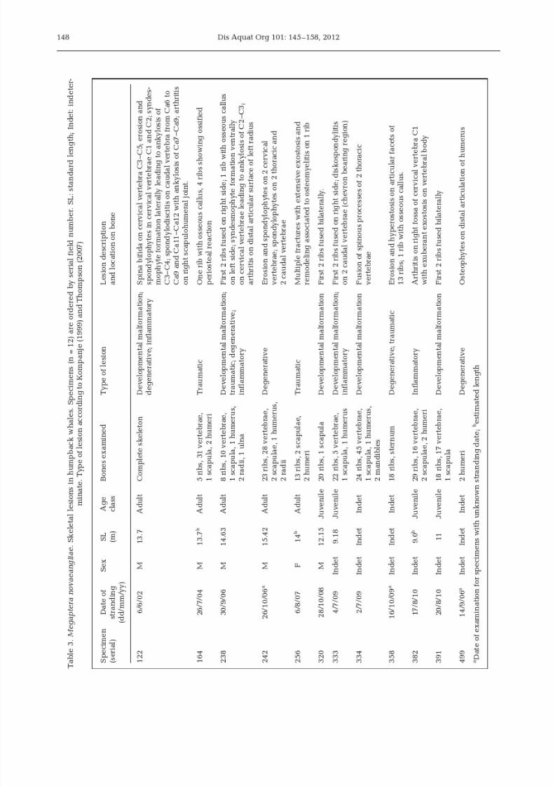
caudal vertebrae. Specimen 238 presented syndesmo-
phyte formation ventrally in the cervical region lead-
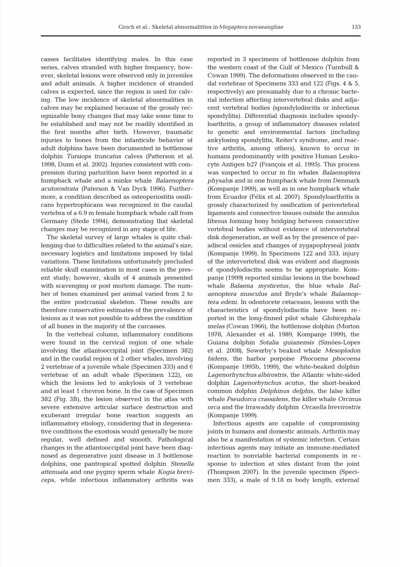
ing to ankylosis of C2 and C3 (Fig. 1A). Specimen 122
presented degenerative changes on cervical vertebrae
C1 to C4. Erosion was observed in the caudal epiphy-
ses and spondylophytes in the right lateral margin of
C1 and C2. Ankylosis of C3 and C4 was observed due
to syndesmophyte formation in the right lateral of the
vertebral body (Fig. 1B). in the same specimen, de-
generative changes were also observed in the right
scapulohumeral joint, with erosion in the head of the
humerus and focally extensive exostosis in the margin
of the scapular glenoid fossa.
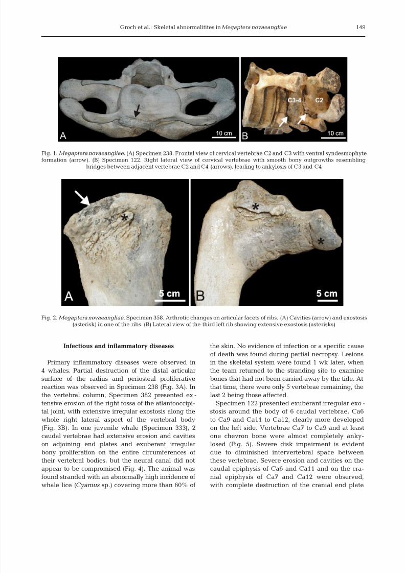
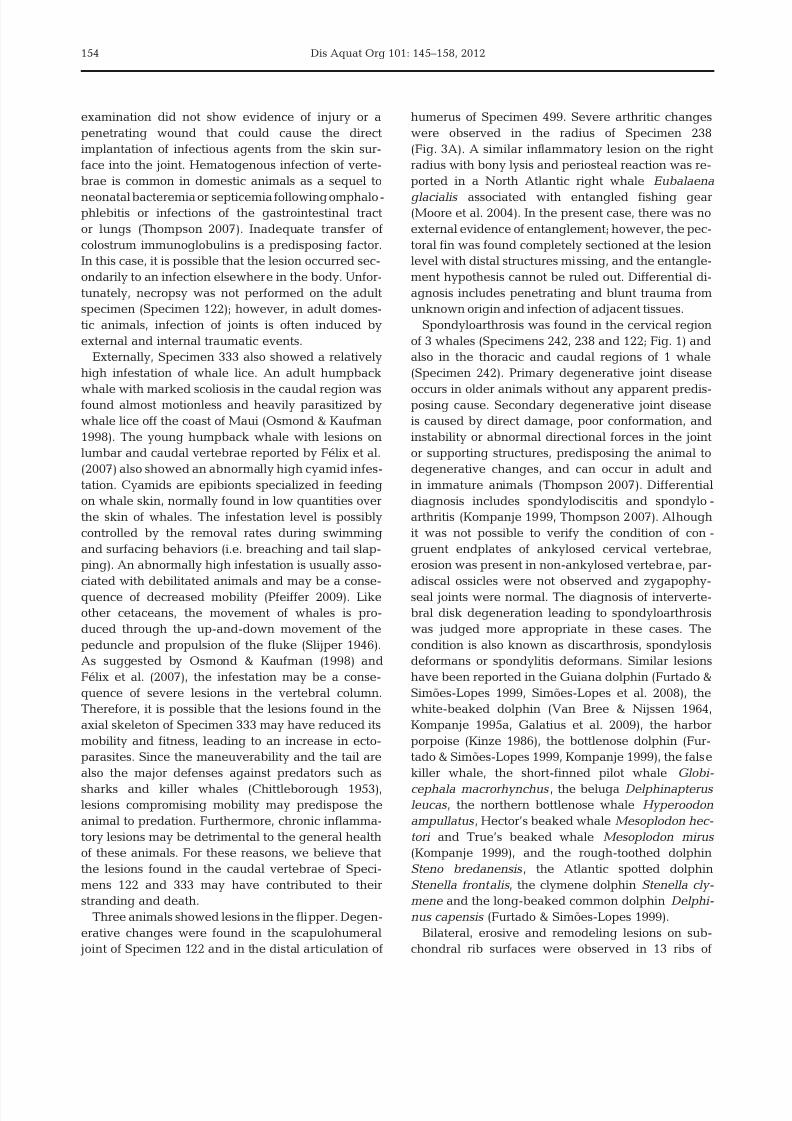
Arthrotic changes were observed in 13 ribs ofSpecimen 358. The articular facet of ribs presented
variable degrees of erosion and/or cavities and mul-
tiple areas of irregular periosteal new bone formation
around the neck (Fig. 2A). The third rib on each side
presented the most severe lesions with lytic changes
and exostosis, leading to deformation of articular
facets (Fig. 2B). Unfortunately, vertebrae were not
present in the carcass and could not be examined.
Specimen 499 presented osteophyte formation in the
distal articulation of the humerus.
147
No. of animals examined Prev. (%)Total With pathology
Sex
Male 14 5 35.7
Female 11 1 9.1Undetermined 24 6 25.0
Age class
Calf 19 0 0
Juvenile 10 3 30.0Adult 13 6 46.2
Undetermined 7 3 42.9
Table 1. Megaptera novaeangliae. Totals of animals exam-
ined by sex and age class and prevalence of pathological
changes
Skeleton No. of animals examined Prev. (%)region Total With pathology
Vertebral columnCervical 14 4 28.6Thoracic 20 2 10.0Lumbar 16 0 0Caudal 19 3 15.8
Ribs 41 7 17.1Scapulohumeral joint 27 1 3.7Humerus, distal joint 11 1 9.1Radius 7 1 14.3Ulna 6 0 0.0
Table 2. Megaptera novaeangliae. Prevalence of pathologi-
cal changes in skeletons of humpback whales
8/12/2019 Groch Et Al 2012_Skeletal Abnormalities in Humpback Whales
http://slidepdf.com/reader/full/groch-et-al-2012skeletal-abnormalities-in-humpback-whales 4/14
8/12/2019 Groch Et Al 2012_Skeletal Abnormalities in Humpback Whales
http://slidepdf.com/reader/full/groch-et-al-2012skeletal-abnormalities-in-humpback-whales 5/14
Groch et al.: Skeletal abnormalitites in Megaptera novaeangliae
Infectious and inflammatory diseases
Primary inflammatory diseases were observed in
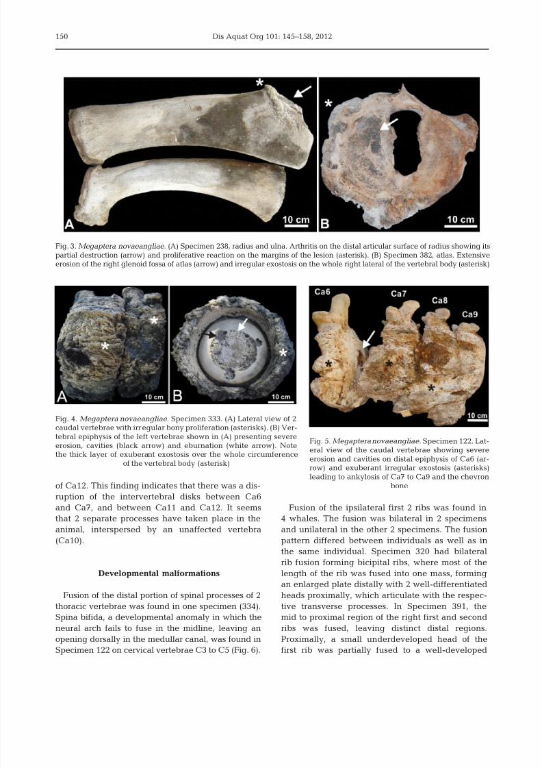
4 whales. Partial destruction of the distal articular
surface of the radius and periosteal proliferative
reaction was observed in Specimen 238 (Fig. 3A). In
the vertebral column, Specimen 382 presented ex-
tensive erosion of the right fossa of the atlantooccipi-tal joint, with extensive irregular exostosis along the
whole right lateral aspect of the vertebral body
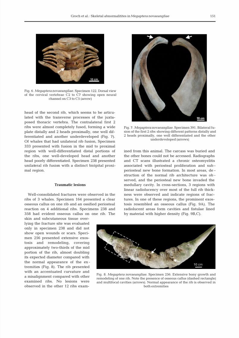
(Fig. 3B). In one juvenile whale (Specimen 333), 2
caudal vertebrae had extensive erosion and cavities
on adjoining end plates and exuberant irregular
bony proliferation on the entire circumferences of
their vertebral bodies, but the neural canal did not
appear to be compromised (Fig. 4). The animal was
found stranded with an abnormally high incidence of
whale lice (Cyamus sp.) covering more than 60% of
the skin. No evidence of infection or a specific cause
of death was found during partial necropsy. Lesions
in the skeletal system were found 1 wk later, when
the team returned to the stranding site to examine
bones that had not been carried away by the tide. At
that time, there were only 5 vertebrae remaining, the
last 2 being those affected.
Specimen 122 presented exuberant irregular exo-stosis around the body of 6 caudal vertebrae, Ca6
to Ca9 and Ca11 to Ca12, clearly more developed
on the left side. Vertebrae Ca7 to Ca9 and at least
one chevron bone were almost completely anky-
losed (Fig. 5). Severe disk impairment is evident
due to diminished intervertebral space between
these vertebrae. Severe erosion and cavities on the
caudal epiphysis of Ca6 and Ca11 and on the cra-
nial epiphysis of Ca7 and Ca12 were observed,
with complete destruction of the cranial end plate
149
Fig. 1. Megaptera novaeangliae . (A) Specimen 238. Frontal view of cervical vertebrae C2 and C3 with ventral syndesmophyteformation (arrow). (B) Specimen 122. Right lateral view of cervical vertebrae with smooth bony outgrowths resembling
bridges between adjacent vertebrae C2 and C4 (arrows), leading to ankylosis of C3 and C4
Fig. 2. Megaptera novaeangliae . Specimen 358. Arthrotic changes on articular facets of ribs. (A) Cavities (arrow) and exostosis
(asterisk) in one of the ribs. (B) Lateral view of the third left rib showing extensive exostosis (asterisks)
8/12/2019 Groch Et Al 2012_Skeletal Abnormalities in Humpback Whales
http://slidepdf.com/reader/full/groch-et-al-2012skeletal-abnormalities-in-humpback-whales 6/14
Dis Aquat Org 101: 145–158, 2012
of Ca12. This finding indicates that there was a dis-
ruption of the intervertebral disks between Ca6
and Ca7, and between Ca11 and Ca12. It seems
that 2 separate processes have taken place in the
animal, interspersed by an unaffected vertebra
(Ca10).
Developmental malformations
Fusion of the distal portion of spinal processes of 2
thoracic vertebrae was found in one specimen (334).
Spina bifida, a developmental anomaly in which the
neural arch fails to fuse in the midline, leaving an
opening dorsally in the medullar canal, was found in
Specimen 122 on cervical vertebrae C3 to C5 (Fig. 6).
Fusion of the ipsilateral first 2 ribs was found in
4 whales. The fusion was bilateral in 2 specimens
and unilateral in the other 2 specimens. The fusion
pattern differed between individuals as well as inthe same individual. Specimen 320 had bilateral
rib fusion forming bicipital ribs, where most of the
length of the rib was fused into one mass, forming
an enlarged plate distally with 2 well-differentiated
heads proximally, which articulate with the respec-
tive transverse processes. In Specimen 391, the
mid to proximal region of the right first and second
ribs was fused, leaving distinct distal regions.
Proximally, a small underdeveloped head of the
first rib was partially fused to a well-developed
150
Fig. 3. Megaptera novaeangliae . (A) Specimen 238, radius and ulna. Arthritis on the distal articular surface of radius showing its
partial destruction (arrow) and proliferative reaction on the margins of the lesion (asterisk). (B) Specimen 382, atlas. Extensiveerosion of the right glenoid fossa of atlas (arrow) and irregular exostosis on the whole right lateral of the vertebral body (asterisk)
Fig. 5. Megaptera novaeangliae . Specimen 122. Lat-eral view of the caudal vertebrae showing severe
erosion and cavities on distal epiphysis of Ca6 (ar-row) and exuberant irregular exostosis (asterisks)
leading to ankylosis of Ca7 to Ca9 and the chevron
bone
Fig. 4. Megaptera novaeangliae . Specimen 333. (A) Lateral view of 2caudal vertebrae with irregular bony proliferation (asterisks). (B) Ver-
tebral epiphysis of the left vertebrae shown in (A) presenting severe
erosion, cavities (black arrow) and eburnation (white arrow). Notethe thick layer of exuberant exostosis over the whole circumference
of the vertebral body (asterisk)
8/12/2019 Groch Et Al 2012_Skeletal Abnormalities in Humpback Whales
http://slidepdf.com/reader/full/groch-et-al-2012skeletal-abnormalities-in-humpback-whales 7/14
Groch et al.: Skeletal abnormalitites in Megaptera novaeangliae
head of the second rib, which seems to be articu-
lated with the transverse processes of the juxta-posed thoracic vertebra. The contralateral first 2
ribs were almost completely fused, forming a wide
plate distally and 2 heads proximally, one well dif-
ferentiated and another underdeveloped (Fig. 7).
Of whales that had unilateral rib fusion, Specimen
333 presented with fusion in the mid to proximal
region with well-differentiated distal portions of
the ribs, one well-developed head and another
head poorly differentiated. Specimen 238 presented
unilateral rib fusion with a distinct bicipital proxi-
mal region.
Traumatic lesions
Well-consolidated fractures were observed in the
ribs of 3 whales. Specimen 164 presented a clear
osseous callus on one rib and an ossified periosteal
reaction on 4 additional ribs. Specimens 238 and
358 had evident osseous callus on one rib. The
skin and subcutaneous tissue over-
lying the fracture site was evaluated
only in specimen 238 and did not
show open wounds or scars. Speci-
men 256 presented extensive exos-tosis and remodeling, covering
approximately two-thirds of the mid
portion of the rib, almost doubling
its expected diameter compared with
the normal appearance of the ex-
tremities (Fig. 8). The rib presented
with an accentuated curvature and
a misalignment compared with other
examined ribs. No lesions were
observed in the other 12 ribs exam-
ined from this animal. The carcass was buried and
the other bones could not be accessed. Radiographs
and CT scans illustrated a chronic osteomyelitis
associated with periosteal proliferation and sub-
periosteal new bone formation. In most areas, de-
struction of the normal rib architecture was ob-
served, and the periosteal new bone invaded the
medullary cavity. In cross-sections, 3 regions with
linear radiolucency over most of the full rib thick-
ness were observed and indicate regions of frac-
tures. In one of these regions, the prominent exos-
tosis resembled an osseous callus (Fig. 9A). The
radiolucent areas form cavities and fistulae lined
by material with higher density (Fig. 9B,C).
151
Fig. 6. Megaptera novaeangliae . Specimen 122. Dorsal viewof the cervical vertebrae C2 to C7 showing open neural
channel on C3 to C5 (arrow)
Fig. 7. Megaptera novaeangliae . Specimen 391. Bilateral fu-
sion of the first 2 ribs showing different patterns distally and
2 heads proximally, one well differentiated and the otherunderdeveloped (arrows)
Fig. 8. Megaptera novaeangliae . Specimen 256. Extensive bony growth and
remodeling of one rib. Note the presence of osseous callus (dashed rectangle)and multifocal cavities (arrows). Normal appearance of the rib is observed in
both extremities

8/12/2019 Groch Et Al 2012_Skeletal Abnormalities in Humpback Whales
http://slidepdf.com/reader/full/groch-et-al-2012skeletal-abnormalities-in-humpback-whales 8/14
Dis Aquat Org 101: 145–158, 2012
DISCUSSION
After the protection of humpback whales in Brazil
(Brazil Federal Decree no. 211, 28 Feb 1962) and the
statutory ban of commercial whaling at the end of the
last century (Brazil Federal Law no. 7.643, 18 Dec
1987), the population of humpback whales has
shown evidence of recovery (Andriolo et al. 2010).
Despite increases in the population, little is known
about the health conditions, threats and impacts on
this species, and how these factors may affect its
recovery.
However, assessing the health of large whales in
the wild is a difficult task. Stranded humpbackwhales are often found dead, in states of advanced
decomposition, with only skeletal remains available
for diagnostic examination. The gross evaluation of
bone may contribute to the understanding of patho-
logic processes that affect the health of these ani-
mals. In fact, like other systems, the skeletal system
reacts to injury and is susceptible to circulatory,
inflammatory, neoplastic, metabolic and develop-
mental disorders (Rosenberg 2005). Evidence of
infectious and degenerative diseases for example,
has been inferred through osseous changes in ceta-
ceans (Cowan 1966, De Smet 1977, Morton 1978,
Kinze 1986, Kompanje 1995a, 1999, Sweeny et al.
2005, Félix et al. 2007, Simões-Lopes et al. 2008).
Skeletal abnormalities have been documented in
several species of cetaceans; however, published
information on osseous pathology in humpback
whales is scarce. Five single cases have been
reported (Stede 1994, Paterson & Van Dyck 1996,
Kompanje 1999, Félix et al. 2007, Hellier et al. 2011).
Analysis usually relies on museum specimens, or
stranded animals dissected with the purpose of col-
lecting the skeleton. To our knowledge, this is the
first report on bone pathology of humpback whalesfrom the southwestern Atlantic Ocean and the first
study on skeletal abnormalities based on the system-
atic analysis of skeletal remains of stranded whales.
Skeletal changes were found in at least 24.5%
(12/49) of the humpback whales examined. A higher
percentage was found in males than in females.
However, it is important to consider that in most
cases, the sex was not determined (due to absent dis-
tinctive features or tissue sample suitable for genetic
analysis) and a prolapsed penis in decomposed car-
152
Fig. 9. Megaptera novaeangliae . Specimen 256. (A) Radiograph of the rib region marked by the dashed rectangle in Fig. 8.
Note the linear radiolucent area (arrow) surrounded by large osseous callus (asterisks). (B,C) Computed tomographs of the rib.
Note periosteal exostosis (asterisks), fistulae (B, arrow) and cavities lined with material with higher density (C, arrow)
8/12/2019 Groch Et Al 2012_Skeletal Abnormalities in Humpback Whales
http://slidepdf.com/reader/full/groch-et-al-2012skeletal-abnormalities-in-humpback-whales 9/14
Groch et al.: Skeletal abnormalitites in Megaptera novaeangliae
casses facilitates identifying males. In this case
series, calves stranded with higher frequency; how-
ever, skeletal lesions were observed only in juveniles
and adult animals. A higher incidence of stranded
calves is expected, since the region is used for calv-
ing. The low incidence of skeletal abnormalities incalves may be explained because of the grossly rec-
ognizable bony changes that may take some time to
be established and may not be readily identified in
the first months after birth. However, traumatic
injuries to bones from the infanticide behavior of
adult dolphins have been documented in bottlenose
dolphin Tursiops truncatus calves (Patterson et al.
1998, Dunn et al. 2002). Injuries consistent with com-
pression during parturition have been reported in a
humpback whale and a minke whale Balaenoptera
acutorostrata (Paterson & Van Dyck 1996). Further-
more, a condition described as osteoperiostitis ossifi-cans hypertrophicans was recognized in the caudal
vertebra of a 6.9 m female humpback whale calf from
Germany (Stede 1994), demonstrating that skeletal
changes may be recognized in any stage of life.
The skeletal survey of large whales is quite chal-
lenging due to difficulties related to the animal’s size,
necessary logistics and limitations imposed by tidal
variations. These limitations unfortunately precluded
reliable skull examination in most cases in the pres-
ent study; however, skulls of 4 animals presented
with scavenging or post mortem damage. The num-
ber of bones examined per animal varied from 2 to
the entire postcranial skeleton. These results are
therefore conservative estimates of the prevalence of
lesions as it was not possible to address the condition
of all bones in the majority of the carcasses.
In the vertebral column, inflammatory conditions
were found in the cervical region of one whale
involving the atlantooccipital joint (Specimen 382)
and in the caudal region of 2 other whales, involving
2 vertebrae of a juvenile whale (Specimen 333) and 6
vertebrae of an adult whale (Specimen 122), on
which the lesions led to ankylosis of 3 vertebrae
and at least 1 chevron bone. In the case of Specimen
382 (Fig. 3B), the lesion observed in the atlas withsevere extensive articular surface destruction and
exuberant irregular bone reaction suggests an
inflammatory etiology, considering that in degenera-
tive conditions the exostosis would generally be more
regular, well defined and smooth. Pathological
changes in the atlantooccipital joint have been diag-
nosed as degenerative joint disease in 3 bottlenose
dolphins, one pantropical spotted dolphin Stenella
attenuata and one pygmy sperm whale Kogia brevi-
ceps , while infectious inflammatory arthritis was
reported in 3 specimens of bottlenose dolphin from
the western coast of the Gulf of Mexico (Turnbull &
Cowan 1999). The deformations observed in the cau-
dal vertebrae of Specimens 333 and 122 (Figs. 4 & 5,
respectively) are presumably due to a chronic bacte-
rial infection affecting intervertebral disks and adja-cent vertebral bodies (spondylodiscitis or infectious
spondylitis). Differential diagnosis includes spondy-
loarthritis, a group of inflammatory diseases related
to genetic and environmental factors (including
ankylosing spondylitis, Reiter’s syndrome, and reac-
tive arthritis, among others), known to occur in
humans predominantly with positive Human Leuko-
cyte Antigen b27 (François et al. 1995). This process
was suspected to occur in fin whales Balaenoptera
physalus and in one humpback whale from Denmark
(Kompanje 1999), as well as in one humpback whale
from Ecuador (Félix et al. 2007). Spondyloarthritis isgrossly characterized by ossification of perivertebral
ligaments and connective tissues outside the annulus
fibrous forming bony bridging between consecutive
vertebral bodies without evidence of intervertebral
disk degeneration, as well as by the presence of par-
adiscal ossicles and changes of zygapophyseal joints
(Kompanje 1999). In Specimens 122 and 333, injury
of the intervertebral disk was evident and diagnosis
of spondylodiscitis seems to be appropriate. Kom-
panje (1999) reported similar lesions in the bowhead
whale Balaena mysticetus , the blue whale Bal-
aenoptera musculus and Bryde’s whale Balaenop-
tera edeni . In odontocete cetaceans, lesions with the
characteristics of spondylodiscitis have been re-
ported in the long-finned pilot whale Globicephala
melas (Cowan 1966), the bottlenose dolphin (Morton
1978, Alexander et al. 1989, Kompanje 1999), the
Guiana dolphin Sotalia guianensis (Simões-Lopes
et al. 2008), Sowerby’s beaked whale Mesoplodon
bidens , the harbor porpoise Phocoena phocoena
(Kompanje 1995b, 1999), the white-beaked dolphin
Lagenorhynchus albirostris , the Atlantic white-sided
dolphin Lagenorhynchus acutus , the short-beaked
common dolphin Delphinus delphis , the false killer
whale Pseudorca crassidens , the killer whale Orcinus orca and the Irrawaddy dolphin Orcaella brevirostris
(Kompanje 1999).
Infectious agents are capable of compromising
joints in humans and domestic animals. Arthritis may
also be a manifestation of systemic infection. Certain
infectious agents may initiate an immune-mediated
reaction to nonviable bacterial components in re-
sponse to infection at sites distant from the joint
(Thompson 2007). In the juvenile specimen (Speci-
men 333), a male of 9.18 m body length, external
153
8/12/2019 Groch Et Al 2012_Skeletal Abnormalities in Humpback Whales
http://slidepdf.com/reader/full/groch-et-al-2012skeletal-abnormalities-in-humpback-whales 10/14
examination did not show evidence of injury or a
penetrating wound that could cause the direct
implantation of infectious agents from the skin sur-
face into the joint. Hematogenous infection of verte-
brae is common in domestic animals as a sequel to
neonatal bacteremia or septicemia following omphalo-phlebitis or infections of the gastrointestinal tract
or lungs (Thompson 2007). Inadequate transfer of
colostrum immunoglobulins is a predisposing factor.
In this case, it is possible that the lesion occurred sec-
ondarily to an infection elsewhere in the body. Unfor-
tunately, necropsy was not performed on the adult
specimen (Specimen 122); however, in adult domes-
tic animals, infection of joints is often induced by
external and internal traumatic events.
Externally, Specimen 333 also showed a relatively
high infestation of whale lice. An adult humpback
whale with marked scoliosis in the caudal region wasfound almost motionless and heavily parasitized by
whale lice off the coast of Maui (Osmond & Kaufman
1998). The young humpback whale with lesions on
lumbar and caudal vertebrae reported by Félix et al.
(2007) also showed an abnormally high cyamid infes-
tation. Cyamids are epibionts specialized in feeding
on whale skin, normally found in low quantities over
the skin of whales. The infestation level is possibly
controlled by the removal rates during swimming
and surfacing behaviors (i.e. breaching and tail slap-
ping). An abnormally high infestation is usually asso-
ciated with debilitated animals and may be a conse-
quence of decreased mobility (Pfeiffer 2009). Like
other cetaceans, the movement of whales is pro-
duced through the up-and-down movement of the
peduncle and propulsion of the fluke (Slijper 1946).
As suggested by Osmond & Kaufman (1998) and
Félix et al. (2007), the infestation may be a conse-
quence of severe lesions in the vertebral column.
Therefore, it is possible that the lesions found in the
axial skeleton of Specimen 333 may have reduced its
mobility and fitness, leading to an increase in ecto-
parasites. Since the maneuverability and the tail are
also the major defenses against predators such as
sharks and killer whales (Chittleborough 1953),lesions compromising mobility may predispose the
animal to predation. Furthermore, chronic inflamma-
tory lesions may be detrimental to the general health
of these animals. For these reasons, we believe that
the lesions found in the caudal vertebrae of Speci-
mens 122 and 333 may have contributed to their
stranding and death.
Three animals showed lesions in the flipper. Degen-
erative changes were found in the scapulohumeral
joint of Specimen 122 and in the distal articulation of
humerus of Specimen 499. Severe arthritic changes
were observed in the radius of Specimen 238
(Fig. 3A). A similar inflammatory lesion on the right
radius with bony lysis and periosteal reaction was re-
ported in a North Atlantic right whale Eubalaena
glacialis associated with entangled fishing gear(Moore et al. 2004). In the present case, there was no
external evidence of entanglement; however, the pec-
toral fin was found completely sectioned at the lesion
level with distal structures missing, and the entangle-
ment hypothesis cannot be ruled out. Differential di-
agnosis includes penetrating and blunt trauma from
unknown origin and infection of adjacent tissues.
Spondyloarthrosis was found in the cervical region
of 3 whales (Specimens 242, 238 and 122; Fig. 1) and
also in the thoracic and caudal regions of 1 whale
(Specimen 242). Primary degenerative joint disease
occurs in older animals without any apparent predis-posing cause. Secondary degenerative joint disease
is caused by direct damage, poor conformation, and
instability or abnormal directional forces in the joint
or supporting structures, predisposing the animal to
degenerative changes, and can occur in adult and
in immature animals (Thompson 2007). Differential
diagnosis includes spondylodiscitis and spondylo-
arthritis (Kompanje 1999, Thompson 2007). Al hough
it was not possible to verify the condition of con-
gruent endplates of ankylosed cervical vertebrae,
erosion was present in non-ankylosed vertebrae, par-
adiscal ossicles were not observed and zygapophy-
seal joints were normal. The diagnosis of interverte-
bral disk degeneration leading to spondyloarthrosis
was judged more appropriate in these cases. The
condition is also known as discarthrosis, spondylosis
deformans or spondylitis deformans. Similar lesions
have been reported in the Guiana dolphin (Furtado &
Simões-Lopes 1999, Simões-Lopes et al. 2008), the
white-beaked dolphin (Van Bree & Nijssen 1964,
Kompanje 1995a, Galatius et al. 2009), the harbor
porpoise (Kinze 1986), the bottlenose dolphin (Fur-
tado & Simões-Lopes 1999, Kompanje 1999), the false
killer whale, the short-finned pilot whale Globi-
cephala macrorhynchus , the beluga Delphinapterus leucas , the northern bottlenose whale Hyperoodon
ampullatus , Hector’s beaked whale Mesoplodon hec-
tori and True’s beaked whale Mesoplodon mirus
(Kompanje 1999), and the rough-toothed dolphin
Steno bredanensis , the Atlantic spotted dolphin
Stenella frontalis , the clymene dolphin Stenella cly-
mene and the long-beaked common dolphin Delphi-
nus capensis (Furtado & Simões-Lopes 1999).
Bilateral, erosive and remodeling lesions on sub-
chondral rib surfaces were observed in 13 ribs of
Dis Aquat Org 101: 145–158, 2012154

8/12/2019 Groch Et Al 2012_Skeletal Abnormalities in Humpback Whales
http://slidepdf.com/reader/full/groch-et-al-2012skeletal-abnormalities-in-humpback-whales 11/14
Groch et al.: Skeletal abnormalitites in Megaptera novaeangliae
Specimen 358 (Fig. 2). Similar lesions have been
reported in sperm whales Physeter macrocephalus
and were diagnosed as osteonecrosis, possibly result-
ing from dysbaric stress (Moore & Early 2004). Differ-
ential diagnosis to this process includes infection
and autoimmune inflammatory reaction. At present,there is no evidence that humpback whales can
undergo lesions associated with dysbaric stress and
complementary studies would be necessary to clarify
this hypothesis. In the present case, the pathological
changes observed on ribs are likely of degenerative
etiology.
The presence of osseous callus was observed in the
ribs of 3 whales (Specimens 164, 238 and 358), with
evidence of fracture or fissure repair. Fractures in
large whales have been reported in ribs of an 18.9 m
fin whale (Hellier et al. 2011), in the mandible of a
11.4 m bowhead whale (Philo et al. 1990) and in theradius and ulna of a skeletally immature fin whale
(Ogden et al. 1981) of unknown origin. Bilaterally
fractured ribs and superficial focally extensive cystic
osseous lesions associated with periosteal new bone
formation dorsolaterally in the supra-orbital region
were observed in a humpback whale and a minke
whale calf from Australia; these lesions were consis-
tent with trauma during parturition (Paterson & Van
Dyck 1996). Fractures on the mandible, skull and
thoracic transverse process of North Atlantic right
whales were attributed to propeller cuts or massive
blunt trauma from ship collision (Moore et al. 2004).
In odontocete cetaceans, rib fractures are apparently
common (Cowan 1966, De Smet 1977, Kompanje
1995c, Dunn et al. 2002, Simões-Lopes et al. 2008). In
our cases, the causes of fractures were not apparent.
The fracture may be pathological or a result of blunt
trauma. A pathological fracture could have been the
result of neoplasia, osteomyelitis or degenerative
bone disease. Blunt trauma could have occurred from
ship collision, a result of aggressive intraspecific or
interspecific behavior, or interaction with predators
(De Smet 1977, Philo et al. 1990, Ross & Wilson 1996,
Patterson et al. 1998, Dunn et al. 2002, Moore et al.
2004). In cases found with a well-formed osseous cal-lous, there was no macroscopic evidence to support
the diagnosis of pathological fracture and a traumatic
event was considered to be more likely. However,
histological and radiological studies would be neces-
sary to confirm this hypothesis.
Specimen 256 (Fig. 8) presented extensive bony
growth and remodeling that covered approximately
two-thirds of the rib, yet both extremities were rela-
tively normal. The rib showed accentuated curvature
and a misalignment. Radiological images evidenced
multiple sites of possible fractures (Fig. 9). Our
hypothesis is that one or more fractures triggered the
infection leading to osteomyelitis; however, it is
uncertain whether osteomyelitis was already pres-
ent, predisposing the bone to pathological fractures.
The formation of the osseous callus was chronicallystimulated, leading to extensive diffuse exostosis and
osseous remodeling. The overall radiological appear-
ance was similar to the infectious form of a condition
known in humans as diffuse sclerosing osteomyelitis
(DSO), with extensive sclerosis and periosteal new
bone formation associated with infection (Groot et al
1996). In this case, it is possible that the secondary
infection of multiple complicated fractures in the rib
induced a chronic reparative reaction, producing
features similar to those of DSO. Lesions with grossly
similar appearance, attributed to several healed frac-
tures with complicating osteomyelitis, were found inone rib of killer whale found in Wieringen Island,
Holland (Kompanje 1991).
Fusion of the first 2 cranial ribs was found in 4
whales, both unilaterally (Specimens 333 and 238)
and bilaterally (Specimens 320 and 391; Fig. 7). A
similar condition has been mentioned in literature
as a bicipital, bifid or double-headed rib (Turner
1871, Allen 1916, Nishiwaki & Kasuya 1971, Omura
et al. 1981, DeLynn et al. 2011). This developmental
abnormality is characterized as the fusion of the first
2 ribs with a cleft in the vertebral end, originating 2
distinct processes that may articulate with the last
cervical and first thoracic vertebra or with the first 2
thoracic vertebrae, which has occasionally been
observed in large whale skeleton museum speci-
mens, including species such as the North Atlantic
right whale (Allen 1916), the southern right whale
Eubalaena australis , the blue whale (Turner 1871),
the sei whale Balaenoptera borealis (Slijper 1936,
Nishiwaki & Kasuya 1971) and Bryde’s whale (Sli-
jper 1936, Omura et al. 1981). In the present study,
bicipital ribs were found unilaterally and bilaterally
in humpback whales. Additionally, another form of
abnormality with fusion of ribs forming 1 or 2 dis-
tinct processes in the vertebral end and 2 processesin the sternal end has been found. To our knowl-
edge, this form of fusion has only been described
bilaterally in a blue whale by Slijper (1936). Unilat-
erally fused fifth and sixth ribs with distinct extrem-
ities, associated with a deformed sternum and rib
cage distortion, have been reported in the bottle-
nose dolphin (DeLynn et al. 2011). The specimen
also presented with bilateral bicipital ribs and
severe developmental malformations in the axial
skeleton (DeLynn et al. 2011). In the present study,
155
8/12/2019 Groch Et Al 2012_Skeletal Abnormalities in Humpback Whales
http://slidepdf.com/reader/full/groch-et-al-2012skeletal-abnormalities-in-humpback-whales 12/14
Dis Aquat Org 101: 145–158, 2012
this condition was found unilaterally and bilaterally
in specimens with either normal or bicipital rib
fusion of the first 2 ribs in the contralateral side.
Spina bifida was observed in 3 cervical vertebrae
of Specimen 122 (Fig. 6). Spina bifida results from
defective closure of dorsal vertebral laminae in a ver-tebral column segment (Thompson 2007). Two cate-
gories of spina bifida malformations may be consid-
ered in the present case: spina bifida occulta, without
protrusion of the spinal cord or meninges, and spina
bifida cystic, in which there is protrusion through the
defective vertebral arch of a cystic swelling, involv-
ing the meninges, spinal cord or both (Dorland 1997).
Defective arch closure was reported in the long-
finned pilot whale, mostly involving thoracic verte-
brae (Cowan 1966). In domestic animals, it has been
reported in canine, feline, bovine, equine and ovine
specimens (Wilson et al. 1979, LeCouteur & Grandy2010) and may be associated with various soft tissue
defects including defective innervations of muscle
groups, kidney fusion and uterine aplasia (Thompson
2007). Degenerative changes were observed in the
spinal column of an 18-yr-old bottlenose dolphin with
multiple developmental abnormalities in the axial
skeleton (DeLynn et al. 2011). In the present case, the
cervical vertebra showing spina bifida also had
spondyloarthrosis, leading to ankylosis. It is unclear
whether the lesions are associated, but it should be
considered. Overall, because it was an adult animal,
the condition may not have caused major disabilities
which would compromise its survival and may have
manifested as the occult form (spina bifida occulta).
The developmental malformations documented
herein are apparently incidental findings of no major
concern to the health of these individuals; however, it
is unknown whether they were associated with
abnormalities in adjacent soft tissues and organs.
Therefore, further studies should be carried out to
elucidate whether skeletal abnormalities affect in-
nervations, vasculature pathways and tendon inser-
tions or predispose animals to degenerative changes.
This study contributes to a better understanding of
the skeletal disorders in humpback whales, addingnew information from 12 cases out of 49 animals,
which shows that approximately one-quarter of the
animals had at least one type of lesion. This is a con-
servative estimate in view of the limitations on the
number of bones examined per animal. Therefore,
efforts to examine skeletal tissues during necropsy
are encouraged and provide additional insights into
the life history and morbid conditions of the animals
found in advanced stage of decomposition. Comple-
mentary diagnostic techniques are needed to better
understand and differentiate pathologic conditions
affecting bones. Diagnostic imaging techniques are
valuable tools (Sweeny et al. 2005). Bacteriology and
molecular techniques are needed for the survey of
possible etiologic agents involved on infectious con-
ditions. Histopathological and immunohistochemicalanalyses of bone and adjacent soft tissue may pro-
vide further understanding on the etiology and
pathogenesis of these conditions.
Acknowledgements . We thank Sr. Ariosvaldo Pinto dos San-tos and volunteers for the invaluable support in the field-
work; Projeto TAMAR, Parque Estadual de Itaúnas and
Instituto Orca for the logistical support in many stranding
events; Prof. Dr. Paulo César Simões-Lopes for providingmany papers for the review of literature; Prof. Dr. Sentiel
Rommel, Dr. Erwin J. O. Kompanje and 2 anonymous refer-
ees for very helpful comments on the manuscript; and Prof.
Dr. Rainer Haetinger and Dr. Enio César Vieira Pereira forhelp with the interpretation of radiographic images. The
support of Fundação de Amparo à Pesquisa do Estado deSão Paulo (FAPESP), processes 2010/50094-3, 2011/08357-0
and 2012/00021-5, is greatly appreciated. Veracel Celulose
provided financial support to the Rescue Program. Projeto
Baleia Jubarte is sponsored by Petroleo Brasileiro (Petrobras).J.L.C.D. is a recipient of a professorship by the Conselho
Nacional de Desenvolvimento Científico e Tecnológico—
CNPq (301517/2006-1). This study was conducted by K.R.G.as partial fulfillment of the requirements for the doctoral
degree at the Departamento de Patologia, Faculdade de
Medicina Veterinária e Zootecnia, Universidade de SãoPaulo.
LITERATURE CITED
Alexander JW, Solangi MA, Riegel LS (1989) Vertebralosteomyelitis and suspected diskospondylitis in an At-
lantic bottlenose dolphin (Tursiops truncatus ). J Wildl Dis
25:118−121Allen GM (1916) The whalebone whales of New England.
Mem Boston Soc Nat Hist 8:105−322
Andriolo A, Martins CCA, Engel MH, Pizzorno JL, and others (2006) The first aerial survey to estimate abun-
dance of humpback whales (Megaptera novaeangliae )in the breeding ground off Brazil (breeding stock A).
J Cetacean Res Manag 8:307−311
Andriolo A, Kinas PG, Engel MH, Albuquerque Martins CC,Rufino AM (2010) Humpback whales within the Brazilian
breeding ground: distribution and population size esti-
mate. Endang Species Res 11:233−243Chittleborough RG (1953) Aerial observations on the hump-
back whale, Megaptera nodosa (Bonaterre), with noteson other species. Aust J Mar Freshw Res 4:219−226
Cowan DF (1966) Pathology of the pilot whale. Globi-
cephala melaena. A comparative survey. Arch Pathol82:178−189
Cypriano-Souza AL, Fernández GP, Lima-Rosa CA, Engel
MH, Bonatto SL (2009) Microsatellite genetic characteri-zation of the humpback whale (Megaptera novae -
angliae ) breeding ground off Brazil (breeding stock A).J Hered 101:189−200
156
8/12/2019 Groch Et Al 2012_Skeletal Abnormalities in Humpback Whales
http://slidepdf.com/reader/full/groch-et-al-2012skeletal-abnormalities-in-humpback-whales 13/14
Groch et al.: Skeletal abnormalitites in Megaptera novaeangliae
De Smet WMA (1977) The fate of old bottle-nosed dolphins,
Tursiops truncatus , in nature as revealed by the condi-
tion of their skeletons. Aquat Mamm 5:48−86DeLynn R, Lovewell G, Wells RS, Early G (2011) Congenital
scoliosis of a bottlenose dolphin. J Wildl Dis 47:979−983Dorland WAN (1997) Dorland’s illustrated medical diction-
ary. Saunders, Philadelphia, PADunn DG, Barco SG, Pabst DA, McLellan WA (2002) Evi-
dence for infanticide in bottlenose dolphins of the west-
ern north Atlantic. J Wildl Dis 38:505−510
Engel MH, Fagundes NJR, Rosenbaum HC, Ott PH, and others (2006) Mitochondrial DNA variability and evalua-
tion of the likely feeding grounds of the humpback whale(Megaptera novaeangliae) population of the Abrolhos
bank, Bahia, Brazil. International Whaling Commission,
HobartFélix F, Haase B, Aguirre WE (2007) Spondylitis in a hump-
back whale (Megaptera novaeangliae) from the south-
east Pacific. Dis Aquat Org 75:259−264François RJ, Eulderink F, Bywaters EG (1995) Commented
glossary for rheumatic spinal diseases, based on pathol-
ogy. Ann Rheum Dis 54:615−625Furtado MHBC, Simões-Lopes PC (1999) Alterações senil-
degenerativas e variações anatômicas na coluna verte-bral de pequenos cetáceos. Biotemas 12:133−147 (with
English abstract)
Galatius A, Sonne C, Kinze CC, Dietz R, Jensen JEB (2009)Occurrence of vertebral osteophytosis in a museum
sample of white-beaked dolphins (Lagenorhynchus albi-
rostris) from Danish waters. J Wildl Dis 45:19−28
Groot RH, van Merkesteyn JP, Bras J (1996) Diffuse scleros-
ing osteomyelitis and florid osseus dysplasia. Oral SurgOral Med Oral Pathol Oral Radiol Endod 81:333–342
Hellier CA, Hufthammer AK, Lislevand T (2011) Osteolo -
gical pathology in a humpback (Megaptera novaean-
gliae) and fin (Balaenoptera physalus) whale skeleton.
Int J Paleopathol 1:117−120Kinze CC (1986) Note on the orcurrence of Spondylitis
deformans in a sample of harbor porpoises (Phocoena
phocoena) taken in Danish waters. Aquat Mamm 12:25−27
Kompanje EJO (1991) Een oud geval van osteomyelitis bij
een orka Orcinus orca [An old case of osteomyelitis in akiller whale Orcinus orca]. Lutra 34:71−76 (with English
abstract)Kompanje EJO (1995a) On the occurrence of spondylosis
deformans in white-beaked dolphins Lagenorhynchus
albirostris (Gray, 1846) stranded on the Dutch coast. ZoolMed Leiden 69:231−250
Kompanje EJO (1995b) Differences between spondylo-
osteomyelitis and spondylosis deformans in small odon-tocetes based on museum material. Aquat Mamm 21:
199−203
Kompanje EJO (1995c) Strandings of killer whales Orcinus
orca in the Netherlands between 1783 and 1995 with
some remarks on skeletal and dental pathology (Mam-malia, Cetacea, Odontoceti). Deinsea 2:67−82
Kompanje EJO (1999) Considerations of the comparative
pathology of the vertebrae in Mysticeti and Odontoceti:evidence for the occurrence of discarthrosis, zygarthro-
sis, infectious spondylitis and spondyloarthritis. Zool
Med Leiden 73:99−130LeCouteur RA, Grandy JL (2010) Diseases of the spinal cord.
In: Ettinger SJ, Feldman EC (eds) Textbook of veterinaryinternal medicine, Vol 2. Expert Consult, ST. Louis, MO
(www.expertconsultbook.com, accessed 15 Oct 2012)
Mazzuca L, Atkinson S, Nitta E (1998) Deaths and entangle-
ments of humpback whales, Megaptera novaeangliae , inthe main Hawaiian Island, 1972–1996. Pac Sci 52:1−13
Moore MJ, Early GA (2004) Cumulative sperm whale bonedamage and the bends. Science 306:2215
Moore M, Knowlton AR, Kraus SD, McLellan WA, Bonde RK(2004) Morphometry, gross morphology and availablehistopathology in North Atlantic right whale (Eubalaena
glacialis) mortalities (1970–2002). J Cetacean Res
Manag 6: 199−214Morton B (1978) Osteomyelitis (pyogenic spondylitis) of
the spine in a dolphin. J Am Vet Med Assoc 173:1119−1120
Nishiwaki M, Kasuya T (1971) Osteological note of an
Antarctic sei whale (Balaenoptera borealis). Sci RepWhales Res Inst Tokyo 23:83−89
Ogden JA, Conlogue GJ, Light TR, Sloan TR (1981) Frac-
tures of the radius and ulna in a skeletally immature finwhale. J Wildl Dis 17:111−116
Omura H, Kasuya T, Kato H, Wada S (1981) Osteological
study of the Brydes whale (Balaenoptera edeni) from thecentral south Pacific and eastern Indian ocean. Sci Rep
Whales Res Inst Tokyo 33:1−26Osmond MG, Kaufman GD (1998) A heavily parasitized
humpback whale (Megaptera novaeangliae). Mar
Mamm Sci 14:146−149Paterson R, Van Dyck SM (1996) Perinatal skeletal injuries
in two balaenopterid whales. Mem Queensl Mus 39:333−337
Patterson IAP, Reid RJ, Wilson B, Grellier K, Ross HM,
Thompson PM (1998) Evidence for infanticide in bottle-nose dolphins: an explanation for violent interactions
with harbour porpoise? Proc R Soc Lond B Biol Sci 265:
1167−1170Pfeiffer CJ (2009) Whale lice. In: Perrin WF, Würsig B,
Thewissen JGM (eds) Encyclopedia of marine mammals.Academic Press, San Diego, CA, p 1220−1223Philo LM, Hanns C, George JC (1990) Fractured mandible
and associated oral lesions in a subsistence-harvestedbowhead whale (Balaena mysticetus). J Wildl Dis 26:
125−128
Rosenberg AE (2005) Bones, joints, and soft tissue tumors.In: Kumar V, Abbas AK, Fausto N (eds) Robbins and
Cotran pathologic basis of disease. Elsevier, Philadel-
phia, PA, p 1273–1324Ross HM, Wilson B (1996) Violent interactions between bot-
tlenose dolphins and harbour porpoises. Proc R Soc LondB Biol Sci 263:283−286
Simões-Lopes PC, Menezes ME, Ferigolo J (2008) Alter-
ações senil-degenerativas, patológicas, traumáticas emalformações ósseas. In: Filho ELAM, Monteiro KDKA
(eds) Biologia, ecologia e conservação do boto-cinza.
Páginas & Letras Editora e Gráfica, São Paulo, p 39−50Slijper EJ (1936) Die Cetaceen vergleichend anatomisch
und systematisch. Capita Zool VI 7:1−590Slijper EJ (1946) Comparative biologic-anatomical investi-
gations on the vertebral column and spinal musculature
of mammals. Verh Kon Nederl Akad Wetensch Amster-dam II 42:1−128
Stede M (1994) Zur Todesursache bei Walen der niedersäch-
sischen Nordseeküste. Drosera 94:7−19Sweeny MM, Price JM, Jones GS, French TW, Early GA,
Moore MJ (2005) Spondylitic changes in long-finnedpilot whales (Globicephala melas) stranded on Cape
157

8/12/2019 Groch Et Al 2012_Skeletal Abnormalities in Humpback Whales
http://slidepdf.com/reader/full/groch-et-al-2012skeletal-abnormalities-in-humpback-whales 14/14
Dis Aquat Org 101: 145–158, 2012
Cod, Massachusetts, USA, between 1982 and 2000.
J Wildl Dis 41:717−727
Thompson K (2007) Bones and joints. In: Maxie MG (ed)Jubb, Kennedy & Palmer’s pathology of domestic ani-
mals, Vol 1. Saunders, Philadelphia, PA, p 1−184Turnbull BS, Cowan DF (1999) Synovial joint disease in wild
cetaceans. J Wildl Dis 35:511−518Turner (1871) On the so-called two-headed ribs in whales
and in man. J Anat Physiol 5:348−361
Van Bree PJH, Nijssen H (1964) On three specimens of
Lagenorhynchus albirostris Gray, 1846 (Mammalia,
Cetacea). Beaufortia 11:85−93
Wedekin LL, Engel MH, Azevedo A, Kinas PG, Andriolo A,Simões-Lopes PC (2010) Density and abundance of
the humpback whale in the Brazilian breeding ground(stock A): aerial survey, 2008. Document SC/62/SH28, 30
May−11 June 2010, Scientific Committee of the Interna-tional Whaling Commision, Agadir
Wilson JW, Kurtz HJ, Leipold HW, Lees GE (1979) Spina
bifida in the dog. Vet Pathol 16:165−179
158
Editorial responsibility: Michael Moore,Woods Hole, Massachusetts, USA
Submitted: March 8, 2012; Accepted: August 14, 2012 Proofs received from author(s): October 16, 2012