Embed Size (px)
Citation preview
Case ReportGrinspan’s Syndrome: A Rare Case withMalignant Transformation
Numan Kokten,1 Lokman Uzun,1 Ayse Serap Karadag,2 Tulay Zenginkinet,3
and M. Tayyar Kalcıoglu 1
1Department of Otolaryngology, Istanbul Medeniyet University Medical Faculty, Goztepe Training and Research Hospital,Istanbul, Turkey2Department of Dermatology, Istanbul Medeniyet University Medical Faculty, Goztepe Training and Research Hospital,Istanbul, Turkey3Department of Pathology, Istanbul Medeniyet University Medical Faculty, Goztepe Training and Research Hospital,Istanbul, Turkey
Correspondence should be addressed to M. Tayyar Kalcıoglu; [email protected]
Received 18 October 2017; Revised 15 January 2018; Accepted 6 February 2018; Published 4 March 2018
Academic Editor: Abrão Rapoport
Copyright © 2018NumanKokten et al.0is is an open access article distributed under theCreative CommonsAttribution License,which permits unrestricted use, distribution, and reproduction in any medium, provided the original work is properly cited.
Aim. Oral lichen planus (OLP) is one of the common chronic inflammatory, noninfectious, and precancerous oral mucosaldiseases that affect the stratified squamous epithelium in adults. Grinspan et al. reported an association of OLP with diabetesmellitus and vascular hypertension and called that Grinspan’s syndrome in 1966.We aim to present a case of Grinspan’s syndromewith malignant transformation. Case Presentation. A 60-year-old man who presented with a ten-year history of OLP diagnosedclinically and histologically was referred to our otolaryngology department with a painless swallowing in the left buccal mucosa for3 months. Clinical examination revealed several plaques, striated white lesions in the buccal mucosa bilaterally, and an exophytictumor in the left buccal mucosa. Histopathological examination showed lichen planus bilaterally and oral squamous cell car-cinoma in the left buccal mucosa. 0e tumor had been developed on the preexisting areas of lichen planus which had beenhistologically proven before. 0e tumor was removed completely, and the tissue defect on the buccal mucosa was repaired witha split-thickness skin graft. Conclusion. Patients with OLP should be followed up periodically in a long term at close intervals forearly diagnosis of malignant transformation.
1. Introduction
Oral lichen planus (OLP) is one of the common chronicinflammatory, noninfectious, and precancerous oral mu-cosal diseases that affect the stratified squamous epitheliumin adults [1, 2]. In general population, OLP prevalence isabout 0.5–2% worldwide and more common in females [2].OLP can present as painless white streaks, raised, lacy-likelesions, or painful and persistent ulcers or plaques or papulesand can resemble leukoplakia [2, 3]. In OLP patients, buccalmucosa, gingiva, tongue, labial mucosa, and vermilion of thelower lip are the most common affected sites [1–4]. 0ebuccal mucosa is affected most commonly, and the tongueand gingiva follow buccal mucosa [4]. 0e lesions are mostlybilateral [5].
Although the etiopathogenesis is still unknown, immu-nological mechanisms are blamed [1, 6]. AutocytotoxicT lymphocytes were arraigned to trigger apoptosis of epi-thelial cells resulting in chronic inflammation. Civatte bodies,acanthosis, and parakeratosis are the results of lymphocyticinfiltration of subepithelial tissue, basal membrane impair-ment, and degenerations of keratinocytes [1]. Malignancypotential of OLP is reported between 0.3 and 10% [7, 8]. 0erelease of inflammatory cytokines caused by oxidative stressis supposed to activate transcription factors that affect pre-malignant cells to turn into malignant cells [1, 3, 4].
OLP diagnosis can be made with clinical features likeclassic oral white lesions, but to confirm diagnosis andexclude the dysplasia or malignancy, the histopathologicalexamination is recommended [5, 6].
HindawiCase Reports in OtolaryngologyVolume 2018, Article ID 9427650, 4 pageshttps://doi.org/10.1155/2018/9427650
OLP has been associated with different systemic diseasessuch as primary biliary cirrhosis, thymoma, ulcerative co-litis, chronic active hepatitis, and myasthenia gravis and withseveral viruses such as hepatitis C virus (HCV), humanpapillomavirus (HPV), human herpes virus 6 (HHV-6), andEpstein-Barr virus (EBV) [6]. 0e lesions related with anidentifiable aetiology which resemble OLP clinically andhistopathologically are called lichenoid reaction [9]. Somedental materials, chronic graft versus host disease, and drugssuch as angiotensin-converting enzyme inhibitors, anti-malarial drugs, and nonsteroidal anti-inflammatory drugsare some examples that cause lichenoid reactions [6, 9].
Anti-inflammatory agents such as the topical cortico-steroids are mainly preferred for OLP treatment; if lesionsare widespread, systemic agents may be required [1, 5, 6].
Patients with OLP should be followed up periodically ina long term at close intervals for early diagnosis of malignanttransformation. Malignant transformation after 40 yearsfrom the initial diagnosis of OLP has been reported [6, 10].
Grinspan et al. reported an association of OLP withdiabetes mellitus (DM) and vascular hypertension (VHT)and called that Grinspan’s syndrome [11]. We present a caseof Grinspan’s syndrome with malignant transformation.
2. Case Presentation
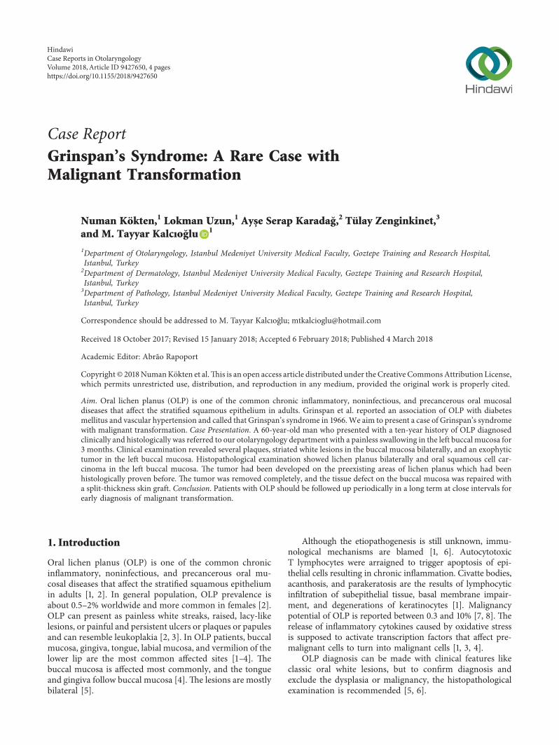
A 60-year-old man who presented with a 10-year history ofOLP diagnosed clinically and histopathologically was re-ferred to our otolaryngology department from dermatologydepartment. 0e complaints of the patient were burningsensation after hot or spicy foods in the left buccal mucosaand painless swallowing in the left buccal mucosa for 3months (Figure 1(a)). 0ere was no history of using tobacco,drinking alcohol, or any other harmful habits. Medical
history of the patient represents diabetes mellitus of 5 yearsand newly diagnosed hypertension accompanying OLP.
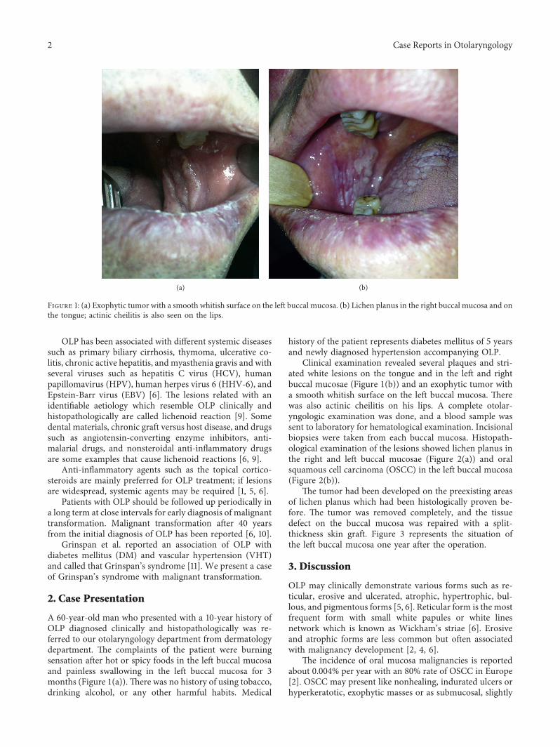
Clinical examination revealed several plaques and stri-ated white lesions on the tongue and in the left and rightbuccal mucosae (Figure 1(b)) and an exophytic tumor witha smooth whitish surface on the left buccal mucosa. 0erewas also actinic cheilitis on his lips. A complete otolar-yngologic examination was done, and a blood sample wassent to laboratory for hematological examination. Incisionalbiopsies were taken from each buccal mucosa. Histopath-ological examination of the lesions showed lichen planus inthe right and left buccal mucosae (Figure 2(a)) and oralsquamous cell carcinoma (OSCC) in the left buccal mucosa(Figure 2(b)).
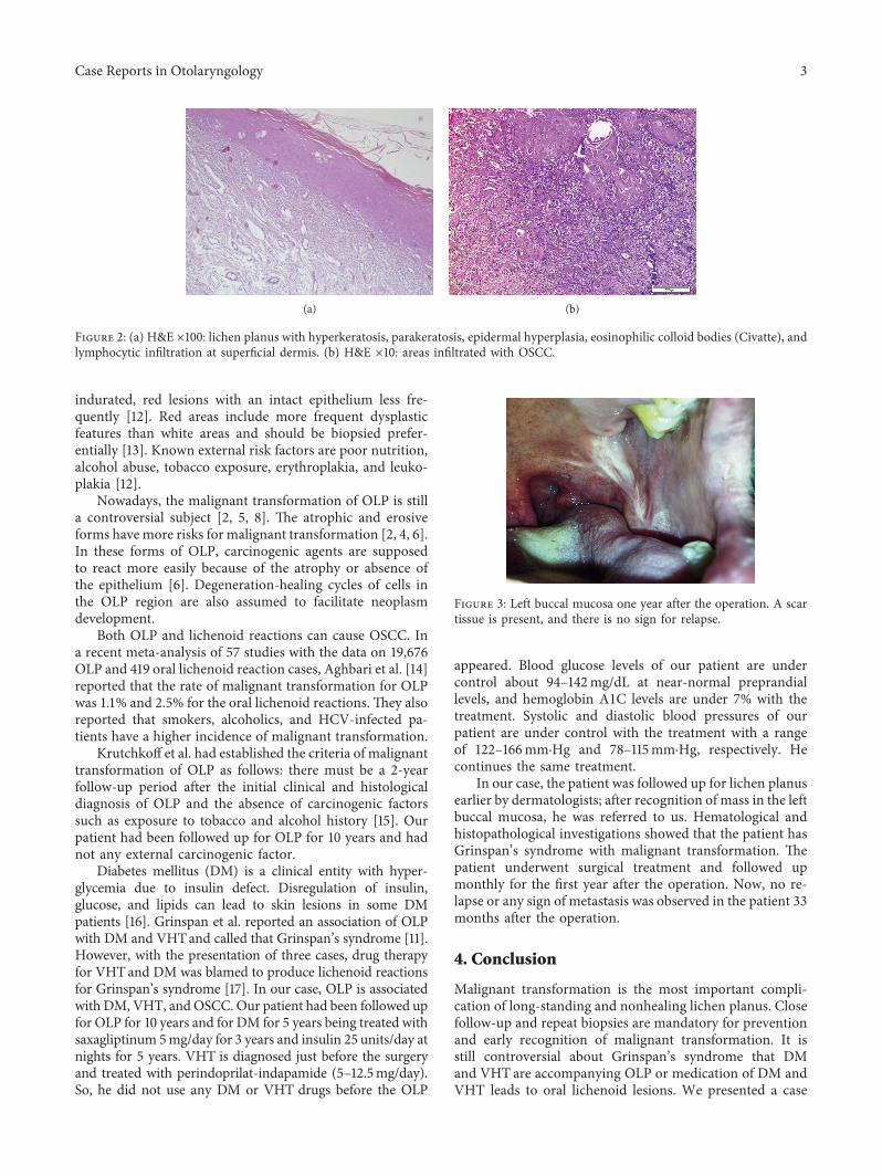
0e tumor had been developed on the preexisting areasof lichen planus which had been histologically proven be-fore. 0e tumor was removed completely, and the tissuedefect on the buccal mucosa was repaired with a split-thickness skin graft. Figure 3 represents the situation ofthe left buccal mucosa one year after the operation.
3. Discussion
OLP may clinically demonstrate various forms such as re-ticular, erosive and ulcerated, atrophic, hypertrophic, bul-lous, and pigmentous forms [5, 6]. Reticular form is the mostfrequent form with small white papules or white linesnetwork which is known as Wickham’s striae [6]. Erosiveand atrophic forms are less common but often associatedwith malignancy development [2, 4, 6].
0e incidence of oral mucosa malignancies is reportedabout 0.004% per year with an 80% rate of OSCC in Europe[2]. OSCC may present like nonhealing, indurated ulcers orhyperkeratotic, exophytic masses or as submucosal, slightly
(a) (b)
Figure 1: (a) Exophytic tumor with a smooth whitish surface on the left buccal mucosa. (b) Lichen planus in the right buccal mucosa and onthe tongue; actinic cheilitis is also seen on the lips.
2 Case Reports in Otolaryngology
indurated, red lesions with an intact epithelium less fre-quently [12]. Red areas include more frequent dysplasticfeatures than white areas and should be biopsied prefer-entially [13]. Known external risk factors are poor nutrition,alcohol abuse, tobacco exposure, erythroplakia, and leuko-plakia [12].
Nowadays, the malignant transformation of OLP is stilla controversial subject [2, 5, 8]. 0e atrophic and erosiveforms havemore risks for malignant transformation [2, 4, 6].In these forms of OLP, carcinogenic agents are supposedto react more easily because of the atrophy or absence ofthe epithelium [6]. Degeneration-healing cycles of cells inthe OLP region are also assumed to facilitate neoplasmdevelopment.
Both OLP and lichenoid reactions can cause OSCC. Ina recent meta-analysis of 57 studies with the data on 19,676OLP and 419 oral lichenoid reaction cases, Aghbari et al. [14]reported that the rate of malignant transformation for OLPwas 1.1% and 2.5% for the oral lichenoid reactions. 0ey alsoreported that smokers, alcoholics, and HCV-infected pa-tients have a higher incidence of malignant transformation.
Krutchkoff et al. had established the criteria of malignanttransformation of OLP as follows: there must be a 2-yearfollow-up period after the initial clinical and histologicaldiagnosis of OLP and the absence of carcinogenic factorssuch as exposure to tobacco and alcohol history [15]. Ourpatient had been followed up for OLP for 10 years and hadnot any external carcinogenic factor.
Diabetes mellitus (DM) is a clinical entity with hyper-glycemia due to insulin defect. Disregulation of insulin,glucose, and lipids can lead to skin lesions in some DMpatients [16]. Grinspan et al. reported an association of OLPwith DM and VHTand called that Grinspan’s syndrome [11].However, with the presentation of three cases, drug therapyfor VHTand DM was blamed to produce lichenoid reactionsfor Grinspan’s syndrome [17]. In our case, OLP is associatedwith DM, VHT, andOSCC. Our patient had been followed upfor OLP for 10 years and for DM for 5 years being treated withsaxagliptinum 5mg/day for 3 years and insulin 25 units/day atnights for 5 years. VHT is diagnosed just before the surgeryand treated with perindoprilat-indapamide (5–12.5mg/day).So, he did not use any DM or VHT drugs before the OLP
appeared. Blood glucose levels of our patient are undercontrol about 94–142mg/dL at near-normal preprandiallevels, and hemoglobin A1C levels are under 7% with thetreatment. Systolic and diastolic blood pressures of ourpatient are under control with the treatment with a rangeof 122–166mm·Hg and 78–115mm·Hg, respectively. Hecontinues the same treatment.
In our case, the patient was followed up for lichen planusearlier by dermatologists; after recognition of mass in the leftbuccal mucosa, he was referred to us. Hematological andhistopathological investigations showed that the patient hasGrinspan’s syndrome with malignant transformation. 0epatient underwent surgical treatment and followed upmonthly for the first year after the operation. Now, no re-lapse or any sign of metastasis was observed in the patient 33months after the operation.
4. Conclusion
Malignant transformation is the most important compli-cation of long-standing and nonhealing lichen planus. Closefollow-up and repeat biopsies are mandatory for preventionand early recognition of malignant transformation. It isstill controversial about Grinspan’s syndrome that DMand VHTare accompanying OLP or medication of DM andVHT leads to oral lichenoid lesions. We presented a case
(a) (b)
Figure 2: (a) H&E ×100: lichen planus with hyperkeratosis, parakeratosis, epidermal hyperplasia, eosinophilic colloid bodies (Civatte), andlymphocytic infiltration at superficial dermis. (b) H&E ×10: areas infiltrated with OSCC.
Figure 3: Left buccal mucosa one year after the operation. A scartissue is present, and there is no sign for relapse.
Case Reports in Otolaryngology 3
with Grinspan’s syndrome which appeared before DM andVHT medication and with malignant transformation.
Disclosure
0is report was presented at the 10th Balkan Congress ofOtorhinolaryngology, Head and Neck Surgery, June 2–5,2016, Tirana, Albania.
Conflicts of Interest
0e authors declare that there are no conflicts of interestregarding the publication of this article.
References
[1] E. M. Otero-Rey, F. Suarez-Alen, M. Peñamaria-Mallon,J. Lopez-Lopez, and A. Blanco-Carrion, “Malignant trans-formation of oral lichen planus by a chronic inflammatoryprocess: use of topical corticosteroids to prevent this pro-gression?,” Acta Odontologica Scandinavica, vol. 72, no. 8,pp. 570–577, 2014.
[2] R. Laeijendecker, T. van Joost, M. C. Kuizinga, B. Tank, andH. A. Neumann, “Premalignant nature of oral lichen planus,”Acta Dermato-Venereologica, vol. 85, no. 6, pp. 516–520, 2005.
[3] A. Marini, M. Wagenmann, E. Ting, and U. R. Hengge,“Squamous cell cancer and human papillomavirus infection inoral lichen planus: case report and literature review,” Der-matologic Surgery, vol. 33, no. 6, pp. 756–760, 2007.
[4] S. G. Fitzpatrick, S. A. Hirsch, and S. C. Gordon, “0e ma-lignant transformation of oral lichen planus and oral lichenoidlesions: a systematic review,” Journal of the American DentalAssociation, vol. 145, no. 1, pp. 45–56, 2014.
[5] J. B. Epstein, L. S. Wan, M. Gorsky, and L. Zhang, “Oral lichenplanus: progress in understanding its malignant potential andthe implications for clinical management,” Oral Surgery, OralMedicine, Oral Pathology, Oral Radiology, and Endodontology,vol. 96, no. 1, pp. 32–37, 2003.
[6] G. Abbate, A. M. Foscolo, M. Gallotti, A. Lancella, andF. Mingo, “Neoplastic transformation of oral lichen: casereport and review of the literature,” Acta Otorhinolar-yngologica Italica, vol. 26, no. 1, pp. 47–52, 2006.
[7] R. W. Katz, J. S. Brahim, and W. D. Travis, “Oral squamouscell carcinoma arising in a patient with long-standing lichenplanus: a case report,” Oral Surgery, Oral Medicine, OralPathology, vol. 70, no. 3, pp. 282–285, 1990.
[8] P. F. Petti, J. V. Bagan, C. Scully, and N. Chaparro, “Malignantturn of oral planus in three new cases,” Acta Otorrinolar-ingologica Española, vol. 55, no. 1, pp. 41–44, 2004.
[9] A. K. Ramachamparambathu, L. Chatra, and P. S. K, “Ma-lignant transformation of a potentially low risk lichenoidreaction,” Journal of the College of Physicians and Surgeons-Pakistan, vol. 22, no. 3, pp. 184-185, 2012.
[10] M. De Vincentiis, A. Gallo, A. Minni, R. Turchetta, andG. Acquaviva, “Le precancerosi del cavo orale,” in Proceedingsof de Campora E. I Tumori del Cavo Orale, V ConvegnoInternazionale, pp. 20-21, Isola Tiberina, Roma, Italy, No-vember 1992.
[11] G. Colella, A. Itro, and G. Corvo, “A case of a carcinomaarising in lichen planus in a subject with diabetes mellitusand arterial hypertension (Grinspan’s syndrome),” MinervaStomatologica, vol. 41, pp. 417–420, 1992.
[12] I. van der Waal, “Tumours of the oral mucosa,” Ned TijdschrDerm Venereol, vol. 12, pp. 368–370, 2002.
[13] C. Scully, “0e oral cavity,” in Textbook of Dermatology,R. H. Champion, J. L. Burton, D. A. Burns, andS. M. Breathnach, Eds., vol. 4, pp. 3074–3076, BlackwellScience, Oxford, UK, 6th edition, 1998.
[14] S. M. H. Aghbari, A. I. Abushouk, A. Attia et al., “Malignanttransformation of oral lichen planus and oral lichenoid le-sions: a meta-analysis of 20095 patient data,” Oral Oncology,vol. 68, pp. 92–102, 2017.
[15] D. J. Krutchkoff, L. Cutler, and S. Laskowski, “Oral lichenplanus: the evidence regarding potential malignant trans-formation,” Journal of Oral Pathology and Medicine, vol. 7,no. 1, pp. 1–7, 1978.
[16] T. Naheed, N. Akbar, M. Shehzad, S. Jamil, and T. Ali, “Skinmanifestations amongst diabetic patients admitted in generalmedical ward for various other medical problems,” PakistanJournal of Medical Sciences, vol. 18, no. 4, pp. 291–296, 2002.
[17] P. J. Lamey, J. Gibson, S. C. Barclay, and S. Miller, “Grinspan’ssyndrome: a drug-induced phenomenon?,” Oral Surgery, OralMedicine, Oral Pathology, vol. 70, no. 2, pp. 184-185, 1990.
4 Case Reports in Otolaryngology

Stem Cells International
Hindawiwww.hindawi.com Volume 2018
Hindawiwww.hindawi.com Volume 2018
MEDIATORSINFLAMMATION
of
EndocrinologyInternational Journal of
Hindawiwww.hindawi.com Volume 2018
Hindawiwww.hindawi.com Volume 2018
Disease Markers
Hindawiwww.hindawi.com Volume 2018
BioMed Research International
OncologyJournal of
Hindawiwww.hindawi.com Volume 2013
Hindawiwww.hindawi.com Volume 2018
Oxidative Medicine and Cellular Longevity
Hindawiwww.hindawi.com Volume 2018
PPAR Research
Hindawi Publishing Corporation http://www.hindawi.com Volume 2013Hindawiwww.hindawi.com
The Scientific World Journal
Volume 2018
Immunology ResearchHindawiwww.hindawi.com Volume 2018
Journal of
ObesityJournal of
Hindawiwww.hindawi.com Volume 2018
Hindawiwww.hindawi.com Volume 2018
Computational and Mathematical Methods in Medicine
Hindawiwww.hindawi.com Volume 2018
Behavioural Neurology
OphthalmologyJournal of
Hindawiwww.hindawi.com Volume 2018
Diabetes ResearchJournal of
Hindawiwww.hindawi.com Volume 2018
Hindawiwww.hindawi.com Volume 2018
Research and TreatmentAIDS
Hindawiwww.hindawi.com Volume 2018
Gastroenterology Research and Practice
Hindawiwww.hindawi.com Volume 2018
Parkinson’s Disease
Evidence-Based Complementary andAlternative Medicine
Volume 2018Hindawiwww.hindawi.com
Submit your manuscripts atwww.hindawi.com