Embed Size (px)
Citation preview
Human Journals
Research Article
November 2017 Vol.:10, Issue:4
© All rights are reserved by Bhusnure O.G et al.
Green Synthesis of Silver Nanoparticle Using Catharanthus
roseus Extract For Pharmacological Activity
www.ijppr.humanjournals.com
Keywords: Silver nanoparticles, Medicinal plants, Green
synthesis, Vinca rosea
ABSTRACT
Nano biotechnology gives emphasis for the synthesis of
nanoparticles using living organisms such as microorganisms,
plant extracts or plant biomass in an eco-friendly way. Among
the various agents used for nanoparticle synthesis, plants have
found the important application. The present study was
designed to screen the neuroprotective effect of Catharanthus
roseus (Linn.) on streptozotocin induced diabetic neuropathy in
rats. Diabetes was induced in rats with a single intraperitoneal
injection of streptozotocin (55 mg/kg b.w). The ethanol extract
of Catharanthus roseus at a dose of 100, 200 and 400 mg/kg of
body weight was administered at the single dose per day to
diabetes-induced rats for a period of 12 weeks. Neuropathic
pain was assessed in diabetic rats with various painful
procedures viz., hot and cold-water tail immersion test
performed to assess the degree of thermal, mechanical, cold
hyperalgesia and locomotor activity as well as motor
coordination. The biomolecules found in plants induce the
reduction of Ag+ ions from silver nitrate to silver nanoparticles
(AgNPs). The aqueous leaves extract of Catharanthus roseus
was used as reducing and stabilizing agent for the synthesis of
the silver nanoparticle. The synthesized nanoparticle is
confirmed by the change of color from transparent yellow to
dark brown indicates the formation of silver nanoparticles.
FTIR spectra were used to monitor the quantitative formation of
silver nanoparticles. The plant-based route could be considered
an environmentally friendly, safe and economic biological
method for the silver nanoparticles production.
Bhusnure O.G1*, Kuthar V.S.
1, Gholve S.B.
1, Giram
P.S.2, Shembekar V.S.
3, Zingade S.G.
4, Jadhav P.P.
1
1. Channabasweshwar Pharmacy College, Dept of
Quality Assurance, Latur (MS), India
2. Channabasweshwar Pharmacy College, Dept of
Pharmacology, Latur (MS), India
3. Rajashri Shahu Mahavidhylay, Department of
Biotechnolgy, (Autonomous), Latur, India.
4. Channabasweshwar Pharmacy College, Dept of
Pharmacognosy, Latur (MS), India
Submission: 23 October 2017
Accepted: 5 November 2017
Published: 30 November 2017
www.ijppr.humanjournals.com
Citation: Bhusnure O.G et al. Ijppr.Human, 2017; Vol. 10 (4): 77-88.
78
INTRODUCTION
In the present scenario, nanotechnology is an important enabling active area of research in
modern material sciences. Nanoparticles deals with the synthesis and control of matter in
scales less than 1µm, normally from 1 to 100 nanometers (nm) 1
. Nanoparticles show
completely new or improved properties and have the wide scope for their diversified
application based on specific characteristics such as size, distribution, and morphology. Silver
nanoparticles have found various and important applications for their bactericidal and
fungicidal activity2. Antimicrobial effect is due to blockage of respiratory enzyme pathways,
alterations of microbial DNA and the cell wall3. Historically, the synthesis of metallic
nanoparticles utilized chemical reducing agents such as hydrazine, sodium citrate, and
sodium borohydride to create uniform suspensions4. However, the chemical method is
harmful in some way as the chemicals used are toxic, flammable, low synthesis rate etc. In
the current phase, green synthesis of nanoparticles is exploited to improve and to protect the
environment by the use of chemicals. Raveendran et al. 2003 suggested three important
factors, which should be considered for the synthesis of nanoparticles, solvent choice, the use
of reducing agent and the use of non-toxic material for nanoparticle stablisation5. Recently,
biological entities serving as both reducing and stabilizing agents for green synthesis of
metallic nanoparticles6. Utilizing biological organisms such as microorganisms
7, enzymes
8
and plant extract or plant biomass could be an excellent alternative to chemical and physical
methods for the production of nanoparticles in a cheap and eco-friendly manner compared to
physical and chemical methods.
Synthesis of nanoparticles using plants can be advantageous over other biological processes
by eliminating the elaborate process of maintaining cell culture9. The microbial enzymes and
secondary metabolites with anti-oxidant or reducing properties are usually reducing metal
compounds into their respective nanoparticles. Plants have been reported to be used for the
synthesis of metal nanoparticles of gold and silver and of a gold-silver-copper alloy 10-14
.
Colloidal silver is of particular interest because of its distinctive properties such as good
conductivity, chemical stability, and catalytic and antibacterial activity15-16
. In during present
study, we found that plant extracts prepared from Catharanthus roses can be used for the
synthesis of silver NPs under bright conditions. The objective of the present study was the
synthesis of silver nanoparticles, reducing the silver ions present in the solution of silver
www.ijppr.humanjournals.com
Citation: Bhusnure O.G et al. Ijppr.Human, 2017; Vol. 10 (4): 77-88.
79
nitrate by the aqueous extract of medicinal plants and evaluation of synthesized silver
nanoparticles against animal toxicity.
MATERIAL AND METHODS
Experimental
Silver nitrate was purchased from Merck Chemicals. All glassware sterilized with nitric acid,
further with distilled water, and dried in the oven before use. Catharanthus roseus leaves
were collected from the college campus in the month of March.
Preparation of leaf extract
The fresh leaves were washed several times with running tap water and after that with
distilled water. Around 20 g of leaves were weighed and boiled for 1h in 100 mL double
distilled water at 60°C and then the extracts were filtered through Whatman filter paper. Then
the filtered extract was stored in the refrigerator at 40C for further use in the synthesis of
silver nanoparticles.
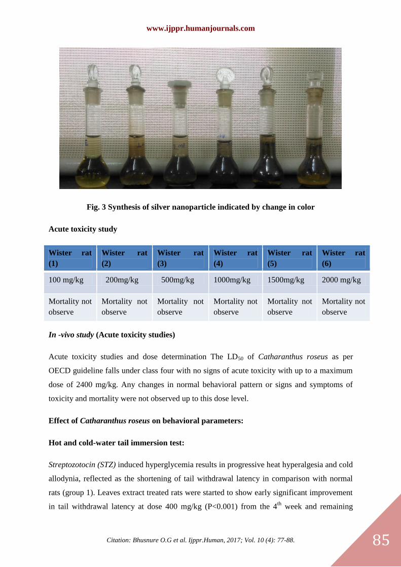
One pot green synthesis of silver nanoparticles
100 mL (1mM) aqueous solution of silver nitrate was prepared in the volumetric flask. Then
1.0, 2.0, 3.0, 4.0 and 5.0 mL of leaf extract were added separately to 10mL aqueous silver
nitrate solution kept in separate beakers at room temperature. The solution was kept in dark
chamber until solution color changes to yellow to dark yellow. After, 15 min, the solution
turns yellow to yellow-red or dark brown indicating the formation of silver nanoparticles.
Sample Plant extract (ml) AgNO3 solution (ml)
A 10 -
1A 1 10
2A 2 10
3A 3 10
4A 4 10
5A 5 S10
Structural studies
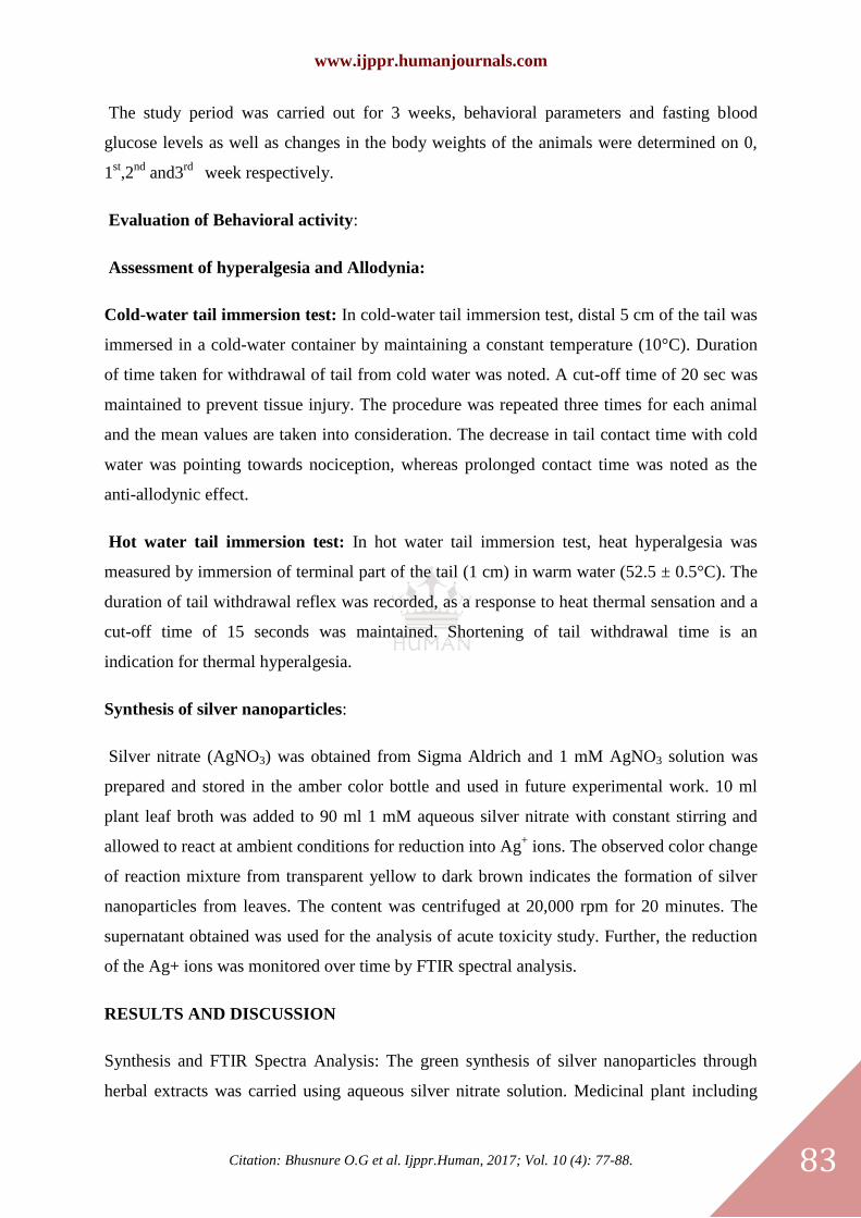
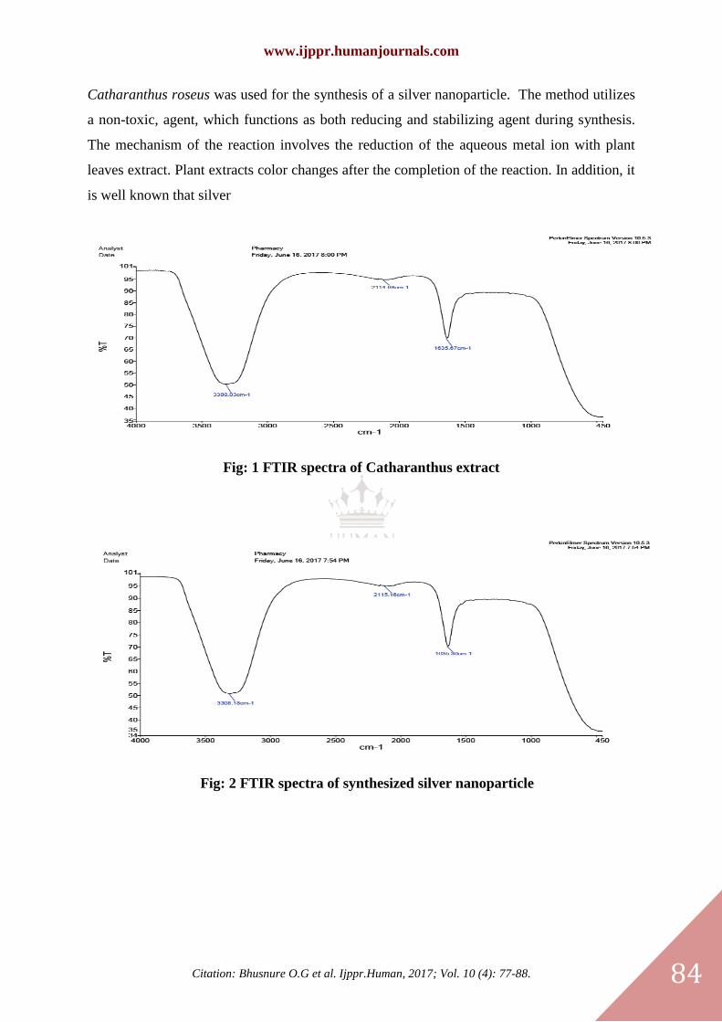
FTIR has become an important tool in understanding the involvement of functional groups in
relation between metal particles and biomolecules, which is used to search the chemical
www.ijppr.humanjournals.com
Citation: Bhusnure O.G et al. Ijppr.Human, 2017; Vol. 10 (4): 77-88.
80
composition of the surface of the silver nanoparticles and identify the biomolecules for
capping and efficient stabilization of the metal nanoparticles. There were many functional
groups present which may have been responsible for the bio-reduction of Ag+ ions. The band
intensities in different regions of the spectrum for plant extract and silver nanoparticles were
analyzed and are shown in Figure.
Acute Toxicity Studies
All the animals were fed with rodent pellet diet and water was allowed ad-libitum under strict
hygienic condition. The animals were fasted overnight prior to the experiment. Fixed-dose
method as per OECD Guideline No. 425 method, given by CPCSEA was adopted for toxicity
studies. The study was conducted by prior permission of institutional animal ethical
committee (IAEC registration no. 731/Po/Re/2002/CPCSEA, approval no.
CBPC/IAEC/2016-17/11). The Wister rats were divided in to control and test group each
containing 6 animals. The test groups of rats were administrated with the dose of 25, 200, 500
&2000 mg/kg of extracts. Carefully observe all the rats and any sign of toxicity in the first
four hours, after the administration of extracts and daily following that for the period of 14
days.
Animal Activity
Selection of animals, caring, and handling
The Wistar rats (Wistar strain 150-200 g) of either sex were used. After randomization into
various groups, animals were accustomed for a period of 10 days under standard husbandry
condition.
Room temperature: 23 ± 3°C
Relative humidity: 50 ± 20%
12 hrs dark and light cycle.
Acute toxicity study
Method
Acute toxicity tests are generally the first tests conducted. They provide data on the relative
toxicity likely to arise from a single drug exposure. The study was conducted after obtaining
www.ijppr.humanjournals.com
Citation: Bhusnure O.G et al. Ijppr.Human, 2017; Vol. 10 (4): 77-88.
81
Institutional animal ethical committee clearance according to Rule 170, Department of
Ayush, Government of India and OECD guidelines 420
Material: 1) Wistar albino rat
2) Catharanthus roseus Linn
3) Gum acacia
4) Distilled water
Procedure
Rats were fasted for 24 hrs prior to drug administration. Six animals were used. MSB
uniformly dispersed in 2% Gum acacia suspension was administered as a single oral dose
equivalent to 2000 mg/kg body weight. Food was withheld for a further 4 hrs. animals were
observed individually at least once during the first 30 min after dosing, and then periodically
during the first 24 hrs (with special attention during first 4 hrs), and daily thereafter for a
period of 14 days. Mortality, if any, was determined over a period of 2 weeks (OECD, 2001).
LD50 was calculated as per OECD guidelines.
Acute toxicity study of Catharanthus roseus on animal model
Plant materials: The leaves of Catharanthus roseus was collected from a college campus in
the month of March.
Extract preparation: The leaves of Catharanthus roseus were collected and dried under
shade and ground into powder. Aqueous extract of Catharanthus roseus leaves was done in
the Department of Pharmacology,
Acute toxicity study: Acute toxicity study of aqueous extract of the leaves of Catharanthus
roseus was determined in Wistar albino rats (150-180 gm) according to the OECD guidelines
No.420. Based on performed toxicity tests the LD50 dose was selected in three doses of 100,
200, 400 mg/kg P.O.
Drugs and Chemicals: Streptozotocin was obtained from Sisco research laboratories Pvt.
Ltd, Mumbai, India, and gabapentin was purchased from Swapnaroop drugs &
www.ijppr.humanjournals.com
Citation: Bhusnure O.G et al. Ijppr.Human, 2017; Vol. 10 (4): 77-88.
82
pharmaceuticals, Aurangabad, Maharastra, India. All other chemicals and reagents used were
of analytical grade.
Animals used: Adult healthy Albino rats of Wistar strain of either sex weighing between
180-250 gm was gathered from the Central animal house, Aurangabad. The rats were housed
in polypropylene cages under standard laboratory conditions 23±20C with 12hr light dark
cycle and had free accesses to water with standard chow diet. Animal care should be taken as
per guidelines of the Committee for the Purpose of Control and Supervision of Experiments
on Animals (CPCSEA). Approval was taken from the Institutional Animal Ethics Committee
for the study.
Experimental design: Experimental design:
In the present investigation, 36 rats were taken and divided into six groups of 6 rats in each.
Out of 6 groups, five were made diabetic with a single dose of the prepared solution of
Streptozotocin 55 mg/kg body weight in cold citrate buffer (PH 4.5, 0.01 M) was
administered intraperitoneally. After 72 hrs blood, glucose level of surviving rats was
measured and rats with fasting blood glucose levels above 250 mg/dl were used for further
study.
The study of test compound and standard drugs were dissolved in distilled water and
administered orally with the help of the gastric oral tube. Rats were divided into the following
groups;
Group-1: Normal control rats (Distilled water 5 ml/kg, p.o)
Group-2: Diabetic control rats (STZ 55 mg/kg, i.p)
Group-3: Diabetic rats served with gabapentin (10 mg/kg, p.o)
Group-4: Diabetic rats served with Catharanthus roseus extract (100 mg/kg, p.o)
Group-5: Diabetic rats served with Catharanthus roseus extract (200 mg/kg, p.o)
Group-6: Diabetic rats served with Catharanthus roseus extract (400 mg/kg, p.o)
www.ijppr.humanjournals.com
Citation: Bhusnure O.G et al. Ijppr.Human, 2017; Vol. 10 (4): 77-88.
83
The study period was carried out for 3 weeks, behavioral parameters and fasting blood
glucose levels as well as changes in the body weights of the animals were determined on 0,
1st,2
nd and3
rd week respectively.
Evaluation of Behavioral activity:
Assessment of hyperalgesia and Allodynia:
Cold-water tail immersion test: In cold-water tail immersion test, distal 5 cm of the tail was
immersed in a cold-water container by maintaining a constant temperature (10°C). Duration
of time taken for withdrawal of tail from cold water was noted. A cut-off time of 20 sec was
maintained to prevent tissue injury. The procedure was repeated three times for each animal
and the mean values are taken into consideration. The decrease in tail contact time with cold
water was pointing towards nociception, whereas prolonged contact time was noted as the
anti-allodynic effect.
Hot water tail immersion test: In hot water tail immersion test, heat hyperalgesia was
measured by immersion of terminal part of the tail (1 cm) in warm water (52.5 ± 0.5°C). The
duration of tail withdrawal reflex was recorded, as a response to heat thermal sensation and a
cut-off time of 15 seconds was maintained. Shortening of tail withdrawal time is an
indication for thermal hyperalgesia.
Synthesis of silver nanoparticles:
Silver nitrate (AgNO3) was obtained from Sigma Aldrich and 1 mM AgNO3 solution was
prepared and stored in the amber color bottle and used in future experimental work. 10 ml
plant leaf broth was added to 90 ml 1 mM aqueous silver nitrate with constant stirring and
allowed to react at ambient conditions for reduction into Ag+ ions. The observed color change
of reaction mixture from transparent yellow to dark brown indicates the formation of silver
nanoparticles from leaves. The content was centrifuged at 20,000 rpm for 20 minutes. The
supernatant obtained was used for the analysis of acute toxicity study. Further, the reduction
of the Ag+ ions was monitored over time by FTIR spectral analysis.
RESULTS AND DISCUSSION
Synthesis and FTIR Spectra Analysis: The green synthesis of silver nanoparticles through
herbal extracts was carried using aqueous silver nitrate solution. Medicinal plant including
www.ijppr.humanjournals.com
Citation: Bhusnure O.G et al. Ijppr.Human, 2017; Vol. 10 (4): 77-88.
84
Catharanthus roseus was used for the synthesis of a silver nanoparticle. The method utilizes
a non-toxic, agent, which functions as both reducing and stabilizing agent during synthesis.
The mechanism of the reaction involves the reduction of the aqueous metal ion with plant
leaves extract. Plant extracts color changes after the completion of the reaction. In addition, it
is well known that silver
Fig: 1 FTIR spectra of Catharanthus extract
Fig: 2 FTIR spectra of synthesized silver nanoparticle
www.ijppr.humanjournals.com
Citation: Bhusnure O.G et al. Ijppr.Human, 2017; Vol. 10 (4): 77-88.
85
Fig. 3 Synthesis of silver nanoparticle indicated by change in color
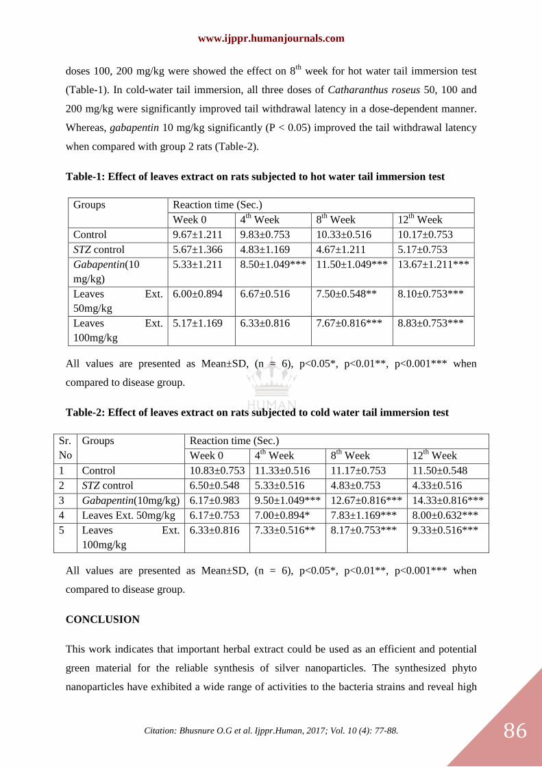
Acute toxicity study
Wister rat
(1)
Wister rat
(2)
Wister rat
(3)
Wister rat
(4)
Wister rat
(5)
Wister rat
(6)
100 mg/kg 200mg/kg 500mg/kg 1000mg/kg 1500mg/kg 2000 mg/kg
Mortality not
observe
Mortality not
observe
Mortality not
observe
Mortality not
observe
Mortality not
observe
Mortality not
observe
In -vivo study (Acute toxicity studies)
Acute toxicity studies and dose determination The LD50 of Catharanthus roseus as per
OECD guideline falls under class four with no signs of acute toxicity with up to a maximum
dose of 2400 mg/kg. Any changes in normal behavioral pattern or signs and symptoms of
toxicity and mortality were not observed up to this dose level.
Effect of Catharanthus roseus on behavioral parameters:
Hot and cold-water tail immersion test:
Streptozotocin (STZ) induced hyperglycemia results in progressive heat hyperalgesia and cold
allodynia, reflected as the shortening of tail withdrawal latency in comparison with normal
rats (group 1). Leaves extract treated rats were started to show early significant improvement
in tail withdrawal latency at dose 400 mg/kg (P<0.001) from the 4th
week and remaining
www.ijppr.humanjournals.com
Citation: Bhusnure O.G et al. Ijppr.Human, 2017; Vol. 10 (4): 77-88.
86
doses 100, 200 mg/kg were showed the effect on 8th
week for hot water tail immersion test
(Table-1). In cold-water tail immersion, all three doses of Catharanthus roseus 50, 100 and
200 mg/kg were significantly improved tail withdrawal latency in a dose-dependent manner.
Whereas, gabapentin 10 mg/kg significantly (P < 0.05) improved the tail withdrawal latency
when compared with group 2 rats (Table-2).
Table-1: Effect of leaves extract on rats subjected to hot water tail immersion test
Groups Reaction time (Sec.)
Week 0 4th
Week 8th
Week 12th
Week
Control 9.67±1.211 9.83±0.753 10.33±0.516 10.17±0.753
STZ control 5.67±1.366 4.83±1.169 4.67±1.211 5.17±0.753
Gabapentin(10
mg/kg)
5.33±1.211 8.50±1.049*** 11.50±1.049*** 13.67±1.211***
Leaves Ext.
50mg/kg
6.00±0.894 6.67±0.516 7.50±0.548** 8.10±0.753***
Leaves Ext.
100mg/kg
5.17±1.169 6.33±0.816 7.67±0.816*** 8.83±0.753***
All values are presented as Mean±SD, (n = 6), p<0.05*, p<0.01**, p<0.001*** when
compared to disease group.
Table-2: Effect of leaves extract on rats subjected to cold water tail immersion test
Sr.
No
Groups Reaction time (Sec.)
Week 0 4th
Week 8th
Week 12th
Week
1 Control 10.83±0.753 11.33±0.516 11.17±0.753 11.50±0.548
2 STZ control 6.50±0.548 5.33±0.516 4.83±0.753 4.33±0.516
3 Gabapentin(10mg/kg) 6.17±0.983 9.50±1.049*** 12.67±0.816*** 14.33±0.816***
4 Leaves Ext. 50mg/kg 6.17±0.753 7.00±0.894* 7.83±1.169*** 8.00±0.632***
5 Leaves Ext.
100mg/kg
6.33±0.816 7.33±0.516** 8.17±0.753*** 9.33±0.516***
All values are presented as Mean±SD, (n = 6), p<0.05*, p<0.01**, p<0.001*** when
compared to disease group.
CONCLUSION
This work indicates that important herbal extract could be used as an efficient and potential
green material for the reliable synthesis of silver nanoparticles. The synthesized phyto
nanoparticles have exhibited a wide range of activities to the bacteria strains and reveal high
www.ijppr.humanjournals.com
Citation: Bhusnure O.G et al. Ijppr.Human, 2017; Vol. 10 (4): 77-88.
87
efficacy of silver Nanoparticle as a strong antibacterial agent. Thus, this phyto nanoparticles
has the potential for the development of drugs for various diseases and useful in biomedical
application.
ACKNOWLEDGEMENT
The work was supported by Department of Biotechnology, Vilasrao Deshmukh
Biotechnology College Latur in Maharashtra. Our special thanks to Dr. Thonte S. S. for
facility provided for the research.
REFERENCES
1. Dahl J.A., Maddux B.L.S. and Hutchison, J.E., “Toward greener nano synthesis.” Chemical Reviews., 107:
2228-2269 (2007)
2. Hutchison, J.E.,“Greener nanoscience: a proactive approach to advancing applications and reducing
implications of nanotechnology.”ACS Nano., 2: 395-402 (2008)
3. Rai M., Yadav A. and Gade A.,“Silver Nanoparticles as A New Generation of Antimicrobial.”
Biotechnology Advances., 27: 76-83 (2009)
4. Zhou J., Ralston J., Sedev R.and Beattie, D.A.,“Functionalized gold nanoparticles: synthesis, structure and
colloid stability”. J. Colloid Interface Sci., 331: 251-262 (2009)
5. Raveendran P., Fu J. and Wallen J.S.L., (2003). „Completely “green” synthesis and stabilization of metal
nanoparticles‟. J. Am. Chem. Soc., 125: 13940-13941(2003)
6. Thakkar K.N., Mhatre S.S. and Parikh R.Y., „Biological synthesis of metallic nanoparticles.‟
Nanomedicine., 6: 257-262 (2010)
7. Schultz S., Smith D.R., Mock J.J. and Schultz D.A., Single-target molecule detection with nonbleaching
multicolor optical immunolabels. Proceedings of the National Academy of Sciences., 97: 996-1001 (2000)
8. Nair B., Pradeep T., Coalescence of nanoclusters and formation of submicron crystallites assisted by
Lactobacillus strains. Cryst Growth Des., 2: 293-298 (2002) 9. Willner I., Baron R., and Willner B., Growing
metal nanoparticles by enzymes. Adv Mater., 18: 1109-1120 (2006)
9. Joerger R., Klaus T. and Granqvist C.G., “Biologically produced silver-carbon composite materials for
optically functional thin-film coatings.” Advanced Materials., 12: 407-409 (2000)
10. Ahmad A., Senapati S., Khan M.I., Kumar R. and Sastry M., “Extracellular biosynthesis of monodisperse
gold nanoparticles by a novel extremophilic actinomycete thermomonospora sp.” Langmuir., 19: 3550-3553
(2003)
11. Anderson C.W.N., Brooks R.R., Stewart R.B. and Simcock R., “Harvesting a crop of gold in plants.”
Nature., 395: 553-554 (1998)
12. Gardea-Torresdey J.L.E., Gomez E., Peralta-Videa J.R., Parsons J.G., Troiani H. and JoseYacaman M.,
“Alfalfa sprouts: a natural source for the synthesis of silver nanoparticles.” Langmuir., 19: 1357-1361 (2003)
13. Romero-Gonzalez J., Walton J.C., Peralta-Videa J.R., Rodrıguez E., Romero J. and GardeaTorresdey J.L.,
“Modeling the adsorption of Cr(III) from aqueous solution onto Agave lechuguilla biomass: the study of the
advective and dispersive transport.” Journal of Hazardous Materials., 161: 360-365 (2009)
14. Tessier P.M., Velev O.D., Kalambur A.T., Rabolt J.F., Lenhoff A.M. and Kaler E.W., “Assembly of gold
nanostructured films templated by colloidal crystals and use in surface-enhanced Raman spectroscopy." Journal
of the American Chemical Society., 122: 9554-9555 (2000)
15. Cao Y.C., Jin R., and Mirkin C.A., “Nanoparticles with Raman spectroscopic fingerprints for DNA and
RNA detection.” Science., 297: 1536-1540 (2002) 17. Perez C., Paul M. and Bazerque P., Antibiotic assay by
the agar well diffusion method. Acta Biol Med Exp., 15: 113-115 (1990)
www.ijppr.humanjournals.com
Citation: Bhusnure O.G et al. Ijppr.Human, 2017; Vol. 10 (4): 77-88.
88
16. Ching T., Hou J. and Fu S., “Biocatalysis and Biomolecules Engineering”, John Willey & sons, Hoboken,
New Jersey, 452-454 (2010).
17. Thirumurgan A., Tomy, N.A., Jai Ganesh, R. and Gobikrishnan, S., Biological reduction of silver
nanoparticles using plant leaf extracts and its effect an increased antimicrobial activity against the clinically
isolated organism. De Phar Chem., 2: 279-284 (2010)
18. Jain D., Sumita K., Rohith J., Srivastava G. and Kothari S.L., Novel microbial route to synthesize silver
nanoparticles using spore-crystal mixture of Bacillus thuringiensis. Indian J Exp Biol., 48: 1152-1156 (2010)
19. Prabhu N., Divya T.R. and Yamuna G., Synthesis of silver phyto nanoparticles and their antibacterial
efficacy. Digest J Nanomater Biostruct., 5: 185-189 (2010)
20. Farooqui, A.M.D., Chauhan, P.S., Moorthy, P.K. and Shaik, J., Extraction of silver nanoparticles from the
left extracts of Clerodendrum incerme. Digest J Nanomater Biostruct., 5: 43-49 (2010)
21. Elumalai E.K., Prasad T.N.V.K.V. and Hemachandran J., Extracellular synthesis of silver nanoparticles
using leaves of Euphorbia hirta and their antibacterial activities. J Pharm Sci Res., 2: 549-554 (2010)
22. Khandelwal N., Singh A., Jain D., Upadhyay M.K. and Verma H.N., Green synthesis of silver
nanoparticles using Argimone mexicana leaf extract and Evaluation of their antimicrobial activities. Digest J
Nanomater Biostruct., 5: 483-489 (2010) 25.