Embed Size (px)
Citation preview
T h i s a rT i c l e i s r e p r i n T e d f ro m T h e j o u r n a l o f wo u n d c a r e vo l 2 0 , n o 6 , j u n e 2 0 1 1
research
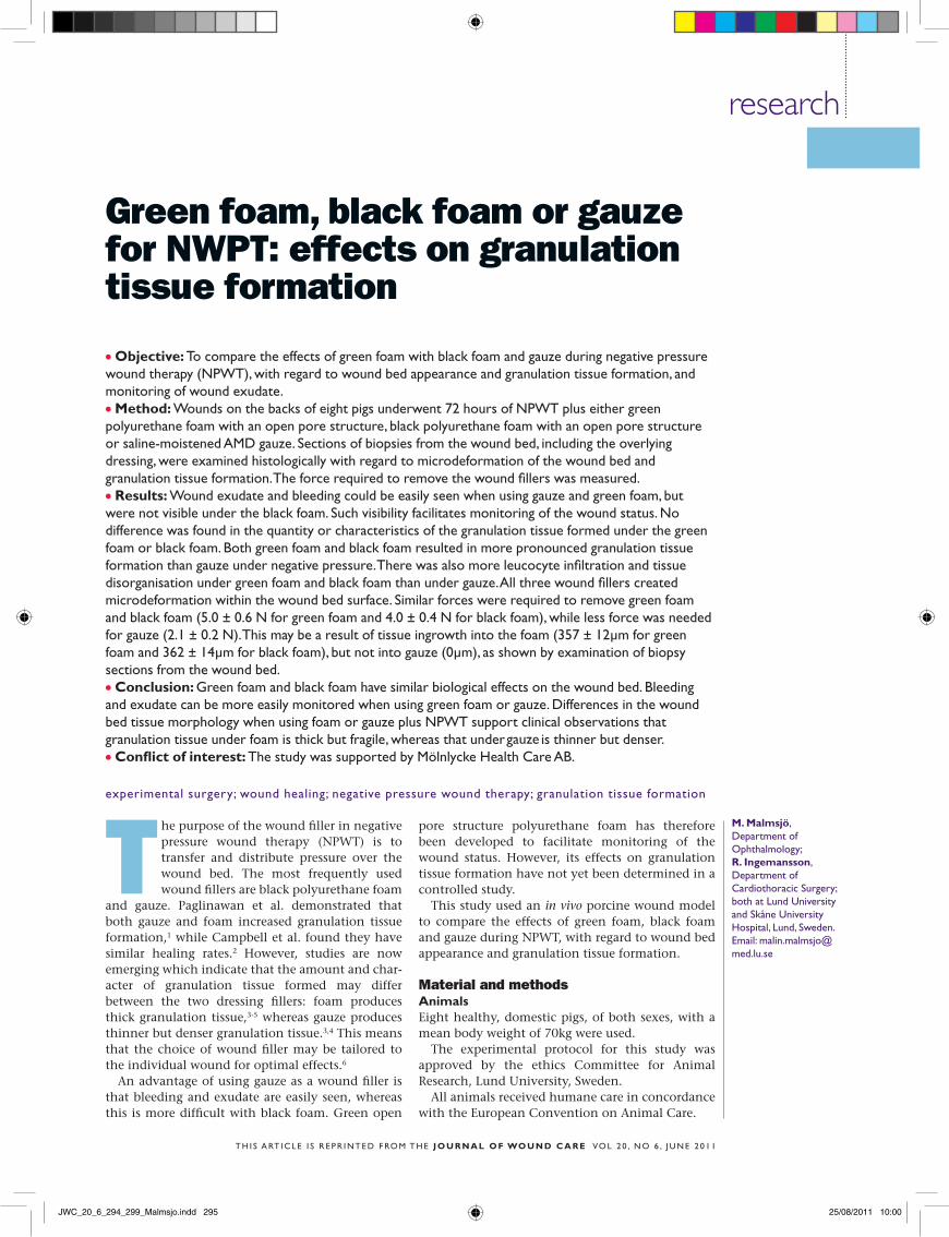
Green foam, black foam or gauze for NWPT: effects on granulation tissue formation
l objective: To compare the effects of green foam with black foam and gauze during negative pressure wound therapy (npWT), with regard to wound bed appearance and granulation tissue formation, and monitoring of wound exudate. l Method: Wounds on the backs of eight pigs underwent 72 hours of npWT plus either green polyurethane foam with an open pore structure, black polyurethane foam with an open pore structure or saline-moistened amd gauze. sections of biopsies from the wound bed, including the overlying dressing, were examined histologically with regard to microdeformation of the wound bed and granulation tissue formation. The force required to remove the wound fillers was measured.l results: Wound exudate and bleeding could be easily seen when using gauze and green foam, but were not visible under the black foam. such visibility facilitates monitoring of the wound status. no difference was found in the quantity or characteristics of the granulation tissue formed under the green foam or black foam. Both green foam and black foam resulted in more pronounced granulation tissue formation than gauze under negative pressure. There was also more leucocyte infiltration and tissue disorganisation under green foam and black foam than under gauze. all three wound fillers created microdeformation within the wound bed surface. similar forces were required to remove green foam and black foam (5.0 ± 0.6 n for green foam and 4.0 ± 0.4 n for black foam), while less force was needed for gauze (2.1 ± 0.2 n). This may be a result of tissue ingrowth into the foam (357 ± 12µm for green foam and 362 ± 14µm for black foam), but not into gauze (0µm), as shown by examination of biopsy sections from the wound bed. l conclusion: Green foam and black foam have similar biological effects on the wound bed. Bleeding and exudate can be more easily monitored when using green foam or gauze. differences in the wound bed tissue morphology when using foam or gauze plus npWT support clinical observations that granulation tissue under foam is thick but fragile, whereas that under foam is thinner but denser. l conflict of interest: The study was supported by mölnlycke health care aB.
experimental surgery; wound healing; negative pressure wound therapy; granulation tissue formation
The purpose of the wound filler in negative pressure wound therapy (NPWT) is to transfer and distribute pressure over the wound bed. The most frequently used wound fillers are black polyurethane foam
and gauze. Paglinawan et al. demonstrated that both gauze and foam increased granulation tissue formation,1 while Campbell et al. found they have similar healing rates.2 However, studies are now emerging which indicate that the amount and char-acter of granulation tissue formed may differ between the two dressing fillers: foam produces thick granulation tissue,3-5 whereas gauze produces thinner but denser granulation tissue.3,4 This means that the choice of wound filler may be tailored to the individual wound for optimal effects.6
An advantage of using gauze as a wound filler is that bleeding and exudate are easily seen, whereas this is more difficult with black foam. Green open
pore structure polyurethane foam has therefore been developed to facilitate monitoring of the wound status. However, its effects on granulation tissue formation have not yet been determined in a controlled study.
This study used an in vivo porcine wound model to compare the effects of green foam, black foam and gauze during NPWT, with regard to wound bed appearance and granulation tissue formation.
Material and methodsanimalsEight healthy, domestic pigs, of both sexes, with a mean body weight of 70kg were used.
The experimental protocol for this study was approved by the ethics Committee for Animal Research, Lund University, Sweden.
All animals received humane care in concordance with the European Convention on Animal Care.
M. Malmsjö, department of ophthalmology;r. Ingemansson, department of cardiothoracic surgery; both at lund university and skåne university hospital, lund, sweden.email: [email protected]
JWC_20_6_294_299_Malmsjo.indd 295 25/08/2011 10:00
research
T h i s a rT i c l e i s r e p r i n T e d f ro m T h e j o u r n a l o f wo u n d c a r e vo l 2 0 , n o 6 , j u n e 2 0 1 1
anaesthesia The pigs were fasted overnight, with free access to water. Premedication was performed with an intra-muscular injection of xylazine (Rompun vet. 20mg/ml; Bayer AG, Leverkusen, Germany; 2mg/kg), mixed with ketamine (Ketaminol vet. 100mg/ml; Farmaceutici Gellini S.p.A., Aprilia, Italy; 20mg/kg). Two peripheral veins in the pigs’ ears were cannu-lated for induction and maintenance of anaesthesia, and for fluid administration.
Anaesthesia was maintained with a continuous infusion of ketamine (Ketaminol vet. 50mg/ml; Far-maceutici Gellini S.p.A.; 0.4–0.6mg/kg/h). Com-plete neuromuscular blockade was achieved with a continuous infusion of pancuronium bromide (Pavulon; N.V. Organon, Oss, the Netherlands; 0.3–0.5mg/kg/h).
Fluid loss was compensated for by continuous infusion of Ringer’s solution at a rate of 200ml/kg/h for the first 24 hours, followed by 110ml/h for the remainder of the experiment. The animals received total parenteral nutrition (Kabiven; Fresenius Kabi AB, Uppsala, Sweden). Antibiotics were given once daily as intravenous bolus injections (streptocillin vet. 250mg/ml and 200mg/ml; Boehringer Ingelhe-im Vetmedica, Malmö, Sweden; 10ml).
The animals were orally intubated with cuffed endotracheal tubes. Mechanical ventilation was established with a Siemens-Elema ventilator (Sie-mens-Elema AB, Solna, Sweden) in the volume-con-trolled mode (65% nitrous oxide, 35% oxygen). Ventilatory settings were identical for all animals (respiratory rate, 15 breaths/min; minute ventila-tion, 12 litres/min). A positive end-expiratory pres-sure of 5cm H2O was applied. A Foley catheter was inserted into the urinary bladder through a supra-pubic cystostomy.
After the experiments were completed, the ani-mals were euthanised with a lethal dose (60mmol) of intravenous potassium chloride.
negative pressure wound therapyCircular wounds, 6cm in diameter, extending into the subcutaneous tissue, were created on each pig’s back using a scalpel. The wounds were immediately sealed for NPWT, as described below. The wounds were filled with saline-moistened AMD gauze (Ken-dall Kerlix AMD, Tyco Healthcare Group, Mansfield, MA, USA), green polyurethane foam with an open pore structure (Avance foam, Mölnlycke Health Care AB, Gothenburg, Sweden), or black poly-urethane foam with an open pore structure (VAC Granufoam, KCI, San Antonio, TX, USA). The green and black foam are both an open pore structure polyurethane foam. Besides colour, the only differ-ence between them is that the green foam has high-er tensile strength than the black foam (165kPa ver-sus 108kPa), which may lead to fewer problems with
foam residues in the wound bed. For gauze and green foam, drainage tubes were placed over the top of the wound filler and connected to the vacuum source (Avance NPWT system, Mölnlycke Health Care AB, Gothenburg, Sweden). For the wounds filled with black foam, a drain (T.R.A.C. pad, KCI, San Antonio, TX, USA) was attached to the top of the dressing and connected to the vacuum source (VAC ATS pump, KCI, San Antonio, TX, USA). For all three fillers, this was in accordance with the manu-facturers’ instructions. The wounds were treated for 72 hours at either 0mmHg or -120mmHg.
Quantity of granulation tissue formedThe quantity of granulation tissue formed during NPWT was graded on a scale from 0–5 by two differ-ent surgeons. Grading was performed blinded, and separately, by each surgeon, as described in a previ-ous study.4 The scale ranged from zero (a pale wound bed without granulation) to five (fully granulated tissue and a vascularised wound bed).
force measurementsAfter 72 hours of NPWT (as described above), the adhesive film dressing that covered the wound was cut along the borderline between the tissue and the wound filler, and the drain was cut off. The wound filler was attached to an advanced force gauge (AFG) (Mecmesin, UK) and withdrawn at a constant speed of 4mm/s.4 The force required to remove the wound filler was plotted as a function of time, using a computer.
Histological examinationThe tissue morphology and the cellular infiltrate in the wound bed underlying the wound filler were examined histologically. A strip of the wound filler material (1 x 1 x 2cm) was sutured onto the bottom of each wound before the NPWT dressings were applied. Post-NPWT, the strip and the underlying wound bed tissue were excised with a scalpel. The tissue was then treated in 4% para-formaldehyde, dehydrated and finally embedded in paraffin and left overnight. Biopsies were sectioned (4µm thick) using a rotary microtome (HM 355, ThermoFisher Scientific, MA, USA), mounted on glass slides and stained with routine haematoxylin and eosin stain-ing. All wound samples were subject to histological examination.l characteristics of the granulation tissue formed Biopsy sections were evaluated with regard to microdeformation (wound bed surface undulations), ingrowth into the wound filler, and morphology of the underlying tissue, including disorganisation of the cells in the wound bed (disruption of the contacts between the cells and differences in cell size) and leucocyte count (number per µm2).
JWC_20_6_294_299_Malmsjo.indd 296 25/08/2011 10:00
research
T h i s a rT i c l e i s r e p r i n T e d f ro m T h e j o u r n a l o f wo u n d c a r e vo l 2 0 , n o 6 , j u n e 2 0 1 1
Statistical analysisResults are presented as means ± the standard error of the mean (SEM).
Statistical analysis was performed using the Mann-Whitney test when comparing two groups, and the Kruskal-Wallis test with Dunn’s post-test for multiple comparisons when comparing three groups or more. All differences referred to in the text have been statistically verified.
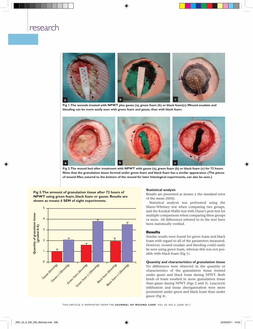
ResultsSimilar results were found for green foam and black foam with regard to all of the parameters measured. However, wound exudate and bleeding could easily be seen using green foam, whereas this was not pos-sible with black foam (Fig 1).
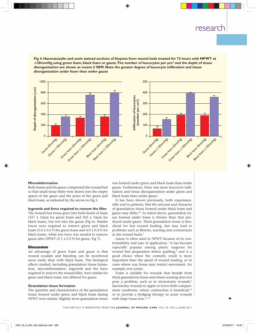
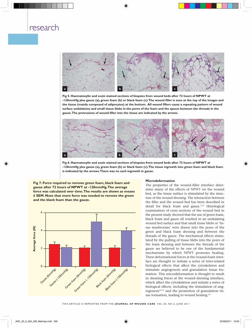
Quantity and characteristics of granulation tissue No differences were observed in the quantity or characteristics of the granulation tissue formed under green and black foam during NPWT. Both kinds of foam resulted in more granulation tissue than gauze during NPWT (Figs 2 and 3). Leucocyte infiltration and tissue disorganisation were more prominent under green and black foam than under gauze (Fig 4).
fig 2. The wound bed after treatment with nPwT with gauze (a), green foam (b) or black foam (c) for 72 hours. note that the granulation tissue formed under green foam and black foam has a similar appearance. (The pieces of wound filler, sutured to the bottom of the wound for later histological experiments, can also be seen.)
a b c
a b c
fig 1. The wounds treated with nPwT plus gauze (a), green foam (b) or black foam(c). wound exudate and bleeding can be more easily seen with green foam and gauze, than with black foam.
5
4
3
2
1
0
Qua
ntit
y o
f gra
nula
ion
tiss
ue
(gra
ded
0–5)
fig 3. The amount of granulation tissue after 72 hours of nPwT using green foam, black foam or gauze. results are shown as means ± SeM of eight experiments.
Gauze
(0mmhg)
Gauze
(-12
0mmhg)
Green
foam
(0mmhg)
Green
foam
(-12
0mmhg)
Black
foam
(0mmhg)
Black
foam
(-12
0mmhg)
JWC_20_6_294_299_Malmsjo.indd 298 25/08/2011 10:00
researchs
T h i s a rT i c l e i s r e p r i n T e d f ro m T h e j o u r n a l o f wo u n d c a r e vo l 2 0 , n o 6 , j u n e 2 0 1 1
s
MicrodeformationBoth foams and the gauze compressed the wound bed so that small tissue blebs were drawn into the empty spaces of the gauze and the pores of the green and black foam, as indicated by the arrows in Fig 5.
Ingrowth and force required to remove the fillerThe wound bed tissue grew into both kinds of foam (357 ± 12µm for green foam and 362 ± 14µm for black foam), but not into the gauze (Fig 6). Similar forces were required to remove green and black foam (5.0 ± 0.6 N for green foam and 4.0 ± 0.4 N for black foam), while less force was needed to remove gauze after NPWT (2.1 ± 0.2 N for gauze, Fig 7).
DiscussionAn advantage of green foam and gauze is that wound exudate and bleeding can be monitored more easily than with black foam. The biological effects studied, including granulation tissue forma-tion, microdeformation, ingrowth and the force required to remove the wound filler, were similar for green and black foam, but differed for gauze.
Granulation tissue formationThe quantity and characteristics of the granulation tissue formed under green and black foam during NPWT were similar. Slightly more granulation tissue
was formed under green and black foam than under gauze. Furthermore, there was more leucocyte infil-tration and tissue disorganisation under green and black foam than under gauze.
It has been shown previously, both experimen-tally and in patients, that the amount and character of granulation tissue formed under black foam and gauze may differ.4,5 As stated above, granulation tis-sue formed under foam is thicker than that pro-duced under gauze. Thick granulation tissue is ben-eficial for fast wound healing, but may lead to problems such as fibrosis, scarring and contractures as the wound heals.5
Gauze is often used in NPWT because of its con-formability and ease of application.7 It has become especially popular among plastic surgeons for wound bed preparation before grafting,8 and is a good choice when the cosmetic result is more important than the speed of wound healing, or in cases where scar tissue may restrict movement, for example over joints.
Foam is suitable for wounds that benefit from thick granulation tissue and where scarring does not pose a problem, such as in sternotomy wounds,9 fascio tomy wounds in upper or lower limb compart-ment syndrome, where contraction is beneficial,10 or to provide a bridging therapy in acute wounds with large tissue loss.11,12
dep
th o
f dis
org
anis
atio
n (μ
m)
1000
800
600
400
200
0
Gauze
(0mmhg)
Gauze
(-12
0mmhg)
Green
foam
(0mmhg)
Green
foam
(-12
0mmhg)
Black
foam
(0mmhg)
Black
foam
(-12
0mmhg)
Infi
ltra
tio
n o
f leu
cocy
tes
(nu
mbe
r pe
r μ
m2 )
500
400
300
200
100
0
Gauze
(0mmhg)
Gauze
(-12
0mmhg)
Green
foam
(0mmhg)
Green
foam
(-12
0mmhg)
Black
foam
(0mmhg)
Black
foam
(-12
0mmhg)
fig 4. Haematoxylin and eosin stained sections of biopsies from wound beds treated for 72 hours with nPwT at -120mmHg using green foam, black foam or gauze. The number of leucocytes per µm2 and the depth of tissue disorganisation are shown as means ± SeM. note the greater degree of leucocyte infiltration and tissue disorganisation under foam than under gauze
JWC_20_6_294_299_Malmsjo.indd 299 25/08/2011 10:00
research
T h i s a rT i c l e i s r e p r i n T e d f ro m T h e j o u r n a l o f wo u n d c a r e vo l 2 0 , n o 6 , j u n e 2 0 1 1
MicrodeformationThe properties of the wound–filler interface deter-mine many of the effects of NPWT on the wound bed, as the tissue surface is stimulated by the struc-ture of the wound dressing. The interaction between the filler and the wound bed has been described in detail for black foam and gauze.3,13 Histological examination of cross sections of the wound bed in the present study showed that the use of green foam, black foam and gauze all resulted in an undulating wound bed surface and that small tissue blebs or ‘tis-sue mushrooms’ were drawn into the pores of the green and black foam dressing and between the threads of the gauze. The mechanical effects stimu-lated by the pulling of tissue blebs into the pores of the foam dressing and between the threads of the gauze are believed to be one of the fundamental mechanisms by which NPWT promotes healing. These deformational forces at the wound-foam inter-face are thought to initiate a series of inter-related biological effects that affect the cytoskeleton and stimulate angiogenesis and granulation tissue for-mation. This microdeformation is thought to result in shearing forces at the wound–dressing interface, which affect the cytoskeleton and initiate a series of biological effects, including the stimulation of ang-iogenesis14,15 and the promotion of granulation tis-sue formation, leading to wound healing.16
fig 5. Haematoxylin and eosin stained sections of biopsies from wound beds after 72 hours of nPwT at -120mmHg plus gauze (a), green foam (b) or black foam (c). The wound filler is seen at the top of the images and the tissue (mainly composed of adipocytes) at the bottom. all wound fillers cause a repeating pattern of wound surface undulations and small tissue blebs in the pores of the foam and the spaces between the threads in the gauze. The protrusions of wound filler into the tissue are indicated by the arrows.
fig 6. Haematoxylin and eosin stained sections of biopsies from wound beds after 72 hours of nPwT at -120mmHg plus gauze (a), green foam (b) or black foam (c). The tissue ingrowth into green foam and black foam is indicated by the arrows. There was no such ingrowth in gauze.
Gauze
(0mmhg)
Gauze
(-12
0mmhg)
Green
foam
( -1
20mmhg)
Black
foam
(-12
0mmhg)
Green
foam
(0mmhg)
Black
foam
(0mmhg)
6
4
2
0
ave
rage
forc
e (n
)
fig 7. force required to remove green foam, black foam and gauze after 72 hours of nPwT at -120mmHg. The average force was calculated over time. The results are shown as means ± SeM. note that more force was needed to remove the green and the black foam than the gauze.
a b c
a b c
JWC_20_6_294_299_Malmsjo.indd 300 25/08/2011 10:00
research
T h i s a rT i c l e i s r e p r i n T e d f ro m T h e j o u r n a l o f wo u n d c a r e vo l 2 0 , n o 6 , j u n e 2 0 1 1
Ingrowth and force needed to remove the fillerThe results show that the wound bed tissue grew into both black and green foam, but not into gauze. Simi-lar forces were required to remove green and black foam, while less force was needed to remove gauze. This is probably due to the tissue ingrowth into the green and black foam, but not into the gauze, as seen in biopsy specimens from the wound bed.
A number of complications are associated with ingrowth into foam. First, the patient may experi-ence pain during dressing changes as the ingrown tissue is torn away from the wound, requiring the administration of strong analgesics. 17,18 Second, wound-bed disruption and mechanical tissue dam-age may arise as the foam is removed from the wound bed during dressing changes. Finally, pieces of foam may become stuck in the wound bed and, if not removed, will act as foreign bodies that may impede wound healing.
A non-adherent wound contact layer, therefore, is often placed over the wound bed, underneath the wound filler, when the clinician anticipates such complications.19 A wound contact layer may be placed over vulnerable structures, such as blood ves-sels or nerves,19 and over the wound bed itself to
prevent the ingrowth of granulation tissue into the wound filler.
The mechanism governing ingrowth into foam is likely related to the interaction between the tissue and dressing at a microscopic level.20 The differenc-es in ingrowth observed in this study are probably due to differences in the physical properties of the foam and gauze dressings. The chemical nature of the material may also be of importance.
ConclusionGreen foam and black foam have similar biological effects on the wound bed. However, the wound sta-tus (bleeding and exudate) can easily be monitored with green foam, but not with black foam.
Differences were observed in the morphology of the wound bed tissue when using foam and gauze plus NPWT, which is in accordance with clinical observations that granulation tissue under foam is thick but fragile, whereas that under gauze is thin-ner but denser.
Treatment of the patient can be optimised by choosing the appropriate wound filler material for individual wounds and patients, depending on the desired effects in the wound bed. n
references1 paglinawan, r., colic, m., simon, m. a. comparative study of the influence of different pressure levels combined with various wound dressings on negative pressure wound therapy driven wound healing. european Tissue repair society. malta, 2008.2 campbell, p.e., smith, G.s., smith, j.m. retrospective clinical evaluation of gauze-based negative pressure wound therapy. int Wound j 2008; 5: 2, 280–286.3 Borgquist, o., Gustafsson, l., ingemansson, r., malmsjö, m. micro- and macromechanical effects on the wound bed by negative pressure wound therapy using gauze and foam. ann plast surg 2010; 64: 6, 789–793.4 Borgquist, o., Gustafsson, l., ingemansson, r. et al. Tissue ingrowth into foam but not into gauze during negative pressure wound therapy. Wounds 2009; 21: 11, 302–309.5 fraccalvieri, m., Zingarelli, e., ruka, e. et al. negative pressure wound therapy using the gauze and the foam: immunohistological and ultrasonography morphological analysis of the granulation tissue and the scar
tissue. preliminary report of a clinical study. int Wound j 2011; 12: in press.6 malmsjö, m., Borgquist, o. npWT settings and dressing choices made easy. Wounds international 2010; 1: 3:7 jeffery, l.c. advanced wound therapies in the management of severe military lower limb trauma: a new perspective. eplasty 2009; 9: e28.8 chariker, m.e., Gerstle, T.l., morrison, c.s. an algorithmic approach to the use of gauze-based negative-pressure wound therapy as a bridge to closure in pediatric extremity trauma. plast reconstr surg 2009; 123: 5, 1510–1520.9 Gustafsson, r.i., sjogren, j., ingemansson, r. deep sternal wound infection: a sternal-sparing technique with vacuum-assisted closure therapy. ann Thorac surg 2003; 76: 6, 2048–2053.10 Zannis, j., angobaldo, j., marks, m. et al. comparison of fasciotomy wound closures using traditional dressing changes and the vacuum-assisted closure device. ann plast surg 2009; 62: 4,
407–409.11 Bollero, d., carnino, r., risso, d. et al. acute complex traumas of the lower limbs: a modern reconstructive approach with negative pressure therapy. Wound repair regen 2007; 15: 4, 589–594.12 stannard, j.p., robinson, j.T., anderson, e.r. et al. negative pressure wound therapy to treat hematomas and surgical incisions following high-energy trauma. j Trauma 2006; 60: 6, 1301–1306.13 Wilkes, r., Zhao, Y., Kieswetter, K. et al. effects of dressing type on 3d tissue microdeformations during negative pressure wound therapy: a computational study. j Biomech eng 2009; 131: 3, 031012.14 evans, d., land, l. Topical negative pressure for treating chronic wounds: a systematic review. Br j plast surg 2001; 54: 3, 238–242.15 Greene, a.K., puder, m., roy, r. et al. microdeformational wound therapy: effects on angiogenesis and matrix metalloproteinases in chronic wounds of 3 debilitated patients. ann plast surg 2006; 56: 4, 418–422.
16 morykwas, m.j., argenta, l.c., shelton-Brown, e.i. et al. vacuum-assisted closure: a new method for wound control and treatment: animal studies and basic foundation. ann plast surg 1997; 38: 6, 553–562.17 franczyk, m., lohman, r.f., agarwal, j.p. et al. The impact of topical lidocaine on pain level assessment during and after vacuum-assisted closure dressing changes: a double-blind, prospective, randomized study. plast reconstr surg 2009; 124: 3, 854–861.18 Krasner, d.l. managing wound pain in patients with vacuum-assisted closure devices. ostomy Wound manage 2002; 48: 5, 38–43.19 dunbar, a., Bowers, d.m., holderness, h. jnr. silicone net dressing as an adjunct with negative pressure wound therapy. ostomy Wound manage 2005; 51: (11a suppl), 21–22.20 morykwas, m.j., simpson, j., punger, K. et al. vacuum-assisted closure: state of basic research and physiologic foundation. plast reconstr surg 2006; 117: (7 suppl), 121s–126s.
JWC_20_6_294_299_Malmsjo.indd 301 25/08/2011 10:00