Embed Size (px)
Citation preview
RESEARCH ARTICLE Open Access
Grape seed proanthocyanidins inhibit the invasivepotential of head and neck cutaneous squamouscell carcinoma cells by targeting EGFR expressionand epithelial-to-mesenchymal transitionQian Sun1, Ram Prasad1, Eben Rosenthal2,3 and Santosh K Katiyar1,3,4,5*
Abstract
Background: Head and neck squamous cell carcinoma (HNSCC) is responsible for over 20,000 deaths every year inUnited States. Most of the deaths are due, in large part, to its propensity to metastasize. We have examined theeffect of bioactive component grape seed proanthocyanidins (GSPs) on human cutaneous HNSCC cell invasion andthe molecular mechanisms underlying these effects using SCC13 cell line as an in vitro model.
Methods: The therapeutic effects of GSPs on cancer cell invasion were studied using Boyden chamber and woundhealing assays. The effects of GSPs on the levels of various proteins related with cancer cell invasion weredetermined using western blot analysis.
Results: Using in vitro cell invasion assays, we observed that treatment of SCC13 cells with GSPs resulted in aconcentration-dependent inhibition of cell invasion of these cells, which was associated with a reduction in thelevels of epidermal growth factor receptor (EGFR). Treatment of cells with gefitinib and erlotinib, inhibitors of EGFR,or transient transfection of SCC13 cells with EGFR small interfering RNA, also inhibited invasion of these cells. Theinhibition of cell invasion by GSPs was associated with the inhibition of the phosphorylation of ERK1/2, a memberof mitogen-activated protein kinase family. Treatment of cells with UO126, an inhibitor of MEK, also inhibited theinvasion potential of SCC13 cells. Additionally, inhibition of human cutaneous HNSCC cell invasion by GSPs wasassociated with reversal of epithelial-to-mesenchymal transition (EMT) process, which resulted in an increase in thelevels of epithelial biomarker (E-cadherin) while loss of mesenchymal biomarkers (vimentin, fibronectin and N-cadherin) in cells. Similar effect on EMT biomarkers was also observed when cells were treated with erlotinib.
Conclusion: The results obtained from this study indicate that grape seed proanthocyanidins have the ability toinhibit the invasion of human cutaneous HNSCC cells by targeting the EGFR expression and reversing the processof epithelial-to-mesenchymal transition. These data suggest that GSPs can be developed as a complementary andalternative medicine for the prevention of invasion/metastasis of HNSCC cells.
BackgroundHead and neck squamous cell carcinoma (HNSCC)affects more than 40,000 people in the United Statesannually and is responsible for over 20,000 deaths everyyear [1,2]. HNSCC often generates from critical organsincluding the oral cavity, larynx, pharynx, and tongue
that play indispensable roles in increased mortality rate[1]. Head and neck cutaneous SCC is also very common.Advances in surgical and medical therapies for HNSCChave only modestly improved the mortality rate, whichhas remained at 50% for the last three decades [3-6]. Ithas been demonstrated that epidermal growth factorreceptor (EGFR), one of the ErbB family of receptors,which is overexpressed in over 90% of HNSCC tumors,is a marker of poor prognosis in patients with HNSCC[7-9]. Mortality rate due to HNSCC is closely associated
* Correspondence: [email protected] of Dermatology, University of Alabama at Birmingham,Birmingham, AL, USAFull list of author information is available at the end of the article
Sun et al. BMC Complementary and Alternative Medicine 2011, 11:134http://www.biomedcentral.com/1472-6882/11/134
© 2011 Sun et al; licensee BioMed Central Ltd. This is an Open Access article distributed under the terms of the Creative CommonsAttribution License (http://creativecommons.org/licenses/by/2.0), which permits unrestricted use, distribution, and reproduction inany medium, provided the original work is properly cited.
with its potent capacity to metastasize distantly. There-fore, an approach that decreases the metastatic ability ofHNSCC cells may facilitate the development of an effec-tive strategy for its treatment and/or prevention.Naturally occurring agents, particularly bioactive diet-
ary phytochemicals, may serve as appropriate candidatesfor the prevention or therapy of HNSCC metastasis. Ifthese phytochemicals are safe and devoid of toxicities,these can be considered for the prevention of cancercell invasion, migration or metastasis and thus can beutilized as complementary and alternative medicine and/or as adjuvant therapy for conventional cytotoxic thera-pies. Grape seed proanthocyanidins (GSPs) are suchpromising bioactive phytochemicals that have shownanti-carcinogenic effects in some tumor models andexhibit no apparent toxicity in vivo animal models[10-12]. GSPs contain primarily proanthocyanidins(89%), which constitute dimers, trimers, tetramers, andoligomers of monomeric catechins and/or (-)-epicate-chins, as described previously [11]. Although GSPs havebeen shown to have anti-tumor effects [10], their che-motherapeutic effects on the invasive potential ofHNSCC cells have not been explored.In the current study, we assessed the chemotherapeu-
tic effects of GSPs on the invasion potential of humanhead and neck cutaneous squamous cell carcinoma cells,as the invasion of cancer cells is a major event in themetastatic cascade. The invasion potential of cutaneousSCC cells was also compared with the invasion potentialof human epidermoid carcinoma cells which were notfound on head and neck sub-sites. For this purpose, twocutaneous SCC cells lines were selected: one is SCC13which was generated from the squamous cell carcinomaof the facial (head) skin. Second cell line is A431 whichis well known human epidermoid carcinoma cell lineand is not related with head and neck sub-sites. In thisstudy, we characterized the role of EGFR on the migra-tion of head and neck cutaneous SCC cells and ascer-tained whether GSPs have any suppressive effects on theinvasion of these cells and whether EGFR is involved inthis process. Epithelial-to-mesenchymal transition(EMT), the process whereby epithelial cells transforminto mesenchymal cells, has been shown to be relevantfor cancer and cancer cell metastasis. During EMT, can-cer cells lose expression of proteins that promote cell-cell contact such as E-cadherin and acquire mesenchy-mal markers such as vimentin, fibronectin and N-cad-herin, which promote tumor progression, cell invasionand metastasis [13,14]. The EMT has also been asso-ciated with higher expression levels of EGFR and EGFR-mediated signaling, therefore we have also checkedwhether inhibition of EGFR expression by GSPs in headand neck cutaneous SCC cells is associated with reversalof EMT and that leads to inhibitory effect on cell
invasion of head and neck cutaneous SCC cells. Here,we present evidence that GSPs inhibit the invasivepotential or migratory behavior of head and neck cuta-neous squamous cell carcinoma cells through inhibitionor reversal of EMT and that GSPs do so through a pro-cess that involves the reduction in EGFR expressionlevel.
MethodsGSPs, source and compositionGSPs were received from Kikkoman Biochemifa Com-pany, Japan (no financial conflict of interest). Qualitycontrol of GSPs is maintained by the company on lot-to-lot basis. GSPs contain approximately 89% proantho-cyanidins, with dimers (6.6%), trimers (5.0%), tetramers(2.9%) and oligomers (74.8%), as described earlier[10-12]. Based on vendor’s information and analysis thisproduct is stable for at least two years when refrigeratedat 4°C. We have particularly selected this product as asource of proanthocyanidins because it is commerciallyavailable in purified form, and their composition isknown and maintained on lot-to-lot basis by the com-mercial vendor. Some proanthocyanidins products arealso commercially available in the market and theircompositions are more or less comparable with the pro-duct used in this study.
Cell lines and cell culture conditionsHuman epidermoid carcinoma cells (A431) and humanhead and neck cutaneous SCC cells SCC13 wereobtained from the American Type Culture Collection(Manassas, VA) and normal human epidermal keratino-cytes (NHEK) were obtained from Cell Culture CoreFacility of Skin Diseases Research Centre at the Univer-sity of Alabama at Birmingham, AL. The cells were cul-tured as monolayers in DMEM supplemented with 10%heat inactivated fetal bovine serum, 100 μg/ml penicil-lin-streptomycin (Invitrogen, Carlsbad, CA), and kept ina humidified atmosphere of 5% CO2 at 37°C. TheNHEK were cultured in keratinocyte growth mediumsupplemented with 5 ng/ml human recombinant epider-mal growth factor and 0.05 mg/ml bovine pituitaryextract (Gibco/Invitrogen, Carlsbad, CA) and maintainedin an incubator under the identical conditions. Cellswere seeded at a density of 1 × 10⁶ cells per petri dishand allowed to attach for 24 h before treatment withGSPs or other treatment agents. The sub-confluent cellswere treated with either various concentrations of GSPsor other agents such as gefitinib or erlotinib. The GSPs,erlotinib or gefitinib were dissolved in a small amountof dimethylsulfoxide (DMSO), which was added to thecomplete cell culture medium. The maximum concen-tration of DMSO in media was 0.1% (v/v). Cells treatedwith DMSO only served as a vehicle control. To
Sun et al. BMC Complementary and Alternative Medicine 2011, 11:134http://www.biomedcentral.com/1472-6882/11/134
Page 2 of 12
determine the effect of GSPs on epidermal growth factor(EGF)-mediated effects, GSPs were added in cell culturemedium at least 30 minutes before the treatment of thecells with EGF.
Antibodies, chemicals and reagentsBoyden Chambers and polycarbonate membranes (8 μmpore size) for cell invasion assays were obtained fromNeuroprobe, Inc. (Gaithersburg, MD). The antibodiesspecific to N-cadherin, fibronectin, EGF, EGFR, ERK1/2and b-actin were obtained from Santa Cruz Biotechnol-ogy (Santa Cruz, CA), while antibodies for vimentin andE-cadherin were purchased from Cell Signaling Tech-nology (Beverly, MA). The appropriate secondary anti-bodies conjugated with horseradish peroxidase wereobtained from Invitrogen (Carlsband, CA).
Cell invasion assayThe invasion capacity of SCC cells was determined invitro using Boyden Chambers (Gaithersburg, MD). Inthis assay, two chambers were separated with matrigelcoated Millipore membranes (6.5 mm diameter filters, 8μM pore size), as detailed previously [15,16]. Briefly,cancer cells (1.5 × 10⁴ cells/100 μl serum-reduced med-ium) were placed in the upper chamber of Boydenchambers, test agents were added alone, or in combina-tion, to the upper chamber (200 μl), and the lowerchamber contained the medium alone (150 μl). Cham-bers were assembled and kept in an incubator fordesired time points. After incubation, cells from theupper surface of Millipore membranes were removedwith gentle swabbing and the migratory cells on thelower surface of membranes were fixed and stained withcrystal violet. The membranes were examined microsco-pically and cellular invasion was determined by countingthe number of cells on membranes in at least 4-5 ran-domly selected fields using an Olympus BX41 micro-scope. Representative photomicrographs were obtainedusing a Qcolor5 digital camera system fitted to anOlympus BX41 microscope. Each cell invasion experi-ment was repeated at least three times.
Scratch assay or wound healing assayScratch assay was performed to detect the cell migrationability of SCC13 cells, as detailed previously [15]. Briefly,SCC13 cells were grown to full confluency in six-wellplates and incubated overnight in starvation medium,which contained only 0.5% FBS in DMEM cell culturemedium. Cell monolayers were wounded with a sterile100 μl pipette tip, washed with starvation medium toremove detached cells from the plates. Cells were lefteither untreated or treated with selected concentrationsof GSPs in full medium and kept in a cell culture incu-bator for 48 h. After 48 h, medium was replaced with
phosphate-buffered saline (PBS) buffer, the wound gapwas examined and cells were photographed using anOlympus BX41 microscope fitted with digital camera.
Western blot analysisFollowing treatment of cells for the indicated time peri-ods with or without the treatment of GSPs or any otheragent, the cells were harvested, washed with cold PBSand lysed with ice-cold lysis buffer supplemented withprotease inhibitors, as detailed previously [15,16]. Equalamounts of proteins (50 μg) were resolved on 10% Tris-Glycine gels and transferred onto a nitrocellulose mem-brane. After blocking the non-specific binding sites, themembrane was incubated with the primary antibody at4°C overnight. The membrane was then incubated withthe appropriate peroxidase-conjugated secondary anti-body and the protein bands were visualized using theenhanced chemiluminescence reagents. The equal load-ing of protein samples on the gel was verified after re-probing the membrane with anti-b actin antibody.
Statistical analysisFor cell invasion assays, the control and GSPs, gefitinibor erlotinib treatment groups or combined-treatmentgroups separately were compared using one-way analysisof variance (ANOVA) followed by post hoc Dunn’s testusing GraphPad Prism version 4.00 for Windows,GraphPad Software, San Diego, California, USA, http://www.graphpad.com. All quantitative data for cell migra-tion are shown as the mean number of migrating cells ±SD/microscopic field, n = 3. In each case P < 0.05 wasconsidered statistically significant.
ResultsThe invasive potential of head and neck cutaneous SCC13cells was greater than A431 cellsFirst, we checked the invasive potential of head and neckcutaneous SCC13 cells and compared it with that ofhuman epidermoid carcinoma cell line A431, which arenot head and neck cancer cells, under identical experi-mental conditions. As shown in Figure 1A and 1B, thecell invasion ability of SCC13 cells was significantlyhigher (P < 0.0001) than A431 cells. The number of inva-sive SCC13 cells was 2000 ± 205 cells/microscopic fieldwhile the invasion of A431 cells was 12 ± 2 cells/micro-scopic field. These data indicate that cutaneous head andneck SCC cells are strongly aggressive in terms of theirinvasive potential than A431 cells which are not from thehead and neck sites. Under identical conditions, the inva-sion potential of normal human epidermal keratinocyteswas not observed (data not shown).As SCC13 cells were highly invasive in nature, we exam-
ined the invasion ability of SCC13 cells at the early timepoints. As shown in Figure 1C, we could see the invasion
Sun et al. BMC Complementary and Alternative Medicine 2011, 11:134http://www.biomedcentral.com/1472-6882/11/134
Page 3 of 12
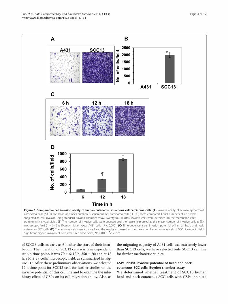
of SCC13 cells as early as 6 h after the start of their incu-bation. The migration of SCC13 cells was time dependent.At 6 h time point, it was 70 ± 6; 12 h, 350 ± 20; and at 18h, 850 ± 29 cells/microscopic field, as summarized in Fig-ure 1D. After these preliminary observations, we selected12 h time point for SCC13 cells for further studies on theinvasive potential of this cell line and to examine the inhi-bitory effect of GSPs on its cell migration ability. Also, as
the migrating capacity of A431 cells was extremely lowerthan SCC13 cells, we have selected only SCC13 cell linefor further mechanistic studies.
GSPs inhibit invasive potential of head and neckcutaneous SCC cells: Boyden chamber assayWe determined whether treatment of SCC13 humanhead and neck cutaneous SCC cells with GSPs inhibited
A431 SCC13
A B2500
dA431 SCC13
0
500
1000
1500
2000 *
No.
of c
ells
/fiel
d
18 h6 h 12 h
CA431 SCC13
D1000
No.
of c
ells
/fiel
d
200
400
600
800
1000 *
N
Time in h6 12 18
0
Figure 1 Comparative cell invasion ability of human cutaneous squamous cell carcinoma cells. (A) Invasive ability of human epidermoidcarcinoma cells (A431) and head and neck cutaneous squamous cell carcinoma cells (SCC13) were compared. Equal numbers of cells weresubjected to cell invasion using standard Boyden chamber assay. Twenty-four h later, invasive cells were detected on the membrane afterstaining with crystal violet. (B) The number of invasive cells were counted and the results expressed as the mean number of invasive cells ± SD/microscopic field (n = 3). Significantly higher versus A431 cells, *P < 0.0001. (C) Time-dependent cell invasion potential of human head and neckcutaneous SCC cells. (D) The invasive cells were counted and the results expressed as the mean number of invasive cells ± SD/microscopic field.Significant higher invasion of cells versus 6 h time point, *P < 0.001; ¶P < 0.01.
Sun et al. BMC Complementary and Alternative Medicine 2011, 11:134http://www.biomedcentral.com/1472-6882/11/134
Page 4 of 12
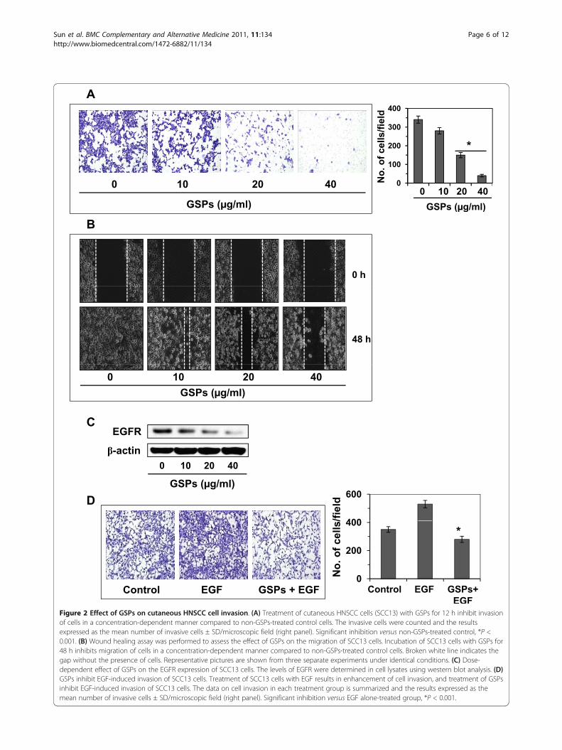
their invasiveness using Boyden chamber cell invasionassays. First, screening experiments were performed todetermine the effects of lower (non-death inducing)concentrations of GSPs (μg/ml). As shown in Figure 2A,relative to untreated control cells, treatment of cellswith GSPs at concentrations of 0, 10, 20 and 40 μg/mlreduced the invasive potential of SCC13 cells in a con-centration-dependent manner. The density of the inva-sive cells on the membrane after staining with crystalviolet is shown in Figure 2A, and the numbers of inva-sive cells/microscopic field are summarized in Figure 2A(right panel). The cell invasion was inhibited by18-85%(P < 0.01-0.001) in SCC13 cells in a concentration-dependent manner after treatment with GSPs for 12 h.To verify that the inhibition of invasion of SCC13 cellsby GSPs was a direct effect on invasion ability, and thatwas not due to a reduction in cell viability/cell death, atrypan blue and/or MTT assays were performed [15]using cells that were treated identically to those used inthe invasion assays. Treatment of SCC13 cells with var-ious concentrations of GSPs (0, 10, 20 and 40 μg/ml)for 12 h had no significant effect on cell viability or celldeath (data not shown).
GSPs inhibit the migration of head and neck cutaneousSCC cells: Scratch or wound healing assayAs shown in Figure 2B, relative to untreated control cells,treatment of cells with various concentrations of GSPs(10, 20 and 40 μg/ml) reduced the migration capacity ofSCC13 cells in a concentration-dependent manner afterthe treatment of cells for 48 h. The major part of gap orwounding space between cell layers after making awound was occupied by the migrating SCC13 cells whichwere not treated with GSPs. However, the healing of thewound or the empty space between the cell layers waslargely not occupied by the migrating cells treated withGSPs and this effect was dose-dependent. The gap orwounding space between the cells is highlighted by bro-ken white lines (Figure 2B). These observations suggestthat GSPs inhibited the migration of SCC13 cells. Tofurther confirm that the inhibition of cancer cell migra-tion by GSPs after 48 h was a direct effect on cell migra-tion and not due to a reduction in cell viability, a trypanblue assay was performed using cells that were treatedidentically to those used in the migration assays. Treat-ment of SCC13 cells with various concentrations of GSPs(10, 20 and 40 μg/ml) for 48 h had no significant effecton cell viability or cell death (data not shown).
The inhibitory effect of GSPs on invasive potential ofSCC13 cells is associated with the reduction of EGFRexpressionTo determine whether the inhibitory effect of GSPs onthe invasion of the SCC13 cells is associated with
inhibition of EGFR expression, we determined the levelsof EGFR in lysates of cells from the various treatmentgroups using western blot analysis. As shown in Figure2C, treatment of SCC13 cells with GSPs for 12 hreduced the levels of EGFR expression in a concentra-tion-dependent manner as compared to the expressionin non-GSPs-treated controls. These results suggest thatGSPs-induced reduction in EGFR expression may beassociated with an inhibitory effect of the GSPs on thecell invasion of these cells.
EGF, a ligand of EGFR, enhances the invasion of SCC13cells, and GSPs inhibit EGF-induced cell invasionEGF is a well known ligand of EGFR and has beenshown to stimulate the activity of EGFR; therefore, thehead and neck cutaneous SCC13 cells were treated withEGF (10 ng/ml) for EGFR stimulation, and thereafterdetermined the effect of EGF on the invasion of SCC13cells. As shown in Figure 2D, treatment of SCC13 cellswith EGF for 12 h resulted in significantly enhanced cellinvasion (P < 0.01) compared to non-EGF-treated con-trol cells. To determine whether GSPs inhibit EGF-induced cell invasion in human head and neck cuta-neous SCC13 cells, SCC13 cells were treated with EGF(10 ng/ml) with and without the treatment of GSPs for12 h. We found that the treatment of SCC13 cells withGSPs (40 μg/ml) resulted in significant inhibition (P <0.001) of EGF-induced invasion of SCC13 cells. A sum-mary of the cell invasion data for the different treatmentgroups is shown in Figure 2D (right panel).
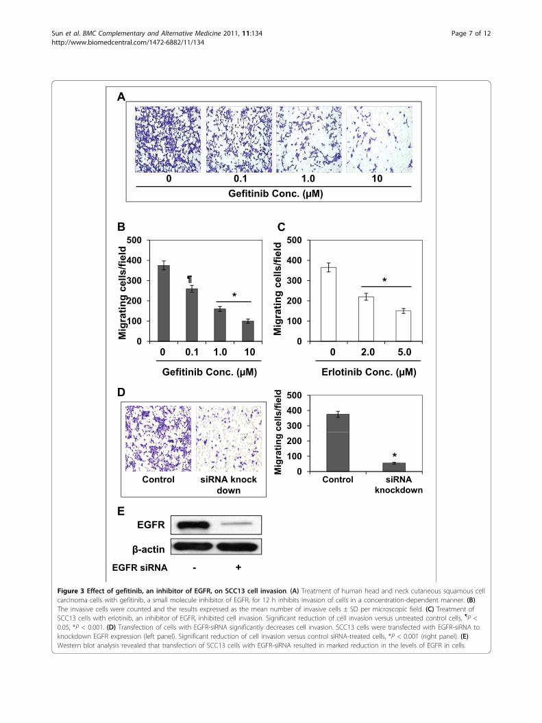
Selective EGFR inhibitors, gefitinib and erlotinib, inhibitthe invasion of SCC13 cellsThis experiment was performed to determine whether theinhibitory effect of GSPs on the cell invasion of head andneck cutaneous squamous cell carcinoma cells is mediatedthrough its inhibitory effect on EGFR expression. For thispurpose, SCC13 cells were subjected to the cell invasionassay after treatment with various concentrations of gefiti-nib (0, 0.1, 1.0 and 10.0 μM), a well known inhibitor ofEGFR, for 12 h. As shown in Figure 3A, treatment of thecells with gefitinib resulted in a dose-dependent reductionin the cell invasion capacity of SCC13 cells as comparedwith non-gefitinib-treated controls (P < 0.05-0.001). Thesedata suggested that the inhibition of constitutive levels ofEGFR expression is associated with the inhibition of cellinvasion of head and neck cutaneous squamous cell carci-noma cells. The resultant data on cell invasion/micro-scopic field at different doses of gefitinib are summarizedin Figure 3B. Similar results were obtained when SCC13cells were treated with another inhibitor of EGFR, erloti-nib. Treatment of SCC13 cells with erlotinib for 12 hinhibited the invasion capacity of these cells, as shown bydata summarized in Figure 3C.
Sun et al. BMC Complementary and Alternative Medicine 2011, 11:134http://www.biomedcentral.com/1472-6882/11/134
Page 5 of 12
300
400
/fiel
d
A
0 10 20 40
GSPs ( g/ml)
0
100
200
0 10 20 40GSPs ( g/ml)
No.
of c
ells
/
*
( g ) GSPs ( g/ml)
0 h
B
48 h
0 10 20 40 GSPs ( g/ml)
EGFRC
-actin
GSPs ( g/ml)
0 10 20 40
D
400
600
/fiel
d
Control EGF GSPs + EGF0
200
400
No.
of c
ells
/
Control EGF GSPs+ EGF
*
EGFFigure 2 Effect of GSPs on cutaneous HNSCC cell invasion. (A) Treatment of cutaneous HNSCC cells (SCC13) with GSPs for 12 h inhibit invasionof cells in a concentration-dependent manner compared to non-GSPs-treated control cells. The invasive cells were counted and the resultsexpressed as the mean number of invasive cells ± SD/microscopic field (right panel). Significant inhibition versus non-GSPs-treated control, *P <0.001. (B) Wound healing assay was performed to assess the effect of GSPs on the migration of SCC13 cells. Incubation of SCC13 cells with GSPs for48 h inhibits migration of cells in a concentration-dependent manner compared to non-GSPs-treated control cells. Broken white line indicates thegap without the presence of cells. Representative pictures are shown from three separate experiments under identical conditions. (C) Dose-dependent effect of GSPs on the EGFR expression of SCC13 cells. The levels of EGFR were determined in cell lysates using western blot analysis. (D)GSPs inhibit EGF-induced invasion of SCC13 cells. Treatment of SCC13 cells with EGF results in enhancement of cell invasion, and treatment of GSPsinhibit EGF-induced invasion of SCC13 cells. The data on cell invasion in each treatment group is summarized and the results expressed as themean number of invasive cells ± SD/microscopic field (right panel). Significant inhibition versus EGF alone-treated group, *P < 0.001.
Sun et al. BMC Complementary and Alternative Medicine 2011, 11:134http://www.biomedcentral.com/1472-6882/11/134
Page 6 of 12
A
Gefitinib Conc. ( M)0 0.1 1.0 10
300
400
500
ells
/fiel
d
B
300
400
500
ells
/fiel
d
C
*
0
100
200
300
Mig
ratin
g ce
0 0.1 1.0 100
100
200
300M
igra
ting
ce
0 2.0 5.0
*
Gefitinib Conc. ( M)
300400500
cells
/fiel
dD
Erlotinib Conc. ( M)
Control siRNA knock down
0100200
Control siRNA knockdown
Mig
ratin
g
E
*
EGFR siRNA - +
EGFR
-actin
E
Figure 3 Effect of gefitinib, an inhibitor of EGFR, on SCC13 cell invasion. (A) Treatment of human head and neck cutaneous squamous cellcarcinoma cells with gefitinib, a small molecule inhibitor of EGFR, for 12 h inhibits invasion of cells in a concentration-dependent manner. (B)The invasive cells were counted and the results expressed as the mean number of invasive cells ± SD per microscopic field. (C) Treatment ofSCC13 cells with erlotinib, an inhibitor of EGFR, inhibited cell invasion. Significant reduction of cell invasion versus untreated control cells, ¶P <0.05, *P < 0.001. (D) Transfection of cells with EGFR-siRNA significantly decreases cell invasion. SCC13 cells were transfected with EGFR-siRNA toknockdown EGFR expression (left panel). Significant reduction of cell invasion versus control siRNA-treated cells, *P < 0.001 (right panel). (E)Western blot analysis revealed that transfection of SCC13 cells with EGFR-siRNA resulted in marked reduction in the levels of EGFR in cells.
Sun et al. BMC Complementary and Alternative Medicine 2011, 11:134http://www.biomedcentral.com/1472-6882/11/134
Page 7 of 12
siRNA knock-down of EGFR reduces the invasion ofSCC13 cellsWe further verified the role of EGFR in cell invasionthrough siRNA knock-down of EGFR in the SCC13 cellsusing siRNA Transfection Reagent Kit (Santa Cruz Bio-technology, Inc., Santa Cruz, CA), and examinedwhether it would lead to the inhibition of the cell inva-sion in these cells. The data from cell invasion assayrevealed that transfection of SCC13 cells with EGFRsiRNA resulted in significant reduction of cell invasion(84%, P < 0.001) after 12 h as compared to the invasionof control siRNA-transfected SCC13 cells (Figure 3D,left and right panels). We also confirmed using westernblot analysis that EGFR siRNA transfection of SCC13cells resulted in marked reduction in the levels of EGFRprotein (> 80%) in these cells (Figure 3E).
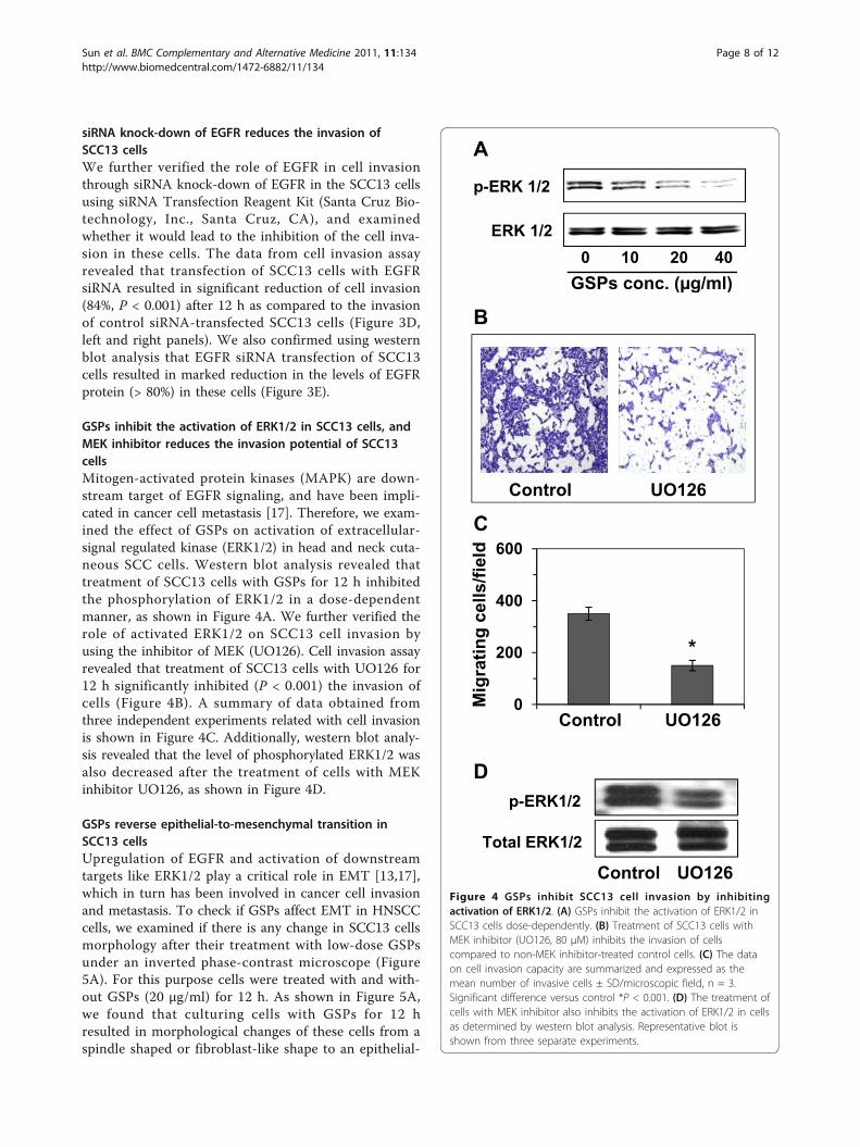
GSPs inhibit the activation of ERK1/2 in SCC13 cells, andMEK inhibitor reduces the invasion potential of SCC13cellsMitogen-activated protein kinases (MAPK) are down-stream target of EGFR signaling, and have been impli-cated in cancer cell metastasis [17]. Therefore, we exam-ined the effect of GSPs on activation of extracellular-signal regulated kinase (ERK1/2) in head and neck cuta-neous SCC cells. Western blot analysis revealed thattreatment of SCC13 cells with GSPs for 12 h inhibitedthe phosphorylation of ERK1/2 in a dose-dependentmanner, as shown in Figure 4A. We further verified therole of activated ERK1/2 on SCC13 cell invasion byusing the inhibitor of MEK (UO126). Cell invasion assayrevealed that treatment of SCC13 cells with UO126 for12 h significantly inhibited (P < 0.001) the invasion ofcells (Figure 4B). A summary of data obtained fromthree independent experiments related with cell invasionis shown in Figure 4C. Additionally, western blot analy-sis revealed that the level of phosphorylated ERK1/2 wasalso decreased after the treatment of cells with MEKinhibitor UO126, as shown in Figure 4D.
GSPs reverse epithelial-to-mesenchymal transition inSCC13 cellsUpregulation of EGFR and activation of downstreamtargets like ERK1/2 play a critical role in EMT [13,17],which in turn has been involved in cancer cell invasionand metastasis. To check if GSPs affect EMT in HNSCCcells, we examined if there is any change in SCC13 cellsmorphology after their treatment with low-dose GSPsunder an inverted phase-contrast microscope (Figure5A). For this purpose cells were treated with and with-out GSPs (20 μg/ml) for 12 h. As shown in Figure 5A,we found that culturing cells with GSPs for 12 hresulted in morphological changes of these cells from aspindle shaped or fibroblast-like shape to an epithelial-
A
p-ERK 1/2
ERK 1/20 10 20 40
GSPs conc. ( g/ml)
B( g )
600ld
CControl UO126
200
400
grat
ing
cells
/fiel
*
p-ERK1/2
0Mi
Control UO126
D
Total ERK1/2
Control UO126Figure 4 GSPs inhibit SCC13 cell invasion by inhibitingactivation of ERK1/2. (A) GSPs inhibit the activation of ERK1/2 inSCC13 cells dose-dependently. (B) Treatment of SCC13 cells withMEK inhibitor (UO126, 80 μM) inhibits the invasion of cellscompared to non-MEK inhibitor-treated control cells. (C) The dataon cell invasion capacity are summarized and expressed as themean number of invasive cells ± SD/microscopic field, n = 3.Significant difference versus control *P < 0.001. (D) The treatment ofcells with MEK inhibitor also inhibits the activation of ERK1/2 in cellsas determined by western blot analysis. Representative blot isshown from three separate experiments.
Sun et al. BMC Complementary and Alternative Medicine 2011, 11:134http://www.biomedcentral.com/1472-6882/11/134
Page 8 of 12
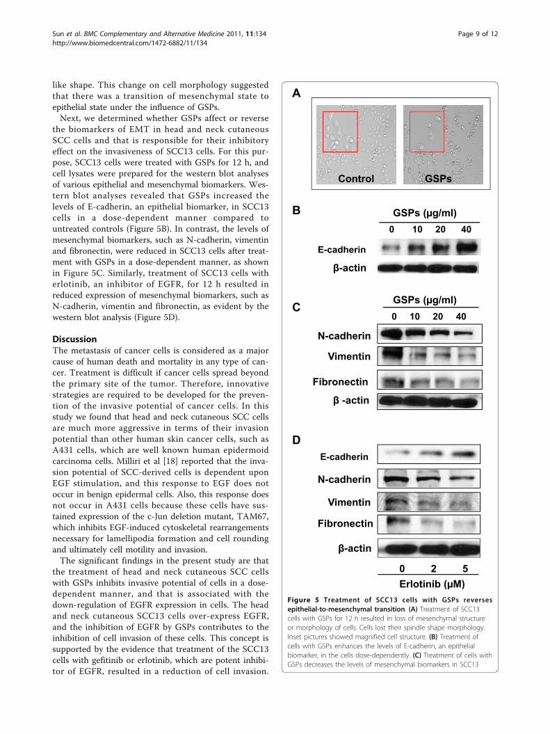
like shape. This change on cell morphology suggestedthat there was a transition of mesenchymal state toepithelial state under the influence of GSPs.Next, we determined whether GSPs affect or reverse
the biomarkers of EMT in head and neck cutaneousSCC cells and that is responsible for their inhibitoryeffect on the invasiveness of SCC13 cells. For this pur-pose, SCC13 cells were treated with GSPs for 12 h, andcell lysates were prepared for the western blot analysesof various epithelial and mesenchymal biomarkers. Wes-tern blot analyses revealed that GSPs increased thelevels of E-cadherin, an epithelial biomarker, in SCC13cells in a dose-dependent manner compared tountreated controls (Figure 5B). In contrast, the levels ofmesenchymal biomarkers, such as N-cadherin, vimentinand fibronectin, were reduced in SCC13 cells after treat-ment with GSPs in a dose-dependent manner, as shownin Figure 5C. Similarly, treatment of SCC13 cells witherlotinib, an inhibitor of EGFR, for 12 h resulted inreduced expression of mesenchymal biomarkers, such asN-cadherin, vimentin and fibronectin, as evident by thewestern blot analysis (Figure 5D).
DiscussionThe metastasis of cancer cells is considered as a majorcause of human death and mortality in any type of can-cer. Treatment is difficult if cancer cells spread beyondthe primary site of the tumor. Therefore, innovativestrategies are required to be developed for the preven-tion of the invasive potential of cancer cells. In thisstudy we found that head and neck cutaneous SCC cellsare much more aggressive in terms of their invasionpotential than other human skin cancer cells, such asA431 cells, which are well known human epidermoidcarcinoma cells. Milliri et al [18] reported that the inva-sion potential of SCC-derived cells is dependent uponEGF stimulation, and this response to EGF does notoccur in benign epidermal cells. Also, this response doesnot occur in A431 cells because these cells have sus-tained expression of the c-Jun deletion mutant, TAM67,which inhibits EGF-induced cytoskeletal rearrangementsnecessary for lamellipodia formation and cell roundingand ultimately cell motility and invasion.The significant findings in the present study are that
the treatment of head and neck cutaneous SCC cellswith GSPs inhibits invasive potential of cells in a dose-dependent manner, and that is associated with thedown-regulation of EGFR expression in cells. The headand neck cutaneous SCC13 cells over-express EGFR,and the inhibition of EGFR by GSPs contributes to theinhibition of cell invasion of these cells. This concept issupported by the evidence that treatment of the SCC13cells with gefitinib or erlotinib, which are potent inhibi-tor of EGFR, resulted in a reduction of cell invasion.
A
Control GSPs
E-cadherin
0 10 20 40GSPs ( g/ml)B
-actin
N-cadherin
Vimentin
0 10 20 40
GSPs ( g/ml)C
Vimentin
Fibronectin
-actin
Vimentin
N-cadherin
DE-cadherin
Fibronectin
-actin
Erlotinib ( M)0 2 5
( )Figure 5 Treatment of SCC13 cells with GSPs reversesepithelial-to-mesenchymal transition. (A) Treatment of SCC13cells with GSPs for 12 h resulted in loss of mesenchymal structureor morphology of cells. Cells lost their spindle shape morphology.Inset pictures showed magnified cell structure. (B) Treatment ofcells with GSPs enhances the levels of E-cadherin, an epithelialbiomarker, in the cells dose-dependently. (C) Treatment of cells withGSPs decreases the levels of mesenchymal biomarkers in SCC13
Sun et al. BMC Complementary and Alternative Medicine 2011, 11:134http://www.biomedcentral.com/1472-6882/11/134
Page 9 of 12
Similar effects were also noted when the SCC13 cellswere transfected with EGFR-siRNA. Treatment of cellswith EGF stimulates EGFR, and we observed that treat-ment of SCC13 cells with EGF enhances cell invasionability, and that this EGF-induced cell invasion wasblocked by the treatment of cells with GSPs. These
observations support the evidence that inhibition ofhead and neck cutaneous squamous cell carcinoma cellinvasion by GSPs is mediated through their inhibitoryeffects on EGFR expression. It has been reported thatinhibitors of EGFR can prevent the growth and progres-sion of HNSCC; however, long term use may alsoinduce some form of toxicity [19,20]. This possibility isnot expected with the use of GSPs as these are dietarycomponents and toxicity has not been observed in ani-mal models [11,12].Proteins of MAPK family are a downstream target of
EGFR, and have been shown to play a crucial role in
cells, such as N-cadherin, vimentin and fibronectin, in a dose-dependent manner. (D) Treatment of SCC13 cells with erlotinib, aninhibitor of EGFR, for 12 h decreases the levels of mesenchymalbiomarkers, such as N-cadherin, vimentin and fibronectin.
Gefitinib
MEKGSPs
UO126
EMT
ERK
Cell invasion
GSPs
Low Mortality Rate
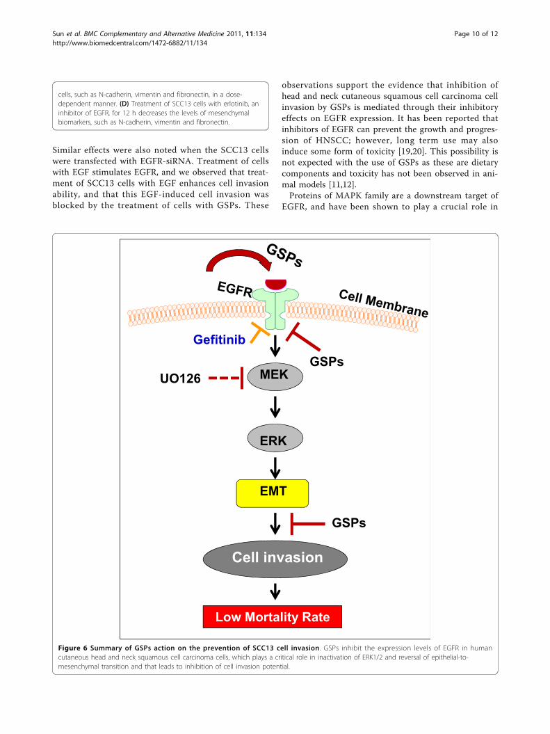
Figure 6 Summary of GSPs action on the prevention of SCC13 cell invasion. GSPs inhibit the expression levels of EGFR in humancutaneous head and neck squamous cell carcinoma cells, which plays a critical role in inactivation of ERK1/2 and reversal of epithelial-to-mesenchymal transition and that leads to inhibition of cell invasion potential.
Sun et al. BMC Complementary and Alternative Medicine 2011, 11:134http://www.biomedcentral.com/1472-6882/11/134
Page 10 of 12

cancer cell invasion. Our results show that inhibition ofinvasion of SCC13 cells by GSPs is associated with theinhibition of ERK1/2 phosphorylation. The inhibition ofMEK with UO126, a MEK inhibitor, blocked the inva-sion capacity of SCC13 cells which is similar to theaction of GSPs. These observations suggest a possibleinvolvement of ERK1/2/MAPK pathway in inhibition ofthe invasion of cutaneous HNSCC cells. Activation ofthe proteins of MAPK family leads to the activation NF-�B which play an important role in multiple biologicalprocesses, including inflammation, cell proliferation andangiogenesis [21-23]. Importantly, NF-�B has been iden-tified as an important regulator of EMT in several can-cer cell types [21-24]. EMT has been implicated ininvasion and metastasis of epithelial tumors. EMT canrender tumor cells migratory and invasive through theinvolvement of all stages: invasion, intravasation andextravasation [13,14]. During the process of EMT, cellscan change from an epithelial to a mesenchymal state.They lose their characteristic epithelial traits and insteadgain properties of mesenchymal cells. This process isprimarily coordinated by the disappearance or loss ofepithelial biomarkers such as E-cadherin with the con-comitant appearance or gain of mesenchymal markerssuch as vimentin, fibronectin and N-cadherin, etc. Inthe present study, GSPs treatment of SCC13 cellsshowed the suppression of mesenchymal biomarkers,such as vimentin, fibronectin and N-cadherin whilerestored the levels of epithelial biomarker such as, E-cadherin, in human cutaneous head and neck SCC cellswhich suggest that GSPs have the ability to reverse theEMT process in HNSCC cells. These information sug-gest that reversal of EMT in SCC13 cells by GSPs mayalso be one of the possible mechanisms through whichGSPs reduce the invasiveness of cutaneous head andneck squamous cell carcinoma cells and that lead toinhibition of invasion of SCC13 cells in our system. Arecent study showed that GSPs inhibit invasion of mela-noma cancer cells and this inhibitory effect of GSPs onmelanoma cell invasion was associated with their inhibi-tory effect on COX-2 overexpression and successivedown-regulation of NF-�B and reversal of EMT process[25]. Similar to GSPs, other phytochemicals, such asberberine, have also been shown to inhibit the invasionpotential of cancer cells. Berberine inhibits the invasionof melanoma cancer cells through its inhibitory effecton endogenous COX-2 overexpression and successivedown-regulation of prostaglandin E2 and prostaglandinE2 receptors [16].
ConclusionThe results from this study have identified for the firsttime that GSPs inhibit the invasiveness of human cuta-neous HNSCC cells and that involves: (i) the inhibitory
effect of GSPs on endogenous EGFR overexpression, (ii)the inhibitory effect of GSPs on the activation of theERK1/2 proteins of MAPK family, and (iii) the reversalof EMT process, as summarized in Figure 6. Moredetailed studies are needed to develop GSPs as a phar-macologically safe agent either alone or in combinationwith other anti-metastatic drugs for the treatment ofcutaneous head and neck SCCs in humans.
List of abbreviationsEGF: epidermal growth factor; EGFR: epidermal growth factor receptor; EMT:epithelial-to-mesenchymal transition; ERK1/2: extracellular signal relatedkinases; GSPs: grape seed proanthocyanidins; HNSCC: head and necksquamous cell carcinoma; MAPK: mitogen-activated protein kinases; NF-κB:nuclear factor-kappaB; NHEK: normal human epidermal keratinocytes; SCC:squamous cell carcinoma.
Acknowledgements and fundingThis work was supported by the funds from Veterans Administration MeritReview Award (S.K.K.) and National Cancer Institute/NCCAM/NIH (CA140197,CA140832). NHEK were obtained from the UAB Skin Diseases ResearchCenter (AR050948-01). The content of this article does not necessarily reflectthe views or policies of the funding sources.
Author details1Department of Dermatology, University of Alabama at Birmingham,Birmingham, AL, USA. 2Department of Surgery-Otolaryngology, University ofAlabama at Birmingham, Birmingham, AL, USA. 3Comprehensive CancerCenter, University of Alabama at Birmingham, Birmingham, AL, USA.4Nutrition Obesity Research Center, University of Alabama at Birmingham,Birmingham, AL, USA. 5Birmingham Veterans Affairs Medical Center,Birmingham, AL, 35294, USA.
Authors’ contributionsThe work reported in this manuscript was done in collaboration with allauthors. QS and RP have performed all experimental work, cell migrationsassays, western blot analysis and statistical analysis of data. SKK is a principalinvestigator of the study, has designed the study, provided all supervisionon daily basis, data analysis and write the final draft of the manuscript. ERwas associated in data interpretation, discussion and study design, as well asfinal drafting of the manuscript. All authors read and approved the finalmanuscript.
Competing interestsThe authors declare that they have no competing interests.
Received: 30 September 2011 Accepted: 21 December 2011Published: 21 December 2011
References1. Hunter KD, Parkinson EK, Harrison PR: Profiling early head and neck
cancer. Nat Rev Cancer 2005, 5(2):127-135.2. Arbes SJ Jr, Olshan AF, Caplan DJ, Schoenbach VJ, Slade GD, Symons MJ:
Factors contributing to the poorer survival of black Americansdiagnosed with oral cancer (United States). Cancer Causes Control 1999,10:513-523.
3. Posner MR, Hershock DM, Blajman CR, Mickiewicz E, Winquist E,Gorbounova V, Tjulandin S, Shin DM, Cullen K, Ervin TJ: Cisplatin andfluorouracil alone or with docetaxel in head and neck cancer. N Engl JMed 2007, 357:1705-1715.
4. Vermorken JB, Remenar E, van Herpen C, Gorlia T, Mesia R, Degardin M,Stewart JS, Jelic S, Betka J, Preiss JH: Cisplatin, fluorouracil, and docetaxelin unresectable head and neck cancer. N Engl J Med 2007, 357:1695-1704.
5. Leon X, Quer M, Orus C, del Prado Venegas M: Can cure be achieved inpatients with head and neck carcinomas? The problem of secondneoplasm. Expert Rev Anticancer Ther 2001, 1:125-133.
Sun et al. BMC Complementary and Alternative Medicine 2011, 11:134http://www.biomedcentral.com/1472-6882/11/134
Page 11 of 12
6. Casiglia J, Woo SB: A comprehensive review of oral cancer. Gen Dent2001, 49:72-82.
7. He Y, Zeng Q, Drenning SD, Melhem MF, Tweardy DJ, Huang L, Grandis JR:Inhibition of human squamous cell carcinoma growth in vivo byepidermal growth factor receptor antisense RNA transcribed from theU6 promoter. J Natl Cancer Inst 1998, 90(14):1080-1087.
8. Grandis JR, Melhem MF, Barnes EL, Tweardy DJ: Quantitativeimmunohistochemical analysis of transforming growth factor-α andepidermal growth factor receptor in patients with squamous cellcarcinoma of the head and neck. Cancer 1996, 78:1284-1292.
9. Grandis JR, Melhem MF, Gooding WE, Day R, Holst VA, Wagener MM,Drenning SD, Tweardy DJ: Levels of TGF-α and EGFR protein in head andneck squamous cell carcinoma and patient survival. J Natl Cancer Inst1998, 90:824-832.
10. Nandakumar V, Singh T, Katiyar SK: Multi-targeted prevention and therapyof cancer by proanthocyanidins. Cancer Lett 2008, 269:378-387.
11. Mittal A, Elmets CA, Katiyar SK: Dietary feeding of proanthocyanidins fromgrape seeds prevents photocarcinogenesis in SKH-1 hairless mice:relationship to decreased fat and lipid peroxidation. Carcinogenesis 2003,24:1379-1388.
12. Meeran SM, Vaid M, Punathil T, Katiyar SK: Dietary grape seedproanthocyanidins inhibit 12-O-tetradecanoyl phorbol-13-acetate-causedskin tumor promotion in 7, 12-dimethylbenz(a)anthracene-initiatedmouse skin, which is associated with the inhibition of inflammatoryresponses. Carcinogenesis 2009, 30:520-528.
13. Maier HJ, Wirth T, Beug H: Epithelial-mesenchymal transition in pancreaticcarcinoma. Cancers 2010, 2:2058-2083.
14. Thiery JP: Epithelial-mesenchymal transitions in tumor progression. NatRev Cancer 2002, 2:442-454.
15. Mantena SK, Sharma SD, Katiyar SK: Berberine inhibits growth, induces G1arrest and apoptosis in human epidermoid carcinoma A431 cells byregulating Cdki-Cdk-cyclin cascade, disruption of mitochondrialmembrane potential and cleavage of caspase-3 and PARP. Carcinogenesis2006, 27:2018-2027.
16. Singh T, Vaid M, Katiyar N, Sharma S, Katiyar SK: Berberine, an isoquinolinealkaloid, inhibits melanoma cancer cell migration by reducing theexpressions of cyclooxygenase-2, prostaglandin E2 and prostaglandin E2receptors. Carcinogenesis 2011, 32:86-92.
17. Zuo JH, Zhu W, Li MY, Li XH, Yi H, Zeng GQ, Wan XX, He QY, Li JH, Qu JQ,Chen Y, Xiao ZQ: Activation of EGFR promotes squamous carcinomaSCC10A cell migration and invasion via inducing EMT-like phenotypechange and MMP-9-mediated degradation of E-cadherin. J Cell Biochem2011, 112(9):2508-2517.
18. Malliri A, Symons M, Hennigan RF, Hurlstone AF, Lamb RF, Wheeler T,Ozanne BW: The transcription factor AP-1 is required for EGF-inducedactivation of rho-like GTPases, cytoskeletal rearrangements, motility, andin vitro invasion of A431 cells. J Cell Biol 1998, 143:1087-1099.
19. Leeman-Neill RJ, Cai Q, Joyce SC, Thomas SM, Bhola NE, Neill DB, Arbiser JL,Grandis JR: Honokiol inhibits epidermal growth factor receptor signalingand enhances the antitumor effects of epidermal growth factor receptorinhibitors. Clinical Cancer Res 2011, 16(9):2571-2579.
20. Leeman-Neill RJ, Seethala RR, Singh SV, Freilino ML, Bednash JS,Thomas SM, Panahandeh MC, Gooding WE, Joyce SC, Lingen MW, Neill DB,Grandis JR: Inhibition of EGFR-STAT3 signaling with erlotinib preventscarcinogenesis in a chemically-induced mouse model of oral squamouscell carcinoma. Cancer Prev Res (Phila) 2011, 4(2):230-237.
21. Min C, Eddy SF, Sherr DH, Sonenshein GE: NF-kappaB and epithelial tomesenchymal transition of cancer. J Cell Biochem 2008, 104:733-744.
22. Huber MA, Azoitei N, Baumann B, Grünert S, Sommer A, Pehamberger H,Kraut N, Beug H, Wirth T: NF-κB is essential for epithelial-mesenchymaltransition and metastasis in a model of breast cancer progression. J ClinInvest 2004, 114:569-581.
23. Chua HL, Bhat-Nakshatri P, Clare SE, Morimiya A, Badve S, Nakshatri H: NF-kappaB represses E-cadherin expression and enhances epithelial tomesenchymal transition of mammary epithelial cells: potentialinvolvement of ZEB-1 and ZEB-2. Oncogene 2007, 26:711-724.
24. Huber MA, Beug H, Wirth T: Epithelial-mesenchymal transition: NF-kappaBtakes center stage. Cell Cycle 2004, 3:1477-1480.
25. Vaid M, Singh T, Katiyar SK: Grape seed proanthocyanidins inhibitmelanoma cell invasiveness by reduction of PGE2 synthesis and reversalof epithelial-to-mesenchymal transition. PLoS ONE 2011, 6(6):e21539.
Pre-publication historyThe pre-publication history for this paper can be accessed here:http://www.biomedcentral.com/1472-6882/11/134/prepub
doi:10.1186/1472-6882-11-134Cite this article as: Sun et al.: Grape seed proanthocyanidins inhibit theinvasive potential of head and neck cutaneous squamous cell carcinomacells by targeting EGFR expression and epithelial-to-mesenchymaltransition. BMC Complementary and Alternative Medicine 2011 11:134.
Submit your next manuscript to BioMed Centraland take full advantage of:
• Convenient online submission
• Thorough peer review
• No space constraints or color figure charges
• Immediate publication on acceptance
• Inclusion in PubMed, CAS, Scopus and Google Scholar
• Research which is freely available for redistribution
Submit your manuscript at www.biomedcentral.com/submit
Sun et al. BMC Complementary and Alternative Medicine 2011, 11:134http://www.biomedcentral.com/1472-6882/11/134
Page 12 of 12