Embed Size (px)
Citation preview
lable at ScienceDirect
Respiratory Medicine Case Reports 11 (2014) 4e6
Contents lists avai
Respiratory Medicine Case Reports
journal homepage: www.elsevier .com/locate /rmcr
Case report
Granulomatous PJP presenting as a solitary lung nodule in an immunecompetent femaleq
J. Lam a, M.M. Kelly b, R. Leigh a, M.D. Parkins a,c,*
aDepartment of Medicine, The University of Calgary, Calgary, AB, CanadabDepartment of Pathology and Laboratory Medicine, The University of Calgary, Calgary, AB, CanadacDepartment of Microbiology & Infectious Diseases, The University of Calgary, Calgary, AB, Canada
a r t i c l e i n f o
Article history:Received 2 April 2013Received in revised form4 October 2013Accepted 14 October 2013
Keywords:PCPPneumocystisGranuloma
q This is an open-access article distributed undeCommons Attribution-NonCommercial-ShareAlike Lcommercial use, distribution, and reproduction inoriginal author and source are credited.* Corresponding author. 3330 Hospital Drive NW, C
Tel.: þ1 403 210 7913; fax: þ1 403 270 2772.E-mail address: [email protected] (M.D. Park
2213-0071/$ e see front matter � 2013 The Authors.http://dx.doi.org/10.1016/j.rmcr.2013.10.002
a b s t r a c t
Pneumocystis jiroveci pneumonia (PJP) opportunistically targets immunosuppressed patients, mostnotably those with advanced HIV/AIDS. Radiologically, PJP typically appears as bilateral diffuse pulmo-nary infiltrates. Herein an unusual case of an immunocompetent woman developing granulomatous PJPin the absence of evident risk factors is described. PJP may be an under-recognized cause of pulmonarynodules in immune competent individuals.
� 2013 The Authors. Published by Elsevier Ltd. All rights reserved.
1. Case presentation
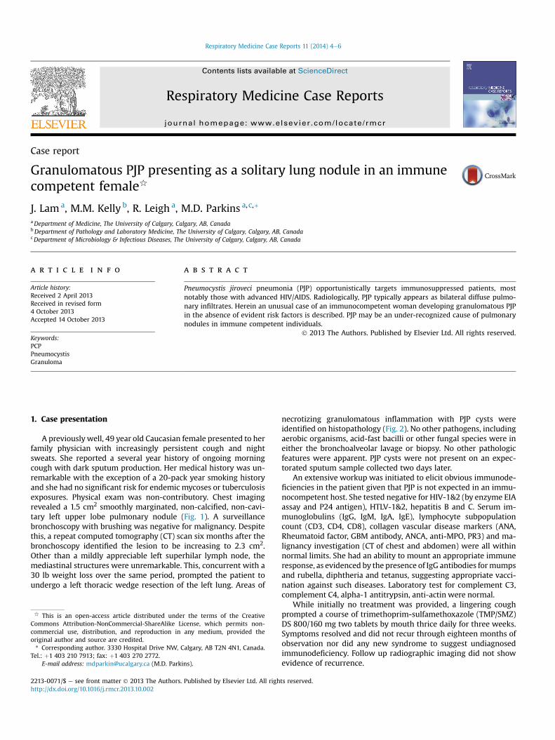
A previously well, 49 year old Caucasian female presented to herfamily physician with increasingly persistent cough and nightsweats. She reported a several year history of ongoing morningcough with dark sputum production. Her medical history was un-remarkable with the exception of a 20-pack year smoking historyand she had no significant risk for endemic mycoses or tuberculosisexposures. Physical exam was non-contributory. Chest imagingrevealed a 1.5 cm2 smoothly marginated, non-calcified, non-cavi-tary left upper lobe pulmonary nodule (Fig. 1). A surveillancebronchoscopy with brushing was negative for malignancy. Despitethis, a repeat computed tomography (CT) scan six months after thebronchoscopy identified the lesion to be increasing to 2.3 cm2.Other than a mildly appreciable left superhilar lymph node, themediastinal structures were unremarkable. This, concurrent with a30 lb weight loss over the same period, prompted the patient toundergo a left thoracic wedge resection of the left lung. Areas of
r the terms of the Creativeicense, which permits non-any medium, provided the
algary, AB T2N 4N1, Canada.
ins).
Published by Elsevier Ltd. All righ
necrotizing granulomatous inflammation with PJP cysts wereidentified on histopathology (Fig. 2). No other pathogens, includingaerobic organisms, acid-fast bacilli or other fungal species were ineither the bronchoalveolar lavage or biopsy. No other pathologicfeatures were apparent. PJP cysts were not present on an expec-torated sputum sample collected two days later.
An extensive workup was initiated to elicit obvious immunode-ficiencies in the patient given that PJP is not expected in an immu-nocompetent host. She tested negative for HIV-1&2 (by enzyme EIAassay and P24 antigen), HTLV-1&2, hepatitis B and C. Serum im-munoglobulins (IgG, IgM, IgA, IgE), lymphocyte subpopulationcount (CD3, CD4, CD8), collagen vascular disease markers (ANA,Rheumatoid factor, GBM antibody, ANCA, anti-MPO, PR3) and ma-lignancy investigation (CT of chest and abdomen) were all withinnormal limits. She had an ability to mount an appropriate immuneresponse, as evidenced by the presence of IgG antibodies formumpsand rubella, diphtheria and tetanus, suggesting appropriate vacci-nation against such diseases. Laboratory test for complement C3,complement C4, alpha-1 antitrypsin, anti-actin were normal.
While initially no treatment was provided, a lingering coughprompted a course of trimethoprim-sulfamethoxazole (TMP/SMZ)DS 800/160 mg two tablets by mouth thrice daily for three weeks.Symptoms resolved and did not recur through eighteen months ofobservation nor did any new syndrome to suggest undiagnosedimmunodeficiency. Follow up radiographic imaging did not showevidence of recurrence.
ts reserved.
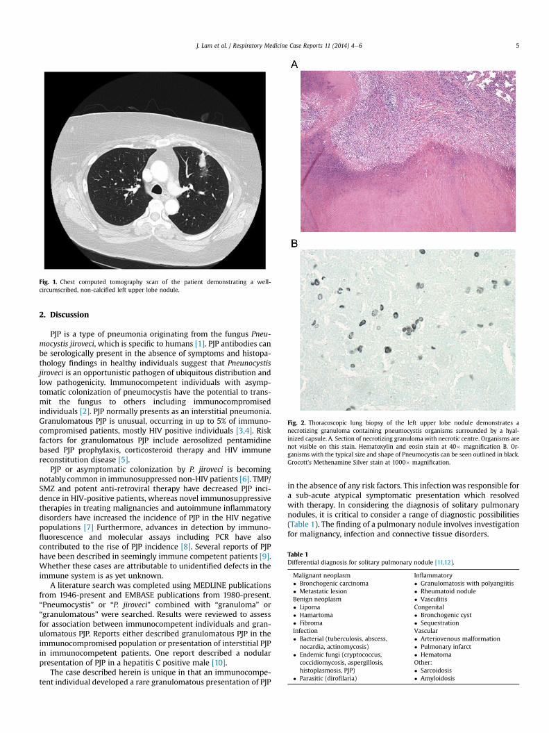
Fig. 1. Chest computed tomography scan of the patient demonstrating a well-circumscribed, non-calcified left upper lobe nodule.
Fig. 2. Thoracoscopic lung biopsy of the left upper lobe nodule demonstrates anecrotizing granuloma containing pneumocystis organisms surrounded by a hyal-inized capsule. A. Section of necrotizing granulomawith necrotic centre. Organisms arenot visible on this stain. Hematoxylin and eosin stain at 40� magnification B. Or-ganisms with the typical size and shape of Pneumocystis can be seen outlined in black.Grocott’s Methenamine Silver stain at 1000� magnification.
Table 1Differential diagnosis for solitary pulmonary nodule [11,12].
Malignant neoplasm� Bronchogenic carcinoma� Metastatic lesionBenign neoplasm� Lipoma� Hamartoma� FibromaInfection� Bacterial (tuberculosis, abscess,
nocardia, actinomycosis)� Endemic fungi (cryptococcus,
coccidiomycosis, aspergillosis,histoplasmosis, PJP)
� Parasitic (dirofilaria)
Inflammatory� Granulomatosis with polyangiitis� Rheumatoid nodule� VasculitisCongenital� Bronchogenic cyst� SequestrationVascular� Arteriovenous malformation� Pulmonary infarct� HematomaOther:� Sarcoidosis� Amyloidosis
J. Lam et al. / Respiratory Medicine Case Reports 11 (2014) 4e6 5
2. Discussion
PJP is a type of pneumonia originating from the fungus Pneu-mocystis jiroveci, which is specific to humans [1]. PJP antibodies canbe serologically present in the absence of symptoms and histopa-thology findings in healthy individuals suggest that Pneunocystisjiroveci is an opportunistic pathogen of ubiquitous distribution andlow pathogenicity. Immunocompetent individuals with asymp-tomatic colonization of pneumocystis have the potential to trans-mit the fungus to others including immunocompromisedindividuals [2]. PJP normally presents as an interstitial pneumonia.Granulomatous PJP is unusual, occurring in up to 5% of immuno-compromised patients, mostly HIV positive individuals [3,4]. Riskfactors for granulomatous PJP include aerosolized pentamidinebased PJP prophylaxis, corticosteroid therapy and HIV immunereconstitution disease [5].
PJP or asymptomatic colonization by P. jiroveci is becomingnotably common in immunosuppressed non-HIV patients [6]. TMP/SMZ and potent anti-retroviral therapy have decreased PJP inci-dence in HIV-positive patients, whereas novel immunosuppressivetherapies in treating malignancies and autoimmune inflammatorydisorders have increased the incidence of PJP in the HIV negativepopulations [7] Furthermore, advances in detection by immuno-fluorescence and molecular assays including PCR have alsocontributed to the rise of PJP incidence [8]. Several reports of PJPhave been described in seemingly immune competent patients [9].Whether these cases are attributable to unidentified defects in theimmune system is as yet unknown.
A literature search was completed using MEDLINE publicationsfrom 1946-present and EMBASE publications from 1980-present.“Pneumocystis” or “P. jiroveci” combined with “granuloma” or“granulomatous” were searched. Results were reviewed to assessfor association between immunocompetent individuals and gran-ulomatous PJP. Reports either described granulomatous PJP in theimmunocompromised population or presentation of interstitial PJPin immunocompetent patients. One report described a nodularpresentation of PJP in a hepatitis C positive male [10].
The case described herein is unique in that an immunocompe-tent individual developed a rare granulomatous presentation of PJP
in the absence of any risk factors. This infectionwas responsible fora sub-acute atypical symptomatic presentation which resolvedwith therapy. In considering the diagnosis of solitary pulmonarynodules, it is critical to consider a range of diagnostic possibilities(Table 1). The finding of a pulmonary nodule involves investigationfor malignancy, infection and connective tissue disorders.
J. Lam et al. / Respiratory Medicine Case Reports 11 (2014) 4e66
Disclosures
No author has any conflicts that impact the data presentedherein. The patient described provided written informed consentfor this case to be prepared and published.
References
[1] Stringer JR. Pneumocystis. Int J Med Microbiol 2002;292:391e404.[2] Ponce CA, Gallo M, Bustamante R, Vargas SL. Pneumocystis colonization is
highly prevalent in the autopsied lungs of the general population. Clin InfectDis 2010;50:347e53.
[3] Travis WD, Pittaluga S, Lipschik GY, Ognibene FP, Suffredini AF, Masur H, et al.Atypical pathologic manifestations of pneumocystis carinii pneumonia in theacquired immune deficiency syndrome. Review of 123 lung biopsies from 76patients with emphasis on cysts, vascular invasion, vasculitis and granulomas.Am J Surg Pathol 1990;14:615e25.
[4] Hartel PH, Shilo K, Klassen-Fischer M, Neafie RC, Ozbudak IH, Galvin JR, et al.Granulomatous reaction to Pneumocystis jiroveci: clinicopathologic review of20 cases. Am J Surg Pathol 2010;34:730e4.
[5] Tolet A, Duwat H, Daste G, Berry A, Escamilla R, Nevez G. Pneumocystis jirovecigenotypes and granulomatous pneumocystosis. Med Mal Infect 2006;36:229e31.
[6] Medrano FJ, Montes-Cano M, Conde M, de la Horra C, Respaldiza N, Gasch A,et al. Pneumocystis jirovecii in general population. Emerg Infect Dis 2005;11:245e50.
[7] Overgaard UM, Helweg-Larsen J. Pneumocystis jiroveci pneumonia (PJP) inHIV-1-negative patients: a retrospective study 2002e2004. Scand J Infect Dis2007;39:589e95.
[8] Fily F, Lachkar S, Thiberville L, Favennec L, Caron F. Pneumocystis jiroveciicolonization and infection among non-HIV infected patients. Med MalInfect 2011;41:526e31.
[9] Cano S, Capote F, Pereira A, Calderon E, Castillo J. Pneumocystis cariniipneumonia in patients without predisposing illnesses. Acute episode andfollow-up of five cases. Chest 1993;104:376e81.
[10] Harris K, Maroun R, Chalhoub M, Elsayegh D. Unusual presentation of pneu-mocystis pneumonia in an immunocompetent patient diagnosed by openlung biopsy. Heart Lung Circ 2012;21:221e4.
[11] Winer-Muram HT. The solitary pulmonary nodule. Radiology 2006;239:34e9.[12] Leef 3rd JL, Klein JS. The solitary pulmonary nodule. Radiol Clin North Am
2002;40:123e43.