Embed Size (px)
Citation preview

G ranulocyte colony-stimulating factor (G-CSF) is a cytokine associated with granulocytosis.
G-CSF-producing malignant tumors have been diag-nosed in various organs, including the lungs, colon, gallbladder, and stomach [1-3]. G-CSF-producing tumors exhibit significant hyperplastic and metastatic properties, and have a very poor prognosis [4]. Interestingly, most cases of G-CSF-producing tumors have been reported in Japan. Here, we report a rare case of a G-CSF-producing gallbladder tumor in a middle-aged female who has survived without recur-rence for 48 months after surgery.
Case Report
A 47-year-old female visited a gynecological clinic approximately one month after developing fever and
general fatigue. She was not diagnosed with a gyneco-logical disorder at that time, although abdominal ultrasonography (US) revealed a mass in the gallblad-der; thus, she was referred to our institution. The patient reported a 3-kg weight loss over the past few months. On a physical examination, her body tem-perature was 38℃. An approximately 10-cm-diame-ter mass was found in the upper right quadrant of the abdomen, with slight tenderness. Laboratory test results revealed leukocytosis (21,200 cells/µl) with 83.8 granulocytes and a platelet count of 67.6×10⁴/µl. The C-reactive protein level was 17.27 mg/dl. Although the serum aspartate aminotransferase level was normal, the alanine aminotransferase and alkaline phosphatase levels were elevated at 29 and 1,017 U/l, respectively. Levels of the tumor markers carcinoem-bryonic antigen and carbohydrate antigen 19-9 were normal; however, the interleukin (IL)-2 receptor
Acta Med. Okayama, 2016Vol. 70, No. 5, pp. 393-396CopyrightⒸ 2016 by Okayama University Medical School.
http ://escholarship.lib.okayama-u.ac.jp/amo/Case Report
Granulocyte Colony-Stimulating Factor-Producing Gallbladder Cancer
Nobuhiko Kanayaa,c*, Hideki Aokia, Rie Yamasakib, Toshiaki Morihiroc, and Hitoshi Takeuchia
Departments of aSurgery and bPathology, Iwakuni Clinical Center, National Hospital Organization, Iwakuni, Yamaguchi 740-0041, Japan, cDepartment of Gastroenterological Surgery, Okayama University Graduate School of Medicine, Dentistry and Pharmaceutical Sciences,
Okayama 700-8558, Japan
We report a case of a granulocyte colony-stimulating factor (G-CSF)-producing gallbladder tumor asso-ciated with fever in a middle-aged female. Preoperative blood analysis showed leukocytosis with ele-vated levels of C-reactive protein and G-CSF. We resected the liver at S4a+S5, with regional lymph node dissection and partial resection of the duodenum. Histology revealed undifferentiated carcinoma with spindle and giant cells and papillary adenocarcinoma. Immunohistochemistry revealed Stage IIIB G-CSF-producing gallbladder cancer. Postoperatively, leukocyte and serum G-CSF levels decreased to within normal limits. Adjuvant gemcitabine chemotherapy was administered for 16 months, and she has been recurrence-free for 48 months.
Key words: gallbladder cancer, G-CSF, unidentified fever, leukocystosis, and adenocarcinoma
Received December 25, 2015 ; accepted April 4, 2016.*Corresponding author. Phone : +81-86-235-7257; Fax : +81-86-221-8775E-mail : [email protected] (N. Kanaya)
Conflict of Interest Disclosures: No potential conflict of interest relevant to this article was reported.
level was markedly elevated at 2,290 U/ml (normal range: 145-519 U/ml). Abdominal US showed an enlarged gallbladder and the presence of a heteroge-neous mass occupying the body and fundus. Contrast-enhanced computed tomography (CT) showed an enhanced solid mass in the gallbladder measuring 8.5×8.0 cm (Fig. 1A) and a cystic ductal lymph node with a diameter of approximately 4.0 cm. This lymph node was compressing the second part of the duode-
num (Fig. 1B). Magnetic resonance cholangiopan-creatography showed no pancreaticobiliary malforma-tions, stenosis, or irregularity of the bile duct. On endoscopic ultrasound-guided fine needle aspiration, the tumor was suspected as a lesion of undifferenti-ated carcinoma of the gallbladder. Prior to surgery, the patientʼs fever did not improve with antibiotics. The cause of these symptoms was suspected to be cytokine production by the tumor cells. Therefore,
394 Kanaya et al. Acta Med. Okayama Vol. 70, No. 5
A B
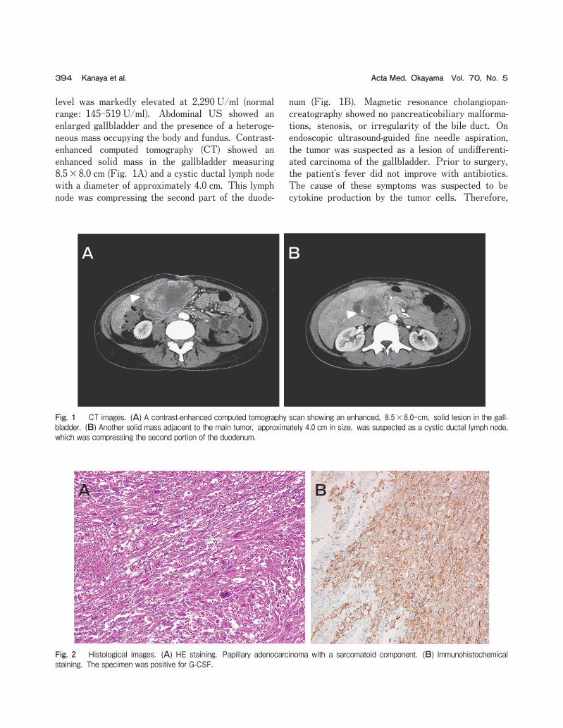
Fig. 2 Histological images. (A) HE staining. Papillary adenocarcinoma with a sarcomatoid component. (B) Immunohistochemical staining. The specimen was positive for G-CSF.
A B
Fig.1
Fig. 1 CT images. (A) A contrast-enhanced computed tomography scan showing an enhanced, 8.5×8.0-cm, solid lesion in the gall-bladder. (B) Another solid mass adjacent to the main tumor, approximately 4.0 cm in size, was suspected as a cystic ductal lymph node, which was compressing the second portion of the duodenum.

the serum G-CSF level was measured and subse-quently found to be elevated at 82.1 pg/ml (normal range<39.0 pg/ml). The patient underwent resection of hepatic S4a+S5 and regional lymph node dissection, along with partial resection of the duodenum. The extrahe-patic bile duct was not resected, since the intraopera-tive histological examination of the cystic duct showed a negative surgical margin and the lymph nodes around the bile duct appeared to be negative for metastasis. Curative resection was achieved. The resected gall-bladder specimen contained a tumor, 10 cm in size, occupying the body and fundus. A cross-section revealed a solid mass with central necrosis. Histologi-cally, the tumor was composed of undifferentiated carcinoma with spindle and giant cells, as well as papillary adenocarcinoma. The undifferentiated carci-noma component comprised almost the complete area of the tumor (Fig. 2A). Immunohistochemically, part of the undifferentiated carcinoma was positive for the cytokeratin markers AE1/3 and CAM5.2, and nega-tive for other markers, such as c-kit, CD34 and S-100. The mass was positive for G-CSF (Fig. 2B). Pathologically, the tumor had invaded the duodenal wall. Another mass adjacent to the main tumor was identified as the only lymph node presenting metasta-sis. The stage of the gallbladder cancer at resection was considered to be Stage IIIB, T3N1M0 (UICC-7). Postoperatively, the patientʼs leukocyte count and body temperature dramatically decreased to normal
levels (Fig. 3). By the time of discharge, approxi-mately 2 weeks after surgery, the serum G-CSF and IL-2 receptor concentrations had also decreased to 20.9 pg/ml and 388 U/ml, respectively. Adjuvant gemcitabine chemotherapy was administered for 16 months. The patient was followed-up with CT scans every 6 months after chemotherapy. No postoperative recurrence had occurred at 48 months.
Discussion
G-CSF-producing tumors were first reported by Asano in 1977 and were initially found in the lung [5]. These tumors are rarely detected in cases of gallblad-der cancer. G-CSF-producing tumors often secrete cytokines, such as IL-1 and IL-6. Therefore, affected patients present with fever and leukocytosis. The current patient had a high fever and leukocytosis on admission; however, no pathogens were detected and her symptoms did not improve with antibiotics. Cholecystitis and other diseases were ruled out, and G-CSF-producing gallbladder cancer was suspected. The diagnostic criteria for G-CSF-producing tumors recommended by Asano include four factors: (i) leuko-cytosis with an unknown cause, (ii) elevated serum G-CSF level, (iii) decreased leukocyte count and serum G-CSF level after tumor resection, and (iv) proof of G-CSF production in the tumor [5]. The present patient met all 4 criteria. In the Japanese literature, we found 22 patients
October 2016 GCSF-Producing Gallbladder Cancer 395
35
36
37
38
39
0
5,000
10,000
15,000
20,000
25,000
WBCBT
WBC(/µl) BT(℃)OP
day1
day8
day13
day15
day17
day19
day21
day25AD
ENTday29
Fig. 3 Clinical course. Changes in the leukocyte count (dot) and body temperature (line) in the present case. AD, admission; OP, operation; ENT, discharge; WBC, white blood cells; BT, body temperature.

with gallbladder cancer producing G-CSF reported from 1983 to 2014 [6,7]. The male: female ratio was 8 : 14. The mean age at diagnosis was 67.4 (range, 47-82) years, with the current patient being the youngest. The mean leukocyte count and serum G-CSF level on admission were 31,809 cells/µl and 234 pg/ml, respectively. These blood parameters can be helpful for diagnosing G-CSF-producing tumors and are also useful as markers of recurrence and tumor exacerbation. The mean tumor size at diagnosis was 96.9 (range, 45-130) mm. Histological examinations of the specimens demonstrated 6 cases of undifferenti-ated or poorly differentiated adenocarcinoma, 6 cases of adenosquamous carcinoma, 3 cases of pleomorphic giant cell carcinoma, 3 cases of moderately differenti-ated adenocarcinoma, 2 cases of squamous cell carci-noma, and three others. Only three of these 22 cases were common types of adenocarcinoma. The prognoses among these cases were generally poor. Eight of 22 patients could not undergo surgery, and 15 of the 22 patients died within 6 months. This is partly because of the characteristics of differentiation. G-CSF-producing gallbladder cancer tends to exhibit high biological malignancy. Despite the poor general prog-nosis, some patients achieve long-term survival follow-ing curative resection. The 2 most important factors for improving the prognosis of G-CSF-producing gall-bladder cancer are early diagnosis and curative resec-tion. Although the significance of postoperative adjuvant chemotherapy for gallbladder carcinoma remains con-troversial, it might be useful for treating gallbladder carcinoma according to the results of a phase III mul-ticenter prospective randomized controlled trial con-ducted by Takada et al. [8]. Also, in a study of biliary tract cancer, Valle et al. [9] reported that therapy with cisplatin plus gemcitabine was associated with a significant survival advantage, compared with gemcit-abine monotherapy alone. Adjuvant chemotherapy trials using gemcitabine, capecitabine, S-1, and com-bination chemotherapy with platinum agents are cur-rently underway. Adjuvant chemotherapy was per-formed in the present patient because of the poor
prognosis and the discovery of metastasis in a lymph node. In summary, for patients with gallbladder cancer exhibiting high fever and leukocytosis, G-CSF-producing gallbladder cancer should be included as a differential diagnosis. Although G-CSF-producing gallbladder cancer demonstrates high biological malig-nancy and is associated with a poor prognosis, cura-tive resection, when possible, may contribute to long-term survival.
References
1. Hasegawa S, Suda T, Negi K and Hattori Y: Lung large cell car-cinoma producing granulocyte-colony-stimulating factor. Ann Thorac Surgery (2007) 83: 308-310.
2. Takahashi H, Yasuda A, Ochi N, Sakamoto M, Takayama S, Wakasugi, Funahashi, H, Sawai, H, Satoh, M, Akamo Y and Takeyama H: Granulocyte-colony stimulating factor producing rec-tal cancer. World J Surg Oncol (2008) 6: 70-74.
3. Kawaguchi M, Asada Y, Terada T, Takehara T, Munemoto Y and Fujisawa K: Aggressive recurrence of gastric cancer as a granulo-cyte-colony-stimulating factor-producing tumor. Int J Clin Oncol (2010) 15: 191-195 (in Japanese).
4. Ikeda T, Ohgaki K, Miura M, Aishima S, Shimizu T and Maehara Y: Granulocyte-colony stimulating factor-producing gallbladder cancer without recurrence more than 2 years after resection: report of a case. Surg Today (2005) 35: 590-593.
5. Asano S, Urabe A, Okabe T, Sato N and Kondo Y: Demonstration of granulopoietic factor in the plasma of nude mice transplanted with a human lung cancer and in the tumor tissue. Blood (1977) 49: 845-852.
6. Ogura T, Takii M, Arisaka Y, Masuda D, Kuwabara H, Egashira Y, Umegaki E and Higuchi K: A case of adenosquamous cell carci-noma of the gallbladder producing G-CSF with diffusely uptake in the spine by FDG-PET. J Hepato-Bilia-Pancr Surg (2011) 25: 759-767 (in Japanese).
7. Suzumura K, Iimuro Y, Asano Y, Kuroda N, Hirano T, Yamanaka J, Okada T, Okamoto T, Torii I and Fujimoto J: Granulocyte-colony stimulating factor-producing gallbladder carcinoma. Int Surg (2014) 99: 577-583.
8. Takada T, Amano H, Yasuda H, Nimura Y, Matsushiro T, Kato H, Nagakawa T and Nakayama T: Is postoperative adjuvant chemo-therapy useful for gallbladder carcinoma? A phase III multicenter prospective randomized controlled trial in patients with resected pancreaticobiliary carcinoma. Cancer (2002) 95: 1685-1695.
9. Valle J, Wasan H, Palmer DH, Cunningham D, Anthoney A, Maraveyas A, Madhusudan S, Iveson T, Hughes S, Pereira SP, Roughton M and Bridgewater J: Cisplatin plus gemcitabine versus gemcitabine for biliary tract cancer. N Engl J Med (2010) 36: 1273-1281.
396 Kanaya et al. Acta Med. Okayama Vol. 70, No. 5