Embed Size (px)
Citation preview

Grand Rounds
Niloofar Piri, MD
Jan 17th 2014

CC: Blind spots and blurry vision OU for more than 2 years (OS more severely affected)
HPI: A 74-y Caucasian gentleman presented with gradually increasing visual field defect and night blindness OU for more than two years. He reports episodes of exacerbation which never returned to normal
He claims a history of blunt trauma OS in 1960s after which the vision OS has been low, he has noticed deterioration of VA OS as well for the past two years.

PMH: CAD - Coronary angioplasty x2 (1999, 2006)
DH: Verapamil, ASA 83 mg/d, Crestor ( Statin)

Ocular examination
BCVA OD: 20/40 (-0.50 +0.25x90)
OS: CF 1m
Anterior segment OD : NS 1+, CC1+
OS: pc-IOL
IOP 14 mmHg 18 mmHg
No cells in the anterior chamber or anterior vitreous
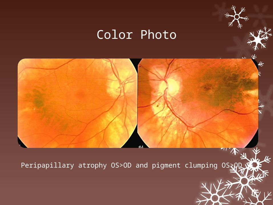
Color Photo
Peripapillary atrophy OS>OD and pigment clumping OS>OD

OCT ODJune 2013
Epiretinal membrane, Loss of outer hyper-reflective bands outside the fovea, central outer layers preserved
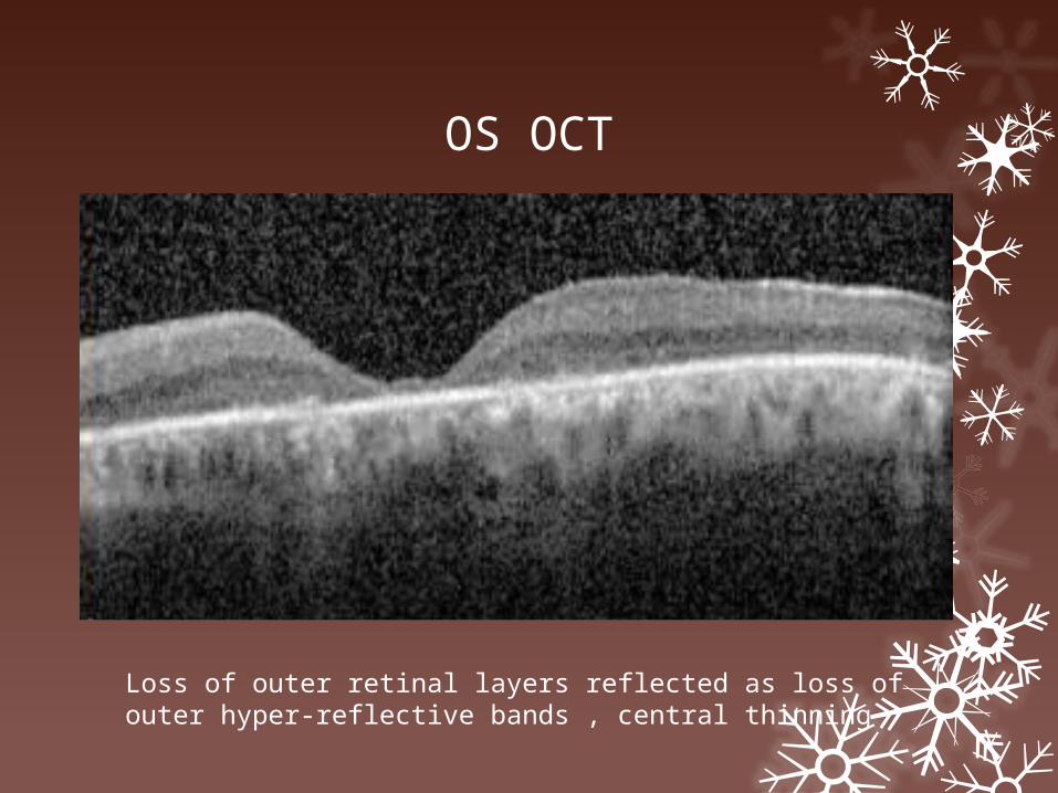
OS OCT
Loss of outer retinal layers reflected as loss of outer hyper-reflective bands , central thinning

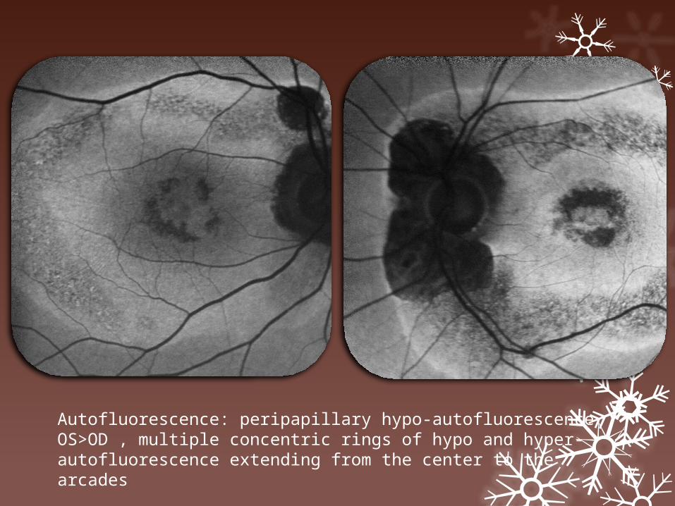
Autofluorescence: peripapillary hypo-autofluorescence OS>OD , multiple concentric rings of hypo and hyper-autofluorescence extending from the center to the arcades

Multifocal ERG
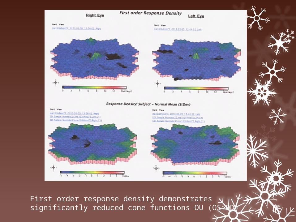
First order response density demonstrates significantly reduced cone functions OU (OS>OD)
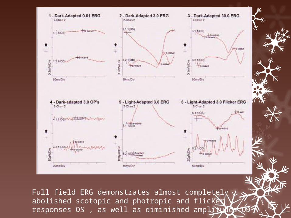
Full field ERG demonstrates almost completely abolished scotopic and photropic and flicker responses OS , as well as diminished amplitudes OD
Assessment: A 74-y old Caucasian gentleman with a history of progressive visual field loss and nyctalopia with some episodes of exacerbations for the past 2 years OS>>OD

Impression: AZOOR (Acute Zonal Occult Outer Retinopathy)
(Acute Annular Outer Retinopathy (AAOR) variant)
AIR (Autoimmune Retinopathy)(CAR, MAR, npAIR)

Monson DM, Smith JR. Acute Zonal Occult Outer Retinopathy (AZOOR) : Major review. Surv of Ophthalmology 2011
In 1992, Gass reported a ‘syndrome’ characterized by the sudden onset of photopsia and acute scotomas related to loss of sectors of outer retinal function

Early in the course of the disease, funduscopic appearance was often normal; however, most patients developed zones of retinal pigment epithelial atrophy or pigment clumping
Changes in the electroretinogram (ERG) and persistent visual field defects were usually observed. Although the retinal dysfunction in AZOOR is focal clinically, the ERG changes suggested a global deficit.

Acute onset of decreased vision in a zone of the visual field, usually accompanied by photopsia
Visual field defect and the photopsia worsened in bright light

76% in women , mean age 37 (17-79)
Starts unilaterally in 62% but progresses to bilateral disease from several weeks to 228 months
In the group of 51 patients followed by Gass et al,20 stabilization of visual field loss occurred within 6 months for 77% of patients.
In 1995 Gass and Stern described acute annular outer retinopathy (AAOR) as a variant of AZOOR

The clinical findings of AAOR were considered to be the same as AZOOR, except for the absence of photopsia and the development of an evanescent annular opacity in the retina.

No known treatment is proved to be effective.