Embed Size (px)
Citation preview

Grand Rounds Vol 6 pages 18–21Speciality: General Surgery, Gynaecology GRArticle Type: Case ReportDOI: 10.1102/1470-5206.2006.0004c© 2006 e-MED Ltd
Differential diagnosis of an incarceratedinguinal hernia: extraperitoneal
endometrioma of the round ligament
Jack J. K. Ku†, Michael Marfan‡, Michael O’ Shea‡, Adam Louie‡, John Lynch‡
and Anthony Griffin†‡
†Surgical Department, Princess Alexandra Hospital, Woolloongabba, Queensland 4102,Australia
‡Queen Elizabeth II Hospital, Cooper Plains, Queensland 4108, Australia
Corresponding address: Dr Jack J K Ku, Department of Surgery, P.O. Box 547,Nambour General Hospital, Nambour Qld 4560, Australia. E-mail: J k [email protected]
Date accepted for publication 1 March 2006
Abstract
General surgeons may encounter endometriosis when extraperitoneal forms of the disease mimic asymptomatic inguinal hernia in premenopausal women. Diagnosis is often difficult, especially in theabsence of menstrual symptoms, and is often made during surgical groin exploration. We presenta case of extraperitoneal endometrioma simulating a symptomatic inguinal hernia and discuss thepathogenesis and management.
Keywords
Inguinal hernia; extraperitoneal endometrioma.
Case report
A 46-year-old lady presented to the Emergency Department of QE II Hospital Queensland Australiain late 2001 with a 2 day history of constant localised right iliac fossa pain. The pain was aggravatedby movement and coughing but relieved by drawing her knees upwards and lying still. She had nogastrointestinal or genitourinary symptoms and was not systemically unwell. There was no historyof intra-abdominal or pelvic surgery.
Her last menstrual period had finished 5 days before presentation. She had been started on oralcontraceptive pills 3 months previously to control ‘menstrual migraines’. She was a Gravida 2,Para 2 (G2P2) and was previously diagnosed with an incompetent cervix, having been hospitalisedfor both of her pregnancies from the 20th week. She denied any symptoms of dysmenorrhea ormenorrhagia.
On examination, she was apyrexial and had normal routine observations. Palpation of herabdomen revealed mild tenderness in the right iliac fossa and inguinal region. Bowel sounds werenormal, the hernial orifices were clear, and vaginal and rectal examinations were unremarkable. Noother abnormalities were detected on physical examination. Routine full blood examination (FBE),urea and electrolytes (U & Es), liver function test (LFTs), ward test urine (WTU), abdominal X-ray anderect chest X-ray were all normal.
This paper is available online at http://www.grandrounds-e-med.com. In the event of a change in the URLaddress, please use the DOI provided to locate the paper.
Differential diagnosis of an incarcerated inguinal hernia 19
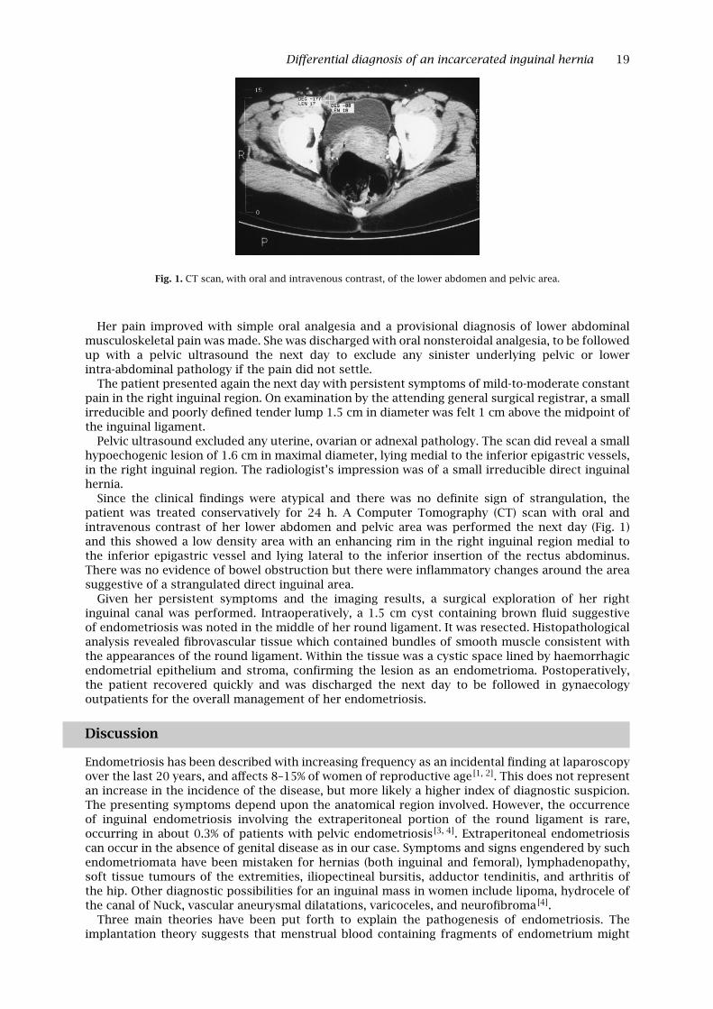
Fig. 1. CT scan, with oral and intravenous contrast, of the lower abdomen and pelvic area.
Her pain improved with simple oral analgesia and a provisional diagnosis of lower abdominalmusculoskeletal pain was made. She was discharged with oral nonsteroidal analgesia, to be followedup with a pelvic ultrasound the next day to exclude any sinister underlying pelvic or lowerintra-abdominal pathology if the pain did not settle.
The patient presented again the next day with persistent symptoms of mild-to-moderate constantpain in the right inguinal region. On examination by the attending general surgical registrar, a smallirreducible and poorly defined tender lump 1.5 cm in diameter was felt 1 cm above the midpoint ofthe inguinal ligament.
Pelvic ultrasound excluded any uterine, ovarian or adnexal pathology. The scan did reveal a smallhypoechogenic lesion of 1.6 cm in maximal diameter, lying medial to the inferior epigastric vessels,in the right inguinal region. The radiologist’s impression was of a small irreducible direct inguinalhernia.
Since the clinical findings were atypical and there was no definite sign of strangulation, thepatient was treated conservatively for 24 h. A Computer Tomography (CT) scan with oral andintravenous contrast of her lower abdomen and pelvic area was performed the next day (Fig. 1)and this showed a low density area with an enhancing rim in the right inguinal region medial tothe inferior epigastric vessel and lying lateral to the inferior insertion of the rectus abdominus.There was no evidence of bowel obstruction but there were inflammatory changes around the areasuggestive of a strangulated direct inguinal area.
Given her persistent symptoms and the imaging results, a surgical exploration of her rightinguinal canal was performed. Intraoperatively, a 1.5 cm cyst containing brown fluid suggestiveof endometriosis was noted in the middle of her round ligament. It was resected. Histopathologicalanalysis revealed fibrovascular tissue which contained bundles of smooth muscle consistent withthe appearances of the round ligament. Within the tissue was a cystic space lined by haemorrhagicendometrial epithelium and stroma, confirming the lesion as an endometrioma. Postoperatively,the patient recovered quickly and was discharged the next day to be followed in gynaecologyoutpatients for the overall management of her endometriosis.
Discussion
Endometriosis has been described with increasing frequency as an incidental finding at laparoscopyover the last 20 years, and affects 8–15% of women of reproductive age [1, 2]. This does not representan increase in the incidence of the disease, but more likely a higher index of diagnostic suspicion.The presenting symptoms depend upon the anatomical region involved. However, the occurrenceof inguinal endometriosis involving the extraperitoneal portion of the round ligament is rare,occurring in about 0.3% of patients with pelvic endometriosis [3, 4]. Extraperitoneal endometriosiscan occur in the absence of genital disease as in our case. Symptoms and signs engendered by suchendometriomata have been mistaken for hernias (both inguinal and femoral), lymphadenopathy,soft tissue tumours of the extremities, iliopectineal bursitis, adductor tendinitis, and arthritis ofthe hip. Other diagnostic possibilities for an inguinal mass in women include lipoma, hydrocele ofthe canal of Nuck, vascular aneurysmal dilatations, varicoceles, and neurofibroma [4].
Three main theories have been put forth to explain the pathogenesis of endometriosis. Theimplantation theory suggests that menstrual blood containing fragments of endometrium might

20 J. J. K. Ku et al.
pass along the Fallopian tubes in a retrograde manner and thus reach the peritoneal cavity. Thecoelomic metaplasia theory suggests that endometriosis originates in the coelomic membranevia a process of metaplasia following a metaplastic induction factor. The lymphatic and vasculardissemination theory proposes that viable endometrial cells gain entry to open basal lymph andblood vessels and embolise to ectopic sites, in order to explain the distant sites of endometriosis [2].
Endometriosis in the inguinal area was first described by Cullen in 1896, and since that timeapproximately 30 cases (mostly in obstetric and gynaecology journals) have been reported, allin women in their reproductive years [5]. 90% of such endometriomas arise from the right roundligament, as in our case [6]. Some authors use the lymphatic and vascular dissemination theory toexplain endometriosis involving the extraperitoneal portion of the round ligament [7]. This could bebecause the lymphatic vessels originate in the uterus and run along the round ligaments [8]. Thisprobably accounts for the rare incidence of ovarian cancer metastasising to the inguinal nodes [9].However, it does not explain its predominant presence on the right. Asymmetrical lymphaticdrainage in favour of the right inguinocrural region would provide a logical explanation, butanatomical descriptions and oncological observations do not seem to support this hypothesis [10].Some authors suggest that the sigmoid colon protects the left groin, which is also felt to accountfor the prevalence of right-sided inguinal hernias [11].
Symptoms of extraperitoneal endometriosis, often diverse and puzzling, usually result fromfunctioning endometrial tissue or scarring at the affected site. The location of the endometriosis,not the size of the implant, determines the symptoms. Many women with extensive endometriosisare asymptomatic, yet a small focus of disease in a particular location can produce disturbingproblems [12].
Clues to the presence of endometriosis include pain, bleeding or menstrual dysfunction. It isalso reported to be associated with infertility, but our patient is a G2P2. Noncyclical symptomscan confuse the clinician. It has been proposed that these extragenital lesions, especially thosefurther away from the uterus, tend to lose their hormonal receptors and response, hence the lackof cyclical symptoms [6, 13]. Ultrasonography has been used as a diagnostic aid and usually reveals acystic-solid lesion. CT scan usually demonstrates a mass which is mainly solid in nature and followsthe course of the round ligament [11, 14]. Treatment is surgical excision of the mass. Local recurrenceat the surgical scar has been reported [15].
Teaching point
Since extraperitoneal endometrioma can mimic an incarcerated inguinal hernia, it is importantthat general surgeons are familiar with this condition. Awareness of endometriosis is essentialto diagnosis and must be considered in any woman of reproductive age with atypical presentationof inguinal hernia. Furthermore, according to a literature review of the 30 documented cases, 32%were associated with an inguinal hernia [6]. In addition, malignant transformation has been reportedto occur in foci of endometriosis. Aggressive mixed clear cell and endometrioid carcinoma resultingin significant mortality despite chemotherapy have been reported [16]. Preoperative diagnosis isoften difficult and groin exploration and excision of lesion are usually diagnostic and curative assuppressive hormonal therapy alone as treatment is usually unsatisfactory. However, the patientshould be referred for a full gynaecological assessment to investigate and manage any concomitantendometriosis elsewhere.
References
1. Mitchell GW. Extrapelvic endometriosis. In: Endometriosis: Contemporary Concepts inClinical Management. Schenken RS, ed. Philadelphia: L.B. Lippincott, 1989: 307–28.
2. Edmonds DK. Chapter 35: Endometriosis. In: Dewhurst’s Textbook of Obstetrics andGynaecology for Postgraduates, 6th edn. London: Blackwell Science, 1999: 420–31.
3. Daro A, Capos N, Baserga R. Endometriosis of the round ligament. J Intern Coll Surg 1953; 21:385.
4. Strasser EJ, Davis RM. Extraperitoneal inguinal endometriosis. Am Surg 1977; 43: 421–2.5. Sataloff DM, La Vorgna KA, McFarland MM. Extrapelvic endometriosis present as a hernia:
clinical reports and review of literature. Surgery 1989; 105: 109–12.6. Clausen I, Nielsen KT. Endometriosis in the groin. Int J Gynaecol Obstet 1987; 25: 469–71.7. Candiani GB, Vercellini P, Fedele L, Vendola N, Carinelli S, Scaglione V. Inguinal endometriosis:
pathogenetic and clinical implications. Obstet Gynecol 1991; 78: 191–4.

Differential diagnosis of an incarcerated inguinal hernia 21
8. Krantz KE. Anatomy of the female reproductive system. In: Current Obstetric and GynaecologicDiagnosis and Treatment, 6th edn. Pernoll ML, Benson RC, eds. Norwalk, Connecticut: Appleton& Lange, 1987: 1–46.
9. Fuks Z. Patterns of spread of ovarian carcinoma: relation to therapeutic strategies. In: OvarianCancer, Newman CE, Ford CHJ, Jordan JH, eds. New York: Pergamon Press, 1980: 39–51.
10. Reiffenstuhl G. The Lymphatics of the Female Genital Organs. Philadelphia: JB Lippincott, 1957.11. Freed KS, Granke DS, Tyre LL, Williams VL, Omert LA. Endometriosis of the extraperitoneal
portion of the round ligament: US and CT findings. J Clin Ultrasound 1996; 24: 540–2.12. Luciano AA, Pitkin RM. Endometrios. Approaches to diagnosis and treatment. Surg Annu 1984;
16: 297–312.13. Markham SM, Carpenter SE, Rock JA. Extrapelvic endometriosis. Obstet Gynecol Clin North Am
1989; 16: 193–219.14. Athey PA, Diment DD. The spectrum of sonographic findings in endometriosis. J Ultrasound
Med 1989; 8: 487–91.15. Koger KE, Shatney CH, Hodge K, McClenathan JH. Surgical scar endometriosis. Surg Gynecol
Obstet 1993; 177: 243–6.16. Elemenoglou J, Skopelitou A, Nomikos I. Carcinoma of the inguinal region arising
from endometriosis of the round ligament. Eur J Gynaecol Oncol 1993; 14: 28–32.