Embed Size (px)
Citation preview
974 | Metallomics, 2020, 12, 974--987 This journal is©The Royal Society of Chemistry 2020
Cite this:Metallomics, 2020,
12, 974
Gold(III) bis(dithiolene) complexes: from molecularconductors to prospective anticancer,antimicrobial and antiplasmodial agents†‡
Diana Fontinha,a Sılvia A. Sousa,b Tania S. Morais, c Miguel Prudencio, a
Jorge H. Leitao, b Yann Le Gal,d Dominique Lorcy, d Rafaela A. L. Silva,e
Mariana F. G. Velho,ef Dulce Belo,e M. Almeida, e Joana F. Guerreiro, e
Teresa Pinheirog and Fernanda Marques *e
The anticancer, antimicrobial and antiplasmodial activities of six gold(III) bis(dithiolene) complexes were
studied. Complexes 1–6 showed relevant anticancer properties against A2780/A2780cisR ovarian cancer
cells (IC50 values of 0.08–2 mM), also being able to overcome cisplatin resistance in A2780cisR cells.
Complex 1 also exhibited significant antimicrobial activity against Staphylococcus aureus (minimum
inhibitory concentration (MIC) values of 12.1 � 3.9 mg mL�1) and both Candida glabrata and Candida albicans
(MICs of 9.7 � 2.7 and 19.9 � 2.4 mg mL�1, respectively). In addition, all complexes displayed antiplasmodial
activity against the Plasmodium berghei parasite liver stages, even exhibiting better results than the ones
obtained using primaquine, an anti-malarial drug. Mechanistic studies support the idea that thioredoxin
reductase, but not DNA, is a possible target of these complexes. Complex 1 is stable under biological
conditions, which would be important if this compound is ever to be considered as a drug. Overall, the
results obtained evidenced the promising biological activity of complex 1, which might have potential as a
novel anticancer, antimicrobial and antiplasmodial agent to be used as an alternative to current therapeutics.
Significance to metallomicsGold(III) complexes have been studied due to their use as precursors of neutral single-component molecular conductors and magnetic materials. More recently,these complexes have been shown to exhibit important biological activities. In this study we have investigated the anticancer, antibacterial, antifungal, andantiplasmodial properties of six gold(III) bis(dithiolene) complexes. The overall results obtained evidenced the promising biological activity of this versatilefamily of complexes, which are potential novel anticancer, antimicrobial and antiplasmodial agents to be used as alternatives to current therapeutics.
Introduction
The worldwide increase in the incidence of cancer and in thenumber of infections caused by microorganisms resistant to
available antibiotics led to the urgent need to develop novelefficient antitumor and antimicrobial agents.1–4 In fact, microbialdrug resistance is responsible for the increasing mortality rateobserved for infectious diseases, due to the lack of effective
a Instituto de Medicina Molecular, Faculdade de Medicina, Universidade de Lisboa, Avenida Professor Egas Moniz, 1649-028 Lisboa, Portugalb iBB-Institute for Bioengineering and Biosciences, Departmento de Bioengenharia, Instituto Superior Tecnico, Universidade de Lisboa, Av. Rovisco Pais, 1049-001 Lisboa,
Portugalc Centro de Quımica Estrutural, Faculdade de Ciencias, Universidade de Lisboa, Campo Grande, 1749-016 Lisboa, Portugald Univ Rennes, CNRS, ISCR (Institut des Sciences Chimiques de Rennes) – UMR 6226, F-35000 Rennes, Francee Centro de Ciencias e Tecnologias Nucleares, Instituto Superior Tecnico, Universidade de Lisboa, Estrada Nacional 10 (km 139,7), 2695-066 Bobadela LRS, Portugal.
E-mail: [email protected] Instituto de Telecomunicaçoes, Instituto Superior Tecnico, Universidade de Lisboa, Av. Rovisco Pais 1, 1049-001, Lisboa, Portugalg iBB-Institute for Bioengineering and Biosciences, Departamento de Engenharia e Ciencias Nucleares, Instituto Superior Tecnico, Universidade de Lisboa,
Av. Rovisco Pais 1, 1049-001 Lisboa, Portugal
† Dedication: D. Fontinha, M. Prudencio – antiplasmodial activity studies; S. A. Sousa, J. H. Leitao – antimicrobial activity studies; Y. Le Gal, D. Lorcy: synthesis ofcomplexes 1–4 and redox property studies; M. Velho, R. Silva, D. Belo, M. Almeida: synthesis of complexes 5 and 6 and stability studies in solution; T. Morais,J. Guerreiro, T. Pinheiro, F. Marques: anticancer activity and mechanistic studies.‡ Electronic supplementary information (ESI) available. See DOI: 10.1039/d0mt00064g
Received 12th March 2020,Accepted 27th April 2020
DOI: 10.1039/d0mt00064g
rsc.li/metallomics
Metallomics
PAPER

This journal is©The Royal Society of Chemistry 2020 Metallomics, 2020, 12, 974--987 | 975
drugs and methods for prevention and treatment. Therefore,novel antimicrobial drugs, possibly acting by different mechan-isms and/or on non-classical targets, may have the potentialto overcome antimicrobial resistance.5,6 The incorporationof transition metal ions into rationally designed ligands offersnew opportunities to develop unique metal-containing compoundsaimed at enhancing their biological activity and bioavailability,overcoming drug resistance and toxicity, and improving theirspecificity.7,8 In this context, cisplatin’s biological ability to inhibitEscherichia coli division paved the way to the first studies of itsantitumor activity, sparking interest in its use in cancer chemother-apy, and it became the most powerful drug prescribed againstdifferent types of solid tumors.9–11 Cisplatin’s mode of action hasbeen attributed mainly to its ability to bind DNA, causing DNAdamage and subsequently triggering cell cycle arrest and apoptosisin cancer cells.11–13 However, due to the development of drugresistance and the severe side effects, novel classes of anticancerdrugs based on other transition metals (e.g., Ru, Cu, V) have beenproposed as prospective alternatives to cisplatin as antitumoragents14 or as antitumor, antimicrobial, antiviral, and anti-inflammatory agents exhibiting multifunctional properties.15–18
Gold complexes have been considered as potential alterna-tives to overcome resistance to cisplatin. Nevertheless, theirclinical use has been limited due to their toxicity and poorphysiological stability. Phosphines, N-heterocyclic carbenes(NHCs) and thiosemicarbazonates became increasingly employedto form stable gold(I) complexes for clinical applications, inparticular as potential anticancer agents.19–23 One of the leadcompounds, auranofin, a thiolate–phosphine gold(I) complex,initially used to treat rheumatoid arthritis, was found to havea broad spectrum of activity, thus paving the way to thedevelopment of other gold(I) complexes with potential clinicalinterest.24,25 Auranofin showed potent antitumor activity,in particular towards some types of solid tumors andleukaemia,26–28 representing a reference compound when newgold compounds are studied as prospective therapeutic drugs.
More recently efforts have focused upon the development ofgold(III) complexes through stabilization of the higher oxidationstate of gold achieved with ligands bearing for instance carbon,nitrogen and oxygen atoms to prevent gold(III) reduction underphysiological conditions. Sulphur-containing ligands are oftenintroduced to improve the stability of gold(III) complexes, withdithiocarbamates receiving considerable attention due to thesubstantial stabilizing effect of these bidentate chelatingmolecules.29 Cyclometallation has been particularly exploredas an effective strategy to increase the stability of gold com-plexes in physiological conditions as a result of the presence ofan Au–C bond. This strategy has been successfully applied tothe synthesis of a plethora of gold complexes containing C^N,C^N^N and C^N^C ligands with potential use as anticanceragents.30
Gold(III) complexes are isostructural and isoelectronic withplatinum(II) complexes, both forming four-coordinated square-planar complexes. Therefore, it was initially expected thatgold(III) would exhibit a mode of action similar to that ofcisplatin for which DNA is the main target. Results reported
so far suggest that the interactions of cytotoxic gold(III) complexeswith DNA are weaker and different from those of platinumanalogues, which indicates that DNA might not represent thesole biological target.31–34 The mechanisms of action displayed bygold(III) complexes are still under investigation to unveil theircellular targets as well as their usefulness as a versatile class ofmetal-based compounds with antitumor, antimicrobial, antiviral,and antiparasitic properties.32,35–39
Compared to various studies exploring the use of goldcomplexes as anticancer agents, studies on their antimicrobialactivity are limited. However, the steady increase of microbialresistance to conventional antibiotics posed a serious threat topublic health and a huge financial burden to public healthsystems.40
The 2014 World Health Organization (WHO) report onAntimicrobial Resistance41 highlighted 9 bacteria as deservingconcern from the international community. This includedEscherichia coli resistant to third-generation cephalosporinsor to fluoroquinolones, Klebsiella pneumoniae resistant tothird-generation cephalosporins or to carbapenems, Staphylococcusaureus resistant to methicillin, Streptococcus pneumoniae non-susceptible to penicillin, nontyphoidal Salmonella and Shigellaresistant to fluoroquinolones, and Neisseria gonorrhoeae resis-tant to cephalosporin.41 Fungal infections, particularly thosecaused by Candida spp., are also an increasing public healthproblem, particularly due to increased resistance to azoles andthe emergence of resistance to echinocandins.41
The above context prompted us to explore a series of mono-nuclear gold(III) bis(dithiolene) complexes in their monoanionicstate as prospective antitumor, antibacterial and antiplasmodialdrugs. Usually, the interest in this class of monoanionicgold(III) complexes is related to their use as precursors ofneutral single-component molecular conductors and magneticmaterials.42–46 As found for cisplatin, these compounds featuresquare planar coordination geometries, and, as such, a similarmechanism of action, e.g., antitumor activity in cells sensitiveto cisplatin and interaction with DNA, should be expectedfor these compounds. Reports on the biological activity ofsimilar gold(III) compounds, however, are relatively scarce inthe literature. Recently, a publication on gold(III) 1,2-dithiolenecomplexes described the potential of this type of compoundsagainst Gram-positive bacteria.47
Gold bis(1,2-dithiolene) complexes can exhibit several oxida-tion states from dianionic, monoanionic, neutral to monocationicones, depending on the electron donating or withdrawingcharacter of the dithiolene ligand. Due to the non-innocentcharacter of the dithiolene ligand,48 owing to the mixing of themetal and ligand orbitals generating an electron delocalizationover the metallacycle, it is possible to deliberately modulatetheir redox properties. So far, only the biological activity of goldcomplexes with electron withdrawing dithiolene ligands hasbeen investigated, leading to monoanionic complexes whichcan be easily reduced to the dianionic state (gold(II) complexes).49
Herein we want to take advantage of the non-innocent characterof the ligand by investigating complexes which can be easilyoxidized (rather than reduced).
Paper Metallomics

976 | Metallomics, 2020, 12, 974--987 This journal is©The Royal Society of Chemistry 2020
In this study, we investigated monoanionic gold bis(dithiolene)complexes containing either N-alkyl-1,3-thiazoline-2-thione dithio-late ligands, with different alkyl groups such as ethyl, propyl,hydroxyethyl and cyclopropyl (R = Et, Pr, EtOH, cPr), namely[Au(R-thiazdt)2]1� 50-52 or alkyl substituted thiophenedithiolateligands53 (Scheme 1). These compounds were assessed towardscisplatin sensitive and resistant ovarian cancer cells, clinicallyrelevant bacteria such as S. aureus and E. coli, and two strains ofthe species Candida albicans and C. glabrata, the most importantopportunistic fungal pathogens.54
In order to overcome the scarcity of compounds targetingthe liver stage of Plasmodium infection, an obligatory andclinically silent phase of the malaria parasite’s life cycle beforesymptoms arise, we also screened our gold(III) complexes fortheir ability to inhibit the infection of human hepatoma cells(Huh7) by Plasmodium (P.) berghei. The biological activities ofthe gold complexes were studied and compared with thoseof auranofin, the reference drug currently being explored forpotential therapeutic application in cancer, bacterial and para-sitic infections.27 The effect of alterations of the ligand skeletonon the biological properties of theses complexes is also discussed.
ExperimentalMaterials
The monoanionic gold bis(dithiolene) complexes 1–6 weresynthesized as tetraethylammonium salts in the case of 150
and 3,52 and as tetraphenylphosphonium salts in all othercases.51 Complexes 1–6 were prepared according to previouslyreported procedures, starting from the corresponding cyano-ethylthio protected dithiolene ligands in the case of 1–4 or fromthe hydrolytic cleavage of the precursor ketones in the case of 5and 6.53
Methods
Cyclic voltammetry studies. Cyclic voltammetry investiga-tions were carried out at room temperature on a 10�3 Msolution of complexes 1–6 in 0.1 M CH2Cl2–[NBu4][PF6]. Cyclic
voltammograms were recorded on a Biologic SP-50 instrumentat 0.1 V s�1 on a platinum disk electrode. Potentials weremeasured versus KCl Saturated Calomel Electrode (SCE).
Cytotoxic activity of gold complexes. The ovarian cancer celllines A2780 and A2780cisR, sensitive and resistant to cisplatin,respectively, were obtained from Sigma-Aldrich. V79 normallung fibroblasts were obtained from the American Type CultureCollection (ATCC). Cells were grown in RPMI-1640 (A2780/A2780cisR) or DMEM containing GlutaMax I (V79) supple-mented with 10% fetal bovine serum and were maintained ina 5% CO2 incubator in a humidified atmosphere at 37 1C. Stocksolutions of all compounds were initially prepared in DMSO,and then diluted in culture medium to a final DMSO concen-tration that did not exceed 1% (v/v) and had no cytotoxic effectin cells. The reference drugs auranofin and cisplatin were firstdiluted in DMSO and water, respectively, followed by theirdilution in culture medium to perform the assays. Cell viabilitywas measured by the colorimetric MTT (3-(4,5-dimethylthiazol-2-yl)-2,5-diphenyltetrazolium bromide) assay which estimatesthe number of metabolically active cells that are able to reducetetrazolium into a purple formazan product.55 For the assays,cells were seeded in 96-well plates at a density of 2 � 104 cells(ovarian cells) or 1 � 104 cells (V79) in 200 mL of the appropriatemedium and incubated overnight for optimal adherence. Aftercareful removal of the medium, 200 mL of a serial dilution ofcompounds in fresh medium was added to the cells andincubation was carried out for 24 and 48 h at 37 1C. At theend of the treatment, the medium was discarded and thecells were incubated with 200 mL of an MTT solution in PBS(0.5 mg mL�1). After 3 h at 37 1C the medium was removed and200 mL of DMSO was applied to each well to solubilize thepurple formazan crystals formed. The absorbance at 570 nmwas measured using a plate spectrophotometer (Power WaveXs, Bio-Tek). The IC50 values were calculated from dose–response curves analyzed with the GraphPad Prism software(version 5.0). The results are shown as the mean � SD of twoindependent experiments done with six replicates.
Bacterial and fungal strains. The bacterial strains Staphylo-coccus aureus Newman and Escherichia coli ATCC25922, and the
Scheme 1 Structures of the complexes studied in this work.
Metallomics Paper

This journal is©The Royal Society of Chemistry 2020 Metallomics, 2020, 12, 974--987 | 977
fungal strains Candida glabrata CBS138 and Candida albicansSC5134 were used. Bacterial and fungal strains were main-tained, respectively, in Lennox Broth (LB) solid medium(10 g L�1 tryptone, 5 g L�1 yeast extract, 5 g L�1 NaCl and15 g L�1 agar) and Yeast Extract-Peptone-Dextrose (YPD) solidmedium (20 g L�1 glucose, 20 g L�1 peptone, 10 g L�1 yeastextract and 15 g L�1 agar). Stock solutions of the testedcompounds were prepared in DMSO, at final concentrationsof 10 mg mL�1 (complexes 1 and 2), 5 mg mL�1 (complexes 3, 4and 6, and auranofin) or 2.5 mg mL�1 (complex 5), dependingon their solubility.
Determination of antimicrobial activity of gold complexes
Antibacterial susceptibility testing of the compounds wascarried out according to standard methods (NCCLS) and aspreviously described.49,56 Briefly, 96-well polystyrene microtiterplates (Greiner Bio-One) were filled with 100 mL of Mueller-Hinton (MH) broth (Fluka Analytical). Sequential 1 : 2 dilutionsof each compound were prepared from the stock solutions inorder to obtain final concentrations ranging from 0.49 mg mL�1
to 125 mg mL�1, except when testing auranofin activity towardsS. aureus Newman. In this case, the concentrations used rangedfrom 0.06 mg mL�1 to 125 mg mL�1. Six-hour grown bacterialcultures (carried out in MH broth at 37 1C and 250 rev. min�1)were diluted with fresh MH medium to a final optical densityof 0.02, measured at 640 nm (OD640) using a Hitachi U-2000UV/Vis spectrophotometer. The wells of the microplates werethen inoculated with 100 mL of diluted bacterial suspension(S. aureus Newman or E. coli ATCC25922) and incubated for24 h at 37 1C. After incubation, the wells were examined forturbidity (growth), resuspended by pipetting, and their opticaldensity was measured using a SPECTROstar Nano microplatereader (BMG Labtech) at 640 nm.
Antifungal susceptibility testing was carried out accordingto the standardized microdilution method recommended byEUCAST (European Committee on Antimicrobial SusceptibilityTesting) for Candida spp. and as previously described.40,57
Briefly, 96-well microtiter plates (Greiner Bio-One) were filledwith 100 mL of RPMI-1640 liquid medium (Sigma) bufferedto pH 7.0 with 0.165 M morpholinepropanesulphonic acid(MOPS, Sigma). Sequential 1 : 2 dilutions of each compoundwere carried out in order to obtain final concentrations rangingfrom 0.49 mg mL�1 to 125 mg mL�1. Overnight grown fungalcultures (carried out in YPD broth at 30 1C and 250 rev. min�1)were diluted with fresh RPMI-1640 liquid medium to a finaloptical density of 0.025, measured at 530 nm (OD530) using aHitachi U-2000 UV/Vis spectrophotometer. The wells were theninoculated by the addition of 100 mL of fungal suspensions(C. glabrata CBS138 or C. albicans SC5134) and incubated for24 h at 35 1C. After incubation, the wells were examined forturbidity (growth), resuspended and their optical densitywas measured using a SPECTROstar Nano microplate reader(BMG Labtech) at 530 nm.
All the compounds were tested in three independent experi-ments and in duplicate wells. Minimum inhibitory concentra-tions (MICs) were estimated after data fitting of the OD640 or
OD530 mean values using a modified Gompertz equation, usingthe GraphPad Prism software (version 6.07).58 In each experi-ment, positive (without compound) and negative controls(no organism inoculum) were included. The effect of 5% (v/v)or 1.25% (v/v) DMSO on bacterial or fungal growth, respectively,was also assessed.
In vitro activity of gold complexes against the hepatic stageof P. berghei infection. The in vitro activity of the compoundsagainst infection of a human hepatoma cell line (Huh7) bythe rodent P. berghei malaria parasite was assessed by a bio-luminescence method, as previously described.59 Briefly, Huh7cells were routinely cultured in RPMI-1640 medium supple-mented with 10% (v/v) fetal bovine serum, 1% (v/v) penicillin/streptomycin, 1% (v/v) glutamine, 1% (v/v) nonessential aminoacids, and 10 mM 4-(2-hydroxyethyl)-1-piperazineethane-sulfonic acid (HEPES), pH 7, and maintained at 37 1C with5% CO2. Stock solutions of compounds were prepared in DMSOat 10 mM concentration. In order to assess their activity againstthe hepatic stage of P. berghei infection, the compounds wereserially diluted in infection medium, i.e. culture mediumsupplemented with 50 mg mL�1 of gentamicin and 0.8 mg mL�1
of amphotericin B. One day prior to infection, Huh7 cells wereseeded at 1 � 104 cells per well in 96-well plates. On the day ofinfection, the culture medium was replaced by the adequatecompound dilutions and cells were incubated for 1 h at 37 1C,5% CO2. Firefly luciferase-expressing P. berghei sporozoites werefreshly isolated from the salivary glands of female Anophelesstephensi mosquitoes and added to the cells at a 1 : 1 ratio,followed by centrifugation at 1800 � g for 5 min, and subsequentincubation for 48 h at 37 1C, 5% CO2. The effect of eachcompound concentration on the viability of Huh7 cells wasassessed by the CellTiter-Blue assay (Promega, USA), accordingto the manufacturer’s protocol. The infection load was measuredby a bioluminescence assay (Biotium, USA) following the manu-facturer’s instructions, using a multi-plate reader Infinite M200(Tecan, Switzerland).
Analysis of cellular uptake of gold complexes. The concen-trations of gold (Au) were determined by the particle-inducedX-ray emission (PIXE) technique, installed at the Van de Graafaccelerator of Instituto Superior Tecnico, in A2780 cells incu-bated with complexes 1, 2, 3, 4, and the reference drugauranofin at equimolar concentrations of 50 mM for 2 h. Thecell pellets were obtained by centrifugation after washing thecells with PBS to remove the culture medium, freeze-dried anddigested using suprapure reagents, nitric and hydrochloricacids (1 : 3 molar ratio), together with yttrium (Y) (100 mg l�1)as an internal standard. The procedure combined ultrasoundcycles of 30 min at 60 1C and microwave-assisted acid digestion(350 W, 15 s). The detailed methodology encompassing PIXEanalysis, concentration calculation and quality control hasbeen previously described.60 The elemental concentrationswere obtained in mg g�1 (of dry material) and converted to mgper 106 cells.
In vitro thioredoxin (TrxR) activity assay. To determine theinhibition of TrxR by the gold complexes, a commerciallyavailable Thioredoxin Reductase Assay Kit (Sigma-Aldrich)
Paper Metallomics

978 | Metallomics, 2020, 12, 974--987 This journal is©The Royal Society of Chemistry 2020
was used with minor modifications for a 96-well plate formatassay. The compounds were first dissolved in DMSO at variousconcentrations in the range of 0.1 nM–20 mM. In a 96-well plate2 mL of enzyme solution was added to 14 mL of assay buffer(phosphate buffer pH 7.0 containing 50 mM EDTA), 180 mL ofworking buffer (phosphate buffer containing 0.25 mM NADPH)and 2 mL of the complexes’ solutions. The enzymatic reactionwas started with the addition of 6 mL of DTNB (0.1 M in DMSO).A blank sample (without enzyme) and a positive control(without compounds) were included in the assays. After propershaking for 15 min, the formation of TNB was monitoredat 412 nm using a plate spectrophotometer (Power Wave Xs,Bio-Tek). The IC50 values were calculated using the GraphPadPrism software (version 5.0).
Gold complex–DNA interactionsSample preparation for spectrofluorometric experiments. Stock
solutions of CT-DNA were prepared by dissolving ca. 1 mg ofCT-DNA fibers in 1 mL of 10 mM Hepes buffer pH 7.4 andleaving the mixture at 4 1C on an orbital stirrer with slowgentle shaking for 3 days. The concentration of DNA solutionsis expressed in nucleotide units (nuc), determined spectro-photometrically using the molar absorption coefficient of6600 M�1 cm�1 at 260 nm.61 The purity of the CT-DNA solu-tions was checked by monitoring the ratio of UV absorbance at260 and 280 nm, with the ratio A260nm/A280nm Z 1.8 indicatingthat the DNA was sufficiently free of proteins.61 DNA solutionswere stored at 4 1C and used after 4 days at most. Ethidiumbromide (EB) stock solutions were prepared by dissolving aknown amount of the probe in Millipores water and were keptin the dark for sample preparation. CT-DNA (80 mM nuc�1) andEB (20 mM) were mixed in 10 mM Hepes buffer pH 7.4 and wereincubated in the dark for 1 h at 37 1C. Due to the limitedsolubility of gold complexes in aqueous media, DMSO was usedto prepare concentrated stock solutions of each complex. Thecompetitive interaction between EB and gold complexes wasstudied by adding different concentrations of gold complexesin DMSO (0–40 mM) to the EB–DNA solution. After the additionof each gold complex solution to the EB–DNA solution, the finalDMSO concentration was 1%. The mixtures were stirred toensure the formation of a homogeneous solution and then keptin an incubator at 37 1C for 24 h in the dark. The referencesolutions were also prepared according to the above proceduresand without EB and DNA. Spectroscopic measurements werecarried out on individually prepared samples to ensure thesame pre-incubation time at 25 1C for all samples in each assay.
Fluorescence spectroscopic measurements. Steady state fluores-cence measurements were carried out at 25 1C on a Fluorolog-3spectrofluorometer (Horiba Jobin Yvon) equipped with doublegrating monochromators in both excitation and emission lightpaths from Horiba Jobin Yvon. The excitation wavelength wasset at 510 nm (with 4 nm/4 nm slit widths for excitation andemission) and emission spectra were recorded in the range of520–650 nm. Buffer solutions of the gold complexes in corres-ponding concentrations were used as reference when measur-ing the fluorescence spectra of {EB–DNA}-complex mixtures.
The fluorescence intensities were corrected for the self-absorptionof the exciting light and reabsorption of the emitted light todecrease the inner filter effect.62,63 Using UV-visible absorption,data were recorded for each sample on a Jasco V-660 spectro-photometer in the range of 500 to 900 nm with 1 cm path quartzcells. Corrections were carried out according to eqn (1)
Icorr = Imeas � 10(AEX + AEM)/2 (1)
where Icorr and Imeas are the corrected and measured fluores-cence intensities, and the AEX and AEM are the absorbancevalues at the excitation and emission wavelengths in thesamples, respectively.64
Results and discussionRedox properties of the gold complexes
The redox properties of the six gold(III) bis(dithiolene) complexeswith structurally related manifolds (1–6) have been previouslydescribed in several publications under different conditions.50–53
However, in order to achieve a better comparison of all thecomplexes, their redox properties were investigated by cyclicvoltammetry using the same conditions (dichloromethanesolutions and NBu4PF6 as the supporting electrolyte). Theoxidation potentials are shown in Table 1 together with thoseof [Au(mnt)2]1� for comparison purposes. For all the investi-gated complexes upon anodic scan two oxidation waves wereobserved corresponding to the oxidation of the monoanionicspecies into the neutral one and to the monocationic complexas shown in Fig. 1 for 1 and 6. Comparison of the redoxpotentials shows that complexes 5 and 6 are easier to oxidizeinto the neutral species than complexes 1–4 while, in contrast,neutral complexes 1–4 are easier to oxidize to the monocationicspecies than complexes 5 and 6. As a consequence, thepotential stability window of the neutral species is higher for5 and 6 than for 1–4. This difference is assigned to the natureof the dithiolene ligand as the thiazoline core in 1–4 is notaromatic while the thiophene one in 5 and 6 is aromatic andstabilizes the neutral complex. Another difference betweenthese two complex families is that all the 1–4 complexes presentadsorption phenomena of the oxidized species at the electrodefollowed by a sharp desorption reduction peak while this is notthe case for 5 and 6 complexes. Another interesting feature is
Table 1 Redox potentials of complexes 1–6 (E in V vs. SCE) in CH2Cl2,NBu4PF6, Pt, scan rate 100 mV s�1
Complexes E�2/�11/2 E�1/0
1/2 Epa2/E0/+1pc2
[Au(Et-thiazdt)2]1� 1 �0.90b 0.55/0.49a 0.71/0.61a
[Au(Pr-thiazdt)2]1� 2 �0.85b 0.56/0.53a 0.73/0.64[Au(EtOH-thiazdt)2]1� 3 �0.99b 0.47/0.35a 0.67/0.46a
[Au(cPr-thiazdt)2]1� 4 �0.95b 0.52/0.46a 0.65b
[Au(a-tb-tpdt)2]1� 5 — 0.35 0.96/0.89[Au(a-db-tpdt)2]1� 6 — 0.32 0.92/0.85[Au(mnt)2]1� c �0.705 1.325b
a Adsorption. b Irreversible process. c The redox potentials are given inV vs. Fc,65 E in V vs. SCE were estimated by adding +405 mV to the givenredox potentials.
Metallomics Paper

This journal is©The Royal Society of Chemistry 2020 Metallomics, 2020, 12, 974--987 | 979
the influence of the ligands on the electrochemical propertieswithin these series of complexes compared to the derivativepreviously studied, [Au(mnt)]1�.49 Indeed, all new ligands havea strong electron donating effect that results in easier oxidation(by 0.75 to 1 V) of the gold complexes to the neutral state.Besides this effect on the 1�/0 oxidation potential, the electrondonating character of the ligand makes all the investigatedcomplexes more difficult to reduce to the Au(II) species than[Au(mnt)]1�.
Anticancer activity of the gold complexes
The anticancer activity of complexes 1–6 was evaluated in A2780and A2780cisR ovarian cancer cells. Cells were exposed toincreasing concentrations of the complexes (10�8–10�4 M) for24 and 48 h, at 37 1C. The IC50 values were calculated fromdose–response curves using the colorimetric MTT (3-(4,5-dimethylthiazol-2-yl)-2,5-diphenyltetrazolium bromide) assay.The results show that at 48 h incubation the complexesdisplayed high cytotoxic activity for both cancer cell lines with
IC50 values ranging from 0.4 to 1.3 mM (A2780) and 0.08 to2.0 mM (A2780cisR). Interestingly, these gold complexes 1–6,with electron donating ligands, display higher cytotoxic activitythan the previously studied gold complexes with electron with-drawing dithiolene ligands (0.9–4.4 mM (A2780) and 1.3–5.5 mM(A2780cisR)).49 Remarkably, the gold complexes exhibited amore favorable cytotoxic profile than cisplatin and, mostimportantly, were able to overcome resistance to this referencechemotherapeutic drug in the resistant A2780cisR cell line.In relation to auranofin, the cytotoxic activity of the goldcomplexes, in particular 3, 4 and 5, was found to be similarto this reference drug (Table 2).
All compounds were also evaluated against normal fibro-blasts in order to determine their selectivity for cancer cells, inparticular against the ovarian cancer cells used in this study.The results obtained showed that the gold complexes weremore active against the ovarian cancer cells than the normalfibroblasts, presenting selectivity indexes (SI) of 4–7 for 1–4, 2for 5 and 6, and r2 for cisplatin and auranofin. Clearly,these gold complexes can be divided into two groups: thosehaving SI close to the reference drugs (5 and 6) and thosehaving a higher selectivity against cancer cells (1–4) whichconstitutes a clear advantage of these gold compounds relative tothe reference drugs.
It has been previously demonstrated that if decoordinationof the dithiolene ligand occurs in 1, the free ligand can be easilyoxidized into an organic acceptor, the DEBTTT, as depicted inScheme 2.66 Thus, DEBTTT’s activity against A2780 cells after48 h treatment was tested, in order to rule out the possibilitythat, if formation of this species was occurring, it was contri-buting to the anticancer activity observed for complex 1. Theoxidized form of the free ligand of complex 1 was found to benon-toxic in A2780 cells (IC50 values 4 100 mM), which demon-strates that the relevant cytotoxic effects observed for thecomplex could be ascribed to the coordination of gold withthe two dithiolene ligands.
Complexes 1–6 are known to be redox active moleculeswhich can be oxidized into the neutral gold complexes.50–53
Therefore, we also analysed the cytotoxicity of the corres-ponding neutral gold complexes. However, these neutral com-plexes presented low activity in accordance with their poorsolubility even in DMSO. For that reason, they were excludedfrom this study.
Fig. 1 Cyclic voltammograms of [Au(Et-thiazdt)2]1� 1 (red) and [Au(a-db-tpdt)2]1� 6 (black) in 0.1 M Bu4NPF6/CH2Cl2, scan rate 100 mV s�1.
Table 2 IC50 values (mM) calculated for the gold complexes 1–6 and the reference drugs against ovarian cancer cells (A2780 and A2780cisR) and normalfibroblasts (V79) upon 24 and 48 h incubation. The results are shown as the mean � SD of two independent experiments done with six replicates
Complexes
A2780 A2780cisR V79
24 h 48 h 24 h 48 h 24 h 48 h
1 4.23 � 0.85 1.30 � 0.34 1.46 � 0.88 1.36 � 0.27 12.2 � 2.5 5.40 � 1.092 4.60 � 1.15 0.52 � 0.23 3.81 � 1.07 1.96 � 0.50 5.36 � 0.9 3.05 � 0.703 0.42 � 0.01 0.44 � 0.05 0.54 � 0.02 0.66 � 0.15 3.60 � 0.8 3.27 � 1.154 4.38 � 0.07 0.52 � 0.15 1.00 � 0.38 0.77 � 0.18 13.6 � 5.5 3.39 � 0.965 3.90 � 2.7 0.70 � 0.10 0.93 � 0.06 0.08 � 0.02 4.34 � 1.8 1.34 � 0.806 5.24 � 1.2 1.24 � 0.29 4.80 � 1.0 0.96 � 0.37 10.3 � 2.0 2.75 � 0.91Cisplatin — 3.60 � 1.25 — 35.8 � 13.5 — 6.70 � 2.70Auranofin — 0.43 � 0.27 — 0.52 � 0.17 — 0.66 � 0.35
Paper Metallomics

980 | Metallomics, 2020, 12, 974--987 This journal is©The Royal Society of Chemistry 2020
Antimicrobial activity of the gold complexes
The antimicrobial properties of complexes 1–6 were assessedbased on the determination of the MIC values towards theGram-positive S. aureus Newman, the Gram-negative E. coliATCC25922, and the fungal strains C. glabrata CBS138 andC. albicans SC5134, using the microdilution method. The finalconcentrations of DMSO used to dissolve the compounds understudy did not affect the growth of bacterial or fungal strains(data not shown).
Complex 1 was able to inhibit S. aureus Newman growth,with an estimated MIC value of 12.1 mg mL�1. The other fivecomplexes tested did not inhibit the growth of the S. aureusstrain tested for concentrations r125 mg mL�1. However,at 125 mg mL�1, complex 3 reduced 87% of the S. aureusNewman growth. Contrastingly, no E. coli growth inhibitionwas detected for all the compounds tested at concentrations upto 125 mg mL�1. At concentrations higher than 125 mg mL�1 thecompounds precipitated during the assay and the results werethus not considered.
As a control, the antibacterial activity of auranofin wasevaluated. Auranofin was very active in inhibiting the growthof Gram-positive S. aureus Newman, with a MIC of 0.2 mg mL�1
(Table 3). In contrast, the MIC value for Gram-negative E. coliATCC25922 was 35.6 mg mL�1 (Table 3). These results are inaccordance with previous reports describing a high potency ofauranofin against Gram-positive clinical isolates tested, withinhibitory activities in the range 0.12–2 mg mL�1 and withhigher MIC values for Gram-negative bacteria (416 mg mL�1),67,68
and suggest a higher antibacterial activity of complex 1 towardsGram-positive bacteria.
We have also assessed the antimicrobial activities of thegold(III) complexes against the pathogenic strains C. glabrataCBS138 and C. albicans SC5134 (Table 3). The results obtainedshow that complex 1 was able to inhibit these fungi, with MICvalues of 9.7 and 19.9 mg mL�1, respectively. The other fivecompounds tested were unable to inhibit the growth of theseCandida strains for concentrations below those indicated inTable 3. Higher concentrations were not tested since the finalconcentrations of DMSO necessary to solubilize the complexeswere higher than 1%, which has been reported to affect fungalgrowth.69 These results indicate that complex 1 has the highestantifungal activity towards the Candida clinical isolates. As acontrol, the antifungal activity of auranofin was also tested againstthe clinical Candida spp. isolates. Auranofin was very active ininhibiting the growth of the Candida spp. strains tested, with theMIC ranging from 7.9 to 15.3 mg mL�1 (Table 3). These results arein good agreement with a recent study on auranofin activityagainst several clinical Candida spp. isolates.70
Among the complexes tested, all the [Au(R-thiazdt)2]1� com-plexes, 1–4, present similar redox activities50–52 while the oneswith a thiophene backbone are easier to oxidize.53 The fact thatonly complex 1 is the most efficient of the series suggeststhat the antibacterial and antifungal activity is not uniquelydependent on the redox properties of these complexes. Thecomplexes differ in their geometry and in the presence ofsubstituents of variable bulkiness on the ligands. The lackof bulky substituents facilitates intermolecular interactionsbetween the complexes and molecular components of targetcells. Indeed, all these complexes are sulfur-rich molecules,especially [Au(R-thiazdt)2]1� complexes 1–4, and intermolecularchalcogen�chalcogen interactions, S� � �S, are susceptible tooccur.50–52 In this sense, complex 1 is the less sterically hin-dered of the whole series, which could contribute to its highereffectiveness.
The MIC values determined for complex 1 towards S. aureus,C. albicans and C. glabrata (16.3 � 5.3, 26.1 � 3.2, and 13.1 �3.6 mM, respectively) are similar or higher than the IC50 valuesdetermined for normal fibroblasts (12.2 � 2.5 and 5.4 �1.09 mM, at 24 and 48 h, respectively). These results indicatethat additional chemical modifications should be introducedin complex 1, in order to either lower its cytotoxicity or increaseits antimicrobial activity.
Antiplasmodial activity of the gold complexes
The hepatic stage of Plasmodium infection is a privileged targetfor antimalarial intervention strategies, not only because of itsasymptomatic nature, but also because the liver can serve as areservoir for dormant parasite forms that may lead to relapsesafter the elimination of the initial blood infection.71 Prima-quine (PQ) is the only clinically approved drug known toeliminate liver forms of Plasmodium and to prevent relapse ofmalaria and, for that reason, it was used as a positive control inthis study.72 Although the mechanism of action of this drugremains to be fully elucidated, it has been attributed in part tothe reduction of PQ active metabolites by the liver cytochromeP450 NADPH oxidoreductase (CPR), leading to the genera-tion of hydrogen peroxide, and consequent killing of theparasite.73,74 However, PQ often induces severe side effects,
Scheme 2 Oxidation of the ligand to give DEBTTT.
Table 3 Estimated minimum inhibitory concentration (MIC) towards theGram-positive bacteria S. aureus Newman, Gram-negative E. coliATCC25922, and the Candida glabrata and C. albicans fungal strains.The results are shown as the mean� SD of three independent experimentsdone with two replicates
Compound
MIC (mg mL�1)
S. aureusNewman
E. coliATCC25922
C. glabrataCBS138
C. albicansSC5134
1 12.1 � 3.9 4125 9.7 � 2.7 19.9 � 2.42 4125 4125 4125 41253 4125 4125 462.5 462.54 4125 4125 462.5 462.55 462.5 462.5 431.3 431.36 4125 4125 462.5 462.5Auranofin 0.2 35.6 � 0.5 15.3 � 0.4 7.9 � 0.6
Metallomics Paper

This journal is©The Royal Society of Chemistry 2020 Metallomics, 2020, 12, 974--987 | 981
associated to its toxic metabolites, and causes complicationssuch as hemolytic anemia.75
As depicted in Fig. 2, the six compounds evaluated in theseassays, in particular 2, 4 and 6, as well as auranofin, were activeagainst P. berghei infection at 1 mM. At 10 mM the effectobserved was masked by the cytotoxicity of the compoundsagainst the Huh7 hepatic cells, as observed by the decreasedconfluency of cells treated with this concentration of thecompounds, when compared to that of cells incubated withthe drug vehicle control (0.1% DMSO).
The gold compounds displayed better antiplasmodialactivity than PQ, which was inactive at 1 mM and partially activeat 10 mM. Comparing complexes 1, 2 and 3, which bear differentalkyl chain lengths, the one having a shorter alkyl chain lengthhad the lowest activity on Huh7 cells (1 o 2 o 3). Thecytotoxicity of the complexes also increased in the same order,thus diminishing their potential to reduce infection withoutcompromising the viability of the hepatic host cells.
Mechanistic insights
The mode of action of cytotoxic gold complexes, even for themost promising ones, remains to be fully elucidated. Goldcomplexes show a much lower affinity for DNA than cisplatin,and are often found to overcome resistance to cisplatin. There-fore, it has been assumed that the apoptotic cell death observedwould result from DNA-independent processes, suggesting amode of action substantially distinct from that of cisplatin.Instead of targeting the DNA most studies of gold compoundshave revealed that mitochondria are the most likely biologicaltarget among several cellular organelles, as cancer cells presenthighly activated mitochondrial activity. However, the molecularmechanism of gold compounds towards mitochondria is
deeply dependent on the nature of the ligands. For instance,gold complexes bearing sulphur-containing ligands possess ahigher association with cellular components that are rich insulfhydryl groups like thioredoxin reductase (TrxR).34,76
Inhibition of thioredoxin reductase (TrxR) by the goldcomplexes
Cancer cells often overexpress thiol-dependent antioxidantsystems such as thioredoxin and thioredoxin reductase (TrxR)that control the intracellular redox balance. Gold, as well asother transition metals, is a selective inhibitor of thioredoxinreductase, binding with high affinity to the Cysteine (Cys) orSelenocysteine (Sec) residues of the two catalytically importantredox sites of the enzyme. Accordingly, several different classesof gold complexes, including auranofin, are recognized aspotent inhibitors of this enzymatic system.76–78
The effect of the gold complexes studied in this work on theactivity of TrxR was measured by a colorimetric DTNB assay.This assay is based on the reduction of DTNB (5,50-dithiobis-(2-nitrobenzoic acid)) into TNB (5-thio-2-nitrobenzoic acid) withthe concomitant oxidation of NADPH, in a reaction catalyzed byTrxR. The reduction produces a strong yellow color that ismeasured at 412 nm.79 Table 4 shows the IC50 values obtainedfor the complexes tested in the concentration range of 0.1 nM–20 mM. The inhibitory effect of complexes 1–3 was similar, withlow IC50 values (0.2–0.4 mM), suggesting TrxR as a possiblebiological target of these complexes. In contrast, complexes5 and 6 showed a weak inhibition of TrxR, for the range ofconcentrations used. As expected, auranofin displayed thelowest IC50 value, one order of magnitude lower than that ofcomplexes 1–3. Altogether, these results support the idea thatthe thioredoxin system is a possible target for some of the gold
Fig. 2 In vitro activity of gold(III) bis(dithiolene) complexes against P. berghei hepatic stages. Huh7 cells were infected with luciferase-expressingP. berghei sporozoites in the presence of 1 and 10 mM concentration of each compound or in the presence of equivalent amounts of DMSO, employed asa negative control. Primaquine (PQ) was used as a positive control. Infection load (bars) and cell confluence (dots) were assessed at 48 h post infection.The results are shown as the mean � SD from two independent experiments.
Paper Metallomics
982 | Metallomics, 2020, 12, 974--987 This journal is©The Royal Society of Chemistry 2020
complexes under study. Indeed, all of these complexes aresulfur-rich molecules, especially [Au(R-thiazdt)2]1� complexes(1–4), and interactions with the two redox sites of the enzymeare susceptible to occur. Therefore, in the case of complexes1–4, the peripheral thione group can potentiate S� � �S shortinteractions, while in the case of complexes 5 and 6, the bulkiersubstituents may prevent intermolecular interactions.
Cellular uptake of gold complexes
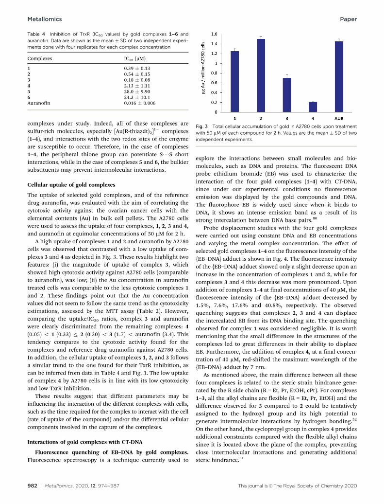
The uptake of selected gold complexes, and of the referencedrug auranofin, was evaluated with the aim of correlating thecytotoxic activity against the ovarian cancer cells with theelemental contents (Au) in bulk cell pellets. The A2780 cellswere used to assess the uptake of four complexes, 1, 2, 3 and 4,and auranofin at equimolar concentrations of 50 mM for 2 h.
A high uptake of complexes 1 and 2 and auranofin by A2780cells was observed that contrasted with a low uptake of com-plexes 3 and 4 as depicted in Fig. 3. These results highlight twofeatures: (i) the magnitude of uptake of complex 3, whichshowed high cytotoxic activity against A2780 cells (comparableto auranofin), was low; (ii) the Au concentration in auranofintreated cells was comparable to the less cytotoxic complexes 1and 2. These findings point out that the Au concentrationvalues did not seem to follow the same trend as the cytotoxicityestimations, assessed by the MTT assay (Table 2). However,comparing the uptake/IC50 ratios, complex 3 and auranofinwere clearly discriminated from the remaining complexes: 4(0.05) o 1 (0.33) r 2 (0.30) o 3 (1.7) o auranofin (3.4). Thistendency compares to the cytotoxic activity found for thecomplexes and reference drug auranofin against A2780 cells.In addition, the cellular uptake of complexes 1, 2, and 3 followsa similar trend to the one found for their TxrR inhibition, ascan be inferred from data in Table 4 and Fig. 3. The low uptakeof complex 4 by A2780 cells is in line with its low cytotoxicityand low TxrR inhibition.
These results suggest that different parameters may beinfluencing the interaction of the different complexes with cells,such as the time required for the complex to interact with the cell(rate of uptake of the compound) and/or the differential cellularcomponents involved in the capture of the complexes.
Interactions of gold complexes with CT-DNA
Fluorescence quenching of EB–DNA by gold complexes.Fluorescence spectroscopy is a technique currently used to
explore the interactions between small molecules and bio-molecules, such as DNA and proteins. The fluorescent DNAprobe ethidium bromide (EB) was used to characterize theinteraction of the four gold complexes (1–4) with CT-DNA,since under our experimental conditions no fluorescenceemission was displayed by the gold compounds and DNA.The fluorophore EB is widely used since when it binds toDNA, it shows an intense emission band as a result of itsstrong intercalation between DNA base pairs.80
Probe displacement studies with the four gold complexeswere carried out using constant DNA and EB concentrationsand varying the metal complex concentration. The effect ofselected gold complexes 1–4 on the fluorescence intensity of the{EB–DNA} adduct is shown in Fig. 4. The fluorescence intensityof the {EB–DNA} adduct showed only a slight decrease upon anincrease in the concentration of complexes 1 and 2, while forcomplexes 3 and 4 this decrease was more pronounced. Uponaddition of complexes 1–4 at final concentrations of 40 mM, thefluorescence intensity of the {EB–DNA} adduct decreased by1.5%, 7.6%, 17.6% and 40.8%, respectively. The observedquenching suggests that complexes 2, 3 and 4 can displacethe intercalated EB from its DNA binding site. The quenchingobserved for complex 1 was considered negligible. It is worthmentioning that the small differences in the structures of thecomplexes led to great differences in their ability to displaceEB. Furthermore, the addition of complex 4, at a final concen-tration of 40 mM, red-shifted the maximum wavelength of the{EB–DNA} adduct by 7 nm.
As mentioned above, the main difference between all thesefour complexes is related to the steric strain hindrance gene-rated by the R side chain (R = Et, Pr, EtOH, cPr). For complexes1–3, all the alkyl chains are flexible (R = Et, Pr, EtOH) and thedifference observed for 3 compared to 2 could be tentativelyassigned to the hydroxyl group and its high potential togenerate intermolecular interactions by hydrogen bonding.52
On the other hand, the cyclopropyl group in complex 4 providesadditional constraints compared with the flexible alkyl chainssince it is located above the plane of the complex, preventingclose intermolecular interactions and generating additionalsteric hindrance.51
Table 4 Inhibition of TrxR (IC50 values) by gold complexes 1–6 andauranofin. Data are shown as the mean � SD of two independent experi-ments done with four replicates for each complex concentration
Complexes IC50 (mM)
1 0.39 � 0.132 0.54 � 0.153 0.18 � 0.084 2.13 � 1.115 28.0 � 9.906 24.3 � 10.1Auranofin 0.016 � 0.006
Fig. 3 Total cellular accumulation of gold in A2780 cells upon treatmentwith 50 mM of each compound for 2 h. Values are the mean � SD of twoindependent experiments.
Metallomics Paper

This journal is©The Royal Society of Chemistry 2020 Metallomics, 2020, 12, 974--987 | 983
Complex–DNA binding constants. The mechanism offluorescence quenching can be described by means of thefollowing Stern–Volmer equation.64
I0
I¼ 1þ Kqt0 Q½ � ¼ 1þ KSV Q½ � (2)
where I0 and I are defined as the emission intensity in theabsence and presence of a quencher, respectively, and [Q] is theconcentration of the quencher. In this study, compounds 2–4were used as a quencher. Kq is the bimolecular quenching rateconstant; t0 is the lifetime of the fluorophore in the absence of
Fig. 4 Fluorescence emission spectra of EB bound to DNA in the absence (black lines) and presence of Au compounds (a) 1; (b) 2; (c) 3 and (d) 4.Experimental conditions: [DNA] = 80 mM; [EB] = 20 mM; [Au] = 0–40 mM; 10 mM Hepes buffer, pH 7.4; incubation at 37 1C for 24 h; lexc = 510 nm.
Fig. 5 Stern–Volmer plots for complexes (a) 2; (b) 3 and (c) 4.
Paper Metallomics

984 | Metallomics, 2020, 12, 974--987 This journal is©The Royal Society of Chemistry 2020
the quencher and KSV is the Stern–Volmer quenching constantwhose values were obtained from the slopes of the plots of I0/Ivs. [Q]. Fig. 5 depicts the plots of I0/I vs. [Q] for the competitionstudies between compounds 2–4 and EB.
The binding constants determined from the Stern–Volmerequation, presented in Table 5, indicate that the complexesstudied have a weak capacity to substitute EB from the[EB–DNA] adduct.
Stability studies in solution by UV-visible spectroscopy. Thestability in solution of complex 1 was assessed by UV-visiblespectroscopy along the time up to 48 h. The spectra of 1 wererecorded in DMSO and in colourless MEM medium in theabsence and presence of fetal bovine serum (FBS). The resultsfrom this study suggested that complex 1 (Fig. 6a–c) is verystable in DMSO, and unstable in MEM medium in the absenceof FBS, after t0, where a slight shift and a decrease in theintensity of the maxima are observed. In the medium with 10%FBS, to simulate the conditions of the cellular studies, the
solutions of 1 remained translucent at least until 48 h, withno visible precipitation. The stability of 1 in the presence ofcellular antioxidants glutathione (GSH) and ascorbate (AsA) wasalso evaluated. Both reducing agents were assayed at 100 mM inthe presence of the gold complex at 1 : 1 molar ratio. The resultspresented in Fig. 6(d and e) suggested that neither GSH norAsA is able to reduce the gold complex, since neither shapemodification nor the maximum shift of the band at 365 nm wasobserved.
Conclusions
The present study reports the screening of 6 gold(III) bis-(dithiolene) complexes for their potential anticancer, anti-microbial and antiplasmodial activities. This is the first reportdealing with this type of complexes as antiplasmodial agents.Although the synthesis and structures of this class of mono-anionic compounds have already been described for theirimportance as precursors of neutral molecular conductorsand magnetic materials, their antiplasmodial activity has notbeen assessed before, and studies on their antimicrobial andanticancer properties are scarce. These monoanionic goldcomplexes present some similarities such as their redox activity,as they can all be oxidized to the neutral species. Thesecomplexes also share the feature of being composed of hetero-atoms like sulfur, which is not common, which are ableto participate in chalcogen interactions. The main structuraldifferences among the complexes under study correspond tothe alkyl groups grafted on their backbones, resulting in dis-tinct steric hindrances and different abilities to establishintermolecular interactions. Another difference relies on thearomatic character of the thiophene dithiolene ligand (tpdt) atvariance with the non-aromatic character of the thiazolinedithiolene ligand (thiazdt).
The cytotoxic properties of this class of gold complexeshave attracted attention only recently. In particular, gold(III)dithiolenes can be considered as an emerging class of metalcomplexes with potential antitumor properties to be used asalternatives to cisplatin due to their exceptional cytotoxicproperties, involving both common and specific mechanisms.In addition, they can reduce the susceptibility to opportunisticmicrobial infections, since they also present antimicrobialactivity. This work also highlights the importance of this typeof complexes as prospective precursors for the development ofantimalarial drugs.
Both experimental and clinical studies support the notionthat the TrxR status has a close relationship to the onset anddevelopment of several diseases including cancer. Therefore,there is an increasing interest in the development of novel TrxRmodulators. The results from this work also highlight therelevance of gold(III) bis-dithiolenes as promising TrxR inhibi-tors. In summary, the gold complexes presented herein exhibitthe ability to overcome resistance in cisplatin-resistant cells,presenting lower cytotoxicity compared to auranofin in non-cancerous cells, and possessing the potential to selectively
Table 5 Stern–Volmer constant (KSV), bimolecular quenching rateconstant (Kq), and correlation coefficient (R) for the quenchers 2, 3 and 4
Compound KSV (�103 L mol�1) Kq (�1011 L mol�1 s�1) R2
2 2.1 � 0.02 2.1 � 0.02 0.99193 4.3 � 0.01 4.3 � 0.01 0.99124 16.8 � 0.02 16.8 � 0.02 0.9904
Fig. 6 Time-dependent UV-vis spectra of complex 1 in DMSO (a), inthe cellular MEM medium (b), in the MEM medium in the presence ofFBS (c), in the MEM medium in the presence of FBS with GSH (d) and in theMEM medium in the presence of FBS with AsA (e) at 0 h (t0), 1 h (t1), 24 h(t24) and 48 h (t48). The concentration of 1 and that of the antioxidantswas 100 mM.
Metallomics Paper

This journal is©The Royal Society of Chemistry 2020 Metallomics, 2020, 12, 974--987 | 985
inhibit thiol-containing enzymes, such as TrxR. Complex 1, inparticular, displays a favourable biological profile that warrantsits further exploitation as a drug lead for the development ofnovel molecules with antitumor, antimicrobial and anti-plasmodial activities as alternatives to current therapeutics.
Conflicts of interest
The authors declare no conflict of interest.
Acknowledgements
We thank the Fundaçao para a Ciencia e Tecnologia (FCT) forthe financial support UIDB/00100/2020 (CQE), PTDC/QUI-QIN/29834/2017 (BD), UID/BIO/04565/2020 (IBB) and PTDC/SAU/INF/29550/2017 (MP), UID/MULTI/04349/2019 (FM). T. S. Moraisthanks FCT for the CEECIND 2017 Initiative for the project CEECIND/00630/2017 (acknowledging FCT, as well as POPH andFSE – European Social Fund).
References
1 P. Kanavos, The rising burden of cancer in the developingworld, Ann. Oncol., 2006, 17, viii15–viii23.
2 A. Urruticoechea, R. Alemany, J. Balart, A. Villanueva,F. Vinals and G. Capella, Recent advances in cancer therapy:an overview, Curr. Pharm. Des., 2010, 16, 3–10.
3 M. Plummer, C. de Martel, J. Vignat, J. Ferlay, F. Bray andS. Franceschi, Global burden of cancers attributable toinfections in 2012: a synthetic analysis, Lancet GlobalHealth, 2016, 4, e609–e616.
4 K. V. Rolston, Infections in Cancer Patients with SolidTumors: A Review, Infect. Dis. Ther., 2017, 6, 69–83.
5 R. J. Fair and Y. Tor, Antibiotics and bacterial resistance inthe 21st century, Perspect. Med. Chem., 2014, 6, 25–64.
6 I. Romero-Canelon and P. J. Sadler, Next-generation metalanticancer complexes: multitargeting via redox modulation,Inorg. Chem., 2013, 52, 12276–12291.
7 K. D. Mjos and C. Orvig, Metallodrugs in medicinal inorganicchemistry, Chem. Rev., 2014, 114, 4540–4563.
8 G. Palermo, A. Magistrato, T. Riedel, T. von Erlach, C. A.Davey, P. J. Dyson and U. Rothlisberger, Fighting cancerwith transition metal complexes: from naked DNA to Proteinand Chromatin Targeting Strategies, ChemMedChem, 2016,11, 1199–1210.
9 T. C. Johnstone, S. M. Alexander, W. Lin and S. J. Lippard,Effects of monofunctional platinum agents on bacterialgrowth: a retrospective study, J. Am. Chem. Soc., 2014, 136,116–118.
10 K. Joyce, S. Saxena, A. Williams, C. Damurjian, N. Auricchio,S. Aluotto, H. Tynan and A. L. Demain, Antimicrobialspectrum of the antitumor agent, cisplatin, J. Antibiot.,2010, 63, 530–532.
11 F. M. Muggia, A. Bonetti, J. D. Hoeschele, M. Rozencweigand S. B. Howell, Platinum antitumor complexes: 50 years
since Barnett Rosenberg’s discovery, J. Clin. Oncol., 2015, 33,4219–4226.
12 S. Dasari and P. B. Tchounwou, Cisplatin in cancer therapy:molecular mechanisms of action, Eur. J. Pharmacol., 2014,740, 364–378.
13 G. Housman, S. Byler, S. Heerboth, K. Lapinska, M. Longacre,N. Snyder and S. Sarkar, Drug resistance in cancer: an over-view, Cancers, 2014, 6, 1769–1792.
14 T. Makovec, Cisplatin and beyond: molecular mechanismsof action and drug resistance development in cancer chemo-therapy, Radiol. Oncol., 2019, 53, 148–158.
15 F. Li, J. G. Collins and F. R. Keene, Ruthenium complexes asantimicrobial agents, Chem. Soc. Rev., 2015, 44, 2529–2542.
16 S. Amer, N. El-Wakiel and H. El-Ghamry, Synthesis, spectral,antitumor and antimicrobial studies on Cu(II) complexes ofpurine and triazole Schiff base derivatives, J. Mol. Struct.,2013, 1049, 326–335.
17 G. Scalese, M. F. Mosquillo, S. Rostan, J. Castiglioni, I. Alho,L. Perez, I. Correia, F. Marques, J. Costa Pessoa andD. Gambino, Heteroleptic oxidovanadium(IV) complexes of2-hydroxynaphtylaldimine and polypyridyl ligands againstTrypanosoma cruzi and prostate cancer cells, J. Inorg. Biochem.,2017, 175, 154–166.
18 M. Selvaganapathy and N. J. Raman, Pharmacological activityof a few transition metal complexes: a short review, Chem. Biol.Ther., 2016, 1, 108.
19 T. Zou, C. T. Lum, C. N. Lok, J. J. Zhang and C. M. Che,Chemical biology of anticancer gold(III) and gold(I) complexes,Chem. Soc. Rev., 2015, 44, 8786–8801.
20 J. C. Lima and L. Rodriguez, Phosphine–gold(I) compoundsas anticancer agents: general description and mechanismsof action, Adv. Anticancer Agents Med. Chem., 2011, 11,921–928.
21 M. Porchia, M. Pellei, M. Marinelli, F. Tisato, F. Del Belloand C. Santini, New insights in Au-NHCs complexes asanticancer agents, Eur. J. Med. Chem., 2018, 146, 709–746.
22 J. A. Lessa, J. C. Guerra, L. F. de Miranda, C. F. Romeiro,J. G. Da Silva, I. C. Mendes, N. L. Speziali, E. M. Souza-Fagundes and H. Beraldo, Gold(I) complexes with thiosemi-carbazones: cytotoxicity against human tumor cell lines andinhibition of thioredoxin reductase activity, J. Inorg. Bio-chem., 2011, 105, 1729–1739.
23 A. Molter, J. Rust, C. W. Lehmann, G. Deepa, P. Chiba andF. Mohr, Synthesis, structures and anti-malaria activity ofsome gold(I) phosphine complexes containing seleno-and thiosemicarbazonato ligands, Dalton Trans., 2011, 40,9810–9820.
24 B. M. Sutton, Gold compounds for rheumatoid arthritis,Gold Bull., 1986, 19, 15–16.
25 M. L. Barrett and G. P. Lewis, Unique properties of aurano-fin as a potential anti-rheumatic drug, Agents Actions, 1986,19, 109–115.
26 W. Fiskus, N. Saba, M. Shen, M. Ghias, J. Liu, S. D. Gupta,L. Chauhan, R. Rao, S. Gunewardena, K. Schorno, C. P. Austin,K. Maddocks, J. Byrd, A. Melnick, P. Huang, A. Wiestner andK. N. Bhalla, Auranofin induces lethal oxidative and
Paper Metallomics

986 | Metallomics, 2020, 12, 974--987 This journal is©The Royal Society of Chemistry 2020
endoplasmic reticulum stress and exerts potent preclinicalactivity against chronic lymphocytic leukemia, Cancer Res.,2014, 74, 2520–2532.
27 C. Roder and M. J. Thomson, Auranofin: repurposing an olddrug for a golden new age, Drugs R&D, 2015, 15, 13–20.
28 I. Landini, A. Lapucci, A. Pratesi, L. Massai, C. Napoli,G. Perrone, P. Pinzani, L. Messori, E. Mini and S. Nobili,Selection and characterization of a human ovariancancer cell line resistant to auranofin, Oncotarget, 2017, 8,96062–96078.
29 T. S. Reddy, D. Pooja, S. H. Priver, R. B. Luwor, N. Mirzadeh,S. Ramesan, S. Ramakrishna, S. Karri, M. Kuncha andS. K. Bhargava, Potent and selective cytotoxic and anti-inflammatory gold(III) compounds containing cyclometa-lated phosphine sulfide ligands, Chem. – Eur. J., 2019, 25,14089–14100.
30 S. Carboni, A. Zucca, S. Stoccoro, L. Maiore, M. Arca, F. Ortu,C. Artner, B. K. Keppler, S. M. Meier-Menches, A. Casini andM. A. Cinellu, New Variations on the Theme of Gold(III)C4N4N Cyclometalated Complexes as Anticancer Agents:Synthesis and Biological Characterization, Inorg. Chem.,2018, 57, 14852–14865.
31 X. Wanga and Z. Guo, Towards the rational design ofplatinum(II) and gold(III) complexes as antitumour agents,Dalton Trans., 2008, 1521–1532.
32 P. I. Maia, V. M. Deflon and U. Abram, Gold(III) complexesin medicinal chemistry, Future Med. Chem., 2014, 6,1515–1536.
33 S. Medici, M. Peana, V. M. Nurchi, J. I. Lachowicz,G. Crisponi and M. A. Zoroddu, Noble metals in medicine:latest advances, Coord. Chem. Rev., 2015, 284, 329–350.
34 C. I. Yeo, K. K. Ooi and E. R. T. Tiekink, Gold-basedmedicine: A paradigm shift in anti-cancer therapy?, Molecules,2018, 23, 1410.
35 A. Casini, R. W. Sun and I. Ott, Medicinal Chemistry of GoldAnticancer Metallodrugs, Met. Ions Life Sci., 2018, 18.
36 B. Bertrand, M. R. M. Williams and M. Bochmann, Gold(III)complexes for antitumor applications: an overview, Chemistry,2018, 4, 11840–11851.
37 M. R. M. Williams, B. Bertrand, D. L. Hughes, Z. A. E.Waller, C. Schmidt, I. Ott, M. O’Connell, M. Searcey andM. Bochmann, Cyclometallated Au(III) dithiocarbamatecomplexes: synthesis, anticancer evaluation and mechanisticstudies, Metallomics, 2018, 10, 1655–1666.
38 S. J. Berners-Price and A. Filipovska, Gold compounds astherapeutic agents for human diseases, Metallomics, 2011,3, 863–873.
39 B. Ð. Glisic and M. Djuran, Gold complexes as antimicrobialagents: an overview of different biological activities inrelation to the oxidation state of the gold ion and the ligandstructure, Dalton Trans., 2014, 43, 5950–5969.
40 J. P. Costa, M. J. F. Pinheiro, S. A. Sousa, A. M. Botelho doRego, F. Marques, M. C. Oliveira, J. H. Leitao, N. P. Mira andM. F. N. N. Carvalho, Antimicrobial activity of silver cam-phorimine complexes against Candida strains, Antibiotics,2019, 8, 144.
41 World Health Organization (2014). Antibiotic Resistance:Global Report on Surveillance 2014, World Health Organi-zation Press, Switzerland.
42 N. C. Schiødt, T. Bjørnholm, K. Bechgaard, J. J. Neumeier,C. Allgeier, C. S. Jacobsen and N. Thorup, Structural,electrical, magnetic, and optical properties of bis-benzene-1,2-dithiolato-Au(IV) crystals, Phys. Rev. B: Condens. MatterMater. Phys., 1996, 53, 1773–1778.
43 D. Belo, H. Alves, E. B. Lopes, M. T. Duarte, V. Gama,R. T. Henriques, M. Almeida, A. Perez-Benıtez, C. Roviraand J. Veciana, Gold complexes with dithiothiophenedigands: A metal based on a neutral molecule, Chem. –Eur. J., 2001, 7, 511–519.
44 Y. Le Gal, T. Roisnel, P. Auban-Senzier, N. Bellec, J. Iniguez,E. Canadell and D. Lorcy, Stable metallic state of a neutralradical single-component conductor at ambient pressure,J. Am. Chem. Soc., 2018, 140, 6998–7004.
45 G. Yzambart, N. Bellec, G. Nasser, O. Jeannin, T. Roisnel,P. Auban-Senzier, J. Iniguez, E. Canadell and D. Lorcy,Anisotropic Chemical Pressure Effects in Single-Component Molecular Metals Based on Thiazole Dithiolateand Diselenolate Gold Complexes, J. Am. Chem. Soc., 2012,134, 17138–17148.
46 D. Belo and M. Almeida, Transition metal complexes basedon thiophene–dithiolene ligands, Coord. Chem. Rev., 2010,254, 1479–1492.
47 A. Pintus, M. C. Aragoni, M. A. Cinellu, L. Maiore, F. Isaia,V. Lippolis, G. Orru, E. Tuveri, A. Zucca and M. Arca,[Au(pyb-H)(mnt)]: A novel gold(III) 1,2-dithiolene cyclometa-lated complex with antimicrobial activity (pyb-H =C-deprotonated 2-benzylpyridine; mnt = 1,2-dicyanoethene-1,2-dithiolate), J. Inorg. Biochem., 2017, 170, 188–194.
48 A. Vlcek, Dithiolenes and non-innocent redox-activeligands, Coord. Chem. Rev., 2010, 254, 1357–1588.
49 S. A. Sousa, J. H. Leitao, R. A. L. Silva, D. Belo, I. C. Santos,J. F. Guerreiro, M. Martins, D. Fontinha, M. Prudencio,M. Almeida, D. Lorcy and F. Marques, On the path to gold:monoanionic Au bisdithiolate complexes with antimicro-bial and antitumor activities, J. Inorg. Biochem., 2020, 202,110904.
50 N. Tenn, N. Bellec, O. Jeannin, L. Piekara-Sady, P. Auban-Senzier, J. Iniguez, E. Canadell and D. Lorcy, A single-component molecular metal based on a thiazole dithiolategold complex, J. Am. Chem. Soc., 2009, 131, 16961–16967.
51 A. Filatre-Furcate, T. Roisnel, M. Fourmigue, O. Jeannin,N. Bellec, P. Auban-Senzier and D. Lorcy, Subtle stericdifferences impact the structural and conducting propertiesof radical gold bis(dithiolene) complexes, Chem. – Eur. J.,2017, 23, 16004–16013.
52 Y. Le Gal, T. Roisnel, P. Auban-Senzier, T. Guizouarn andD. Lorcy, Hydrogen bonding interactions in a single componentmolecular conductor: hydroxyethyl-substituted radical golddithiolene complex, Inorg. Chem., 2014, 53, 8755–8761.
53 M. M. Andrade, R. A. L. Silva, I. C. Santos, E. B. Lopes,S. Rabaça, L. C. J. Pereira, J. T. Coutinho, J. P. Telo,C. Rovira, M. Almeida and D. Belo, Gold and nickel alkyl
Metallomics Paper

This journal is©The Royal Society of Chemistry 2020 Metallomics, 2020, 12, 974--987 | 987
substituted bis-thiophenedithiolene complexes: anionicand neutral forms, Inorg. Chem. Front., 2017, 4, 270–280.
54 J. A. Hendrickson, C. Hu, S. L. Aitken and N. Beyda, Anti-fungal resistance: a concerning trend for the present andfuture, Curr. Infect. Dis. Rep., 2019, 21, 47.
55 G. Fotakis and J. A. Timbrell, In vitro cytotoxicity assays:comparison of LDH, neutral red, MTT and protein assay inhepatoma cell lines following exposure to cadmium chloride,Toxicol. Lett., 2006, 160, 171–177.
56 J. M. S. Cardoso, A. M. Galvao, S. I. Guerreiro, J. H. Leitao,A. C. Suarez and M. F. N. N. Carvalho, Antibacterial activityof silver camphorimine coordination polymers, DaltonTrans., 2016, 45, 7114–7123.
57 M. C. Arendrup, M. Cuenca-Estrella, C. Lass-Florl andW. Hope, EUCAST technical note on the EUCAST definitivedocument EDef 7.2: method for the determination of brothdilution minimum inhibitory concentrations of antifungalagents for yeasts EDef 7.2 (EUCAST-AFST), Clin. Microbiol.Infect., 2012, 18, E246–E247.
58 R. J. W. Lambert and J. Pearson, Susceptibility testing:accurate and reproducible minimum inhibitory concen-tration (MIC) and non-inhibitory concentration (NIC)values, J. Appl. Microbiol., 2000, 88, 784–790.
59 I. H. Ploemen, M. Prudencio, B. G. Douradinha, J. Ramesar,J. Fonager, G. J. van Gemert, A. J. Luty, C. C. Hermsen,R. W. Sauerwein, F. G. Baptista, M. M. Mota, A. P. Waters,I. Que, C. W. Lowik, S. M. Khan, C. J. Janse and B. M.Franke-Fayard, Visualisation and quantitative analysis ofthe rodent malaria liver stage by real time imaging, PLoSOne, 2009, 4, e7881.
60 M. A. Barreiros, T. Pinheiro, M. F. Araujo, M. M. Costa,M. Palha and R. C. da Silva, Quality assurance of X-rayspectrometry for chemical analysis, Spectrochim. Acta,Part B, 2001, 56, 2095–2106.
61 S. R. Gallagher, Quantitation of DNA and RNA with absorp-tion and fluorescence spectroscopy, in Current Protocols inMolecular Biology, ed. F. M. Ausubel, Greene and Wiley-Interscience, New York, 1994.
62 A. Coutinho and M. Prieto, Ribonuclease T1 and alcoholdehydrogenase fluorescence quenching by acrylamide:A laboratory experiment for undergraduate students, J. Chem.Educ., 1993, 70, 425–428.
63 M. Kubista, R. Sjoback, S. Eriksson and B. Albinsson,Experimental correction for the inner-filter effect influorescence-spectra, Analyst, 1994, 119, 417–419.
64 J. R. Lakowicz, Principles of Fluorescence Spectroscopy,Springer Science, New York, 3rd edn, 2006.
65 R. J. LeSuer and W. E. Geiger, Ligand–solvent interactions ina highly reduced metal chelate complex: medium dependenceof the one-electron reduction of the bis(maleonitriledithio-lato)gold dianion, Inorg. Chem., 2009, 48, 10826–10836.
66 Y. Le Gal, N. Bellec, F. Barriere, R. Clerac, M. Fourmigue,V. Dorcet, T. Roisnel and D. Lorcy, A Sulfur rich electronacceptor and its [Fe(Cp*)2]+ charge transfer salt with ferro-magnetic interactions, Dalton Trans., 2013, 42, 16672–16679.
67 L. Aguinagalde, R. Dıez-Martınez, J. Yuste, I. Royo, C. Gil,I. Lasa, M. Martın-Fontecha, N. I. Marın-Ramos, C. Ardanuy,J. Linares, P. Garcıa, E. Garcıa and J. M. Sanchez-Puelles,Auranofin efficacy against MDR Streptococcus pneumoniaeand Staphylococcus aureus infections, J. Antimicrob. Chemother.,2015, 70, 2608–2617.
68 M. B. Harbut, C. Vilcheze, X. Luo, M. E. Hensler, H. Guo,B. Yang, A. K. Chatterjee, V. Nizet, W. R. Jacobs, P. G. Schultz,F. Wang and F. Wang, Auranofin exerts broad-spectrumbactericidal activities by targeting thiol-redox homeostasis,Proc. Natl. Acad. Sci. U. S. A., 2015, 112, 4453–4458.
69 K. C. Hazen, Influence of DMSO on antifungal activityduring susceptibility testing in vitro, Diagn. Microbiol. Infect.Dis., 2013, 75, 60–63.
70 S. Thangamani, M. Maland, H. Mohammad, P. E. Pascuzzi,L. Avramova, C. M. Koehler, T. R. Hazbun and M. N. Seleem,Repurposing approach identifies auranofin with broadspectrum antifungal activity that targets Mia40-Erv1 pathway,Front. Cell. Infect. Microbiol., 2017, 7, 4.
71 M. Prudencio, M. M. Mota and A. M. Mendes, A toolbox tostudy liver stage malaria, Trends Parasitol., 2011, 27, 565–574.
72 F. P. da Cruz, C. Martin, K. Buchholz, M. J. Lafuente-Monasterio, T. Rodrigues, B. Sonnichsen, R. Moreira, F. J.Gamo, M. Marti, M. M. Mota, M. Hannus and M. Prudencio,Drug screen targeted at Plasmodium liver stages identifies apotent multistage antimalarial drug, J. Infect. Dis., 2012,205, 1278–1286.
73 P. A. Stocks, V. Barton, T. Antoine, G. A. Biagini, S. A. Wardand P. M. O’Neill, Novel inhibitors of the Plasmodium falci-parum electron transport chain, Parasitology, 2014, 141, 50–65.
74 G. Camarda, P. Jirawatcharadech, R. S. Priestley, A. Saif,S. March, M. H. L. Wong, S. Leung, A. B. Miller, D. A. Baker,P. Alano, M. J. Paine, S. N. Bhatia, P. M. O’Neill, S. A. Wardand G. A. Biagini, Antimalarial activity of primaquine oper-ates via a two-step biochemical relay, Nat. Commun., 2019,10, 3226.
75 N. Vale, R. Moreira and P. Gomes, Primaquine revisited sixdecades after its discovery, Eur. J. Med. Chem., 2009, 44,937–953.
76 A. Bindoli, M. P. Rigobello, G. Scutari, C. Gabbiani, A. Casiniand L. Messor, Thioredoxin reductase: A target for goldcompounds acting as potential anticancer drugs, Coord.Chem. Rev., 2009, 253, 1692–1707.
77 V. Gandin and A. P. Fernandes, Metal- and semimetal-containing inhibitors of thioredoxin reductase as anticanceragents, Molecules, 2015, 20, 12732–12756.
78 J. Zhang, B. Zhang, X. Li, X. Han, R. Liu and J. Fang, Smallmolecule inhibitors of mammalian thioredoxin reductaseas potential anticancer agents: An update, Med. Res. Rev.,2019, 39, 5–39.
79 A. Holmgren and M. Bjornstedt, Thioredoxin and thioredoxinreductase, Methods Enzymol., 1995, 252, 199–208.
80 J. Olmsted and D. R. Kearns III, Mechanism of ethidiumbromide fluorescence enhancement on binding to nucleicacids, Biochemistry, 1977, 16, 3647–3654.
Paper Metallomics