Embed Size (px)
Citation preview
lable at ScienceDirect
Biomaterials 78 (2016) 27e39
Contents lists avai
Biomaterials
journal homepage: www.elsevier .com/locate/biomater ia ls
Gold-nanorods-siRNA nanoplex for improved photothermal therapyby gene silencing
Bei-Ke Wang a, Xue-Feng Yu b, *, Jia-Hong Wang c, Zhi-Bin Li b, Peng-Hui Li b, e,Huaiyu Wang b, Li Song d, Paul K. Chu e, Chengzhang Li a, **
a The State Key Laboratory Breeding Base of Basic Science of Stomatology, Hubei-MOST & Key, Laboratory of Oral Biomedicine of Ministry of Education,School and Hospital of Stomatology, Wuhan University, Wuhan, 430079, PR Chinab Institute of Biomedicine and Biotechnology, Shenzhen Institutes of Advanced Technology, Chinese Academy of Sciences, Shenzhen, 518055, PR Chinac School of Physics and Technology, Wuhan University, Wuhan, 430072, PR Chinad Department of Stomatology, The Second Affiliated Hospital to Nanchang University, Nanchang, 330006, PR Chinae Department of Physics and Materials Science, City University of Hong Kong, Tat Chee Avenue, Kowloon, Hong Kong, China
a r t i c l e i n f o
Article history:Received 1 August 2015Received in revised form26 October 2015Accepted 12 November 2015Available online 19 November 2015
Keywords:Gold nanorodsPhotothermal therapyRNA interferenceOral cancerBiomedical applications
* Corresponding author.** Corresponding author.
E-mail addresses: [email protected] (X.-F. Yu), lcz56
http://dx.doi.org/10.1016/j.biomaterials.2015.11.0250142-9612/© 2015 Elsevier Ltd. All rights reserved.
a b s t r a c t
Nanomaterials-mediated photothermal therapy (PTT) often suffers from the fundamental cellular de-fense mechanism of heat shock response which leads to therapeutic resistance of cancer cells and re-duces the therapeutic efficacy. Herein, a gold nanorods (GNRs)-siRNA platform with gene silencingcapability is produced to improve the PTT efficiency. After surface modification, the GNRs show theability to deliver siRNA oligos targeting BAG3 which is an efficient gene to block the heat-shock response.The synthesized GNRs-siRNA nanoplex exhibits excellent ability in the delivery of siRNA into cancer cellswith high silencing efficiency which is even better than that of commercial Lipofectamine 2000. Thein vitro and in vivo studies demonstrate the ability of the GNRs-siRNA nanoplex to sensitize the cancercells to PTT under moderate laser irradiation by down-regulating the increased BAG3 expression andenhancing apoptosis. The GNRs-siRNA mediated PTT has large potential in clinical cancer therapy due tothe elimination of therapeutic resistance and enhanced photothermal therapeutic efficacy by means ofgene silencing. It also suggests an efficient platform for gene delivery and controllable gene therapy.
© 2015 Elsevier Ltd. All rights reserved.
1. Introduction
Nanomaterials-mediated photothermal therapy (PTT) by meansof near-infrared (NIR) illumination is an emerging tool in cancertherapy. Owing to small light scattering and absorption fromintrinsic chromophores in tissues, NIR light can penetrate tissueswith sufficient intensity and high spatial precision [1e3]. Therefore,PTT treatment provides an efficient approach to convert photonenergy into cytotoxic heat to destroy cancer cells with high selec-tivity, especially in vital regions where surgery is difficult [1,4,5].Compared with conventional cancer treatment approaches, PTTwith highly localized heat delivery is minimally invasive and rapidand can be combined with chemotherapy and drug/gene delivery.Despite these advantages, the full potential of PTT is still hindered
@163.com (C. Li).
by some challenges. In particular, to achieve sufficient heating forcomplete killing of cancer cells, a high-power laser or large agentconcentration has been suggested [6e8]. However, a high laserpower or agent concentration produces safety risks including un-certain cytotoxicity, unnecessary morbidity due to collateral dam-age, and lack of patient tolerance at a high temperature [9e11]. It isespecially true for some special cancer such as head and neckcancer due to their special anatomic sites, vital biological functions,and cosmetic requirements [12]. Therefore, it is imperative todevelop a general strategy to improve the PTT efficiency without ahigh laser power or large agent amount.
Heat shock response is a mechanism to prevent cancer cellsfrom hyperthermia and has been shown to undermine the thera-peutic efficacy of thermal therapy due to their cytoprotective andantiapoptotic effects, termed as thermoresistance [13e15]. Recentstudies indicate that PTT can trigger the heat shock response incancer cells consequently decreasing the therapeutic efficacy bysuppressing apoptosis [16e19]. Several families of heat shockproteins (such as HSP70) and BAG3 (Bcl-2 associated athanogene
B.-K. Wang et al. / Biomaterials 78 (2016) 27e3928
domain 3, also known as Bis and CAIR) induced by the heat shockresponse exert central cytoprotective effects preventing cell deathand play a major role in the thermoresistance [20e23]. The appli-cation of RNA interference to cancer therapy has recently attractedattention as a promising therapeutic modality [24,25]. By inter-fering with the expression of specific genes, the small-interferingRNA (siRNA) acts as an effective vehicle in RNA interferencethereby suggesting a possible strategy to inhibit the heat shockresponse and renders the cancer cells more susceptible to PTT bysilencing the expression of HSPs or BAG3.
Among the various nano-agents for PTT, gold nanorods (GNRs)with the unique surface plasmon resonance (SPR) bands have beenstudied extensively on account of their strong NIR absorption andhigh photothermal conversion efficiency [26], and PTT mediated byGNRs has been suggested to be a promising treatment for oralcancer [27]. Moreover, possessing a large surface area to volumeratio and being easily and controllably surface functionalized, GNRsare attractive nanocarriers for different types of drugs to cells[28e31], photosensitizers [27,32e35], and small biomolecules[36,37]. The effectiveness of GNRs in a gene delivery system hasbeen demonstrated in vitro and also preliminarily in vivo [38e45]. Ithas further been shown that GNRs can penetrate the blood brainbarrier and silence expression of DARPP-32 in the delivery systemfor specific siRNAs into the neuron cells [46]. Owing to the versa-tility, GNRs can play an important role in a single multifunctionalnanotherapeutic platform for combined PTT and gene delivery.With regard to cancer therapy, it has shown that different kinds ofsynthesized multifunctional GNRs-siRNA nanoplex can be appliedto treat different types of cancer such as breast cancer [38,47e49],pancreatic adenocarcinoma [50], as well as head and neck cancer[51]. In these studies, GNRs-siRNA complexes not only function asnanovectors [52e54] for siRNA and chemotherapy agents delivery[55e57], but also act as photothermal or imaging agents fortheranostic purposes (summarized in Table S1) [55e57].
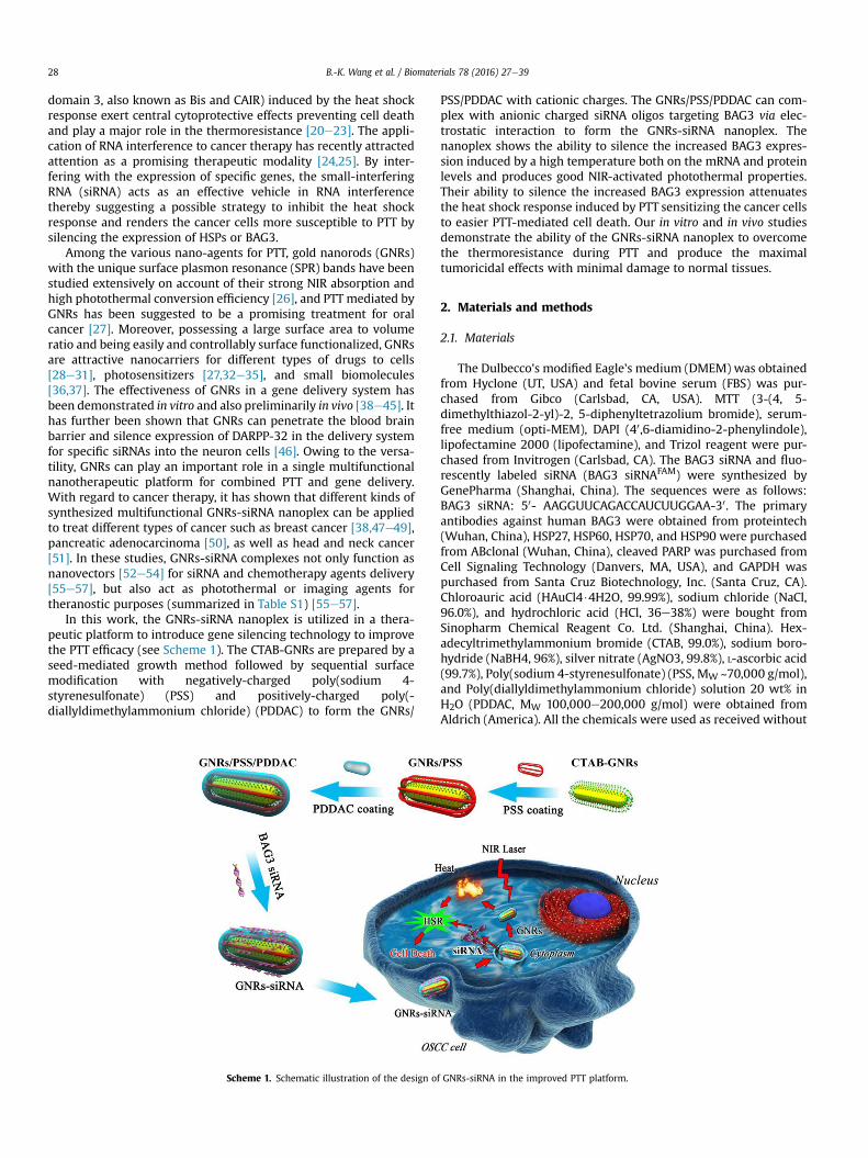
In this work, the GNRs-siRNA nanoplex is utilized in a thera-peutic platform to introduce gene silencing technology to improvethe PTT efficacy (see Scheme 1). The CTAB-GNRs are prepared by aseed-mediated growth method followed by sequential surfacemodification with negatively-charged poly(sodium 4-styrenesulfonate) (PSS) and positively-charged poly(-diallyldimethylammonium chloride) (PDDAC) to form the GNRs/
Scheme 1. Schematic illustration of the design o
PSS/PDDAC with cationic charges. The GNRs/PSS/PDDAC can com-plex with anionic charged siRNA oligos targeting BAG3 via elec-trostatic interaction to form the GNRs-siRNA nanoplex. Thenanoplex shows the ability to silence the increased BAG3 expres-sion induced by a high temperature both on the mRNA and proteinlevels and produces good NIR-activated photothermal properties.Their ability to silence the increased BAG3 expression attenuatesthe heat shock response induced by PTT sensitizing the cancer cellsto easier PTT-mediated cell death. Our in vitro and in vivo studiesdemonstrate the ability of the GNRs-siRNA nanoplex to overcomethe thermoresistance during PTT and produce the maximaltumoricidal effects with minimal damage to normal tissues.
2. Materials and methods
2.1. Materials
The Dulbecco's modified Eagle's medium (DMEM) was obtainedfrom Hyclone (UT, USA) and fetal bovine serum (FBS) was pur-chased from Gibco (Carlsbad, CA, USA). MTT (3-(4, 5-dimethylthiazol-2-yl)-2, 5-diphenyltetrazolium bromide), serum-free medium (opti-MEM), DAPI (40,6-diamidino-2-phenylindole),lipofectamine 2000 (lipofectamine), and Trizol reagent were pur-chased from Invitrogen (Carlsbad, CA). The BAG3 siRNA and fluo-rescently labeled siRNA (BAG3 siRNAFAM) were synthesized byGenePharma (Shanghai, China). The sequences were as follows:BAG3 siRNA: 50- AAGGUUCAGACCAUCUUGGAA-30. The primaryantibodies against human BAG3 were obtained from proteintech(Wuhan, China), HSP27, HSP60, HSP70, and HSP90 were purchasedfrom ABclonal (Wuhan, China), cleaved PARP was purchased fromCell Signaling Technology (Danvers, MA, USA), and GAPDH waspurchased from Santa Cruz Biotechnology, Inc. (Santa Cruz, CA).Chloroauric acid (HAuCl4$4H2O, 99.99%), sodium chloride (NaCl,96.0%), and hydrochloric acid (HCl, 36e38%) were bought fromSinopharm Chemical Reagent Co. Ltd. (Shanghai, China). Hex-adecyltrimethylammonium bromide (CTAB, 99.0%), sodium boro-hydride (NaBH4, 96%), silver nitrate (AgNO3, 99.8%), L-ascorbic acid(99.7%), Poly(sodium 4-styrenesulfonate) (PSS, MW ~70,000 g/mol),and Poly(diallyldimethylammonium chloride) solution 20 wt% inH2O (PDDAC, MW 100,000e200,000 g/mol) were obtained fromAldrich (America). All the chemicals were used as received without
f GNRs-siRNA in the improved PTT platform.
B.-K. Wang et al. / Biomaterials 78 (2016) 27e39 29
purification. Ultrapure water with a resistivity of about18.25 MU cm was used as the solvent in the experiments.
2.2. Synthesis of GNRs-siRNA
The GNRs were synthesized in an aqueous solution using a seed-mediated growth method as previously described [58]. For prepa-ration of the gold seed particles with a size of 3e4 nm, 5 ml of0.5 mM HAuCl4 were mixed with 5 ml of 0.2 M CTAB and 600 ml offreshly prepared ice-cold 10 mM NaBH4 were immediately injectedinto the solution under vigorously stirring. The color changed fromyellow to bright brown. The seed solution was left at least 2 hbefore use. In the GNR synthesis, 1.2 ml of 5 mMHAuCl4 and 15 ml of0.1 M AgNO3 were added to 6 ml of 0.2 M CTAB and then 15 ml of1.2 M HCl and 720 ml of 10 mM ascorbic acid were added and gentlyswirled until the color changed from dark orange to colorless. Af-terwards, 12 ml of the seed solution were added. The resulting so-lution was gently mixed and left undisturbed for 8 h. Finally, theGNRs were collected by centrifugation at 12,000 rpm for 15 min.The supernatant was removed and precipitate was resuspended inultrapurewater. The GNRs concentrationwas estimated to be about0.65 nM. In the synthesis of GNRs-siRNA, GNRs were first coatedwith PSS by the method previously reported [59]. 2 ml of 10 mg/mLPSS dispersed in 1 mMNaCl and 1 ml of 10 mMNaCl were added to10 ml of GNRs and stirred for 1 h at room temperature. A centri-fugation cycle of 10,000 rpm for 10 min was performed to removethe excess PSS and NaCl, obtaining PSS-coated GNRs (GNR/PSS). ThePDDAC was further coated using a similar method. The preparedcationic GNRs/PSS/PDDAC (GNRs) were mixed with differentamounts of siRNAs by pipetting and incubated for 30 min at roomtemperature before the operation to form the nanoplexelectrostatically.
2.3. Characterizations
Transmission electron microscopy (TEM) was performed on aJEOL 2010 (HT) transmission electron microscope at an acceleratingvoltage of 200 kV. The zeta potentials of the samples were deter-mined on a Zeta sizer (Nano ZS90, Malvern Instruments, UK) at25 �C and the absorption spectra were taken on a TU-1810UVeViseNIR spectrophotometer (Purkinje General InstrumentCo. Ltd. Beijing, China).
2.4. Agarose gel electrophoresis
The GNRs-siRNA nanoplexes with different GNRs/siRNA ratioswere prepared by addition of the 1.5 mg BAG3 siRNA to the assignedamounts of 0.65 nM GNRs solution (ranging from 0 to 27 ml) for30min at room temperature. After incubation, the RNase freewaterwas added to the GNR-siRNA nanoplexes with different bindingratios to form the sample solution (30 ml in total). The solutionswere centrifuged and the supernatants were loaded onto 1%agarose gels containing ethidium bromide (0.5 mg/ml) in the1 � TAE buffer (Tris-acetate-EDTA buffer) and electrophoresed at100 V for 20 min. The gel was imaged under UV light on a GelDoc2000 imager system (Bio-Rad, Munich, Germany).
2.5. Photothermal conversion measurement
The photothermal efficiency was measured on a homemadesetup as described previously [27]. A 1 cm quartz cuvette con-taining 2 ml of the sample was covered with a foam cap. Thecuvette was clamped on the top part above the sample surface andthe bottom of the cuvette was kept at approximately 0.5 cm abovethe magnetic stirrer. A fiber-coupled continuous semiconductor
diode laser (810 nm, KS-810F-8000, Kai Site Electronic TechnologyCo., Ltd. Shaanxi, China) with a power density of 2.7 W cm�2 andbeam diameter of approximately 0.5 cm illuminated the cuvette. Adigital thermometer (TX3001, Xintengxing, Wuhan, China) wasused to monitor the temperature change. The head of the ther-mometer was completely submerged in the solution and carefullyprevented from direct illumination by the laser. Each sample in thecuvette was irradiated for 20 min under rigorous stirring and thetemperature was recorded per 30 s.
2.6. Cell culture
The human oral squamous cell carcinoma cell line Cal-27 (CRL-2095, ATCC) was obtained from the Shanghai Research Institute ofStomatology, Affiliated Ninth People's Hospital, Shanghai JiaotongUniversity, Shanghai, China. The cells were maintained in DMEMsupplemented with 10% FBS in a humidified atmosphere of 5% CO2at 37 �C.
2.7. Selection of the moderate laser irradiation power density forPTT
To select the optimal light irradiation conditions for PTT, the cellviability of Cal-27 cells in the presence of GNRs irradiated withdifferent laser power density was assessed by the MTT assay. TheGNRs were diluted in the complete DEME medium to achieve theconcentration of 97.5 pM. 8 � 103 Cal-27 cells/well were seededonto 96-well plates and incubated overnight. Afterwards, the cellswere treated with the prepared GNRs medium for 24 h and irra-diated by the 810 nm laser (VELAS30B-CHEESE, Gigaa OptronicsTechnology Co., Ltd., Wuhan, China) at power densities of 0 J cm�2,200 J cm�2, 400 J cm�2, 600 J cm�2, 800 J cm�2, and 1000 J cm�2.The untreated cells were irradiated with a power density of1000 J cm�2. Afterwards, the cells were incubated in a completemedium up to 24 h. Finally, the cell viability was determined using3-(4,5-dimethylthiazol-2-yl)-2,5-diphenyltetrazolium bromide(MTT, SigmaeAldrich) assay as previously described [60]. The for-mula, (ODtreated/ODcontrol) � 100%, was used to calculate the cellviability and the experiments were conducted in triplicate. Thecells without any treatment serve as the viability control.
2.8. Heat shock response detection
To confirm the occurrence of heat shock response in PTT at amoderate laser power, real-time PCR and western blots were per-formed to determine the expression of HSP27, HSP60, HSP70,HSP90, BAG3 at different time points after the photothermaltreatment. Briefly, the Cal-27 cells were precultured on 24-wellplates at the confluence of 70% and allowed to adhere overnight.The cells were treated with the 97.5 pM GNRs medium and incu-bated for 24 h. After incubation, the treated cells were illuminatedby the 810 nm laser with a power density of 600 J cm�2. The irra-diated cells were incubated in the complete medium and collectedat 0 h, 2 h, 4 h, 8 h, and 12 h for real time PCR and western blots.
2.9. Cell uptake of GNRs-siRNA
According to the fluorescence of BAG3 siRNAFAM (492 nm exci-tation and 518 nm emission), the cellular uptake of GNR-siRNAwasanalyzed using both fluorescence microscopy and flow cytometry.In fluorescence microscopy, the Cal-27 cells were seeded on glasscoverslips at 40e50% confluency on 24-well plates and incubatedovernight before transfection. On the next day, the GNRs (97.5 pM),naked siRNAFAM (0.28 pM), and GNR-siRNAFAM nanoplexes (con-taining 97.5 pM GNRs and 0.28 pM siRNAFAM) were added to the
B.-K. Wang et al. / Biomaterials 78 (2016) 27e3930
complete DMEM medium and incubated with cells for 6 h,respectively. The untreated cells served as the negative control andcells transfected with BAG3 siRNAFAM by lipofectamine 2000(invitrogen) served as the positive control following the manufac-turer‘s instructions. Thereafter, the cells were rinsed twice with1 � cold phosphate buffered saline (PBS) and fixed with 4% para-formaldehyde for 15 min at room temperature. After washing withPBS buffer solution, the cells were mounted using an aqueousmounting medium with DAPI (Invitrogen). The cells were imagedunder the Leica DM4000B fluorescence microscope (Leica, Nus-sloch, Germany). In flow cytometry, the Cal-27 cells were seeded on6-well plates at a density of 5 � 105 cells/ml and incubated understandard conditions. After attachment, the cells were transfectedfor fluorescent microscopy. Six hours after treatment, the cells wereharvested and analyzed on the FACS Calibur flowcytometer (BectonDickinson, Franklin Lakes, NJ) at the excitation wavelength of488 nm and emission wavelength range of 515e545 nm andanalyzed by the Cell Quest Software (Becton Dickinson).
2.10. Evaluation of gene silencing efficiency of BAG3 siRNA viadifferent vectors
Before transfection, the Cal-27 cells were seeded on 24-wellplates at the density of 5 � 104 cells/well and allowed to adhereovernight. 1.875 mg of the BAG3 siRNA oligos were added to onealiquot of the 48.75 fmol GNRs in the eppendorf tubes and incu-bated for 30 min at room temperature to form the GNR-siRNAnanoplexes. The GNRs, siRNA, and prepared GNR-siRNA weregently dipped into the complete DMEM medium making sure thatthe GNRs concentration and siRNA concentration in the final me-dium was 97.5 pM and 0.28 pM, respectively. After aspirating themedia, the cells were treated with the prepared GNRs medium,siRNA medium, and GNRs-siRNA medium for 24 h according to theindicated treatment. The untreated cells were regarded as thecontrol. Lipofectamine 2000 was used as the positive control totransfect BAG3 siRNA and was performed according to the manu-facturer's instruction. Briefly, 3 ml Lipofectamine diluted in 50 mlopti-MEMwere mixed with 5 mg of the siRNA diluted in 50 ml of theopti-MEM. The mixture was incubated at 37 �C for 20 min andadded to the wells producing a final siRNA concentration of0.375 pM for the positive control. Six hours after incubation, themedium was exchanged by the 97.5 pM GNRs medium and incu-bated for 24 h. On the following day, all the groups except controlwere washed by PBS and irradiated by the 810 nm laser with apower density of 600 J cm�2. Eight hours after irradiation, all thecells were harvested for mRNA isolation and protein extraction andthe expressions of BAG3 and GAPDH on the mRNA and proteinlevels were analyzed by real time PCR and western blotting,respectively.
2.11. Analysis of cytotoxicity and apoptosis induced by combinedtreatment of GNRs-siRNA in vitro
The efficacy of the GNR-siRNA in PTT of cancer cells irradiatedwith a moderate-power laser was evaluated by the MTT assay, cellapoptotic detection, and western blotting for cleaved PARP. In theMTT assay, the Cal-27 cells on 96-well plates were either left un-treated or treated with fresh media containing siRNA oligos, GNRs,and GNRs-siRNA at the siRNA concentration of 0.28 pM and GNRsconcentration of 97.5 pM for 24 h. In the positive group, 0.375 pMsiRNA delivered by lipofectamine 2000 was added to the cells,incubated for 6 h at 37 �C, and then was replaced by the 97.5 pMGNRs medium. 810 nm laser irradiation with a power density of600 J cm�2 was performed on most cells at the end of incubation.The cells without treatment were used as a control. After
incubation for 24 h, the cell viability was assessed by the MTT assayas previously described. In analyzing the apoptotic response, thecells were treated in the same manner as in the MTT assay andharvested for apoptosis and necrosis assay and western blotting forcleaved PARP. The apoptosis and necrosis assay was performed aspreviously reported and finally analyzed on a flow cytometer(Becton Dickinson, Franklin Lakes, NJ) using the Cell Quest software(Becton Dickinson) [27].
2.12. Western blots
The harvested cells were lysed in M-PER (Pierce Chemical,Rockford, IL) containing Halt Protease Inhibitor cocktail (Pierce).The denatured protein was collected and the concentration wasdetermined by the BCA Protein Assay Kit (Pierce). Subsequently,20 mg of proteinwere separated using a 10% sodium dodecylsulfate-polyacrylamide gel electrophoresis (SDS-PAGE) and transferredonto polyvinylidene fluoride (PVDF) membranes (Millipore Cor-poration, Billerica, MA). The membranes were blocked by 5% non-fat dry milk for 1 h under room temperature to prevent nonspe-cific binding. The blocked membranes were incubated overnight at4 �C with the primary anti-HSP27 (1:2000, A0240, ABclonal), anti-HSP60 (1:2000, A0969, ABclonal), anti-HSP70 (1:2000, A0284,ABclonal), anti-HSP90 (1:2000, A0365, ABclonal), anti-BAG3(1:2000, 10599-1-AP, proteintech), cleaved PARP (1:1000, #5625,CST), and anti-GAPDH (1:5000, sc-365062, Santa Cruz) antibodies.The immunoblots were probed with Horseradish peroxidase-conjugated goat anti-rabbit or antimouse secondary antibodies(Pierce) for 1 h at room temperature. Finally, the protein werevisualized with a chemiluminescence kit (Pierce) andphotographed.
2.13. RNA extraction and real-time quantitative PCR
The total RNA was isolated from the cells using TRIzol Reagent(Invitrogen). 1 mg total RNA from each sample was reverse tran-scribed to cDNA (20 ml) primed by oligo (dT) using M-MuLV reversetranscriptase (Fermentas, Glen Burnie, MD). The mRNA wasanalyzed quantitatively by real-time PCR. The quantitative PCRreaction was carried out in a 20 ml reaction volume containing 2 mlcDNA as a template, 10 ml 2 � Maxima SYBR Green PCR master mix(Takara, Kyoto, Japan), and 0.8 ml of the primer mix (10 mM forwardprimer, 10 mM reverse primer). All the reactions were conducted intriplicate on an ABI 7500 Real-Time PCR System (Applied Bio-systems, Foster City, CA). The expressionwas normalized to GAPDHwhich was used as the endogenous control in each experiment. Theprimer nucleotide sequences for PCR are presented in Table S2.
2.14. Establishment of tumor xenografts model
The male BALB/c nude mice (4e6 weeks old, 18e20 g, 40 ani-mals) were obtained from Hunan SJA Laboratory Animal Co., Ltd.(Hunan, China). The animals were housed in sterilized cages and a12 h light/dark cycle. All the experiments were approved by theethics committee of Wuhan University and were performed ac-cording to institutional animal use and care regulations.2� 107 Cal-27 cells in 200 ml PBS were subcutaneously injected intothe right flank of mice to initiate tumor growth. The longest andshortest dimension of tumors were detected by a digital calipers.After the longest dimension of the tumor reached 5 mm, the tumorbearing mice were chosen for in vivo treatment.
2.15. Distribution of GNR-siRNA inside the tumor
Twelve mice with established tumors were randomly divided
B.-K. Wang et al. / Biomaterials 78 (2016) 27e39 31
into 4 groups (n ¼ 3 per group): control group, siRNA group, GNRsgroup, GNRs-siRNA group. In the control group, the mouse wasintratumorally injected with sterilized 10 mM PBS solusion. In theother three group, the mice were intratumorally injected with thesiRNAFAM dispersion (15.2 pM siRNAFAM), GNRs dispersion (5.3 nMGNRs), and GNR-siRNAFAM dispersion (containing 5.3 nM GNRsand 15.2 pM siRNAFAM), respectively. The injected volume of eachtype of dispersionwas 200 mL per 50 mm3 tumor volume. After 6 hpost-administration, all the mice were sacrificed and the tumorswere extracted immediately. The ex vivo tumors were subse-quently embedded in the Tissue-Tek OCT (Sakura Finetek), frozenin liquid nitrogen, and cut into sections 5 mm thick on the CM1850UV cryostat (Leica Microsystems). The sections were mountedwith DAPI containing mounting solution (Invitrogen) and imagedby the Leica DM4000B fluorescence microscope (Leica, Nussloch,Germany) at excitationwavelengths of 405 nm (DAPI) and 488 nm(siRNAFAM). The cell uptake of siRNAFAM in vivo was quantified bycalculating the percent of the cells that took up siRNA among thetotal cells in each group.
2.16. In vivo photothermal treatment
After the longest dimension of the inoculated tumors reached~5 mm, the mice were randomized into 5 groups (n ¼ 10 pergroup). The mice in group 1 were kept in the naïve state and servedas the control. The mice in groups 3 and 4 were intratumorallyinjected with the siRNA solution (15.2 pM, 200 mL per 50 mm3
tumor volume) and GNRs solution (5.3 nM, 200 mL per 50 mm3
tumor volume), respectively. The mice in groups 2 and 5 wereintratumorally injected with the prepared GNRs-siRNA solution(200 mL per 50 mm3 tumor volume) at the same concentration ofsiRNA and GNRs as in the group 3 and group 4, respectively. After24 h post-injection, the mice in groups 3, 4, and 5 were irradiatedby the 810 nm near-infrared laser (Gigaa Optronics Technology Co.,Ltd., Wuhan, China) with a power density of 600 J cm�2. Aftewards,the mice were returned to the animal housing and 24 h later, threemice in each group were randomly selected for the BAG3 immu-nohistochemistry and TUNEL assay. The remaining mice were usedto measure the tumor growth volume. The largest and shortesttumor diameters were determined every other day with the digitalcaliper. The formula: V ¼ 1/2 (L � W2) was used to calculate thetumor volume, where L and W were the length (longest diameter)and width (shortest diameter), respectively. The increased tumorpercent (Ti) was calculated according to the formula: Ti (%) ¼ (DP -DC)/DC � 100%, where DC and DP were the tumor volumes beforeand after treatment, respectively. At day 18 after the treatment, themice were euthanatized and the tumors were extracted andphotographed.
2.17. Immunohistochemistry and TUNEL assay
24 h after the treatment, the collected tumors were fixed in 4%paraffin or frozen in liquid nitrogen for subsequent BAG3 immu-nohistochemical analysis and terminal deoxynucleotidyltransferase-mediated dUTP-biotin nick end labeling (TUNEL)assay. In BAG3 immunohistochemistry, the slices were stainedwith the rabbit anti-BAG3 polyclonal antibody (1:1000, Pro-teintech) overnight at 4 �C. A streptavidin-biotin-peroxidasecomplex with diaminobenzidine (DAB) as the peroxidase sub-strate was used to visualize immunoreactivity in accordance withthe manufacturer's instructions (Dako REAL TM Detection System,Peroxidase/DABþ, Rabbit/Mouse). The stained sections werecounterstained with hematoxylin. In the TUNEL assay, the slideswere stained with the TUNEL technique on the in situ cell deathdetection kit (7 sea pharmtech, Shanghai, China) according to the
manufacture's instructions. The fluorescent images were acquiredon the Leica DM4000B fluorescence microscope (Leica, Nussloch,Germany).
2.18. Statistical analysis
All the data were expressed as means ± standard deviationsfrom three independent experiments. The One-Way ANOVA andStudent-Newman-Keuls were used in the statistical analysis withp < 0.05 being considered to be statistically significant.
3. Results and discussion
The CTAB-coated GNRs with an aspect ratio of about 4.2 weresynthesized by a seed-mediated growth method [58]. Negatively-charged PSS was introduced to the surface of the GNRs followedby absorption of positively-charged PDDAC onto the PSS-coatedGNRs (GNRs/PSS) [59]. Afterwards, the positively-charged GNRs/PSS/PDDAC possesses the ability to bind with negatively-chargedBAG3 siRNA electrostatically. The optimal binding ratio of GNRsto BAG3 siRNA is investigated by the agarose gel electrophoresisassay (see Fig. S1).
Fig. 1 shows the characteristics of the products in the GNRs-siRNA synthesis. As shown in Fig. 1a, the original CTAB-GNRsexhibit two SPR bands: a weak transverse SPR (TSPR) band atabout 510 nm and a strong longitudinal SPR (LSPR) band at about808 nm. A slight red-shift is observed from the LSPR band in boththe PSS and PDDAC modification process. Following complexationwith BAG3 siRNA, a further red-shift of about 4 nm in the LSPRband can be observed. It is well known that the LSPR band peakshift from the GNRs results from the local refractive index aroundthe GNRs, which is sensitive to the changes on the surface of theGNRs [61]. Here, the redshift of the LSPR band is probably due tobinding of siRNA with the GNRs/PSS/PDDAC forming GNRs/PSS/PDDAC-siRNA (named GNRs-siRNA). Similar results have beenreported showing that binding of biological molecules can inducethe LSPR band shift from the GNRs [62,63]. In the process, noobvious broadening in the LSPR band can be found thus demon-strating good dispersion of the products. The zeta potentials inFig. 1b present more evidence about successful modification of theGNRs in different stages, i.e. þ43.9 mV (CTAB-GNRs), �33.0 mV(GNRs/PSS), andþ38.0 mV (GNRs/PSS/PDDAC). After incubation ofthe cationic GNRs/PSS/PDDAC substrate with anionic BAG3 siRNA,the zeta potentials reverse from positive (þ38.0 mV) to negative(�24.2 mV), indicating binding of siRNA with GNRs in the finalGNRs-siRNA. The TEM image of GNRs-siRNA in Fig. 1c shows thatthe GNRmorphology is almost unchanged and no aggregation canbe observed, further confirming that the modification andconjugation processes do not affect the dispersion status of theGNRs.
The photothermal conversion ability of the GNRs-siRNA under810 nm laser irradiation is examined by using the original CTAB-GNRs and pure water as the control samples. As shown in Fig. 1d,the temperature of the CTAB-GNRs and GNRs-siRNA increases by38.1 �C and 37.5 �C after NIR light irradiation for 20 min, respec-tively. In contrast, the temperature of pure water only increases by3.4 �C. The results indicate that the GNRs-siRNA retains the goodphotothermal performance of the original GNRs.
The heat shock response in oral cancer cells induced by PTT isinvestigated. The photothermal effect of the GNRs on Cal-27 cells isfirst assessed by the MTT assay (see Fig. S2). The cytotoxic effect ofthe GNRs on cancer cells is hampered at an irradiation powerdensity of 600 J cm�2 (3.3 W cm�2 for 3 min), suggesting that theheat shock response may be induced under these conditions. Theexpression of heat shock response related genes in the GNRs-
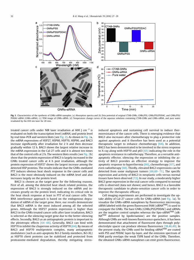
Fig. 1. Characteristics of the synthesis of GNRs-siRNA nanoplex. (a) Absorption spectra and (b) Zeta potential of original CTAB-GNRs, GNRs/PSS, GNRs/PSS/PDDAC, and GNRs/PSS/PDDAC-siRNA (GNRs-siRNA). (c) TEM image of GNRs-siRNA. (d) Temperature change curves of the aqueous solutions containing CTAB-GNRs and GNRs-siRNA, and pure waterirradiated by the 810 nm laser for 20 min.
B.-K. Wang et al. / Biomaterials 78 (2016) 27e3932
treated cancer cells under NIR laser irradiation at 600 J cm�2 isevaluated on both the transcription level (mRNA) and protein levelby real time-PCR and western blots (see Fig. 2). As shown in Fig. 2a,the mRNA expressions of HSP27, HSP60, HSP70, HSP90, and BAG3increase significantly after irradiation for 2 h and then decreasegradually within 12 h. BAG3 shows the largest relative increase inthe mRNA expression in the Cal-27 cells and it is almost ten timesthat of the control cells at 2 h. The western blots results (see Fig. 2b)show that the protein expression of BAG3 is largely increased in theGNRs treated cancer cells at 4 h post irradiation, although theprotein expression of HSP27 shows the largest increase among thedetected HSP proteins. The results indicate that the GNRs-mediatedPTT induces obvious heat shock response in the cancer cells andBAG3 is the most obviously induced on the mRNA level and alsoincreases largely on the protein level.
BAG3 is chosen as the target gene for the following reasons.First of all, among the detected heat shock related proteins, theexpression of BAG3 is strongly induced on the mRNA and in-creases greatly on the protein level, indicating its critical role inthe heat shock response at least in PTT of oral cancer cells. TheRNA interference approach is based on the endogenous degra-dation of mRNA of the target gene. Here, our results demonstratethat BAG3 mRNA is the most induced among all the selectedgenes. Although the HSP27 protein expression is higher thanBAG3 in the GNRs treated cancer cells after laser irradiation, BAG3is selected as the silencing target gene due to the better silencingeffects. Secondly, BAG3 as an antiapoptotic protein is important tothe pleiotropic effects [64e66]. Generally, BAG3 can bind withHSP70 to inhibit the chaperone activity of HSP70 [64]. By means ofBAG3 and HSP70 multiprotein complex, many antiapoptoticmodulators (such as anti-apoptotic Bcl-2 family members, Bcl-XL)as HSP70 client proteins can be stabilized and protected fromproteasome-mediated degradation, thereby mitigating stress-
induced apoptosis and sustaining cell survival to induce ther-moresistance of the cancer cells. There is emerging evidence thatBAG3 also increases after chemotherapy to play a protective roleagainst apoptosis and it therefore has been used as a potentialtherapeutic target to enhance chemotherapy [64]. In addition,BAG3 has been demonstrated to be involved in the stress responseto X-ray along with HSP70 and p63 [65] indicating the role in theapoptosis resistance in radiotherapy. Therefore, as a versatile anti-apoptotic effector, silencing the expression or inhibiting the ac-tivity of BAG3 provides an effective strategy to improve theapoptotic response to hyperthermia [66], chemotherapy [67], andeven radiotherapy [68]. Thirdly, elevated BAG3 expressions can bedetected from some malignant tumors [66,69e71]. The specificexpression and activity of BAG3 in neoplastic cells versus normaltissues have been observed [72]. In our study, a moderately higherBAG3 gene expression in the oral cancer cells compared to normalcells is observed (data not shown) and hence, BAG3 is a favorabletherapeutic candidate to photo-sensitize cancer cells in order toimprove the therapeutic efficacy.
Cell imaging experiments are performed to investigate the up-take ability of Cal-27 cancer cells for GNRs-siRNA (see Fig. 3a). Tovisualize the GNRs-siRNA nanoplexes by fluorescence microscopy,siRNA labeled with the green fluorescent FAM (siRNAFAM) is used toconjugate the GNRs. The bare GNRs (GNRs/PSS/PDDAC) and siRNAsare the negative control samples whereas the lipo-siRNAFAM (siR-NAFAM delivered by lipofectamine) are the positive samples.Although GNRs arewell-known fluorescence quenchers, it has beendemonstrated that attachment of fluorescent dyes through poly-electrolyte spacer can produce obvious fluorescence [38,46,49]. Inthe present study, the GNRs used for binding siRNAFAM are coatedwith PSS and PDDAC layer-by-layer, and the emission spectrum ofFAM partly overlaps the weak TSPR band of the GNRs. Therefore,the obtained GNRs-siRNA nanoplexes can emit green fluorescence.
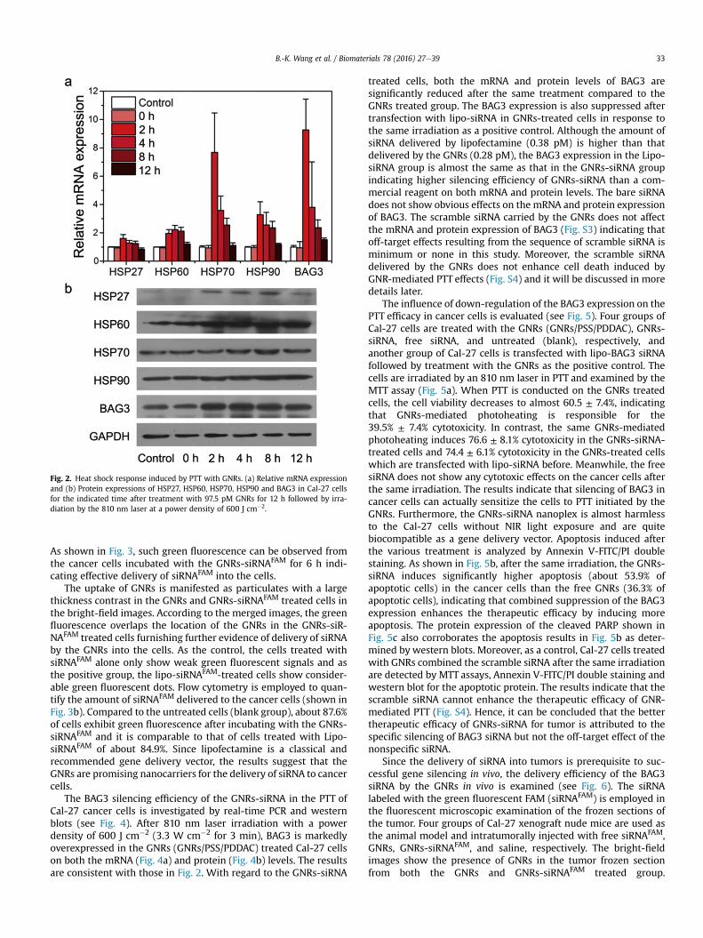
Fig. 2. Heat shock response induced by PTT with GNRs. (a) Relative mRNA expressionand (b) Protein expressions of HSP27, HSP60, HSP70, HSP90 and BAG3 in Cal-27 cellsfor the indicated time after treatment with 97.5 pM GNRs for 12 h followed by irra-diation by the 810 nm laser at a power density of 600 J cm�2.
B.-K. Wang et al. / Biomaterials 78 (2016) 27e39 33
As shown in Fig. 3, such green fluorescence can be observed fromthe cancer cells incubated with the GNRs-siRNAFAM for 6 h indi-cating effective delivery of siRNAFAM into the cells.
The uptake of GNRs is manifested as particulates with a largethickness contrast in the GNRs and GNRs-siRNAFAM treated cells inthe bright-field images. According to the merged images, the greenfluorescence overlaps the location of the GNRs in the GNRs-siR-NAFAM treated cells furnishing further evidence of delivery of siRNAby the GNRs into the cells. As the control, the cells treated withsiRNAFAM alone only show weak green fluorescent signals and asthe positive group, the lipo-siRNAFAM-treated cells show consider-able green fluorescent dots. Flow cytometry is employed to quan-tify the amount of siRNAFAM delivered to the cancer cells (shown inFig. 3b). Compared to the untreated cells (blank group), about 87.6%of cells exhibit green fluorescence after incubating with the GNRs-siRNAFAM and it is comparable to that of cells treated with Lipo-siRNAFAM of about 84.9%. Since lipofectamine is a classical andrecommended gene delivery vector, the results suggest that theGNRs are promising nanocarriers for the delivery of siRNA to cancercells.
The BAG3 silencing efficiency of the GNRs-siRNA in the PTT ofCal-27 cancer cells is investigated by real-time PCR and westernblots (see Fig. 4). After 810 nm laser irradiation with a powerdensity of 600 J cm�2 (3.3 W cm�2 for 3 min), BAG3 is markedlyoverexpressed in the GNRs (GNRs/PSS/PDDAC) treated Cal-27 cellson both the mRNA (Fig. 4a) and protein (Fig. 4b) levels. The resultsare consistent with those in Fig. 2. With regard to the GNRs-siRNA
treated cells, both the mRNA and protein levels of BAG3 aresignificantly reduced after the same treatment compared to theGNRs treated group. The BAG3 expression is also suppressed aftertransfection with lipo-siRNA in GNRs-treated cells in response tothe same irradiation as a positive control. Although the amount ofsiRNA delivered by lipofectamine (0.38 pM) is higher than thatdelivered by the GNRs (0.28 pM), the BAG3 expression in the Lipo-siRNA group is almost the same as that in the GNRs-siRNA groupindicating higher silencing efficiency of GNRs-siRNA than a com-mercial reagent on both mRNA and protein levels. The bare siRNAdoes not show obvious effects on the mRNA and protein expressionof BAG3. The scramble siRNA carried by the GNRs does not affectthe mRNA and protein expression of BAG3 (Fig. S3) indicating thatoff-target effects resulting from the sequence of scramble siRNA isminimum or none in this study. Moreover, the scramble siRNAdelivered by the GNRs does not enhance cell death induced byGNR-mediated PTT effects (Fig. S4) and it will be discussed in moredetails later.
The influence of down-regulation of the BAG3 expression on thePTT efficacy in cancer cells is evaluated (see Fig. 5). Four groups ofCal-27 cells are treated with the GNRs (GNRs/PSS/PDDAC), GNRs-siRNA, free siRNA, and untreated (blank), respectively, andanother group of Cal-27 cells is transfected with lipo-BAG3 siRNAfollowed by treatment with the GNRs as the positive control. Thecells are irradiated by an 810 nm laser in PTT and examined by theMTT assay (Fig. 5a). When PTT is conducted on the GNRs treatedcells, the cell viability decreases to almost 60.5 ± 7.4%, indicatingthat GNRs-mediated photoheating is responsible for the39.5% ± 7.4% cytotoxicity. In contrast, the same GNRs-mediatedphotoheating induces 76.6 ± 8.1% cytotoxicity in the GNRs-siRNA-treated cells and 74.4 ± 6.1% cytotoxicity in the GNRs-treated cellswhich are transfected with lipo-siRNA before. Meanwhile, the freesiRNA does not show any cytotoxic effects on the cancer cells afterthe same irradiation. The results indicate that silencing of BAG3 incancer cells can actually sensitize the cells to PTT initiated by theGNRs. Furthermore, the GNRs-siRNA nanoplex is almost harmlessto the Cal-27 cells without NIR light exposure and are quitebiocompatible as a gene delivery vector. Apoptosis induced afterthe various treatment is analyzed by Annexin V-FITC/PI doublestaining. As shown in Fig. 5b, after the same irradiation, the GNRs-siRNA induces significantly higher apoptosis (about 53.9% ofapoptotic cells) in the cancer cells than the free GNRs (36.3% ofapoptotic cells), indicating that combined suppression of the BAG3expression enhances the therapeutic efficacy by inducing moreapoptosis. The protein expression of the cleaved PARP shown inFig. 5c also corroborates the apoptosis results in Fig. 5b as deter-mined by western blots. Moreover, as a control, Cal-27 cells treatedwith GNRs combined the scramble siRNA after the same irradiationare detected by MTT assays, Annexin V-FITC/PI double staining andwestern blot for the apoptotic protein. The results indicate that thescramble siRNA cannot enhance the therapeutic efficacy of GNR-mediated PTT (Fig. S4). Hence, it can be concluded that the bettertherapeutic efficacy of GNRs-siRNA for tumor is attributed to thespecific silencing of BAG3 siRNA but not the off-target effect of thenonspecific siRNA.
Since the delivery of siRNA into tumors is prerequisite to suc-cessful gene silencing in vivo, the delivery efficiency of the BAG3siRNA by the GNRs in vivo is examined (see Fig. 6). The siRNAlabeled with the green fluorescent FAM (siRNAFAM) is employed inthe fluorescent microscopic examination of the frozen sections ofthe tumor. Four groups of Cal-27 xenograft nude mice are used asthe animal model and intratumorally injected with free siRNAFAM,GNRs, GNRs-siRNAFAM, and saline, respectively. The bright-fieldimages show the presence of GNRs in the tumor frozen sectionfrom both the GNRs and GNRs-siRNAFAM treated group.
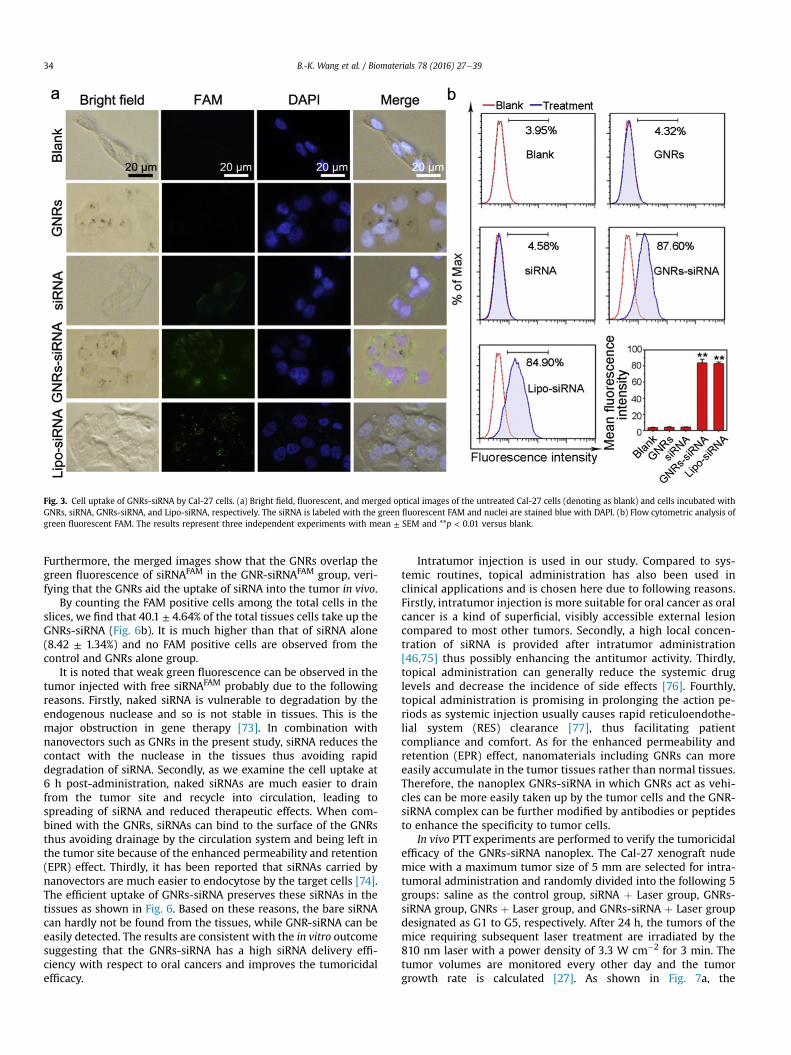
Fig. 3. Cell uptake of GNRs-siRNA by Cal-27 cells. (a) Bright field, fluorescent, and merged optical images of the untreated Cal-27 cells (denoting as blank) and cells incubated withGNRs, siRNA, GNRs-siRNA, and Lipo-siRNA, respectively. The siRNA is labeled with the green fluorescent FAM and nuclei are stained blue with DAPI. (b) Flow cytometric analysis ofgreen fluorescent FAM. The results represent three independent experiments with mean ± SEM and **p < 0.01 versus blank.
B.-K. Wang et al. / Biomaterials 78 (2016) 27e3934
Furthermore, the merged images show that the GNRs overlap thegreen fluorescence of siRNAFAM in the GNR-siRNAFAM group, veri-fying that the GNRs aid the uptake of siRNA into the tumor in vivo.
By counting the FAM positive cells among the total cells in theslices, we find that 40.1 ± 4.64% of the total tissues cells take up theGNRs-siRNA (Fig. 6b). It is much higher than that of siRNA alone(8.42 ± 1.34%) and no FAM positive cells are observed from thecontrol and GNRs alone group.
It is noted that weak green fluorescence can be observed in thetumor injected with free siRNAFAM probably due to the followingreasons. Firstly, naked siRNA is vulnerable to degradation by theendogenous nuclease and so is not stable in tissues. This is themajor obstruction in gene therapy [73]. In combination withnanovectors such as GNRs in the present study, siRNA reduces thecontact with the nuclease in the tissues thus avoiding rapiddegradation of siRNA. Secondly, as we examine the cell uptake at6 h post-administration, naked siRNAs are much easier to drainfrom the tumor site and recycle into circulation, leading tospreading of siRNA and reduced therapeutic effects. When com-bined with the GNRs, siRNAs can bind to the surface of the GNRsthus avoiding drainage by the circulation system and being left inthe tumor site because of the enhanced permeability and retention(EPR) effect. Thirdly, it has been reported that siRNAs carried bynanovectors are much easier to endocytose by the target cells [74].The efficient uptake of GNRs-siRNA preserves these siRNAs in thetissues as shown in Fig. 6. Based on these reasons, the bare siRNAcan hardly not be found from the tissues, while GNR-siRNA can beeasily detected. The results are consistent with the in vitro outcomesuggesting that the GNRs-siRNA has a high siRNA delivery effi-ciency with respect to oral cancers and improves the tumoricidalefficacy.
Intratumor injection is used in our study. Compared to sys-temic routines, topical administration has also been used inclinical applications and is chosen here due to following reasons.Firstly, intratumor injection is more suitable for oral cancer as oralcancer is a kind of superficial, visibly accessible external lesioncompared to most other tumors. Secondly, a high local concen-tration of siRNA is provided after intratumor administration[46,75] thus possibly enhancing the antitumor activity. Thirdly,topical administration can generally reduce the systemic druglevels and decrease the incidence of side effects [76]. Fourthly,topical administration is promising in prolonging the action pe-riods as systemic injection usually causes rapid reticuloendothe-lial system (RES) clearance [77], thus facilitating patientcompliance and comfort. As for the enhanced permeability andretention (EPR) effect, nanomaterials including GNRs can moreeasily accumulate in the tumor tissues rather than normal tissues.Therefore, the nanoplex GNRs-siRNA in which GNRs act as vehi-cles can be more easily taken up by the tumor cells and the GNR-siRNA complex can be further modified by antibodies or peptidesto enhance the specificity to tumor cells.
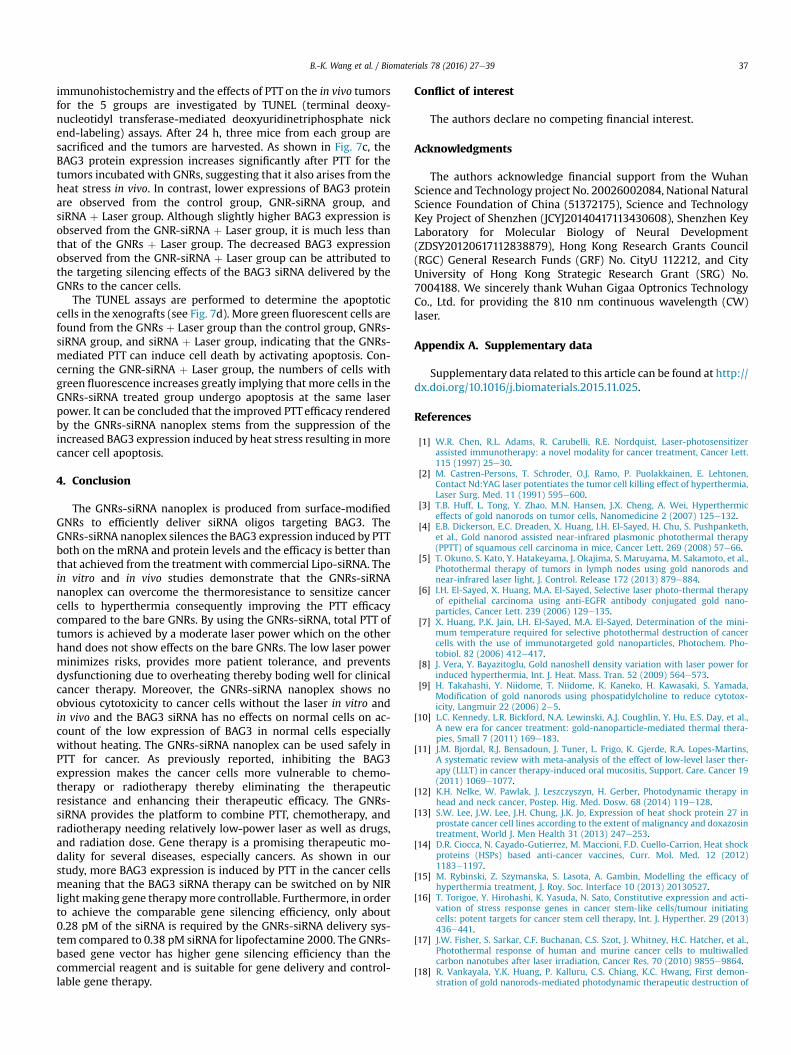
In vivo PTT experiments are performed to verify the tumoricidalefficacy of the GNRs-siRNA nanoplex. The Cal-27 xenograft nudemice with a maximum tumor size of 5 mm are selected for intra-tumoral administration and randomly divided into the following 5groups: saline as the control group, siRNA þ Laser group, GNRs-siRNA group, GNRs þ Laser group, and GNRs-siRNA þ Laser groupdesignated as G1 to G5, respectively. After 24 h, the tumors of themice requiring subsequent laser treatment are irradiated by the810 nm laser with a power density of 3.3 W cm�2 for 3 min. Thetumor volumes are monitored every other day and the tumorgrowth rate is calculated [27]. As shown in Fig. 7a, the
Fig. 4. Expression of BAG3 mRNA in Cal-27 cells treated with GNRs-siRNA after laserirradiation. (a) mRNA and protein expression of BAG3 after indicated treatments (“þ”
indicates with, “�” indicates without). GAPDH is used as the protein loading control.The results represent three independent experiments with mean ± SEM, **p < 0.01versus blank, and ##p < 0.01 versus GNRs þ Laser group.
Fig. 5. Photothermal effect of GNRs-siRNA on Cal-27 cells at an irradiation power of 600 Jcytometry analysis of the Cal-27 cells after the indicated treatment and the corresponding prepresent three independent experiments with mean ± SEM and **p < 0.01 versus blank.
B.-K. Wang et al. / Biomaterials 78 (2016) 27e39 35
siRNA þ Laser group shows no obvious tumor growth inhibitioncompared to the control group, suggesting no obvious toxicity ofthe BAG3 siRNA or laser alone to themice. In the GNRs-siRNA group(without laser), the growth of the tumors decreases slightly afterinjection reaching 79.5% ± 13.0% versus control group, suggestingthat the BAG3 siRNA delivered by the GNRs can suppress tumorgrowth by silencing the BAG3 expression in cancer cells. In theGNRs þ Laser group, the tumor growth is efficiently suppressedafter laser irradiation and the tumor volume is 39.8 ± 10.3% after 18days. However, the tumor resumes growing on the 10th day aftertreatment. It is probably due to the low laser power (600 J cm�2) inthe study. In contrast, in the GNRs-siRNA þ Laser group, thetreatment significantly and effectively inhibits the growth of thetumor xenograft and no regrowth is observed fromG5 until the endof the experiment. The tumor volume is 3.8 ± 2.4% after 18 daysdemonstrating greatly improved PTT efficacy compared to the bareGNRs. At 18 days after treatment, all the mice are sacrificed and thetumors are harvested. Fig. 7b shows the most significant tumorreduction from the GNRs-siRNA þ Laser group (G5) versus tumorsfrom the GNRs þ Laser group (G4). Two out of seven tumors in theGNRs-siRNA group are completely regressed (represented as notumor in the photograph) while the tumors in the GNRs-siRNAgroup (G3) exhibit the slight reduction compared to thesiRNAþ Laser group (G2) and control group (G1). It should be notedthat the laser power is only 600 J cm�2 in our in vivo study.Although this laser power is obviously lower than that used in thepreviously reported GNRs-mediated PTT studies [27,78,79], it isalready sufficient to produce ~97% inhibition in the GNRs-siRNAþ laser group, revealing that the laser power required for theeffective PTT can be reduced tominimize risks rendered by a higherpower laser.
In order to verify the silencing efficacy of GNRs-siRNA in the Cal-27 xenograft models, the expression of BAG3 is determined by
cm�2. (a) Cal-27 cells viability after the treatment. (b) Representative images of flowrotein expression levels of the cleaved PARP in Cal-27 cells are shown in (c). The results
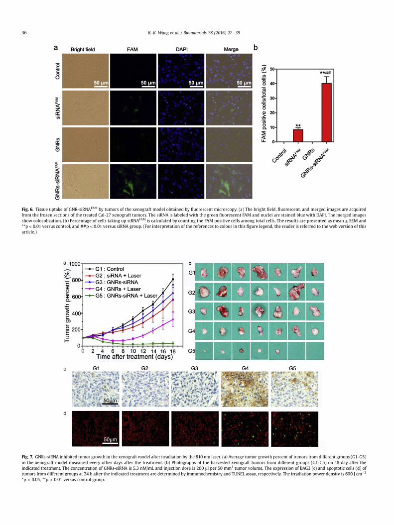
Fig. 6. Tissue uptake of GNR-siRNAFAM by tumors of the xenograft model obtained by fluorescent microscopy. (a) The bright field, fluorescent, and merged images are acquiredfrom the frozen sections of the treated Cal-27 xenograft tumors. The siRNA is labeled with the green fluorescent FAM and nuclei are stained blue with DAPI. The merged imagesshow colocolization. (b) Percentage of cells taking up siRNAFAM is calculated by counting the FAM positive cells among total cells. The results are presented as mean ± SEM and**p < 0.01 versus control, and ##p < 0.01 versus siRNA group. (For interpretation of the references to colour in this figure legend, the reader is referred to the web version of thisarticle.)
Fig. 7. GNRs-siRNA inhibited tumor growth in the xenograft model after irradiation by the 810 nm laser. (a) Average tumor growth percent of tumors from different groups (G1-G5)in the xenograft model measured every other days after the treatment. (b) Photographs of the harvested xenograft tumors from different groups (G1-G5) on 18 day after theindicated treatment. The concentration of GNRs-siRNA is 5.3 nM/mL and injection dose is 200 ml per 50 mm3 tumor volume. The expression of BAG3 (c) and apoptotic cells (d) oftumors from different groups at 24 h after the indicated treatment are determined by immunochemistry and TUNEL assay, respectively. The irradiation power density is 600 J cm�2
*p < 0.05, **p < 0.01 versus control group.
B.-K. Wang et al. / Biomaterials 78 (2016) 27e3936
B.-K. Wang et al. / Biomaterials 78 (2016) 27e39 37
immunohistochemistry and the effects of PTT on the in vivo tumorsfor the 5 groups are investigated by TUNEL (terminal deoxy-nucleotidyl transferase-mediated deoxyuridinetriphosphate nickend-labeling) assays. After 24 h, three mice from each group aresacrificed and the tumors are harvested. As shown in Fig. 7c, theBAG3 protein expression increases significantly after PTT for thetumors incubated with GNRs, suggesting that it also arises from theheat stress in vivo. In contrast, lower expressions of BAG3 proteinare observed from the control group, GNR-siRNA group, andsiRNA þ Laser group. Although slightly higher BAG3 expression isobserved from the GNR-siRNA þ Laser group, it is much less thanthat of the GNRs þ Laser group. The decreased BAG3 expressionobserved from the GNR-siRNA þ Laser group can be attributed tothe targeting silencing effects of the BAG3 siRNA delivered by theGNRs to the cancer cells.
The TUNEL assays are performed to determine the apoptoticcells in the xenografts (see Fig. 7d). More green fluorescent cells arefound from the GNRs þ Laser group than the control group, GNRs-siRNA group, and siRNA þ Laser group, indicating that the GNRs-mediated PTT can induce cell death by activating apoptosis. Con-cerning the GNR-siRNA þ Laser group, the numbers of cells withgreen fluorescence increases greatly implying that more cells in theGNRs-siRNA treated group undergo apoptosis at the same laserpower. It can be concluded that the improved PTT efficacy renderedby the GNRs-siRNA nanoplex stems from the suppression of theincreased BAG3 expression induced by heat stress resulting in morecancer cell apoptosis.
4. Conclusion
The GNRs-siRNA nanoplex is produced from surface-modifiedGNRs to efficiently deliver siRNA oligos targeting BAG3. TheGNRs-siRNA nanoplex silences the BAG3 expression induced by PTTboth on the mRNA and protein levels and the efficacy is better thanthat achieved from the treatment with commercial Lipo-siRNA. Thein vitro and in vivo studies demonstrate that the GNRs-siRNAnanoplex can overcome the thermoresistance to sensitize cancercells to hyperthermia consequently improving the PTT efficacycompared to the bare GNRs. By using the GNRs-siRNA, total PTT oftumors is achieved by a moderate laser power which on the otherhand does not show effects on the bare GNRs. The low laser powerminimizes risks, provides more patient tolerance, and preventsdysfunctioning due to overheating thereby boding well for clinicalcancer therapy. Moreover, the GNRs-siRNA nanoplex shows noobvious cytotoxicity to cancer cells without the laser in vitro andin vivo and the BAG3 siRNA has no effects on normal cells on ac-count of the low expression of BAG3 in normal cells especiallywithout heating. The GNRs-siRNA nanoplex can be used safely inPTT for cancer. As previously reported, inhibiting the BAG3expression makes the cancer cells more vulnerable to chemo-therapy or radiotherapy thereby eliminating the therapeuticresistance and enhancing their therapeutic efficacy. The GNRs-siRNA provides the platform to combine PTT, chemotherapy, andradiotherapy needing relatively low-power laser as well as drugs,and radiation dose. Gene therapy is a promising therapeutic mo-dality for several diseases, especially cancers. As shown in ourstudy, more BAG3 expression is induced by PTT in the cancer cellsmeaning that the BAG3 siRNA therapy can be switched on by NIRlight making gene therapymore controllable. Furthermore, in orderto achieve the comparable gene silencing efficiency, only about0.28 pM of the siRNA is required by the GNRs-siRNA delivery sys-tem compared to 0.38 pM siRNA for lipofectamine 2000. The GNRs-based gene vector has higher gene silencing efficiency than thecommercial reagent and is suitable for gene delivery and control-lable gene therapy.
Conflict of interest
The authors declare no competing financial interest.
Acknowledgments
The authors acknowledge financial support from the WuhanScience and Technology project No. 20026002084, National NaturalScience Foundation of China (51372175), Science and TechnologyKey Project of Shenzhen (JCYJ20140417113430608), Shenzhen KeyLaboratory for Molecular Biology of Neural Development(ZDSY20120617112838879), Hong Kong Research Grants Council(RGC) General Research Funds (GRF) No. CityU 112212, and CityUniversity of Hong Kong Strategic Research Grant (SRG) No.7004188. We sincerely thank Wuhan Gigaa Optronics TechnologyCo., Ltd. for providing the 810 nm continuous wavelength (CW)laser.
Appendix A. Supplementary data
Supplementary data related to this article can be found at http://dx.doi.org/10.1016/j.biomaterials.2015.11.025.
References
[1] W.R. Chen, R.L. Adams, R. Carubelli, R.E. Nordquist, Laser-photosensitizerassisted immunotherapy: a novel modality for cancer treatment, Cancer Lett.115 (1997) 25e30.
[2] M. Castren-Persons, T. Schroder, O.J. Ramo, P. Puolakkainen, E. Lehtonen,Contact Nd:YAG laser potentiates the tumor cell killing effect of hyperthermia,Laser Surg. Med. 11 (1991) 595e600.
[3] T.B. Huff, L. Tong, Y. Zhao, M.N. Hansen, J.X. Cheng, A. Wei, Hyperthermiceffects of gold nanorods on tumor cells, Nanomedicine 2 (2007) 125e132.
[4] E.B. Dickerson, E.C. Dreaden, X. Huang, I.H. El-Sayed, H. Chu, S. Pushpanketh,et al., Gold nanorod assisted near-infrared plasmonic photothermal therapy(PPTT) of squamous cell carcinoma in mice, Cancer Lett. 269 (2008) 57e66.
[5] T. Okuno, S. Kato, Y. Hatakeyama, J. Okajima, S. Maruyama, M. Sakamoto, et al.,Photothermal therapy of tumors in lymph nodes using gold nanorods andnear-infrared laser light, J. Control. Release 172 (2013) 879e884.
[6] I.H. El-Sayed, X. Huang, M.A. El-Sayed, Selective laser photo-thermal therapyof epithelial carcinoma using anti-EGFR antibody conjugated gold nano-particles, Cancer Lett. 239 (2006) 129e135.
[7] X. Huang, P.K. Jain, I.H. El-Sayed, M.A. El-Sayed, Determination of the mini-mum temperature required for selective photothermal destruction of cancercells with the use of immunotargeted gold nanoparticles, Photochem. Pho-tobiol. 82 (2006) 412e417.
[8] J. Vera, Y. Bayazitoglu, Gold nanoshell density variation with laser power forinduced hyperthermia, Int. J. Heat. Mass. Tran. 52 (2009) 564e573.
[9] H. Takahashi, Y. Niidome, T. Niidome, K. Kaneko, H. Kawasaki, S. Yamada,Modification of gold nanorods using phospatidylcholine to reduce cytotox-icity, Langmuir 22 (2006) 2e5.
[10] L.C. Kennedy, L.R. Bickford, N.A. Lewinski, A.J. Coughlin, Y. Hu, E.S. Day, et al.,A new era for cancer treatment: gold-nanoparticle-mediated thermal thera-pies, Small 7 (2011) 169e183.
[11] J.M. Bjordal, R.J. Bensadoun, J. Tuner, L. Frigo, K. Gjerde, R.A. Lopes-Martins,A systematic review with meta-analysis of the effect of low-level laser ther-apy (LLLT) in cancer therapy-induced oral mucositis, Support. Care. Cancer 19(2011) 1069e1077.
[12] K.H. Nelke, W. Pawlak, J. Leszczyszyn, H. Gerber, Photodynamic therapy inhead and neck cancer, Postep. Hig. Med. Dosw. 68 (2014) 119e128.
[13] S.W. Lee, J.W. Lee, J.H. Chung, J.K. Jo, Expression of heat shock protein 27 inprostate cancer cell lines according to the extent of malignancy and doxazosintreatment, World J. Men Health 31 (2013) 247e253.
[14] D.R. Ciocca, N. Cayado-Gutierrez, M. Maccioni, F.D. Cuello-Carrion, Heat shockproteins (HSPs) based anti-cancer vaccines, Curr. Mol. Med. 12 (2012)1183e1197.
[15] M. Rybinski, Z. Szymanska, S. Lasota, A. Gambin, Modelling the efficacy ofhyperthermia treatment, J. Roy. Soc. Interface 10 (2013) 20130527.
[16] T. Torigoe, Y. Hirohashi, K. Yasuda, N. Sato, Constitutive expression and acti-vation of stress response genes in cancer stem-like cells/tumour initiatingcells: potent targets for cancer stem cell therapy, Int. J. Hyperther. 29 (2013)436e441.
[17] J.W. Fisher, S. Sarkar, C.F. Buchanan, C.S. Szot, J. Whitney, H.C. Hatcher, et al.,Photothermal response of human and murine cancer cells to multiwalledcarbon nanotubes after laser irradiation, Cancer Res. 70 (2010) 9855e9864.
[18] R. Vankayala, Y.K. Huang, P. Kalluru, C.S. Chiang, K.C. Hwang, First demon-stration of gold nanorods-mediated photodynamic therapeutic destruction of
B.-K. Wang et al. / Biomaterials 78 (2016) 27e3938
tumors via near infra-red light activation, Small 10 (2014) 1612e1622.[19] R. Vankayala, C.C. Lin, P. Kalluru, C.S. Chiang, K.C. Hwang, Gold nanoshells-
mediated bimodal photodynamic and photothermal cancer treatment usingultra-low doses of near infra-red light, Biomaterials 35 (2014) 5527e5538.
[20] A. Rosati, V. Graziano, V. De Laurenzi, M. Pascale, M.C. Turco, BAG3: a multi-faceted protein that regulates major cell pathways, Cell Death Dis. 2 (2011)e141.
[21] D.A. Parsell, S. Lindquist, The function of heat-shock proteins in stress toler-ance: degradation and reactivation of damaged proteins, Annu. Rev. Genet. 27(1993) 437e496.
[22] A. Rosati, M. Ammirante, A. Gentilella, A. Basile, M. Festa, M. Pascale, et al.,Apoptosis inhibition in cancer cells: a novel molecular pathway that involvesBAG3 protein, Int. J. Biochem. Cell B 39 (2007) 1337e1342.
[23] D.D. Mosser, R.I. Morimoto, Molecular chaperones and the stress of onco-genesis, Oncogene 23 (2004) 2907e2918.
[24] A. Ambesajir, A. Kaushik, J.J. Kaushik, S.T. Petros, RNA interference: a futuristictool and its therapeutic applications, Saudi J. Bio. Sci. 19 (2012) 395e403.
[25] L. Aagaard, J.J. Rossi, RNAi therapeutics: principles, prospects and challenges,Adv. Drug Deliv. Rev. 59 (2007) 75e86.
[26] E. Hutter, J.H. Fendler, Exploitation of localized surface plasmon resonance,Adv. Mater. 16 (2004) 1685e1706.
[27] B. Wang, J.H. Wang, Q. Liu, H. Huang, M. Chen, K. Li, et al., Rose-bengal-con-jugated gold nanorods for in vivo photodynamic and photothermal oralcancer therapies, Biomaterials 35 (2014) 1954e1966.
[28] T.S. Hauck, T.L. Jennings, T. Yatsenko, J.C. Kumaradas, W.C.W. Chan, Enhancingthe toxicity of cancer chemotherapeutics with gold nanorod hyperthermia,Adv. Mater. 20 (2008) 3832e3838.
[29] P. Huang, L. Bao, C. Zhang, J. Lin, T. Luo, D. Yang, et al., Folic acid-conjugatedsilica-modified gold nanorods for X-ray/CT imaging-guided dual-mode radi-ation and photo-thermal therapy, Biomaterials 32 (2011) 9796e9809.
[30] S. Shen, H. Tang, X. Zhang, J. Ren, Z. Pang, D. Wang, et al., Targeting meso-porous silica-encapsulated gold nanorods for chemo-photothermal therapywith near-infrared radiation, Biomaterials 34 (2013) 3150e3158.
[31] Y. Xiao, H. Hong, V.Z. Matson, A. Javadi, W. Xu, Y. Yang, et al., Gold nanorodsconjugated with doxorubicin and cRGD for combined anticancer drug deliveryand PET imaging, Theranostics 2 (2012) 2757e2768.
[32] S.B. Kim, T.H. Lee, I. Yoon, Y.K. Shim, W.K. Lee, Gold nanorod-photosensitizercomplex obtained by layer-by-layer method for photodynamic/photothermaltherapy in vitro, Chem. Asian J. 10 (2015) 563e567.
[33] Y.K. Xu, R.Y. He, D.D. Lin, M.B. Ji, J.Y. Chen, Laser beam controlled drug releasefrom Ce6-Gold nanorod composites in living cells: a FLIM Study, Nanoscale 7(2015) 2433e2441.
[34] N.N. Wang, Z.L. Zhao, Y.F. Lv, H.H. Fan, H.R. Bai, H.M. Meng, et al., Goldnanorod-photosensitizer conjugate with extracellular pH-driven tumor tar-geting ability for photothermal/photodynamic therapy, Nano Res. 7 (2014)1291e1301.
[35] Z.Z. Shi, W.Z. Ren, A. Gong, X.M. Zhao, Y.H. Zou, E.M.B. Brown, et al., Stabilityenhanced polyelectrolyte-coated gold nanorod-photosensitizer complexes forhigh/low power density photodynamic therapy, Biomaterials 35 (2014)7058e7067.
[36] C.K. Kim, P. Ghosh, C. Pagliuca, Z.J. Zhu, S. Menichetti, V.M. Rotello, Entrap-ment of hydrophobic drugs in nanoparticle monolayers with efficient releaseinto cancer cells, J. Am. Chem. Soc. 131 (2009) 1360e1361.
[37] X.J. Liu, I. Hamilton, Adsorption of small molecules on helical gold nanorods: arelativistic density functional study, J. Comput. Chem. 35 (2014) 1967e1976.
[38] J.L. Shen, H.C. Kim, C.F. Mu, E. Gentile, J.H. Mai, J. Wolfram, et al., Multifunc-tional gold nanorods for siRNA gene silencing and photothermal therapy, Adv.Healthc. Mater. 3 (2014) 1629e1637.
[39] A.K. Salem, P.C. Searson, K.W. Leong, Multifunctional nanorods for gene de-livery, Nat. Mater. 2 (2003) 668e671.
[40] C.C. Chen, Y.P. Lin, C.W. Wang, H.C. Tzeng, C.H. Wu, Y.C. Chen, et al., DNA-goldnanorod conjugates for remote control of localized gene expression by nearinfrared irradiation, J. Am. Chem. Soc. 128 (2006) 3709e3715.
[41] H.C. Huang, S. Barua, D.B. Kay, K. Rege, Simultaneous enhancement of pho-tothermal stability and gene delivery efficacy of gold nanorods using poly-electrolytes, ACS. Nano 4 (2010), 1769e1752.
[42] K.V. Chakravarthy, A.C. Bonoiu, W.G. Davis, P. Ranjan, H. Ding, R. Hu, et al.,Gold nanorod delivery of an ssRNA immune activator inhibits pandemic H1N1influenza viral replication, Proc. Natl. Acad. Sci. U. S. A. 107 (2010)10172e10177.
[43] A.M. Alkilany, L.B. Thompson, S.P. Boulos, P.N. Sisco, C.J. Murphy, Goldnanorods: their potential for photothermal therapeutics and drug delivery,tempered by the complexity of their biological interactions, Adv. Drug Deliv.Rev. 64 (2012) 190e199.
[44] L.G. Xu, Y. Liu, Z.Y. Chen, W. Li, Y. Liu, L.M. Wang, et al., Surface-engineeredgold nanorods: promising DNA vaccine adjuvant for HIV-1 treatment, NanoLett. 12 (2012) 2003e2012.
[45] L.M. Wang, X.Y. Lin, J. Wang, Z.J. Hu, Y.L. Ji, S. Hou, et al., Novel insights intocombating cancer chemotherapy resistance using a plasmonic nanocarrier:enhancing drug sensitiveness and accumulation simultaneously with local-ized mild photothermal stimulus of femtosecond pulsed laser, Adv. Funct.Mater. 24 (2014) 4229e4239.
[46] A.C. Bonoiu, S.D. Mahajan, H. Ding, I. Roy, K.T. Yong, R. Kumar, et al., Nano-technology approach for drug addiction therapy: gene silencing using deliveryof gold nanorod-siRNA nanoplex in dopaminergic neurons, Proc. Natl. Acad.
Sci. U. S. A. 106 (2009) 5546e5550.[47] J.H. Choi, H.J. Hwang, S.W. Shin, J.W. Choi, S.H. Um, B.K. Oh, A novel albumin
nanocomplex containing both small interfering RNA and gold nanorods forsynergetic anticancer therapy, Nanoscale 7 (2015) 9229e9237.
[48] Z. Yang, T. Liu, Y. Xie, Z. Sun, H. Liu, J. Lin, et al., Chitosan layered gold nanorodsas synergistic therapeutics for photothermal ablation and gene silencing intriple-negative breast cancer, Acta Biomater. 25 (2015) 194e204.
[49] W. Zhang, J. Meng, Y. Ji, X. Li, H. Kong, X. Wu, et al., Inhibiting metastasis ofbreast cancer cells in vitro using gold nanorod-siRNA delivery system,Nanoscale 3 (2011) 3923e3932.
[50] F. Yin, C. Yang, Q. Wang, S. Zeng, R. Hu, G. Lin, et al., A light-driven therapy ofpancreatic adenocarcinoma using gold nanorods-based nanocarriers for co-delivery of doxorubicin and siRNA, Theranostics 5 (2015) 818e833.
[51] R. Masood, I. Roy, S. Zu, C. Hochstim, K.T. Yong, W.C. Law, et al., Gold nanorod-sphingosine kinase siRNA nanocomplexes: a novel therapeutic tool for potentradiosensitization of head and neck cancer, Integr. Biol. Camb. 4 (2012)132e141.
[52] Y. Xiao, R. Jaskula-Sztul, A. Javadi, W. Xu, J. Eide, A. Dammalapati, et al., Co-delivery of doxorubicin and siRNA using octreotide-conjugated gold nanorodsfor targeted neuroendocrine cancer therapy, Nanoscale 4 (2012) 7185e7193.
[53] Y.T. Chang, P.Y. Liao, H.S. Sheu, Y.J. Tseng, F.Y. Cheng, C.S. Yeh, Near-infraredlight-responsive intracellular drug and siRNA release using au nanoensembleswith oligonucleotide-capped silica shell, Adv. Mater. 24 (2012) 3309e3314.
[54] D. Cui, P. Huang, C. Zhang, C.S. Ozkan, B. Pan, P. Xu, Dendrimer-modified goldnanorods as efficient controlled gene delivery system under near-infraredlight irradiation, J. Control Release. 152 (Suppl 1) (2011) e137e9.
[55] A. Taruttis, N. Lozano, A. Nunes, D.A. Jasim, N. Beziere, E. Herzog, et al., siRNAliposome-gold nanorod vectors for multispectral optoacoustic tomographytheranostics, Nanoscale 6 (2014) 13451e13456.
[56] J.H. Choi, B.K. Oh, Development of two-component nanorod complex for dual-fluorescence imaging and siRNA delivery, J. Microbiol. Biotechnol. 24 (2014)1291e1299.
[57] J. Ramos, K. Rege, Poly(aminoether)-gold nanorod assemblies for shRNAplasmid-induced gene silencing, Mol. Pharm. 10 (2013) 4107e4119.
[58] B. Nikoobakht, M.A. El-Sayed, Preparation and growth mechanism of goldnanorods (NRs) using seed-mediated growth method, Chem. Mater. 15 (2003)1957e1962.
[59] A. Gole, C.J. Murphy, Polyelectrolyte-coated gold nanorods: synthesis, char-acterization and immobilization, Chem. Mater. 17 (2005) 1325e1330.
[60] J.H. Wang, B. Wang, Q. Liu, Q. Li, H. Huang, L. Song, et al., Bimodal opticaldiagnostics of oral cancer based on rose bengal conjugated gold nanorodplatform, Biomaterials 34 (2013) 4274e4283.
[61] X.C. Gou, J. Liu, H.L. Zhang, Monitoring human telomere DNA hybridizationand G-quadruplex formation using gold nanorods, Anal. Chim. Acta 668(2010) 208e214.
[62] W. Zhang, Y. Ji, J. Meng, X. Wu, H. Xu, Probing the behaviors of gold nanorodsin metastatic breast cancer cells based on UV-vis-NIR absorption spectros-copy, PLoS One 7 (2012) e31957.
[63] K.H. Park, S. Kim, S.M. Yang, H.G. Park, Detection of DNA immobilization andhybridization on gold/silver nanostructures using localized surface plasmonresonance, J. Nanosci. Nanotechnol. 9 (2009) 1374e1378.
[64] M. Ammirante, A. Rosati, C. Arra, A. Basile, A. Falco, M. Festa, et al., IKK{gamma} protein is a target of BAG3 regulatory activity in human tumorgrowth, Proc. Natl. Acad. Sci. U. S. A. 107 (2010) 7497e7502.
[65] R. Bonaventura, F. Zito, C. Costa, S. Giarrusso, F. Celi, V. Matranga, Stressresponse gene activation protects sea urchin embryos exposed to X-rays, CellStress Chaperon 16 (2011) 681e687.
[66] T. Yunoki, A. Kariya, T. Kondo, A. Hayashi, Y. Tabuchi, The combination ofsilencing BAG3 and inhibition of the JNK pathway enhances hyperthermiasensitivity in human oral squamous cell carcinoma cells, Cancer Lett. 335(2013) 52e57.
[67] J.A. Kim, Y. Kim, B.M. Kwon, D.C. Han, The natural compound cantharidininduces cancer cell death through inhibition of heat shock protein 70 (HSP70)and Bcl-2-associated athanogene domain 3 (BAG3) expression by blockingheat shock factor 1 (HSF1) binding to promoters, J. Biol. Chem. 288 (2013)28713e28726.
[68] R. Cotugno, A. Basile, E. Romano, D. Gallotta, M.A. Belisario, BAG3 down-modulation sensitizes HPV18(þ) HeLa cells to PEITC-induced apoptosis andrestores p53, Cancer Lett. 354 (2014) 263e271.
[69] A. Rosati, S. Bersani, F. Tavano, E. Dalla Pozza, M. De Marco, M. Palmieri, et al.,Expression of the antiapoptotic protein BAG3 is a feature of pancreaticadenocarcinoma and its overexpression is associated with porer survival, Am.J. Pathol. 181 (2012) 1524e1529.
[70] G. Chiappetta, M. Ammirante, A. Basile, A. Rosati, M. Festa, M. Monaco, et al.,The antiapoptotic protein BAG3 is expressed in thyroid carcinomas andmodulates apoptosis mediated by tumor necrosis factor-related apoptosis-inducing ligand, J. Clin. Endocr. Metab. 92 (2007) 1159e1163.
[71] S. Staibano, M. Mascolo, M. Di Benedetto, M.L. Vecchione, G. Ilardi, G. DiLorenzo, et al., BAG3 protein delocalisation in prostate carcinoma, TumourBiol. 31 (2010) 461e469.
[72] H. Zhu, P. Liu, J. Li, BAG3: a new therapeutic target of human cancers? Histol.Histopathol. 27 (2012) 257e261.
[73] J. Wang, Z. Lu, M.G. Wientjes, J.L. Au, Delivery of siRNA therapeutics: barriersand carriers, AAPS J. 12 (2010) 492e503.
[74] D. Peer, J. Lieberman, Special delivery: targeted therapy with small RNAs,

B.-K. Wang et al. / Biomaterials 78 (2016) 27e39 39
Gene Ther. 8 (2011) 1127e1133.[75] A.C. Bonoiu, E.J. Bergey, H. Ding, R. Hu, R. Kumar, K.T. Yong, Gold nano-
rodesiRNA induces efficient in vivo gene silencing in the rat hippocampus,Nanomedicine Lond. 6 (2011) 617e630.
[76] M. Chvapil, Inhibition of breast adenocarcinoma growth by intratumoral in-jection of lipophilic long-acting lathyrogens, Anticancer Drugs 16 (2005)201e210.
[77] X. Li, Y. Chen, M. Wang, Y. Ma, W. Xia, H. Gu, A mesoporous silica
nanoparticleePEIefusogenic peptide system for siRNA delivery in cancertherapy, Biomaterials 34 (2013) 1391e1401.
[78] Z. Heidari, M. Salouti, R. Sariri, Breast cancer photothermal therapy based ongold nanorods targeted by covalently-coupled bombesin peptide, Nanotech-nology 26 (2015) 195101.
[79] Y.C. Chuang, C.J. Lin, S.F. Lo, J.L. Wang, S.C. Tzou, S.S. Yuan, et al., Dual func-tional AuNRs@MnMEIOs nanoclusters for magnetic resonance imaging andphotothermal therapy, Biomaterials 35 (2014) 4678e4687.
1
Supplementary data for
Gold-Nanorods-siRNA Nanoplex for Improved Photothermal
Therapy by Gene Silencing
Bei-Ke Wanga,1, Xue-Feng Yub*, Jia-Hong Wangc, Zhi-Bin Lib, Peng-Hui Lib,e,
Huaiyu Wangb, Li Songd, Paul K. Chue, Chengzhang Lia*
a The State Key Laboratory Breeding Base of Basic Science of Stomatology,
Hubei-MOST & Key, Laboratory of Oral Biomedicine of Ministry of Education,
School and Hospital of Stomatology, Wuhan University, Wuhan 430079, P. R. China
b Institute of Biomedicine and Biotechnology, Shenzhen Institutes of Advanced
Technology, Chinese Academy of Sciences, Shenzhen 518055, P. R. China
c School of Physics and Technology, Wuhan University, Wuhan 430072, P. R. China
d Department of Stomatology, The Second Affiliated Hospital to Nanchang University,
Nanchang 330006, P. R. China
e Department of Physics and Materials Science, City University of Hong Kong, Tat
Chee Avenue, Kowloon, Hong Kong, China
E-mails: [email protected] (C. Li); [email protected] (X. F. Yu)
2
Table S1. Previous studies on the gold nanorods@siRNA complex.
Title Journal Year Type of Nanocomplex Target
Gene Effect
A novel albumin nanocomplex
containing both small interfering
RNA and gold nanorods for
synergetic anticancer therapy.
Nanoscale 2015 GNRs/siRNA Bcl-2
Combined RNA Interference and
Thermal Ablation for breast cancer
therapy
Multifunctional gold nanorods for
siRNA gene silencing and
photothermal therapy.
Adv. Healthc. Mater. 2014 PEI-GNRs/siRNA PKM2 Combined thermal therapy and gene
silencing for breast cancer therapy
siRNA liposome-Gold nanorod
vectors for multispectral optoacoustic
tomography theranostics.
Nanoscale. 2014 NIR-liposome-GNRs/siRNA PLK1
Multispectral optoacoustic tomography
with increased apoptosis (NIR
visualization)
Chitosan layered gold nanorods as
synergistic therapeutics for
photothermal ablation and gene
silencing in triple-negative breast
cancer.
Acta Biomater. 2015 Chit-GNRs/siRNA PKM2
Combining thermal therapy with gene
silencing for synergistic therapeutics of
triple-negative breast cancer
A light-driven therapy of pancreatic
adenocarcinoma using gold
nanorods-based nanocarriers for
co-Delivery of doxorubicin and
siRNA.
Theranostics 2015 GNRs/siRNA/DOX K-Ras(G12
D mutanted)
Combined chemotherapy, RNA
silencing and light-mediated therapy for
pancreatic Adenocarcinoma
Development of two-component
nanorod complex for
dual-fluorescence imaging and siRNA
J Microbiol Biotechnol. 2014 LHRH peptides-Au/Ni siRNA VEGF
Theranostic in breast cancer treatment
with capabilities of dual imaging and
targeted gene delivery

3
delivery.
Poly(aminoether)-gold nanorod
assemblies for shRNA
plasmid-induced gene silencing.
Mol Pharm 2013 1,4C-1,4Bis-GNRs/shRNA 22Rv1-Luc
(shRNA)
Combined gene silencing and
hyperthemia (two-photon luminescence
based imaging)
Co-delivery of doxorubicin and
siRNA using octreotide-conjugated
gold nanorods for targeted
neuroendocrine cancer therapy.
Nanoscale 2012 GNRs-DOX-OCT/siRNA ASCL1 Combined chemotherapy and RNA
silencing
Near-infrared light-responsive
intracellular drug and siRNA release
using au nanoensembles with
oligonucleotide-capped silica shell.
Adv. Mater. 2012 GNRs@mSiO2/siRNA/ DOX GFP
Intracellular drug delivery (NIR
response)
Dendrimer-modified gold nanorods as
efficient controlled gene delivery
system under near-infrared light
irradiation.
J Control Release 2011 PAMAM-GNRs/shRNA BRCAA1
(shRNA)
Controlled release of shRNA under
near-infrared laser irradiation
Gold nanorod-sphingosine kinase
siRNA nanocomplexes: a novel
therapeutic tool for potent
radiosensitization of head and neck
cancer.
Integr Biol. (Camb) 2012 GNRs/siRNA SphK1
Enhancing radiotherapy via gene
silencing for head and neck squamous
cell carcinoma therapy
Inhibiting metastasis of breast cancer
cells in vitro using gold
nanorod-siRNA delivery system.
Nanoscale 2011 GNRs/siRNA PAR-1 Anti-metastasis therapy via gene
silencing for breast cancer
4
Table S2. Primer sequences in the real-time quantitative PCR.
Gene Forward (5’-3’) Reverse (5’-3’)
HSP27 GTCCCTGGATGTCAACCAC GACTGGGATGGTGATCTCG
HSP60 CGCCCTCGTACACCTGGAT GGGCATCTGTAACTCTGTCTT
HSP70 ACCAAGCAGACGCAGATCTTC CGCCCTCGTACACCTGGAT
HSP90 TGGACAGCAAACATGGAGAG AGACAGGAGCGCAGTTTCAT
BAG3 CGACCAGGCTACATTCCCAT TCTGGCTGAGTGGTTTCTGG
GAPDH CCATGTTCGTCATGGGTGTGAACCA GCCAGTAGAGGCAGGGATGATGTTC

5
S1. Agarose gel electrophoresis
To investigate the optimal binding ratio of the GNRs to BAG3 siRNA, agarose gel
electrophoresis assay was performed. After mixing 1.5 μg FAM-labeled siRNA
with different amounts of the GNRs for 30 min at room temperature (RT), the
complexes at different ratios of GNRs to siRNA (expressed as x fmol/1.5 μg) were
obtained (see Figure S1). After centrifuging, the supernatants of the complex were
collected for agarose electrophoresis and examined under UV light after ethidium
bromide staining. As shown in Figure S1, the siRNA bands became weaker with the
increasing amount of GNRs and finally disappeared at the mass ratio of 11.7
(expressed as 17.55 fmol GNRs / 1.5 μg siRNA). According to the above findings,
the complex with mass ratio of 11.7 was used in subsequent experiments. The zeta
potentials, absorption sepctra, and agarose gel electrophoresis results confirm
successful complexing between the GNRs and siRNA (BAG3).
6
Figure S1. Measurement of the optimium binding ratio of GNRs-siRNA. Agarose
gel electrophoresis of GNRs-siRNA complex at the different GNRs (pM) / siRNA
(1.5 μg) ratios. 100 bp ds RNA ladder was used for marker.
S2. Photothermal effect of GNRs-siRNA under the 810 nm laser irradiation
The photothermal effect of GNRs on Cal-27 cells is determined by the MTT assay.
As shown in Figure S2a, the viability decreases significantly to 16.7 ± 3.3% for the
GNRs incubated cells following 810 nm laser irradiation with a power density of
1000 J·cm-2, but neither the GNRs nor NIR laser irradiation (1000 J·cm2) alone show
obvious effects on the cell viability. According to Figure S2b, the GNRs treated
cancer cells show decreased viability in a laser power density dependent manner.
After irradiation by a higher-power laser such as 800 or 1000 J·cm-2, the majority of
the GNRs-treated cancer cells die showing a viability of (25.6 ± 15.9)% and (16.7 ±
3.3)%. After irradiation with a moderate laser density of 600 J·cm-2, the cell
viability is (65.0 ±8.0)%, indicating only about 35.0% cells are dead. The
GNRs-treated cancer cells irradiated by a lower NIR laser density such as 200 and
400 J·cm-2 exhibits comparable cytotoxicity as the control group.
7
Figure S2. Photothermal effects of GNRs on Cal-27 cells. (a) Viability of Cal-27
cells with GNRs incubation alone (G) after 810 nm laser irradiation with a power of
1000 J·cm-2 alone (L), with GNRs incubation and subsequent irradiation by the 810
nm laser with a power density of 1000 J·cm-2 (G+L (1000 J·cm-2)). (b) Viability of
Cal-27 cells with GNRs incubation and subsequent irradiation by the 810 nm laser
with power densities of 200 J·cm-2 (G+L (200 J·cm-2)), 400 J cm-2 (G+L (400 J·cm-2)),
600 J·cm-2 (G+L (600 J·cm-2)), 800 J·cm-2 (G+L (800 J·cm-2)), 1000 J cm-2 (G+L(1000
J·cm-2)). The untreated cells are used as the control. The concentration of GNRs is
97.5 pM and the incubation time is 24 h. The results are presented as means from
three measurements ± SD.
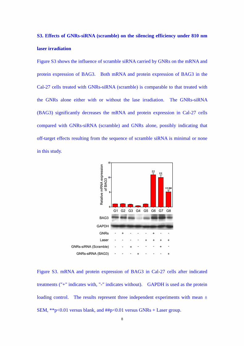
8
S3. Effects of GNRs-siRNA (scramble) on the silencing efficiency under 810 nm
laser irradiation
Figure S3 shows the influence of scramble siRNA carried by GNRs on the mRNA and
protein expression of BAG3. Both mRNA and protein expression of BAG3 in the
Cal-27 cells treated with GNRs-siRNA (scramble) is comparable to that treated with
the GNRs alone either with or without the lase irradiation. The GNRs-siRNA
(BAG3) significantly decreases the mRNA and protein expression in Cal-27 cells
compared with GNRs-siRNA (scramble) and GNRs alone, possibly indicating that
off-target effects resulting from the sequence of scramble siRNA is minimal or none
in this study.
Figure S3. mRNA and protein expression of BAG3 in Cal-27 cells after indicated
treatments ("+" indicates with, "-" indicates without). GAPDH is used as the protein
loading control. The results represent three independent experiments with mean ±
SEM, **p<0.01 versus blank, and ##p<0.01 versus GNRs + Laser group.
9
S4. Effect of GNRs-siRNA (scramble) on photothermal therapy under 810 nm
laser irradiation
Figure S4 shows the influence of scramble siRNA on the therapeutic efficacy of
GNRs-mediated PTT. After laser irradiation, MTT assays reveal that the
GNRs-siRNA (scramble) induces suppression of Cal-27 cell growth similar to the
GNRs, while the GNRs-siRNA (BAG3) causes higher cell death of Cal-27 compared
with GNRs and GNRs-siRNA (scramble). Most importantly, as shown in Figure S4b,
the results from Annexin V-FITC/PI double staining demonstrates that the
GNRs-siRNA (scramble) causes 32.7% cell apoptosis (including early apoptotic
population and late apoptotic population), which is almost the same as 30.1%
apoptosis caused by the GNRs alone, suggesting that the scramble siRNA does not
increase the cell death induced by GNR-mediated thermotherapy. The western blot
for cleaved PARP also confirms the above results that GNRs-siRNA (BAG3) cause
the highest expression of apoptotic protein compared to the GNRs and GNRs-siRNA
(scramble) under the same laser irradiation. These results suggest that the enhanced
therapeutic efficacy induced by the GNRs-siRNA (BAG3) actually depends on the
specific suppression of BAG3, but not the possible “off-target” effects.
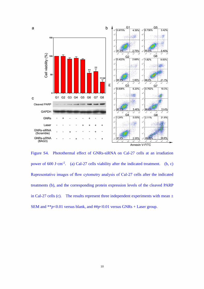
10
Figure S4. Photothermal effect of GNRs-siRNA on Cal-27 cells at an irradiation
power of 600 J·cm-2. (a) Cal-27 cells viability after the indicated treatment. (b, c)
Representative images of flow cytometry analysis of Cal-27 cells after the indicated
treatments (b), and the corresponding protein expression levels of the cleaved PARP
in Cal-27 cells (c). The results represent three independent experiments with mean ±
SEM and **p<0.01 versus blank, and ##p<0.01 versus GNRs + Laser group.