Embed Size (px)
Citation preview
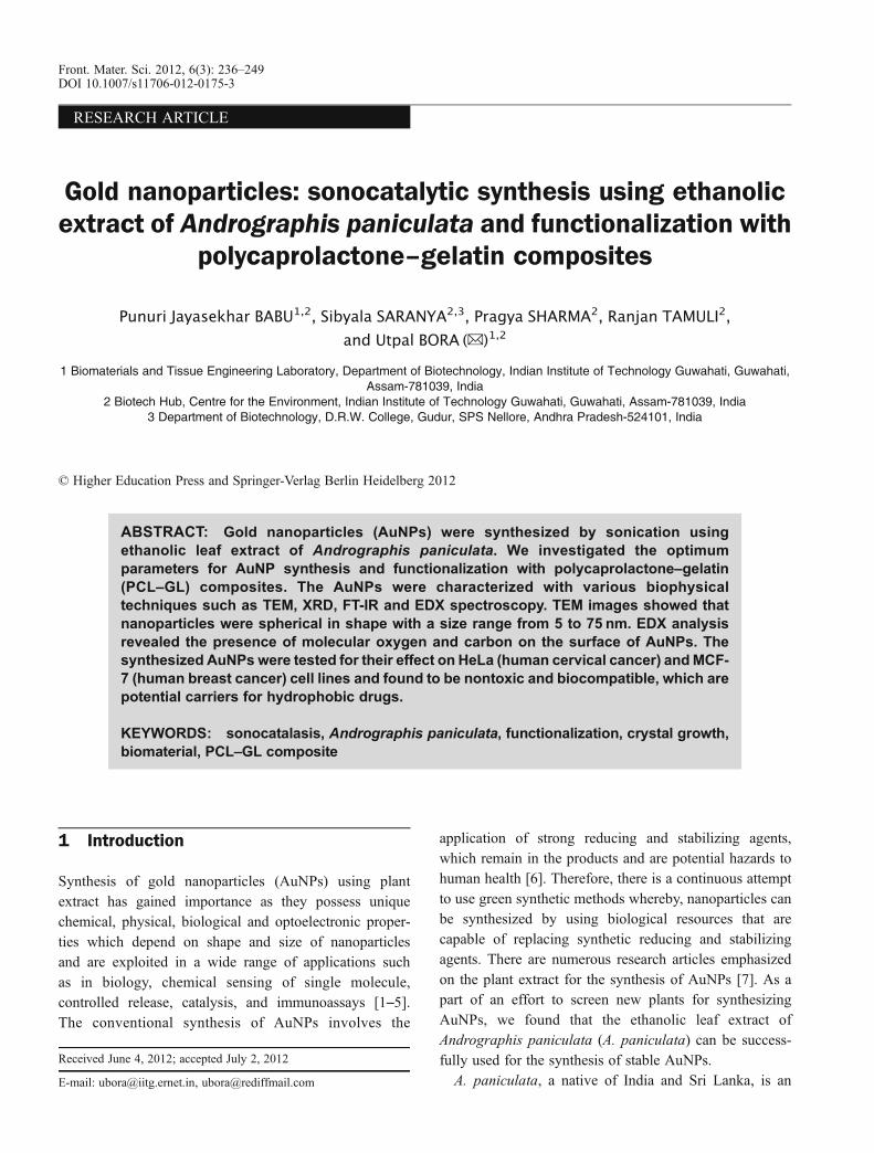
RESEARCH ARTICLE
Gold nanoparticles: sonocatalytic synthesis using ethanolicextract of Andrographis paniculata and functionalization with
polycaprolactone–gelatin composites
Punuri Jayasekhar BABU1,2, Sibyala SARANYA2,3, Pragya SHARMA2, Ranjan TAMULI2,
and Utpal BORA (✉)1,2
1 Biomaterials and Tissue Engineering Laboratory, Department of Biotechnology, Indian Institute of Technology Guwahati, Guwahati,Assam-781039, India
2 Biotech Hub, Centre for the Environment, Indian Institute of Technology Guwahati, Guwahati, Assam-781039, India3 Department of Biotechnology, D.R.W. College, Gudur, SPS Nellore, Andhra Pradesh-524101, India
© Higher Education Press and Springer-Verlag Berlin Heidelberg 2012
ABSTRACT: Gold nanoparticles (AuNPs) were synthesized by sonication usingethanolic leaf extract of Andrographis paniculata. We investigated the optimumparameters for AuNP synthesis and functionalization with polycaprolactone–gelatin(PCL–GL) composites. The AuNPs were characterized with various biophysicaltechniques such as TEM, XRD, FT-IR and EDX spectroscopy. TEM images showed thatnanoparticles were spherical in shape with a size range from 5 to 75 nm. EDX analysisrevealed the presence of molecular oxygen and carbon on the surface of AuNPs. Thesynthesized AuNPs were tested for their effect on HeLa (human cervical cancer) andMCF-7 (human breast cancer) cell lines and found to be nontoxic and biocompatible, which arepotential carriers for hydrophobic drugs.
KEYWORDS: sonocatalasis, Andrographis paniculata, functionalization, crystal growth,biomaterial, PCL–GL composite
1 Introduction
Synthesis of gold nanoparticles (AuNPs) using plantextract has gained importance as they possess uniquechemical, physical, biological and optoelectronic proper-ties which depend on shape and size of nanoparticlesand are exploited in a wide range of applications suchas in biology, chemical sensing of single molecule,controlled release, catalysis, and immunoassays [1–5].The conventional synthesis of AuNPs involves the
application of strong reducing and stabilizing agents,which remain in the products and are potential hazards tohuman health [6]. Therefore, there is a continuous attemptto use green synthetic methods whereby, nanoparticles canbe synthesized by using biological resources that arecapable of replacing synthetic reducing and stabilizingagents. There are numerous research articles emphasizedon the plant extract for the synthesis of AuNPs [7]. As apart of an effort to screen new plants for synthesizingAuNPs, we found that the ethanolic leaf extract ofAndrographis paniculata (A. paniculata) can be success-fully used for the synthesis of stable AuNPs.A. paniculata, a native of India and Sri Lanka, is an
Received June 4, 2012; accepted July 2, 2012
E-mail: [email protected], [email protected]
Front. Mater. Sci. 2012, 6(3): 236–249DOI 10.1007/s11706-012-0175-3
annual herbaceous plant (family: Acanthaceae), extremelybitter, about 3 feet high and one of the most commonlyused plants in traditional systems of Unani and Ayurvedicmedicines. This herb is popularly known as ‘Maha-tita’(king of bitters) or ‘Chirota-tita’ in the northeast of India. Itcan be found in variety of habitats such as planes, hill sides,coastlines, roadsides, farms, and wastelands. It has beenrevealed that A. paniculata leaf extract (ALE) containsditerpenes, flavonoids and stigma sterols [8]. The extractpossesses ent-labdane diterpenoids and andrographolide asthe main bioactive compounds which have wide spectrumof biological applications such as antibacterial [9],antimalarial [10], antithrombotic [11], antitumor [12],immunostimulatory [13] properties. A. paniculata hasbeen reported to have anti-inflammatory [14] and anti-allergic activities. The anti-inflammatory action of the plantis attributed to andrographolide, the major active principleingredient of the plant [15]. A. paniculata also helps inboosting the immune system, protects against cancer andHIV [16], prevents blood clots, diabetes and hypertension[17], and maintains efficient digestive functioning [18]. Ithas many applications, including treatment of dyspepsia,influenza, dysentery, malaria, respiratory infections and asantidote for snake-bite and poisonous stings of someinsects [19]. It is also considered to be used asantiphlogistic antipyretic, detoxicant, analgesic and anagent for the treatment of acute infections of the respiratoryorgans and urinary system and gastrointestinal tract [20].We designed the current work by considering the
bioactive molecules present in A. paniculata such asandrographolide, flavonoids, antioxidants, diterpenes andstigma sterols which can facilitate the reduction of variousmetal salts to their corresponding nanoparticles. For therapid synthesis of AuNPs, we applied ultrasonication(sonics, VC 505 Vibra Cell), which has an advantage offormation of the acoustic bubble and generation ofimmense heat [21] thereby influencing the nucleationprocess of AuNPs. Further, we have functionalized AuNPswith polycaprolactone (PCL), gelatin (GL) and PCL–GLwhich are named as AuNP–PCL, AuNP–GL and AuNP–PCL–GL composites, respectively. The as-synthesizedintact AuNPs were tested for their cytotoxicity usinghuman cancer cell lines HeLa and MCF-7. However,AuNPs showed insignificant effect on cell viability,indicating their biocompatibility. Therefore, this greenapproach is a successful method for the synthesis ofstabilized AuNPs and the functionalization with naturalpolymers without the involvement of chemical agents.
2 Experimental
2.1 Materials
A. paniculata was grown as per traditional agronomicpractice in the experimental field and healthy leaves wereharvested for the AuNP synthesis. Cell lines were obtainedfrom National Centre for Cell Sciences (Pune, India). Cellculture related plasticwares were obtained from the Sigma-Aldrich (Bangalore, India). All the reagents were ofanalytical grade obtained from either Sisco ResearchLaboratories (Mumbai, India) or E. Merck India Ltd.(Mumbai, India). 3-(4, 5-dimethylthiazol-2-yl)-2, 5-diphe-nyltetrazolium bromide (MTT) was purchased fromHiMedia (Bangalore, India).
2.2 Preparation of leaf extract
A. paniculata leaves were washed with deionised water toremove adsorbed dirt. The leaves were chopped into smallpieces (2 cm� 2 cm) and dried at room temperature (25°C)under shade. Then the dried leaves were powdered in amixer grinder (Bajaj Model GX 11, India). About 5 g ofpowder was dissolved in 50 mL of ethanol and kept at 4°Cfor one week to get the leaf extract. The ALE was filteredusing Whatman 50 mm filter papers and the filtrate wasstored at 4°C for various experiments.
2.3 Biochemical test for flavonoids and phenolic
compounds
To the 5 mL of diluted NH3 solution, 2 mL of ALE wasadded, followed by few drops of concentrated H2SO4. Theyellow colour indicated the presence of the flavonoids. Tothe 2 mL of ALE, few drops of 5% FeCl3 were added. Theappearance of dark green colour indicated the presence ofpolyphenolic compounds.
2.4 Synthesis of AuNPs
Synthesis of AuNPs was carried out by varying the ALE(0.5%–5%) against to 1 mmol/L HAuCl4 and the finalvolume was made up to 2 mL with double distilled water.To all the resulting mixtures, we applied ultrasonication for2 min with 30% amplitude at room temperature. To find outthe optimum concentration of HAuCl4, the experimentswere carried out by varying the gold solution concentrationin the range of 0.5–3 mmol/L against 3.5% of the ALE. Theoptimum time for synthesis was determined by incubating
Punuri Jayasekhar BABU et al. AuNPs: sonocatalytic synthesis using ethanolic extract of A. paniculata and functionalization ... 237
3.5% of ALE with 1 mmol/L HAuCl4 by varying thesonication time in the range of 1–4 min with an interval of30 s. All the reactions were done with pulse off and on 4 seach.
2.5 Effect of amplitude and pH on the synthesis of AuNPs
To identify the effect of amplitude and pH, we appliedthe ultrasonication for 120 s to the reaction mixturescomprising of ALE (3.5%) and HAuCl4 (1 mmol/L) byvarying the amplitude from 21% to 35% and adjusting thepH value of reaction mixtures from 1 to 11.
2.6 Functionalization of AuNPs with PCL, GL, and
PCL–GL
Both PCL and GL, 0.25% of each, separately, was added tothe 5 mL of dimethyl formamide (DMF) and 5 mL of thepre-warmed water, respectively, and the total volume ofeach reaction mixture was adjusted to 20 mL with distilledwater containing ALE (3.5%) and HAuCl4 (1 mmol/L). Forfunctionalization of PCL–GL, the reaction mixtures(0.25% of PCL in 5 mL of DMF and 0.25% of GL in5 mL of pre-warmed water) were mixed together and thetotal volume was adjusted to 20 mL of distilled watercontaining ALE (3.5%) and HAuCl4 (1 mmol/L). For allthe reaction mixtures we applied ultrasonication for 10 minat the 35% amplitude.
2.7 Characterization of AuNPs
2.7.1 Ultraviolet-visible (UV-vis) spectroscopy
All UV-vis spectroscopic measurements of synthesizedAuNPs were carried out on a Cary 100 BIO UV-visspectrophotometer (Varian, CA, USA).
2.7.2 Transmission electron microscopy (TEM)
A volume of 10 mL of the AuNP solution was centrifugedat 20,000 rpm for 20 min. The resulted pellet wasresuspended in 3 mL of distilled water and centrifuged at20,000 rpm for 20 min. This process was repeated thriceand the resultant pellet was resuspended in 1 mL of dis-tilled water. Few drops of the redispersed colloidal solutionwere placed over carbon coated copper grid and the waterwas evaporated in hot air oven (Daihan Labtech Co. Ltd.model LDO-150F) at 60°C for 4 h. TEM measurementswere performed on a transmission electron microscope
(TEM-JEOL model 2100) operated at 190 V of 200 kV.
2.7.3 X-ray diffraction (XRD), Fourier transform infrared
(FT-IR) spectroscopy and energy dispersive X-ray (EDX)
spectroscopy
The colloidal solution mixtures obtained in Section 2.6were centrifuged at 20,000 rpm and resulted pellets wereresuspended in appropriate volume (3 mL) of doubledistilled water. This process was repeated for three timesto obtain the pure nanoparticles of the respective type.Finally, each resulted pellets was redispersed in 5 mL ofdouble distilled water and freeze dried in a lyophiliser(Christ Gefriertrocknungsanlagen GmbH Model 1-4) for16 h. The fine dried powder samples were analyzed withthe help of an XRD instrument (Bruker Advance D8 XRDmachine) with Cu source at the wavelength of 1.5406 Å inthin film mode. Infrared spectra were recorded using a FT-IR spectroscope (Spectrum One, Perkin Elmer, MA, USA),from 4000 to 450 cm–1, with a resolution of 2 cm–1 and 5scans/sample by pressing the 1 mg of each finely powderedAuNP, AuNP–PCL, AuNP–GL, and AuNP–PCL–GLpowders with 200 mg KBr.The elemental composition of the intact AuNPs were
obtained by using EDX spectroscopy (LEO 1430 VP) atvariable pressure scanning electron microscope equippedwith INCA Oxford EDX facility, at an acceleration voltageof 10 keV.
2.8 Cytotoxicity studies
The minimal essential medium (MEM) containing1.0 mmol/L C3H3NaO3, 0.1 mmol/L nonessential aminoacids, 1.5 g/L NaHCO3, 2 mmol/L L-glutamine supple-mented with 10% fetal bovine serum (FBS; heat inacti-vated) and 1% antibiotic-antimycotic solution (1000 U/mLpenicillin G, 10 mg/mL streptomycin sulphate, 5 mg/mLgentamycin, and 25 μg/mL amphotericin B) was used tomaintain the HeLa (human cervical cancer) and MCF-7(human breast cancer) cells. The cells were cultured at37°C in a humidified incubator (Heal Force, HF 160W,China) supplemented with 5% CO2.The cytotoxicity of AuNPs on cancer cells was evaluated
by the MTT assay, which is a widely used screeningmethod to measure cell viability and proliferation. Themonocultures of the HeLa and MCF-7 cells were incubatedwith increasing concentrations of filter (0.2 μm) sterilizedAuNPs for 24 h. The cell viability was estimated by MTTdye conversion assay. Cells not exposed to AuNPs were
238 Front. Mater. Sci. 2012, 6(3): 236–249
considered as control. About 1�104 cells were seeded andmaintained in a 96-well plate (Cell Bind, Corning) usingMEM containing serum. After 24 h of incubation, themedium was replaced with the serum free mediumcontaining various concentrations of AuNPs (10–100 μmol/L). The media was removed after 24 h oftreatment and cells were washed with phosphate-bufferedsaline (PBS; 0.01 mol/L, pH = 7.2) followed by theaddition of 100 μL of MTT (0.5 mg/mL) prepared inserum free medium to each well and incubated for 4 h in anincubator. Subsequently, medium was removed and 100 μLof dimethyl sulphoxide (DMSO) was added to each well tosolubilise the formazan crystals. The concentration offormazan was determined using a multiwell plate reader(Tecon micro-plate reader, model 680, CA, USA) at470 nm absorbance. The cell viability was calculatedwith the following equation:
Cell viability=% ¼ Atreated
Acontrol� 100 (1)
where Atreated and Acontrol are the absorbances of treated anduntreated cells, respectively.
3 Results and discussion
3.1 Formation of spherical AuNPs from ALE in the
ultrasonication process under optimum conditions
In an effort to optimize parameters for the synthesis of
spherical AuNPs, different reactions were carried out byvarying the concentration of ALE (0.5%–5%) against1 mmol/L HAuCl4 for 120 s of the ultrasonication time,which resulted in appearance of ruby red colour in all thereaction mixtures indicating the synthesis of AuNPs. Thepeak intensities of reaction mixtures with above 3.5% ofALE were flattened, indicating the insignificant incrementin the formation of AuNPs. This infers that the 3.5% ALEreduces the total 1 mmol/L of HAuCl4. Thus we considerthe 3.5% of ALE as optimum for the AuNP synthesis(Fig. 1(a)). To find out the optimum concentration ofHAuCl4, we carried out various experiments with 3.5%ALE by changing the concentration of the gold salts from0.5 to 1 mmol/L and observed the maximum peak intensityassociated with 1 mmol/L HAuCl4 that we considered as anoptimum (Fig. 1(b)).The peaks associated with 2 mmol/L and above showed
the gradual decrease in their intensities, revealing that 3.5%of the ALE is adequate for the total reduction of HAuCl4.The optimum ultrasonication time required for the AuNPsynthesis was determined by performing various experi-ments with 3.5% ALE and 1 mmol/L HAuCl4 for differentultrasonication time from 60 to 240 s with an interval of30 s and we found 120 s to be the optimum time. Weobserved the progression in the intensities of peaks withrespect to time. As there is no significant change in the peakintensities associated above 120 s, we considered it as anoptimum time (Figs. 2(a) and 2(b) are for easy differentia-tion).
Fig. 1 UV-vis absorption spectra of AuNPs synthesized by (a) varying ALE concentrations (a — 0.5%, b — 1%, c — 1.5%, d — 2%,e— 2.5%, f— 3%, g— 3.5%, h— 4%, i— 4.5%, j— 5%) for 2 min against 1 mmol/L HAuCl4 and (b) varying HAuCl4 concentrations(a — 0.5 mmol/L, b — 1 mmol/L, c — 1.5 mmol/L, d — 2 mmol/L and e — 2.5 mmol/L, f — 3 mmol/L) against 3.5% ALE.
Punuri Jayasekhar BABU et al. AuNPs: sonocatalytic synthesis using ethanolic extract of A. paniculata and functionalization ... 239
We applied ultrasonication for the rapid synthesis ofAuNPs, which can generate the acoustic cavities in themedium (H2O). These acoustic cavities release immenseenergy in the form of high temperature (5000 K) and highpressure (20,000 pounds per square inch (psi)) by under-going implosive collapse [21–23]. The enormous energyreleased during the sonication process assisted reducingagents (phenolic compound and flavonoids) to reduce goldions to AuNPs (Fig. 3).
A. paniculata has a high content of phenolic acids,antioxidants and flavonoids which can act as strongreducing agents. The biochemical test revealed thatflavonoids and phenolic compounds are present in ALEand may be responsible for reductions and stabilisation ofAuNPs. The abundant free hydroxyl groups available inthese compounds participate in the gold bioreduction.Phenolic compounds reduce through the oxidation ofhydroxyl (R –OH) to carbonyl groups (R –C = O) as
Fig. 2 UV-vis absorption spectra of AuNPs synthesized with 3.5% ALE and 1 mmol/L HAuCl4 by different ultrasonication time (a —60 s, b— 90 s, c— 120 s, d— 150 s, e— 180 s, f— 210 s, g— 240 s): (a) wavelength range from 300 to 800 nm; (b) wavelength rangefrom 500 to 600 nm.
Fig. 3 Schematic representation of sonocatalytic synthesis of AuNPs under optimized conditions (3.5% ALE, 120 s ultrasonicationtime, 1 mmol/L HAuCl4, 35% amplitude and pH 5).
240 Front. Mater. Sci. 2012, 6(3): 236–249
shown in the following reaction:
AuCl –4 þ 3R –OH!Au0 þ 3R ¼ Oþ 3Hþþ 4Cl – (2)
Flavonoids interact with Au3+ ions through carbonylgroups or π-electrons and then reduced into AuNPs in theextracellular medium. As soon as they come in contact withAu3+ ions in extracellular medium, their π-electrons aretransferred and Au3+ ions are reduced to Au0, as shown inScheme 1. Flavonoids can also prevent agglomeration andstabilize AuNPs [24]. Thus the involvement of flavonoidsin the rapid reduction of Au3+ ions and as capping agentgives stability to AuNPs.
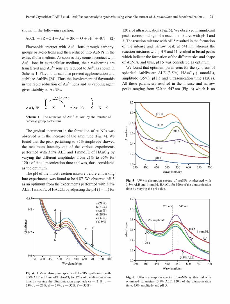
The gradual increment in the formation of AuNPs wasobserved with the increase of the amplitude (Fig. 4). Wefound that the peak pertaining to 35% amplitude showedthe maximum intensity out of the various experimentsperformed with 3.5% ALE and 1 mmol/L of HAuCl4 byvarying the different amplitudes from 21% to 35% for120 s of the ultrasonication time and was, thus, consideredas the optimum.The pH of the intact reaction mixture before embarking
into experiments was found to be 4.87. We observed pH 5as an optimum from the experiments performed with 3.5%ALE, 1 mmol/L of HAuCl4 by adjusting the pH (1 – 11) for
120 s of ultrasonication (Fig. 5). We observed insignificantpeaks corresponding to the reaction mixtures with pH 1 and3. The reaction mixture with pH 5 resulted in the formationof the intense and narrow peak at 541 nm whereas thereaction mixtures with pH 9 and 11 resulted in broad peakswhich indicate the formation of the different size and shapeof AuNPs, and thus, pH 5 was considered as optimum.We found that optimum parameters for the synthesis of
spherical AuNPs are ALE (3.5%), HAuCl4 (1 mmol/L),amplitude (35%), pH 5 and ultrasonication time (120 s).All these parameters resulted in the intense and narrowpeaks ranging from 520 to 547 nm (Fig. 6) which is an
Scheme 1 The reduction of Au3+ to Au0 by the transfer ofcarbonyl group π-electrons.
Fig. 4 UV-vis absorption spectra of AuNPs synthesized with3.5% ALE and 1 mmol/L HAuCl4 for 120 s of the ultrasonicationtime by varying the ultrasonication amplitude (a — 21%, b —
23%, c — 26%, d — 29%, e — 32%, f — 35%).
Fig. 5 UV-vis absorption spectra of AuNPs synthesized with3.5% ALE and 1 mmol/L HAuCl4 for 120 s of the ultrasonicationtime by varying the pH value.
Fig. 6 UV-vis absorption spectra of AuNPs synthesized withoptimized parameters: 3.5% ALE, 120 s of the ultrasonicationtime, 35% amplitude and pH 5.
Punuri Jayasekhar BABU et al. AuNPs: sonocatalytic synthesis using ethanolic extract of A. paniculata and functionalization ... 241
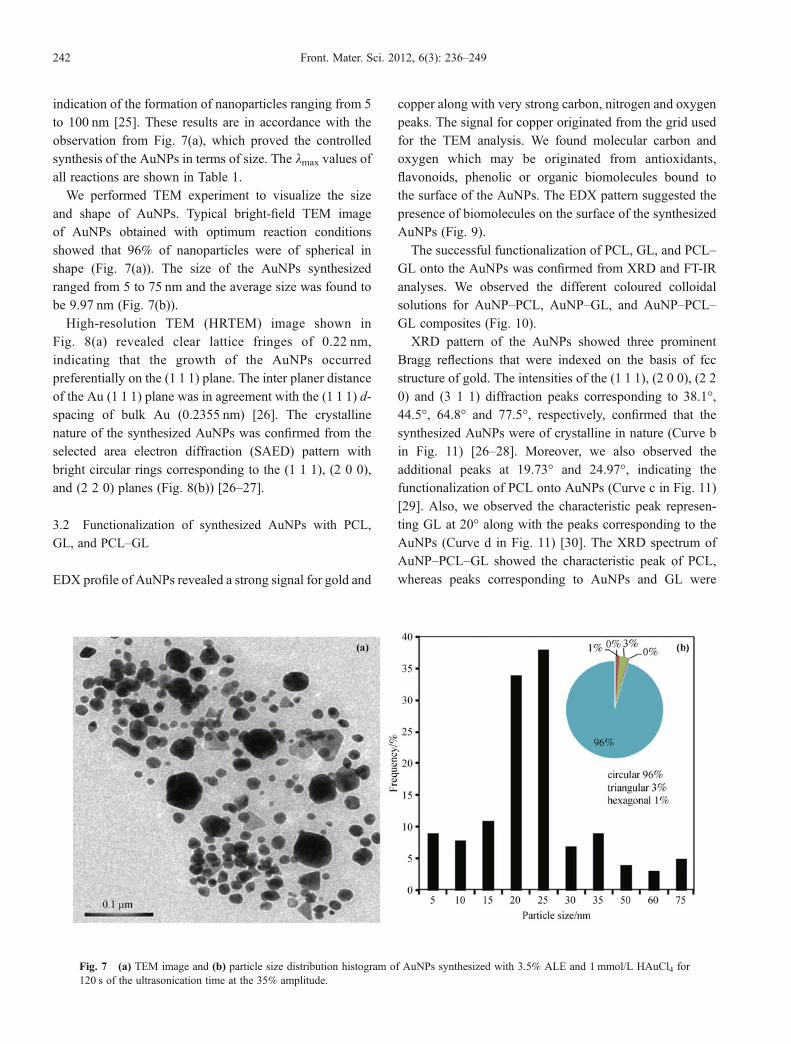
indication of the formation of nanoparticles ranging from 5to 100 nm [25]. These results are in accordance with theobservation from Fig. 7(a), which proved the controlledsynthesis of the AuNPs in terms of size. The lmax values ofall reactions are shown in Table 1.We performed TEM experiment to visualize the size
and shape of AuNPs. Typical bright-field TEM imageof AuNPs obtained with optimum reaction conditionsshowed that 96% of nanoparticles were of spherical inshape (Fig. 7(a)). The size of the AuNPs synthesizedranged from 5 to 75 nm and the average size was found tobe 9.97 nm (Fig. 7(b)).High-resolution TEM (HRTEM) image shown in
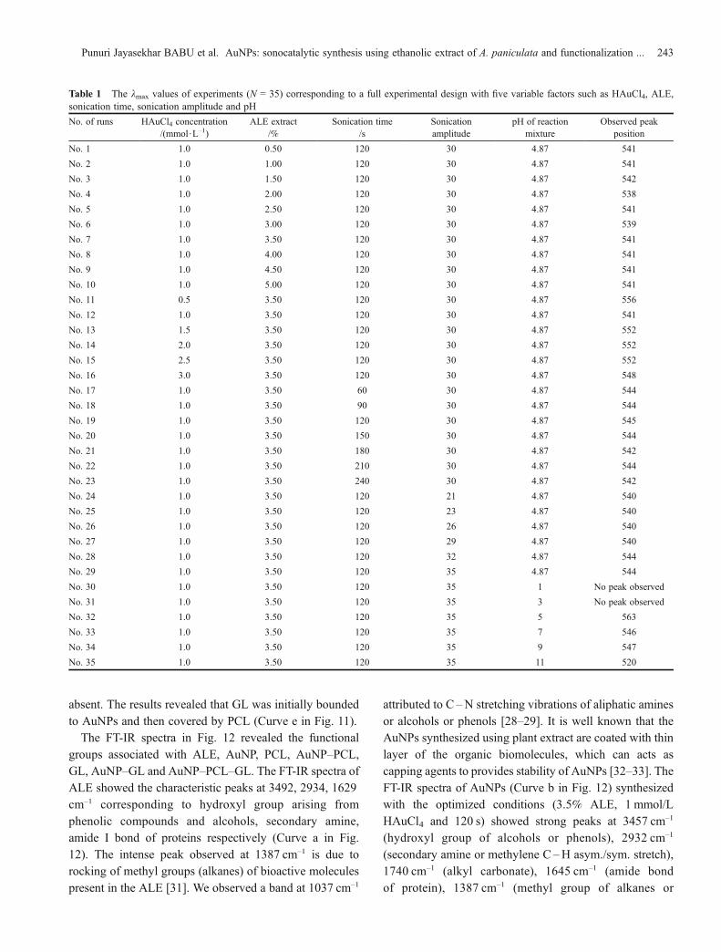
Fig. 8(a) revealed clear lattice fringes of 0.22 nm,indicating that the growth of the AuNPs occurredpreferentially on the (1 1 1) plane. The inter planer distanceof the Au (1 1 1) plane was in agreement with the (1 1 1) d-spacing of bulk Au (0.2355 nm) [26]. The crystallinenature of the synthesized AuNPs was confirmed from theselected area electron diffraction (SAED) pattern withbright circular rings corresponding to the (1 1 1), (2 0 0),and (2 2 0) planes (Fig. 8(b)) [26–27].
3.2 Functionalization of synthesized AuNPs with PCL,
GL, and PCL–GL
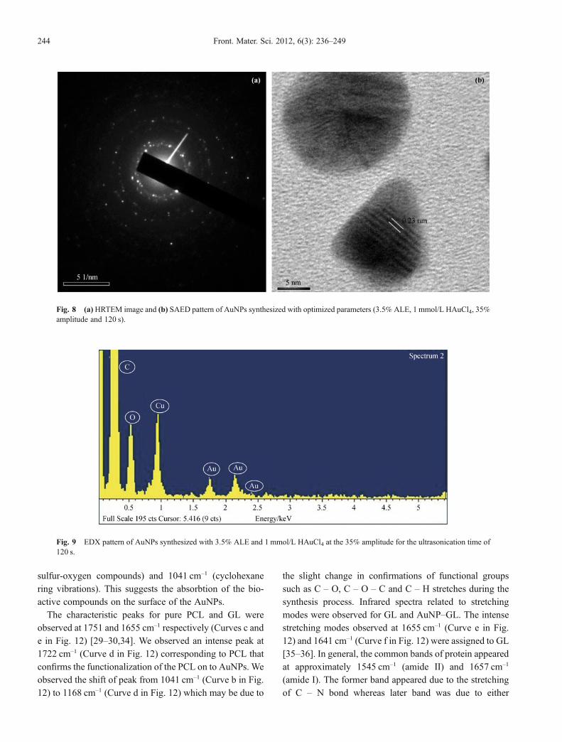
EDX profile of AuNPs revealed a strong signal for gold and
copper along with very strong carbon, nitrogen and oxygenpeaks. The signal for copper originated from the grid usedfor the TEM analysis. We found molecular carbon andoxygen which may be originated from antioxidants,flavonoids, phenolic or organic biomolecules bound tothe surface of the AuNPs. The EDX pattern suggested thepresence of biomolecules on the surface of the synthesizedAuNPs (Fig. 9).The successful functionalization of PCL, GL, and PCL–
GL onto the AuNPs was confirmed from XRD and FT-IRanalyses. We observed the different coloured colloidalsolutions for AuNP–PCL, AuNP–GL, and AuNP–PCL–GL composites (Fig. 10).XRD pattern of the AuNPs showed three prominent
Bragg reflections that were indexed on the basis of fccstructure of gold. The intensities of the (1 1 1), (2 0 0), (2 20) and (3 1 1) diffraction peaks corresponding to 38.1°,44.5°, 64.8° and 77.5°, respectively, confirmed that thesynthesized AuNPs were of crystalline in nature (Curve bin Fig. 11) [26–28]. Moreover, we also observed theadditional peaks at 19.73° and 24.97°, indicating thefunctionalization of PCL onto AuNPs (Curve c in Fig. 11)[29]. Also, we observed the characteristic peak represen-ting GL at 20° along with the peaks corresponding to theAuNPs (Curve d in Fig. 11) [30]. The XRD spectrum ofAuNP–PCL–GL showed the characteristic peak of PCL,whereas peaks corresponding to AuNPs and GL were
Fig. 7 (a) TEM image and (b) particle size distribution histogram of AuNPs synthesized with 3.5% ALE and 1 mmol/L HAuCl4 for120 s of the ultrasonication time at the 35% amplitude.
242 Front. Mater. Sci. 2012, 6(3): 236–249
absent. The results revealed that GL was initially boundedto AuNPs and then covered by PCL (Curve e in Fig. 11).The FT-IR spectra in Fig. 12 revealed the functional
groups associated with ALE, AuNP, PCL, AuNP–PCL,GL, AuNP–GL and AuNP–PCL–GL. The FT-IR spectra ofALE showed the characteristic peaks at 3492, 2934, 1629cm–1 corresponding to hydroxyl group arising fromphenolic compounds and alcohols, secondary amine,amide I bond of proteins respectively (Curve a in Fig.12). The intense peak observed at 1387 cm–1 is due torocking of methyl groups (alkanes) of bioactive moleculespresent in the ALE [31]. We observed a band at 1037 cm–1
attributed to C –N stretching vibrations of aliphatic aminesor alcohols or phenols [28–29]. It is well known that theAuNPs synthesized using plant extract are coated with thinlayer of the organic biomolecules, which can acts ascapping agents to provides stability of AuNPs [32–33]. TheFT-IR spectra of AuNPs (Curve b in Fig. 12) synthesizedwith the optimized conditions (3.5% ALE, 1 mmol/LHAuCl4 and 120 s) showed strong peaks at 3457 cm–1
(hydroxyl group of alcohols or phenols), 2932 cm–1
(secondary amine or methylene C –H asym./sym. stretch),1740 cm–1 (alkyl carbonate), 1645 cm–1 (amide bondof protein), 1387 cm–1 (methyl group of alkanes or
Table 1 The lmax values of experiments (N = 35) corresponding to a full experimental design with five variable factors such as HAuCl4, ALE,sonication time, sonication amplitude and pH
No. of runs HAuCl4 concentration/(mmol$L–1)
ALE extract/%
Sonication time/s
Sonicationamplitude
pH of reactionmixture
Observed peakposition
No. 1 1.0 0.50 120 30 4.87 541
No. 2 1.0 1.00 120 30 4.87 541
No. 3 1.0 1.50 120 30 4.87 542
No. 4 1.0 2.00 120 30 4.87 538
No. 5 1.0 2.50 120 30 4.87 541
No. 6 1.0 3.00 120 30 4.87 539
No. 7 1.0 3.50 120 30 4.87 541
No. 8 1.0 4.00 120 30 4.87 541
No. 9 1.0 4.50 120 30 4.87 541
No. 10 1.0 5.00 120 30 4.87 541
No. 11 0.5 3.50 120 30 4.87 556
No. 12 1.0 3.50 120 30 4.87 541
No. 13 1.5 3.50 120 30 4.87 552
No. 14 2.0 3.50 120 30 4.87 552
No. 15 2.5 3.50 120 30 4.87 552
No. 16 3.0 3.50 120 30 4.87 548
No. 17 1.0 3.50 60 30 4.87 544
No. 18 1.0 3.50 90 30 4.87 544
No. 19 1.0 3.50 120 30 4.87 545
No. 20 1.0 3.50 150 30 4.87 544
No. 21 1.0 3.50 180 30 4.87 542
No. 22 1.0 3.50 210 30 4.87 544
No. 23 1.0 3.50 240 30 4.87 542
No. 24 1.0 3.50 120 21 4.87 540
No. 25 1.0 3.50 120 23 4.87 540
No. 26 1.0 3.50 120 26 4.87 540
No. 27 1.0 3.50 120 29 4.87 540
No. 28 1.0 3.50 120 32 4.87 544
No. 29 1.0 3.50 120 35 4.87 544
No. 30 1.0 3.50 120 35 1 No peak observed
No. 31 1.0 3.50 120 35 3 No peak observed
No. 32 1.0 3.50 120 35 5 563
No. 33 1.0 3.50 120 35 7 546
No. 34 1.0 3.50 120 35 9 547
No. 35 1.0 3.50 120 35 11 520
Punuri Jayasekhar BABU et al. AuNPs: sonocatalytic synthesis using ethanolic extract of A. paniculata and functionalization ... 243
sulfur-oxygen compounds) and 1041 cm–1 (cyclohexanering vibrations). This suggests the absorbtion of the bio-active compounds on the surface of the AuNPs.The characteristic peaks for pure PCL and GL were
observed at 1751 and 1655 cm–1 respectively (Curves c ande in Fig. 12) [29–30,34]. We observed an intense peak at1722 cm–1 (Curve d in Fig. 12) corresponding to PCL thatconfirms the functionalization of the PCL on to AuNPs. Weobserved the shift of peak from 1041 cm–1 (Curve b in Fig.12) to 1168 cm–1 (Curve d in Fig. 12) which may be due to
the slight change in confirmations of functional groupssuch as C – O, C – O – C and C – H stretches during thesynthesis process. Infrared spectra related to stretchingmodes were observed for GL and AuNP–GL. The intensestretching modes observed at 1655 cm–1 (Curve e in Fig.12) and 1641 cm–1 (Curve f in Fig. 12) were assigned to GL[35–36]. In general, the common bands of protein appearedat approximately 1545 cm–1 (amide II) and 1657 cm–1
(amide I). The former band appeared due to the stretchingof C – N bond whereas later band was due to either
Fig. 8 (a)HRTEM image and (b) SAED pattern of AuNPs synthesized with optimized parameters (3.5% ALE, 1 mmol/L HAuCl4, 35%amplitude and 120 s).
Fig. 9 EDX pattern of AuNPs synthesized with 3.5% ALE and 1 mmol/L HAuCl4 at the 35% amplitude for the ultrasonication time of120 s.
244 Front. Mater. Sci. 2012, 6(3): 236–249
stretching vibrations of C – O bond or coupling of bendingof N – H bonds. It is known that amide I band containssignificant information about the secondary structures ofproteins. The β-sheets of the protein absorb in the range1640–1623 cm–1 and near 1675 cm–1, α-helices in the range1660–1653 cm–1, unordered structures and α-helices 1650–1641 cm–1 and β-turns in the range 1695–1659 cm–1 [37].The characteristic peak of GL reappeared at 1641 cm–1
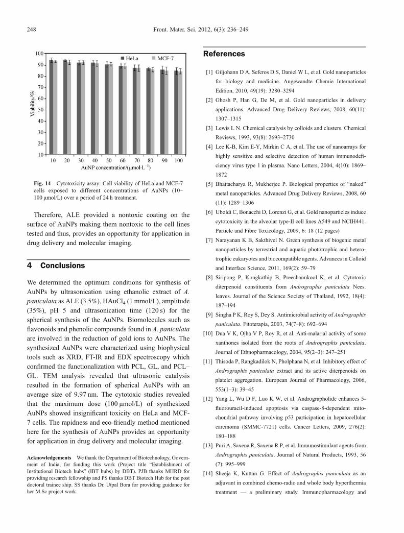
(Curve f in Fig. 12) which confirmed the functionalizationof the AuNPs with GL. Interestingly, in the FT-IR spectraof AuNP–PCL–GL composites, we noticed the peak at1192 cm–1 attributed to the secondary amine and C – Nstretch which may have originated due to the reactionbetween C = O functional group of PCL and amine groupof GL. We also observed a peak at 1738 cm–1 correspon-ding to PCL and the peaks corresponding to both AuNPsand GL were absent (Curve g in Fig. 12). This suggests thatduring the sonication process, GL was initially bound tothe AuNPs and later on it was covered by PCL. Both thepolymers (PCL and GL) can form nanocomposites with thehydrophobic void space inside when they mixed inappropriate amounts. Paclitaxel, an anticancer drug, wassuccessfully delivered into cancer cell line by using thenanocomposites synthesized with PCL and GL [38]. Thecurrent study revealed that these polymers formed intocomposites during the sonication process and were notremoved from the AuNPs even after repeated washing,which confirmed that they were bound covalently. Thetentative mechanisms involved in the binding of PCL–GLcomposites are shown in Fig. 13. The hydrophobic voidspace developed inside the polymers can be used as avehicle for carrying different drugs for different diseases.
3.3 Nontoxic coating on the surface of AuNPs provided by
ALE
The cytotoxicity of intact AuNPs under in vitro conditionsin HeLa and MCF-7 cells was examined in terms of effectof AuNPs on cell viability by MTT assay for 24 h. Onlycells that are viable after 24 h exposure to the AuNPs canmetabolize MTT efficiently and produce purple colouredcrystal that is soluble in DMSO. There was no change inmorphology of both HeLa and MCF-7 after AuNPtreatment. This suggests that AuNPs did not induce anycytotoxic effect for causing significant damage or death ofthe cells (Fig. 14). Both HeLa andMCF-7 cells after 24 h ofpost-treatment showed excellent viability up to as high as100 μmol/L of AuNPs (Fig. 14).
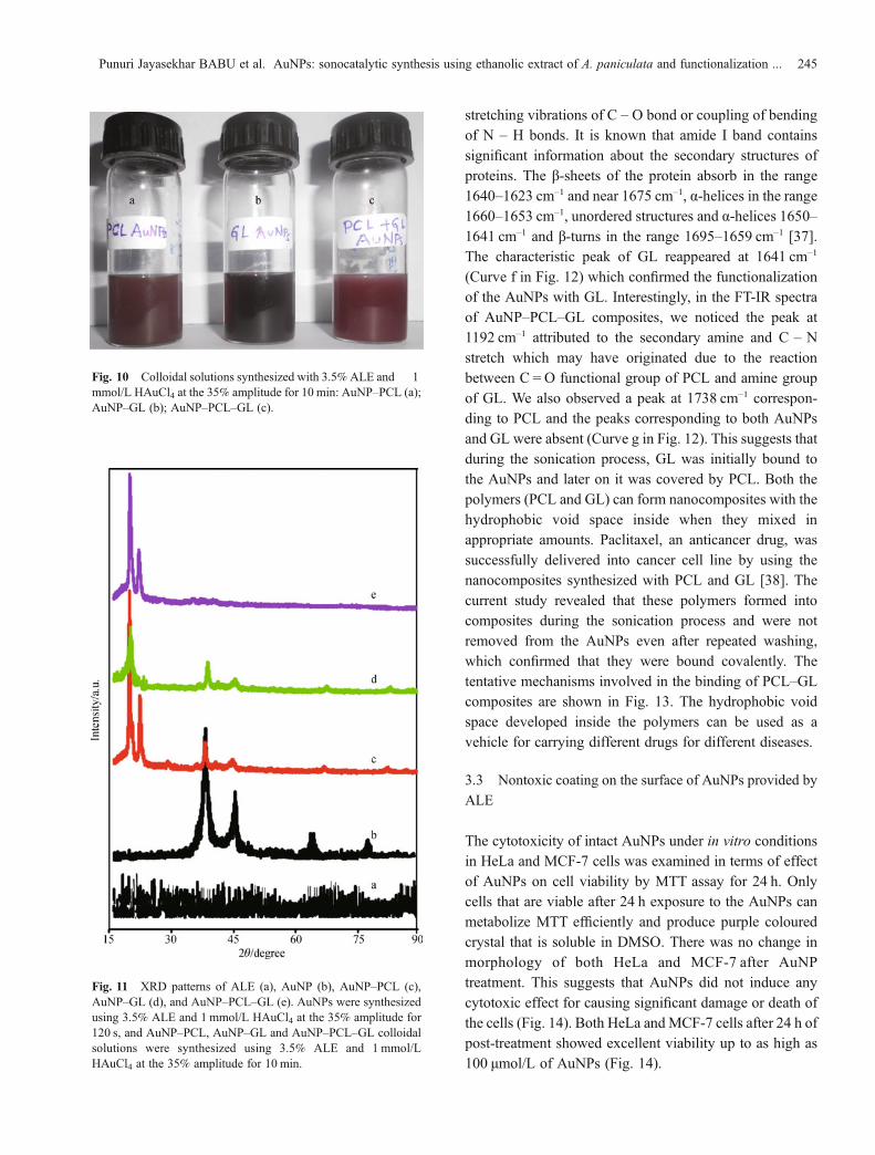
Fig. 10 Colloidal solutions synthesized with 3.5%ALE and 1mmol/L HAuCl4 at the 35% amplitude for 10 min: AuNP–PCL (a);AuNP–GL (b); AuNP–PCL–GL (c).
Fig. 11 XRD patterns of ALE (a), AuNP (b), AuNP–PCL (c),AuNP–GL (d), and AuNP–PCL–GL (e). AuNPs were synthesizedusing 3.5% ALE and 1 mmol/L HAuCl4 at the 35% amplitude for120 s, and AuNP–PCL, AuNP–GL and AuNP–PCL–GL colloidalsolutions were synthesized using 3.5% ALE and 1 mmol/LHAuCl4 at the 35% amplitude for 10 min.
Punuri Jayasekhar BABU et al. AuNPs: sonocatalytic synthesis using ethanolic extract of A. paniculata and functionalization ... 245
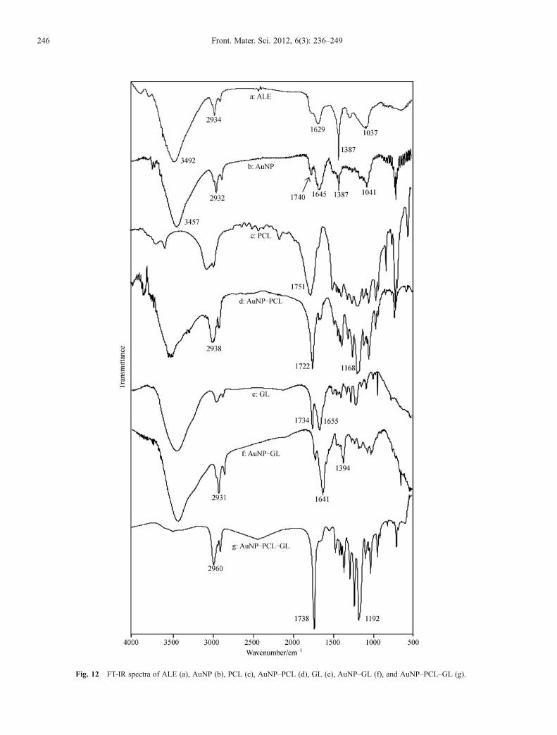
Fig. 12 FT-IR spectra of ALE (a), AuNP (b), PCL (c), AuNP–PCL (d), GL (e), AuNP–GL (f), and AuNP–PCL–GL (g).
246 Front. Mater. Sci. 2012, 6(3): 236–249
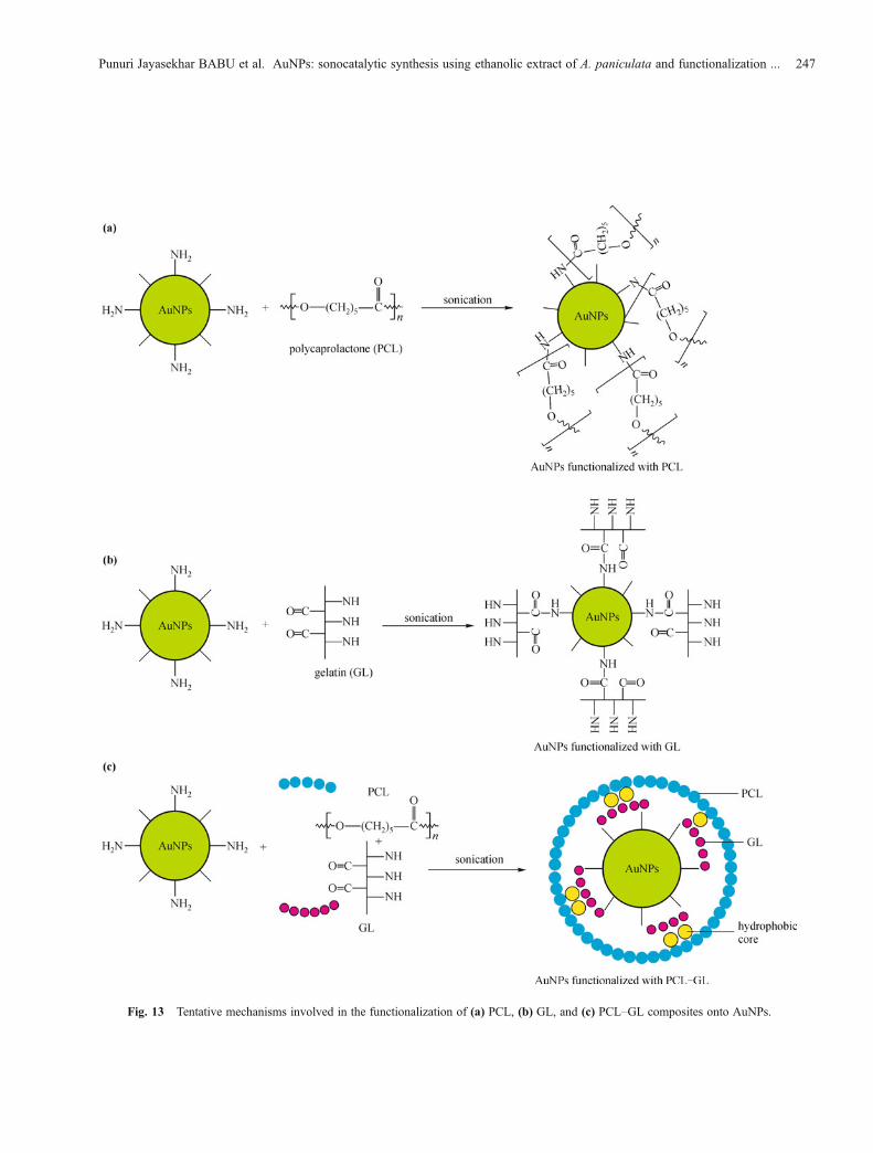
Fig. 13 Tentative mechanisms involved in the functionalization of (a) PCL, (b) GL, and (c) PCL–GL composites onto AuNPs.
Punuri Jayasekhar BABU et al. AuNPs: sonocatalytic synthesis using ethanolic extract of A. paniculata and functionalization ... 247
Therefore, ALE provided a nontoxic coating on thesurface of AuNPs making them nontoxic to the cell linestested and thus, provides an opportunity for application indrug delivery and molecular imaging.
4 Conclusions
We determined the optimum conditions for synthesis ofAuNPs by ultrasonication using ethanolic extract of A.paniculata as ALE (3.5%), HAuCl4 (1 mmol/L), amplitude(35%), pH 5 and ultrasonication time (120 s) for thespherical synthesis of the AuNPs. Biomolecules such asflavonoids and phenolic compounds found in A. paniculataare involved in the reduction of gold ions to AuNPs. Thesynthesized AuNPs were characterized using biophysicaltools such as XRD, FT-IR and EDX spectroscopy whichconfirmed the functionalization with PCL, GL, and PCL–GL. TEM analysis revealed that ultrasonic catalysisresulted in the formation of spherical AuNPs with anaverage size of 9.97 nm. The cytotoxic studies revealedthat the maximum dose (100 μmol/L) of synthesizedAuNPs showed insignificant toxicity on HeLa and MCF-7 cells. The rapidness and eco-friendly method mentionedhere for the synthesis of AuNPs provides an opportunityfor application in drug delivery and molecular imaging.
Acknowledgements We thank the Department of Biotechnology, Govern-ment of India, for funding this work (Project title “Establishment ofInstitutional Biotech hubs” (IBT hubs) by DBT). PJB thanks MHRD forproviding research fellowship and PS thanks DBT Biotech Hub for the postdoctoral trainee ship. SS thanks Dr. Utpal Bora for providing guidance forher M.Sc project work.
References
[1] Giljohann D A, Seferos D S, Daniel W L, et al. Gold nanoparticles
for biology and medicine. Angewandte Chemie International
Edition, 2010, 49(19): 3280–3294
[2] Ghosh P, Han G, De M, et al. Gold nanoparticles in delivery
applications. Advanced Drug Delivery Reviews, 2008, 60(11):
1307–1315
[3] Lewis L N. Chemical catalysis by colloids and clusters. Chemical
Reviews, 1993, 93(8): 2693–2730
[4] Lee K-B, Kim E-Y, Mirkin C A, et al. The use of nanoarrays for
highly sensitive and selective detection of human immunodefi-
ciency virus type 1 in plasma. Nano Letters, 2004, 4(10): 1869–
1872
[5] Bhattacharya R, Mukherjee P. Biological properties of “naked”
metal nanoparticles. Advanced Drug Delivery Reviews, 2008, 60
(11): 1289–1306
[6] Uboldi C, Bonacchi D, Lorenzi G, et al. Gold nanoparticles induce
cytotoxicity in the alveolar type-II cell lines A549 and NCIH441.
Particle and Fibre Toxicology, 2009, 6: 18 (12 pages)
[7] Narayanan K B, Sakthivel N. Green synthesis of biogenic metal
nanoparticles by terrestrial and aquatic phototrophic and hetero-
trophic eukaryotes and biocompatible agents. Advances in Colloid
and Interface Science, 2011, 169(2): 59–79
[8] Siripong P, Kongkathip B, Preechanukool K, et al. Cytotoxic
diterpenoid constituents from Andrographis paniculata Nees.
leaves. Journal of the Science Society of Thailand, 1992, 18(4):
187–194
[9] Singha P K, Roy S, Dey S. Antimicrobial activity of Andrographis
paniculata. Fitoterapia, 2003, 74(7–8): 692–694
[10] Dua V K, Ojha V P, Roy R, et al. Anti-malarial activity of some
xanthones isolated from the roots of Andrographis paniculata.
Journal of Ethnopharmacology, 2004, 95(2–3): 247–251
[11] Thisoda P, Rangkadilok N, Pholphana N, et al. Inhibitory effect of
Andrographis paniculata extract and its active diterpenoids on
platelet aggregation. European Journal of Pharmacology, 2006,
553(1–3): 39–45
[12] Yang L, Wu D F, Luo K W, et al. Andrographolide enhances 5-
fluorouracil-induced apoptosis via caspase-8-dependent mito-
chondrial pathway involving p53 participation in hepatocellular
carcinoma (SMMC-7721) cells. Cancer Letters, 2009, 276(2):
180–188
[13] Puri A, Saxena R, Saxena R P, et al. Immunostimulant agents from
Andrographis paniculata. Journal of Natural Products, 1993, 56
(7): 995–999
[14] Sheeja K, Kuttan G. Effect of Andrographis paniculata as an
adjuvant in combined chemo-radio and whole body hyperthermia
treatment — a preliminary study. Immunopharmacology and
Fig. 14 Cytotoxicity assay: Cell viability of HeLa and MCF-7cells exposed to different concentrations of AuNPs (10–100 μmol/L) over a period of 24 h treatment.
248 Front. Mater. Sci. 2012, 6(3): 236–249
Immunotoxicology, 2008, 30(1): 181–194
[15] Madav S, Tandan S K, Lal J, et al. Anti-inflammatory activity of
andrographolide. Fitoterapia, 1996, 67(5): 452–458
[16] Chang R S, Yeung H W. Inhibition of growth of human
immunodeficiency virus in vitro by crude extracts of Chinese
medicinal herbs. Antiviral Research, 1988, 9(3): 163–175
[17] Ahmad M, Asmawi M Z. Some pharmacological effects of
aqueous extract of Andrographis paniculata Nees. In: Gan E K,
ed. The International Conference on the Use of' Traditional
Medicine & Other Natural Products in Health Care (Abstract).
Penang, Malaysia: School of Pharmaceutical Sciences, University
of Science Malaysia, 1993
[18] Jarukamjorn K, Nemoto N. Pharmacological aspects of Andro-
graphis paniculata on health and its major diterpenoid constituent
Andrographolide. Journal of Health Science, 2008, 54(4): 370–
381
[19] Kirtikar K R, Basu B D. Indian medicinal plants. Periodical
Experts, 1975, 3: 1884–1886
[20] Nazimudeen S K, Ramaswamy S, Kameswaran L. Effect of
Andrographis paniculata on snake venom induced death and its
mechanism. Indian Journal of Pharmaceutical Sciences, 1978, 40
(4): 132–133
[21] Suslick K S. Sonochemistry. Science, 1990, 247(4949): 1439–
1445
[22] Maisonhaute E, Prado C, White P C, et al. Surface acoustic
cavitation understood via nanosecond electrochemistry. Part III:
Shear stress in ultrasonic cleaning. Ultrasonics Sonochemistry,
2002, 9(6): 297–303
[23] Mason T J, Cobley A J, Graves J E, et al. New evidence for the
inverse dependence of mechanical and chemical effects on the
frequency of ultrasound. Ultrasonics Sonochemistry, 2011, 18(1):
226–230
[24] Nune S K, Chanda N, Shukla R, et al. Green nanotechnology from
tea: phytochemicals in tea as building blocks for production of
biocompatible gold nanoparticles. Journal of Materials Chemistry,
2009, 19(19): 2912–2920
[25] Mody V V, Siwale R, Singh A, et al. Introduction to metallic
nanoparticles. Journal of Pharmacy and Bioallied Sciences, 2010,
2(4): 282–289
[26] Babu P J, Sharma P, Borthakur B B, et al. Synthesis of gold
nanoparticles using Mentha arvensis leaf extract. International
Journal of Green Nanotechnology: Physics and Chemistry, 2010, 2
(2): 62–68
[27] Shankar S S, Rai A, Ahmad A, et al. Rapid synthesis of Au, Ag,
and bimetallic Au core–Ag shell nanoparticles using Neem
(Azadirachta indica) leaf broth. Journal of Colloid and Interface
Science, 2004, 275(2): 496–502
[28] Kannan P, Abraham John S. Synthesis of mercaptothiadiazole-
functionalized gold nanoparticles and their self-assembly on Au
substrates. Nanotechnology, 2008, 19(8): 085602
[29] Hiremath J G, Devi V K. Preparation and in vitro characterization
of poly(epsilon-caprolactone)-based tamoxifen citrate-loaded
cylindrical subdermal implant for breast cancer. Asian Journal of
Pharmaceutics, 2011, 5(1): 9–14
[30] Nagahama H, Maeda H, Kashiki T, et al. Preparation and
characterization of novel chitosan/gelatin membranes using
chitosan hydrogel. Carbohydrate Polymers, 2009, 76(2): 255–260
[31] Babu P J, Sharma P, Kalita M C, et al. Green synthesis of
biocompatible gold nanoparticles using Fagopyrum esculentum
leaf extract. Frontiers of Materials Science, 2011, 5(4): 379–387
[32] Shankar S S, Rai A, Ankamwar B, et al. Biological synthesis of
triangular gold nanoprisms. Nature Materials, 2004, 3(7): 482–
488
[33] Song J Y, Kim B S. Rapid biological synthesis of silver
nanoparticles using plant leaf extracts. Bioprocess and Biosystems
Engineering, 2009, 32(1): 79–84
[34] Shah Mohammadi M, Ahmed I, Marelli B, et al. Modulation of
polycaprolactone composite properties through incorporation of
mixed phosphate glass formulations. Acta Biomaterialia, 2010, 6
(8): 3157–3168
[35] Ghasemi-Mobarakeh L, Prabhakaran M P, Morshed M, et al.
Electrospun poly(ε-caprolactone)/gelatin nanofibrous scaffolds
for nerve tissue engineering. Biomaterials, 2008, 29(34): 4532–
4539
[36] Ki C S, Baek D H, Gang K D, et al. Characterization of gelatin
nanofiber prepared from gelatin–formic acid solution. Polymer,
2005, 46(14): 5094–5102
[37] Krimm S, Bandekar J. Vibrational spectroscopy and conformation
of peptides, polypeptides, and proteins. Advances in Protein
Chemistry, 1986, 38: 181–364
[38] Sahoo R, Sahoo S, Sahoo S, et al. Synthesis and characterization
of polycaprolactone–gelatin nanocomposites for control release
anticancer drug paclitaxel. European Journal of Scientific
Research, 2011, 48(3): 527–537
Punuri Jayasekhar BABU et al. AuNPs: sonocatalytic synthesis using ethanolic extract of A. paniculata and functionalization ... 249