Embed Size (px)
Citation preview

Gö-VIP-17: Peter Huppke & Susann Diegmann (Weißbach)
Klinik für Kinder- und Jugendmedizin - Abteilung Neuropädiatrie, UMG
Originalpublikation: Activating de novo mutations in NFE2L2 encoding NRF2 cause a
multisystem disorder.
In: Nat Commun. 2017 Oct 10;8(1):818. doi: 10.1038/s41467-017-00932-7
Autoren: Peter Huppke1,2 * #, Susann Weissbach1 #, Joseph A. Church3, Rhonda Schnur4, Martina Krusen5, Steffi Dreha-Kulaczewski1, W. Nikolaus Kühn-Velten6, Annika Wolf1, Brenda Huppke1, Francisca Millan7, Amber Begtrup7, Fatima Almusafri8, Holger Thiele9, Janine Altmüller9,10, Peter Nürnberg9,11,12, Michael Müller2,13 & Jutta Gärtner1
(1) Department of Pediatrics and Adolescent Medicine, Division of Pediatric Neurology, University Medical Center Göttingen, 37075 Göttingen, Germany
(2) Center for Nanoscale Microscopy and Molecular Physiology of the Brain (CNMPB), 37075 Göttingen, Germany
(3) Divison of Clinical Immunology and Allergy, Childrens Hospital Los Angeles, and Keck School of Medicine University of Southern California, Los Angeles, CA 90027, USA
(4) Division of Genetics, Cooper University Health Care, Cooper Medical School of Rowan University 3, Camden, NJ 08103, USA
(5) Lebenszentrum Königsborn Fachklinik für Kinderneurologie und Sozialpädiatrie mit Sozialpädiatrischem Zentrum, 59425 Unna, Germany
(6) Medical Laboratory Bremen, 28357 Bremen, Germany (7) GeneDx, Gaithersburg, MD 20877, USA. (8) Department of Pediatrics, Clinical and Metabolic Genetics, Hamad Medical Corporation,
3050 Doha, Qatar (9) Cologne Center for Genomics (CCG), University of Cologne, Cologne 50931, Germany (10) Institute of Human Genetics, Universitätsklinik Köln, 50931, Cologne, Germany (11) Center for Molecular Medicine Cologne (CMMC), University of Cologne, 50931 Cologne,
Germany (12) Cologne Excellence Cluster on Cellular Stress Responses in Aging-Associated Diseases
(CECAD), University of Cologne, 50931 Cologne, Germany (13) Zentrum Physiologie und Pathophysiologie, Institut für Neuro- und Sinnesphysiologie,
Georg-August-Universität Göttingen, Universitätsmedizin, 37075 Göttingen, Germany
*Corresponding Author
#Contributed equally to this work
Zusammenfassung des wissenschaftlichen Inhalts
(Peter Huppke & Susann Diegmann)
Bei mehr als der Hälfte der Kinder und Jugendlichen mit Leukoenzephalopathien gelingt es
mit konventionellen Methoden nicht, die Ursache aufzuklären. Im Rahmen eines NGS (Next
Generation Sequencing)-Projektes zu ungeklärten Leukoenzephalopathien der Göttinger
Kinderklinik konnte eine bisher unbekannte kinderneurologische Erkrankung, die durch
Varianten im NFE2L2 Gen verursacht wird, beschrieben und bereits ein erster erfolgreicher
Therapieansatz etabliert werden.
Das NFE2L2 Gen kodiert für NRF2, einen Transkriptionsfaktor, welcher in Säugetierzellen
zentral für die Abwehr von verschiedenen Arten von Stress zuständig ist. In Abwesenheit von
Stress wird NRF2 vom KEAP1-CUL3 Komplex im Zytoplasma gebunden und durch
Ubiquitinierung für den proteasomalen Abbau markiert. Bei oxidativem Stress, Infektionen
oder wenn die Zelle toxischen Substanzen ausgesetzt ist, löst sich NRF2 vom KEAP1-CUL3
Komplex, translokalisiert in den Zellkern und erhöht die Expression von über 200 Genen. Die
Expression der von diesen Genen kodierten Proteine führt dann zur Abwehr des Stresses.
Bisher waren Mutationen im NFE2L2 Gen nur als somatische Mutationen in ganz
unterschiedlichen Tumoren beschrieben worden. Diese Mutationen behindern den Abbau
von NRF2 durch eine Veränderung der Bindungsstelle mit dem KEAP1-CUL3 Komplex. Die
resultierende Akkumulation von NRF2 führt dazu, dass viele Chemotherapeutika nicht mehr
wirksam sein können.
In dem Artikel werden vier Patienten mit angeborenen de novo Mutationen im NFE2L2 Gen
beschrieben. Diese Mutationen betreffen jeweils die Bindungsstelle mit dem KEAP1-CUL3
Komplex. In Patienten-Fibroblasten konnten wir nachweisen, dass die Mutationen zu einer
Akkumulation von NRF2 und einer Überexpression der von NRF2 regulierten Gene führen,
die u.a. zu einer messbaren Verschiebung der zytosolischen Redox Balance führt. Im Blut
der Patienten fand sich eine erhöhte Aktivität der von NRF2 regulierten Enzyme G6P-
Dehydrogenase und Glutathionreduktase sowie eine erniedrigte Homozysteinkonzentration
als Folge der gesteigerten Glutathionsynthese. Klinisch bestanden bei den Patienten neben
der Leukoenzephalopathie eine Immundefizienz, Dystrophie, Wachstumsstörungen,
Muskelschwäche und eine Entwicklungsverzögerung. Analog zu Experimenten in
Tumorzellen wurden Patienten-Fibroblasten mit dem Flavone Luteolin behandelt. In der
Folge kam es zu einer Reduktion der NRF2-Konzentration und einer verminderten
Expression der durch NRF2 regulierten Gene. Die Therapie mit Luteolin bei einem der
Patienten führte zu einer Abnahme der Infektanfälligkeit und einer deutlichen Steigerung der
Muskelkraft und Ausdauer.

Die einzigartige Kombination aus Leukoenzephalopathie, Hypohomozysteinämie und
erhöhter G-6-P-Dehydrogenase Aktivität im Blut ermöglicht eine frühzeitige Diagnose und
Therapie dieser neuen Erkrankung.
Weitere Informationen:
Universitätsmedizin Göttingen
Klinik für Kinder- und Jugendmedizin
Prof. Dr. med. Peter Huppke, Telefon: 0551/39-13872
Robert-Koch-Str. 40, 37075 Göttingen
(Foto des Autors/der Autorin/der Arbeitsgruppe, hohe Auflösung)
ARTICLE
Activating de novo mutations in NFE2L2 encodingNRF2 cause a multisystem disorderPeter Huppke1,2, Susann Weissbach1, Joseph A. Church3, Rhonda Schnur4, Martina Krusen5,
Steffi Dreha-Kulaczewski1, W. Nikolaus Kühn-Velten6, Annika Wolf1, Brenda Huppke1, Francisca Millan7,
Amber Begtrup7, Fatima Almusafri8, Holger Thiele9, Janine Altmüller9,10, Peter Nürnberg9,11,12,
Michael Müller2,13 & Jutta Gärtner1
Transcription factor NRF2, encoded by NFE2L2, is the master regulator of defense against
stress in mammalian cells. Somatic mutations of NFE2L2 leading to NRF2 accumulation
promote cell survival and drug resistance in cancer cells. Here we show that the same
mutations as inborn de novo mutations cause an early onset multisystem disorder with
failure to thrive, immunodeficiency and neurological symptoms. NRF2 accumulation leads
to widespread misregulation of gene expression and an imbalance in cytosolic redox
balance. The unique combination of white matter lesions, hypohomocysteinaemia
and increased G-6-P-dehydrogenase activity will facilitate early diagnosis and therapeutic
intervention of this novel disorder.
DOI: 10.1038/s41467-017-00932-7 OPEN
1 Department of Pediatrics and Adolescent Medicine, Division of Pediatric Neurology, University Medical Center Göttingen, 37075 Göttingen, Germany.2 Center for Nanoscale Microscopy and Molecular Physiology of the Brain (CNMPB), 37075 Göttingen, Germany. 3 Divison of Clinical Immunology andAllergy, Childrens Hospital Los Angeles, and Keck School of Medicine University of Southern California, Los Angeles, CA 90027, USA. 4 Division of Genetics,Cooper University Health Care, Cooper Medical School of Rowan University 3, Camden, NJ 08103, USA. 5 Lebenszentrum Königsborn Fachklinik fürKinderneurologie und Sozialpädiatrie mit Sozialpädiatrischem Zentrum, 59425 Unna, Germany. 6Medical Laboratory Bremen, 28357 Bremen, Germany.7 GeneDx, Gaithersburg, MD 20877, USA. 8Department of Pediatrics, Clinical and Metabolic Genetics, Hamad Medical Corporation, 3050 Doha, Qatar.9 Cologne Center for Genomics (CCG), University of Cologne, Cologne 50931, Germany. 10 Institute of Human Genetics, Universitätsklinik Köln, 50931Cologne, Germany. 11 Center for Molecular Medicine Cologne (CMMC), University of Cologne, 50931 Cologne, Germany. 12 Cologne Excellence Cluster onCellular Stress Responses in Aging-Associated Diseases (CECAD), University of Cologne, 50931 Cologne, Germany. 13 Zentrum Physiologie undPathophysiologie, Institut für Neuro- und Sinnesphysiologie, Georg-August-Universität Göttingen, Universitätsmedizin, 37075 Göttingen, Germany. PeterHuppke and Susann Weissbach contributed equally to this work. Correspondence and requests for materials should be addressed toP.H. (email: [email protected])
NATURE COMMUNICATIONS |8: 818 |DOI: 10.1038/s41467-017-00932-7 |www.nature.com/naturecommunications 1
The survival of cells relies on an immediate reaction todifferent insults such as oxidative stress, hypoxia, toxinsand infections. The broad spectrum of genes involved in
this defense mechanism share an antioxidant response element(ARE) in their regulator region that is recognized by nuclearfactor-erythroid 2-related factor 2 (NRF2). NRF2 belongs to theCap ‘n’ Collar (Cnc) family of basic leucine zipper transcriptionfactors and regulates the expression of more than 200 genes1, 2.Under stressed conditions, NRF2 translocates into the nucleuswhere it accumulates, forming heterodimers with small Maf(musculo aponeurotic fibrosarcoma) proteins which then bind toAREs thereby activating the expression of the respectivetarget genes3. Under non-stressed conditions NRF2 is rapidlyinactivated to avoid unnecessary gene transcription. To achievedownregulation NRF2 is bound in the cytoplasm by a homodimerof the Kelch-like ECH-associated protein 1 (KEAP1), acysteine-rich protein anchored to the actin cytoskeleton4. KEAP1assembles with the Cul3 protein to form a Cullin–RING E3ligase complex leading to ubiquitination of NRF2 therebytargeting it for degradation by the 26S proteasome5. Due toits rapid degradation the half-life of NRF2 is only 20 minunder non-stressed conditions6. KEAP1 binds to NRF2 in theamino-terminal Nrf2 ECH homology 2 (Neh2) domain, one ofseven functional Neh domains identified so far7. Two key aminoacid sequences within Neh2, ETGE and DLG, facilitate thebinding of the two KEAP1 molecules8. A hinge and latchmechanism has been proposed in which the ETGE motif, bindingwith high-affinity, acts as a hinge and the weaker DLG motif asthe latch8. Under stressed conditions reactive cysteines within
KEAP1 are modified by electrophiles and oxidants leading toa conformational change of KEAP1 and release from theDLG binding sequence thereby preventing ubiquitination anddegradation of NRF2. This increases the half-life of NRF2allowing it to transactivate stress response genes.
In this article we describe four patients with a multisystemdisorder characterized by failure to thrive, immunodeficiency andneurological symptoms who carry inborn de novo missensemutations in NFE2L2. The mutations affect the binding sites ofKEAP1 leading to accumulation of NRF2 and consecutiveincreased expression of genes regulated by NRF2.
ResultsIdentification of activating NFE2L2 mutations in four patients.Patient 1 is the second born child of non-consanguineous parentsoriginating from India. He presented to the Pediatric Neurologydepartment in Göttingen, Germany, at an age of 6 years withsuspected multiple sclerosis. Since his first year of life, multiplehospital admissions for poor weight gain, growth retardation andrecurrent lung and skin infections had occurred. Also evidentfrom an early age were a generalized weakness, fatigue andinability to walk long distances. Physical examination revealeda cooperative yet very shy, underweight and growth retarded6-year-old boy (weight 16 kg (< third percentile), length 112 cm(third percentile) body mass index: 12.6 (< third percentile)occipitofrontal circumference (OFC) 50 cm (third percentile)). Hedisplayed no focal neurological signs or dysmorphic features butwas easily fatigued on exertion. Furthermore, intelligence testing
a b cPatient 1 NRF2 p.T80K Patient 2 NRF2 p.G81S Patient 3 NRF2 p.G31R
d Patient 1 NRF2 p.T80K
2.5
0
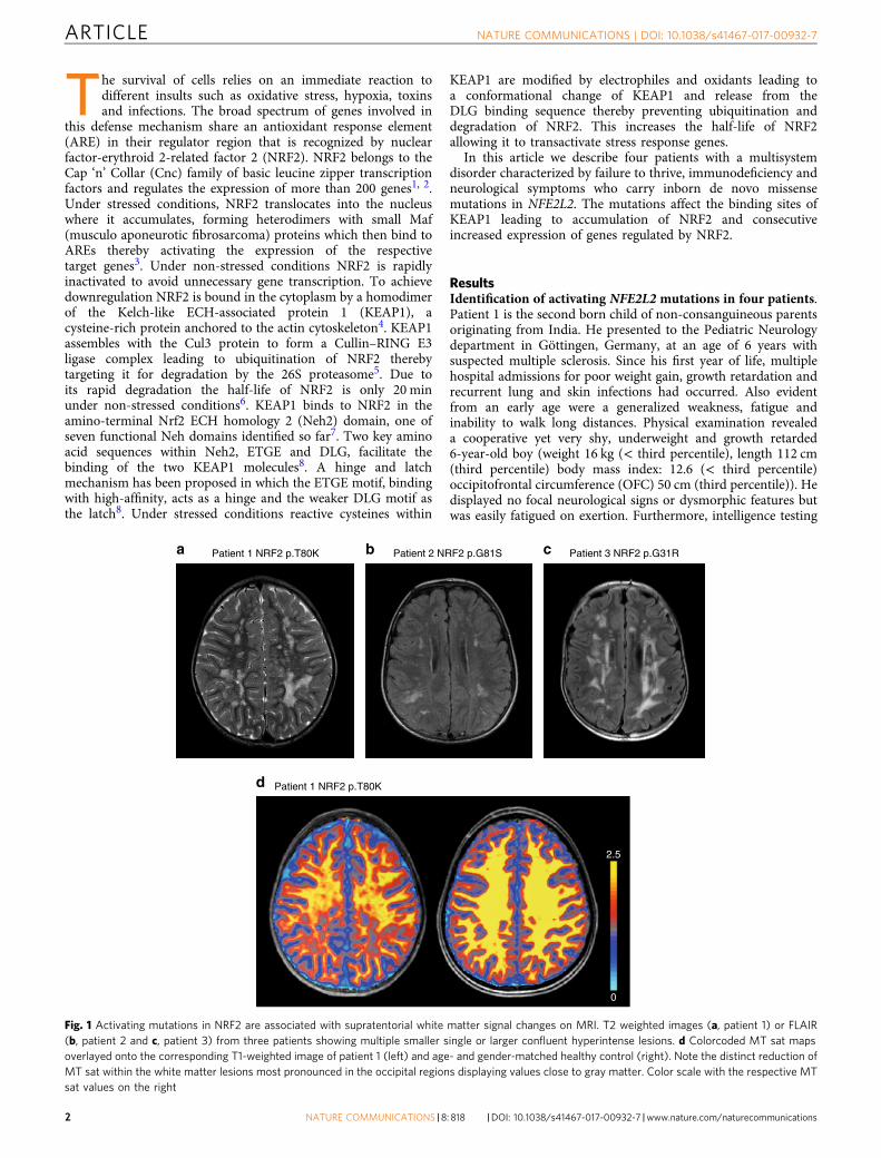
Fig. 1 Activating mutations in NRF2 are associated with supratentorial white matter signal changes on MRI. T2 weighted images (a, patient 1) or FLAIR(b, patient 2 and c, patient 3) from three patients showing multiple smaller single or larger confluent hyperintense lesions. d Colorcoded MT sat mapsoverlayed onto the corresponding T1-weighted image of patient 1 (left) and age- and gender-matched healthy control (right). Note the distinct reduction ofMT sat within the white matter lesions most pronounced in the occipital regions displaying values close to gray matter. Color scale with the respective MTsat values on the right
ARTICLE NATURE COMMUNICATIONS | DOI: 10.1038/s41467-017-00932-7
2 NATURE COMMUNICATIONS | 8: 818 |DOI: 10.1038/s41467-017-00932-7 |www.nature.com/naturecommunications
revealed an IQ of 74. Cerebral magnetic resonance (MR) imagingat age 6.9 years demonstrated bilateral periventricular and sub-cortical white matter signal hyperintensities on T2 weightedimages with sparing of infratentorial and spinal structures(Fig. 1a). No contrast enhancement was demonstrated and onserial studies over 3 years the lesions remained unchanged. Pro-ton magnetic resonance spectroscopy (MRS) showed reducedlevels of N-acetylaspartylglutamate and creatine and normallevels of lactate in gray and white matter. On magnetizationtransfer saturation (MT sat) maps, a quantitative MR parameterfor evaluating myelination, the lesions showed a distinct myelindeficit (Fig. 1d). Laboratory studies showed signs of liver damage,reduced homocysteine (2.9 µmol/l, reference range: 5.5–16.2µmol/l), and low cysteine (5.0 µmol/l, reference range for the ageof the patient: 5–45 µmol/l) levels in blood. Analysis of energymetabolism showed mildly elevated lactate in blood (2.8 mmol/l,reference range: 0.5–2.2 mmol/l), and cerebrospinal fluid (CSF)(2.0 mmol/l, reference range 1.1–1.8 mmol/l).
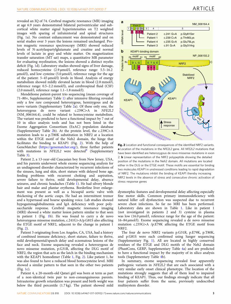
Mendeliome patient-parent trio sequencing (mean coverage of71–80×, Supplementary Table 1) after intensive filtering revealedonly a few rare compound heterozygous, hemizygous and denovo variants (Supplementary Table 2a). Of these only one, theheterozygous de novo variant c.239C>A in NFE2L2(NM_006164.4), could be related to homocysteine metabolism.The variant was predicted to have a functional impact by 7 out of10 in silico analysis tools and has not been listed in theExome Aggregation Consortium (ExAC) population database(Supplementary Table 2b). At the protein level, the c.239C>Amutation leads to a p.T80K substitution in NRF2 at a locationwithin the ETGE motif of the Neh2 domain, the hinge thatfacilitates the binding to KEAP1 (Fig. 2). With the help ofGeneMatcher (https://genematcher.org/), three further patientswith mutations in NFE2L2 were detected9 (SupplementaryTable 3).
Patient 2, a 13-year-old Caucasian boy from New Jersey, USA,and his parents underwent whole exome sequencing analysis foran undiagnosed disorder characterized by recurrent infections ofthe sinuses, lung and skin, short stature with delayed bone age,feeding problems with recurrent choking and aspiration,severe failure to thrive, mild developmental delay, absenceseizures, and chronic headaches (Table 1). He had silvery blondehair and malar and plantar erythema. Borderline liver enlarge-ment was present as well as a bicuspid aortic valve withthickening of the aortic cusps. He had an intermittent tremorand a hypernasal and hoarse speaking voice. Lab studies showedhypogammaglobulinemia and IgA deficiency with poor poly-saccharide response. Cerebral magnetic resonance imaging(MRI) showed a white matter lesion pattern similar to that seenin patient 1 (Fig. 1b). He was found to carry a de novoheterozygous missense mutation, c.241G>A/p.G81S also affectingthe ETGE motif of NRF2, adjacent to the change in patient 1(Fig. 2).
Patient 3 originating from Los Angeles, CA, USA, had a historyof combined immune deficiency, short stature, failure to thrive,mild developmental/speech delay and eczematous lesions of theface and neck. Exome sequencing revealed a heterozygous denovo missense mutation, p.G31R, affecting the DLG motif ofNRF2, the region that acts as the latch in the binding mechanismwith the KEAP1 homodimer (Table 1, Fig. 2). Like patient 1, hewas also found to have a reduced blood homocysteine level. MRIshowed a similar pattern to that seen in the other two patients(Fig. 1c).
Patient 4, a 20-month-old Qatari girl was born at term as partof a non-identical twin pair to non-consanguineous parents.Intrauterine growth retardation was present and birth weight wasbelow the third percentile (1.7 kg). The patient showed mild
dysmorphic features and developmental delay affecting especiallyfine motor skills. Common primary immunodeficiency withnatural killer cell dysfunction was suspected due to recurrentsevere chest infections. So far no MRI has been performed.Laboratory data are shown in Table 1. Like in patient 1(not investigated in patients 2 and 3) cysteine in plasmawas low (16.0 µmol/l, reference range for the age of the patient:16–84 µmol/l). Exome sequencing revealed a de novo missensemutation c.235G>A /p.E79K affecting the ETGE motif fromNRF2.
The four de novo NRF2 variants p.G31R, p.E79K, p.T80K,and p.G81S were each confirmed using Sanger sequencing(Supplementary Fig. 1). All are located in highly conservedresidues of the ETGE and DLG motifs of the Neh2 domain(PhastCons, GERP, Supplementary Table 4a) and are predictedto have a functional impact by the majority of in silico analysistools (Supplementary Table 4b).
In summary, exome sequencing revealed four apparentlypathogenic variants in NFE2L2 that are each associated with avery similar early onset clinical phenotype. The location of themutations strongly suggests that all of them lead to impairedbinding of KEAP1. Thus, our findings strongly indicate that allfour patients suffer from the same, previously undescribedmultisystem disorder.
NM_006164.4
5431 G C G
A A A
Exon 2
N - -C
29
E T G ED L G
KEAP1-binding domain
R K S
31motif 79 82motif NP_006155.2
Patient 3 c.91 G>A p.Gly31Arg
Patient 1 c.239 C>A p.Thr80Lys
Patient 4 c.235 G>A p.Glu79Lys
NRF2
Stressresponse
StabilizedNRF2
AREm
RN
A le
vel
Pro
tein
leve
l
MAF
KEAP1KEAP1
Ub
CUL3
E2
a
b
GA
Patient 2 c.241 G>A p.Gly81Ser
K
Fig. 2 Location and functional consequences of the identified NRF2 variants.a Location of the mutations in the NFE2L2 gene. All NFE2L2 mutations thathave been identified are heterozygous de novo missense mutations in exon2. b Linear representation of the NRF2 polypeptide showing the detailedposition of the mutations in the Neh2 domain. All mutations are locatedeither in the DLG or the ETGE motif. These motifs are essential for bindingtwo molecules KEAP1 in unstressed conditions leading to rapid degradationof NRF2. The mutations inhibit the binding of KEAP1 thereby increasingNRF2 levels in the absence of stress and consecutive chronic activation ofstress response genes
NATURE COMMUNICATIONS | DOI: 10.1038/s41467-017-00932-7 ARTICLE
NATURE COMMUNICATIONS |8: 818 |DOI: 10.1038/s41467-017-00932-7 |www.nature.com/naturecommunications 3
Effect of the NFE2L2 mutations. Each NFE2L2 variant that wasdetected in the patients has previously been reported as a somaticcancer-related pathogenic change in several tumor species10–16
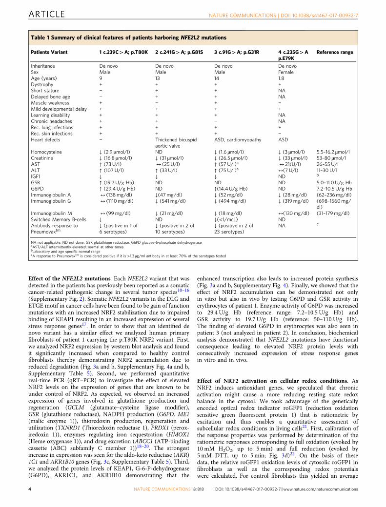
(Supplementary Fig. 2). Somatic NFE2L2 variants in the DLG andETGE motif in cancer cells have been found to be gain of functionmutations with an increased NRF2 stabilization due to impairedbinding of KEAP1 resulting in an increased expression of severalstress response genes17. In order to show that an identified denovo variant has a similar effect we analyzed human primaryfibroblasts of patient 1 carrying the p.T80K NRF2 variant. First,we analyzed NRF2 expression by western blot analysis and foundit significantly increased when compared to healthy controlfibroblasts thereby demonstrating NRF2 accumulation due toreduced degradation (Fig. 3a and b, Supplementary Fig. 4a and b,Supplementary Table 5). Second, we performed quantitativereal-time PCR (qRT–PCR) to investigate the effect of elevatedNRF2 levels on the expression of genes that are known to beunder control of NRF2. As expected, we observed an increasedexpression of genes involved in glutathione production andregeneration (GCLM (glutamate–cysteine ligase modifier),GSR (glutathione reductase), NADPH production (G6PD, ME1(malic enzyme 1)), thioredoxin production, regeneration andutilization (TXNRD1 (Thioredoxin reductase 1), PRDX1 (perox-iredoxin 1)), enzymes regulating iron sequestration (HMOX1(Heme oxygenase 1)), and drug excretion (ABCC1 (ATP-bindingcassette (ABC) subfamily C member 1))18–20. The strongestincrease in expression was seen for the aldo-keto reductase (AKR)1C1 and AKR1B10 genes (Fig. 3c, Supplementary Table 5). Third,we analyzed the protein levels of KEAP1, G-6-P-dehydrogenase(G6PD), AKR1C1, and AKR1B10 demonstrating that the
enhanced transcription also leads to increased protein synthesis(Fig. 3a and b, Supplementary Fig. 4). Finally, we showed that theeffect of NRF2 accumulation can be demonstrated not onlyin vitro but also in vivo by testing G6PD and GSR activity inerythrocytes of patient 1. Enzyme activity of G6PD was increasedto 29.4 U/g Hb (reference range: 7.2–10.5 U/g Hb) andGSR activity to 19.7 U/g Hb (reference: 50–110 U/g Hb).The finding of elevated G6PD in erythrocytes was also seen inpatient 3 (not analyzed in patient 2). In conclusion, biochemicalanalysis demonstrated that NFE2L2 mutations have functionalconsequence leading to elevated NRF2 protein levels withconsecutively increased expression of stress response genesin vitro and in vivo.
Effect of NRF2 activation on cellular redox conditions. AsNRF2 induces antioxidant genes, we speculated that chronicactivation might cause a more reducing resting state redoxbalance in the cytosol. We took advantage of the geneticallyencoded optical redox indicator roGFP1 (reduction oxidationsensitive green fluorescent protein 1) that is ratiometric byexcitation and thus enables a quantitative assessment ofsubcellular redox conditions in living cells21. First, calibration ofthe response properties was performed by determination of theratiometric responses corresponding to full oxidation (evoked by10 mM H2O2, up to 5 min) and full reduction (evoked by5 mM DTT, up to 5 min; Fig. 3d)22. On the basis of thesedata, the relative roGFP1 oxidation levels of cytosolic roGFP1 infibroblasts as well as the corresponding redox potentialswere calculated. For control fibroblasts this yielded an average
Table 1 Summary of clinical features of patients harboring NFE2L2 mutations
Patients Variant 1 c.239C>A; p.T80K 2 c.241G>A; p.G81S 3 c.91G>A; p.G31R 4 c.235G>Ap.E79K
Reference range
Inheritance De novo De novo De novo De novoSex Male Male Male FemaleAge (years) 9 13 14 1.8Dystrophy + + + +Short stature − + + NADelayed bone age − + + NAMuscle weakness + − + −Mild developmental delay + + + +Learning disability + + + NAChronic headaches + + − NARec. lung infections + + + +Rec. skin infections + + + −Heart defects − Thickened bicuspid
aortic valveASD, cardiomyopathy ASD
Homocysteine ↓ (2.9 µmol/l) ND ↓ (1.6 µmol/l) ↓ (3 µmol/l) 5.5–16.2 µmol/lCreatinine ↓ (16.8 µmol/l) ↓ (31 µmol/l) ↓ (26.5 µmol/l) ↓ (33 µmol/l) 53–80 µmol/lAST ↑ (73 U/l) ↔ (25 U/l) ↑ (57 U/l)a ↔ 21(U/l) 26–55 U/lALT ↑ (107 U/l) ↑ (33 U/l) ↑ (75 U/l)a ↔(7 U/l) 11–30 U/lIGF1 ↓ ↓ ↓ ND b
GSR ↑ (19.7 U/g Hb) ND ND ND 5.0–11.0 U/g HbG6PD ↑ (29.4 U/g Hb) ND ↑(14.4 U/g Hb) ND 7.2–10.5 U/g HbImmunoglobulin A ↔ (138mg/dl) ↓(47mg/dl) ↓ (52 mg/dl) ↓ (28mg/dl) (62–236mg/dl)Immunoglobulin G ↔ (1110mg/dl) ↓ (541 mg/dl) ↓ (494mg/dl) ↓ (319 mg/dl) (698–1560mg/
dl)Immunoglobulin M ↔ (99mg/dl) ↓ (21 mg/dl) ↓ (18 mg/dl) ↔(130mg/dl) (31–179mg/dl)Switched Memory B-cells ↓ ND ↓(<1/mcL) NDAntibody response toPneumovaxtm
↓ (positive in 1 of6 serotypes)
↓ (positive in 2 of10 serotypes)
↓ (positive in 2 of23 serotypes)
NA c
NA not applicable, ND not done, GSR glutathione reductase, G6PD glucose-6-phosphate dehydrogenaseaAST/ALT intermittently elevated; normal at other timesbLaboratory and age specific normal rangecA response to Pneumovaxtm is considered positive if it is >1.3 µg/ml antibody in at least 70% of the serotypes tested
ARTICLE NATURE COMMUNICATIONS | DOI: 10.1038/s41467-017-00932-7
4 NATURE COMMUNICATIONS | 8: 818 |DOI: 10.1038/s41467-017-00932-7 |www.nature.com/naturecommunications
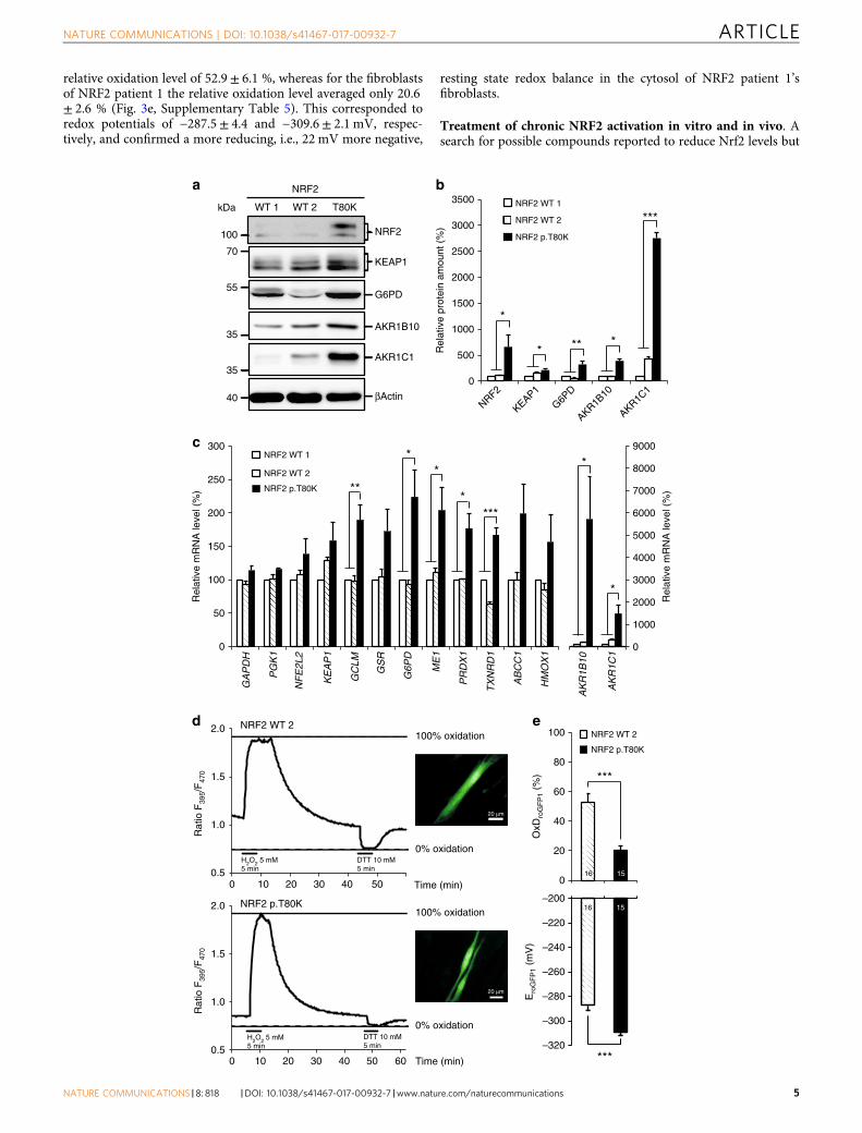
relative oxidation level of 52.9± 6.1 %, whereas for the fibroblastsof NRF2 patient 1 the relative oxidation level averaged only 20.6± 2.6 % (Fig. 3e, Supplementary Table 5). This corresponded toredox potentials of −287.5± 4.4 and −309.6± 2.1 mV, respec-tively, and confirmed a more reducing, i.e., 22 mV more negative,
resting state redox balance in the cytosol of NRF2 patient 1’sfibroblasts.
Treatment of chronic NRF2 activation in vitro and in vivo. Asearch for possible compounds reported to reduce Nrf2 levels but
a
NRF2
KEAP1
G6PD
βActin
70
55
100
kDa
AKR1B1035
AKR1C1
WT 2WT 1 T80K
NRF2
35
40
Rel
ativ
e pr
otei
n am
ount
(%
)
NRF2 WT 1
NRF2 WT 2
NRF2 p.T80K
b
***
0
AKR1B10
G6PD
KEAP1
NRF2
AKR1C1
500
1000
1500
2000
2500
3000
3500
*
****
AK
R1C
1
0
50
100
150
200
250
300
GA
PD
H
PG
K1
NF
E2L
2
KE
AP
1
GC
LM
GS
R
G6P
D
ME
1
PR
DX
1
TX
NR
D1
AB
CC
1
HM
OX
1
0
1000
2000
3000
4000
5000
6000
7000
8000
9000
AK
R1B
10
c
Rel
ativ
e m
RN
A le
vel (
%)
Rel
ativ
e m
RN
A le
vel (
%)
NRF2 WT 1
NRF2 WT 2
NRF2 p.T80K
*
****
***
*
*
0 10 20 30 40 50 60 Time (min)
NRF2 WT 2
NRF2 p.T80K100% oxidation
0% oxidation
0.5
1.0
1.5
2.0
Rat
io F
395/
F47
0R
atio
F39
5/F
470
20 μm
H2O2 5 mM5 min
H2O2 5 mM5 min
DTT 10 mM5 min
DTT 10 mM5 min
100% oxidation
0% oxidation
0.5
1.0
1.5
2.0
20 μm
d
0 10 20 30 40 50 Time (min)
NRF2 WT 2
NRF2 p.T80K
0
20
40
60
80
100
OxD
roG
FP
1 (%
)
–320
–300
–280
–260
–240
–220
–200
Ero
GF
P1
(mV
)
***
***
1516
1516
e
NATURE COMMUNICATIONS | DOI: 10.1038/s41467-017-00932-7 ARTICLE
NATURE COMMUNICATIONS |8: 818 |DOI: 10.1038/s41467-017-00932-7 |www.nature.com/naturecommunications 5

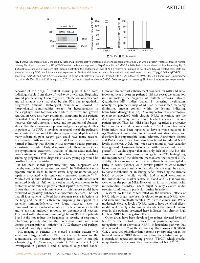
devoid of serious side effects when used in a clinical settingresulted in the identification of two candidates; ascorbic acid andluteolin23. While ascorbic acid is commonly used in clinicalmedicine, luteolin, a flavone that is meant to haveanti-inflammatory and neuroprotective effects, is not commonlyused in Western medicine so far. To test the effect of luteolin andascorbic acid in vitro, fibroblasts from patient 1 were treated for24 h with 50 µM luteolin or different concentrations of ascorbicacid. Subsequent western blotting showed that luteolin treatmentreduced the NRF2 level up to 90% (Fig. 4a and b) while treatmentwith ascorbic acid was less effective and did not consistently lowerNRF2 level to the same degree at the concentrations tested(Supplementary Fig. 5). qRT–PCR analysis of AKR1B10 andAKR1C1 gene expression, expression of both genes is upregulatedby NRF2, in fibroblasts of patient 1 treated with 50 µM luteolinshowed that downregulation of NRF2 resulted in significantlyreduced expression of AKR1B10 and AKR1C1 (Fig. 4c).
Patient 1 was then treated with luteolin (50 mg once daily) andascorbic acid (200 mg once daily). After 6 months of treatmentpreviously elevated liver enzymes had normalized but no effectwas seen on the homocysteine level. Furthermore, the motherreported a very positive development. Infections had been lessfrequent and less severe and the patient was able for the first timeto carry his bag to school and attend the sport lessons. Moreover,overall school performance had improved.
DiscussionIn this article we describe four patients with a novel disordercaused by mutations in NFE2L2 that impair the binding of NRF2by KEAP1. The patients do not have significant dysmorphicfeatures, but display a similar phenotype with several prominentfeatures including developmental delay, failure to thrive, immu-nodeficiency, leukoencephalopathy, and hypohomocysteinaemia.
Somatic missense amino acid substitutions affecting the DLGor ETGE motifs in the regulatory Neh2 domain of NRF2 arepresent in many types of cancer and have been found to beassociated with a poor prognosis17, 24. Analysis of the COSMICdatabank (cancer.sanger.ac.uk) revealed that the mutationsdetected in the four patients have been described previously indifferent kinds of cancer cells (Supplementary Fig. 2b). Shibataet al.17 studied the effect of the p.T80R mutation within the ETGEmotif, the residue affected in patient 1, in 293T cells and showedthat it substantially reduced the ability to interact with KEAP117.An NRF2 protein harboring a mutation in the DLG motif, as seenin patient 3, retained binding to KEAP1 but caused reducedNRF2 ubiquitination17. In 293T cells both mutations led toaccumulation of NRF2 in the nucleus and subsequent increasedexpression of stress response genes. Correspondingly, we foundelevated levels of NRF2 and increased expression of genes that arepart of the NRF2 mediated stress response in fibroblasts of patient
1 (Fig. 3). The strongest increase in expression was seen forAKR1C1 and AKR1B10. This result corresponds well to thefindings of MacLeod et al.20 who knocked down KEAP1 inHaCaT keratinocytes and found that expression of AKR1C1 andAKR1B10 increased 12-and 16-fold.
All experiments on chronic NRF2 activation so far have beendone in vitro. The patients described herein enabled us to analyzethe in vivo effects of NRF2 upregulation. Enzyme activities ofG6PD and GSR were determined in blood of patients 1 and 3(Table 1). 3-fold elevation of G6PD activity and 2-fold of GSRactivity clearly demonstrated an in vivo effect of the mutations.
One of the hallmarks of this novel disorder is hypohomocys-teinaemia. Several disorders with high levels of homocysteinehave been described causing generalized vascular damage andthromboembolic complications but so far no disorder is knownthat is associated with reduced levels. Hypohomocysteinaemia ismost likely a direct effect of NRF2 activation as NRF2 positivelyregulates glutathione synthesis for which homocysteine serves asa precursor. Cysteine, which was also found reduced in the bloodof patient 1 and 4 (not investigated in patient 2 and 3), is gen-erally considered the limiting component of glutathione bio-synthesis25. It has been found in HepG2 cells that as much as halfof the cysteine used for glutathione biosynthesis is generatedfrom homocysteine utilizing the transsulfuration pathway26.In the next step γ-glutamylcysteine is synthesized from cysteineand L-glutamate. This reaction is catalyzed by glutamate–cysteineligase (GCL) and is the rate limiting step in glutathione synthesis.We found that the expression of GCL was increased almost 2-foldin the fibroblasts of patient 1 (Fig. 3). The final step of glutathionesynthesis, the C-terminal addition of glycine, is catalyzed byglutathione synthetase, an enzyme also positively regulated byNRF2.
We speculated that the permanently increased level of NRF2would affect the redox balance in the cytosol because NRF2 isphysiologically upregulated as a response to oxidative stress.Accordingly, we found overexpression of enzymes necessary forthe generation and regeneration of the two major antioxidantsglutathione and thioredoxin. Determination of the redox condi-tions in NRF2 mutant fibroblasts of patient 1 using the opticalredox indicator roGFP1 indeed confirmed a marked reducingshift of the cytosolic redox balance (Fig. 3e) which leads toincreased reduction stress. Proteome reactivity profiling indicatesthat 890 human proteins are potentially sensitive to redoxmodulation resulting in either gain or loss of function27. Thus,potential cellular dysfunction by misregulated proteins will affectmany pathways thereby further enhancing the negative effect ofNFR2 accumulation.
In 2003, Wakabayashi et al.28 reported on a mouse modelcarrying a Keap1-null mutation that led to constitutive chronicNrf2 activation, as seen in the patients presented here. Size and
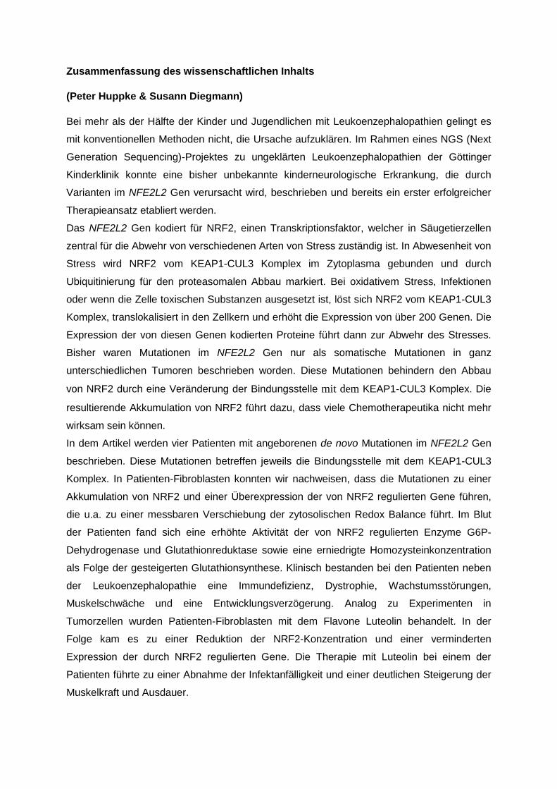
Fig. 3 Increased stabilization and activation of mutant NRF2. a Representative western blot of endogenous level of NRF2, KEAP1, G6PD, AKR1B10 andAKR1C1 in protein lysates of human primary fibroblast cell lines from two controls (NRF2 WT 1, WT 2) and patient 1 with NRF2 p.T80K variant. Full blotsare shown in Supplementary Fig. 6. b Quantitative analysis of western blot images illustrating the endogenous level of NRF2, KEAP1, G6PD, AKR1B10 andAKR1C1 relative normalized to ACTB and NRF2 WT 1. c qRT–PCR analysis of NFE2L2, KEAP1 and target gene expression in primary fibroblast cell lines fromtwo controls (NRF2 WT 1, WT 2) and patient 1 with NRF2 p.T80K variant. AKR1B10 and AKR1C1 are visualized on a separated X axis due to the high range.Expression is normalized to that of ACTB. % of mRNA is equal to 2−ΔΔCT and normalized relative to NRF2 WT 1. Redox calibration confirms fullfunctionality of roGFP1 as well as identical response ranges for NRF2 WT 2 and NRF2 p.T80K fibroblast cells. d Response range calibration of an exemplaryNRF2 WT 2 and NRF2 p.T80K fibroblast cell performed as a continuous recording of the roGFP1 ratio F395/F470 within a ROI of cytoplasm of the cell, scalebar is 20 µM. Plotted traces represent full oxidation (Rox, induced by 5mM H2O2, 5 min) and full reduction (Rred, induced by 10mM DTT, 5 min). Aftercalibration the relative degrees of roGFP1 oxidation and corresponding roGFP1 redox potentials can be calculated. e Baseline redox conditions of NRF2 WT2 and NRF2 p.T80K fibroblasts. Upper diagram shows the relative level of roGFP1 oxidation of NRF2 WT 2 and NRF2 p.T80K cells at rest (OxDroGFP1, Eq. 1).Lower diagram represents corresponding steady-state roGFP1 redox potential (EroGFP1, Eq. 2). b, c Data are given as means± SEM, n≥ 3 independentexperiments. Data were analyzed by one-way analysis of variance with multiple comparisons: *p≤ 0.05, **p≤ 0.01, ***p≤ 0.001. e Data are given asmeans± SEM. Number of measured cells are given within the bar. Statistical differences were obtained with unpaired Welch’s t-test: ***p≤ 0.001
ARTICLE NATURE COMMUNICATIONS | DOI: 10.1038/s41467-017-00932-7
6 NATURE COMMUNICATIONS | 8: 818 |DOI: 10.1038/s41467-017-00932-7 |www.nature.com/naturecommunications
behavior of the Keap1–/– mutant mouse pups at birth wereindistinguishable from those of wild-type littermates. Beginningaround postnatal day 4 severe growth retardation was observedand all mutant mice had died by day P21 due to graduallyprogressive asthenia. Histological examination showed nomorphological abnormalities except for hyperkeratosis inthe esophagus and forestomach. Failure to thrive and growthretardation were also very prominent symptoms in the patientspresented here. Endoscopy performed on patients 1 and 2,however, showed a normal mucosa and no anatomical abnorm-alities other than a narrow esophagus and gastroesophageal refluxin patient 2. As NRF2 is involved in several metabolic pathwaysand constant activation of the stress response will deplete cells ofmany substrates, poor weight gain could have many reasons.Interestingly, birth measurements in all four patients were alsonormal indicating that chronic NRF2 activation causes primarilya postnatal disorder. Early diagnosis could therefore facilitatepre-symptomatic treatment. Analysis of amino acids includinghomocysteine and G6PD activity is included in many newbornscreening programs thus diagnosis at a very young age would bepossible in many countries.
It has been shown previously that Nrf2 suppresses andthereby controls inflammatory reactions29. In Nrf2-deficient micecigarette smoke leads to more severe lung inflammation andsepsis is associated with significantly increased mortality30, 31.Myeloid cell-specific deletion of Keap1 in mice with subsequentenhanced levels of Nrf2 on the other hand, was shown to beprotective of mortality in polymicrobial sepsis32. Moreover, it wasshown that the innate immune cells in this mouse model havepreserved or possibly enhanced antibacterial defenses. The factthat all four patients suffered from frequent severe infections ofthe lung and the skin is therefore surprising. In support of asystemic immunodeficiency we found reduced levels ofimmunoglobulins, a reduced number of switched memory B-cellsand a missing antibody response to pneumococcal vaccine.Treatment with intravenous immunoglobulins (IVIG) in patients2 and 3 did not reduce the frequency or severity of respiratoryinfections, possibly due to the preexisting lung and sinusdisease present prior to initiation of IVIG therapy and perhapscoincident T-cell dysfunction.
MR imaging in patients 1–3 showed a similar pattern withsmall and large confluent T2 hyperintense lesions in thesupratentorial white matter resembling lesions seen in multiplesclerosis (Fig. 1). Moreover, analysis of CSF in patient 1 (notinvestigated in patients 2 and 3) revealed oligoclonal bands.
However, no contrast enhancement was seen on MRI and serialfollow-up over 3 years in patient 1 did not reveal disseminationin time making the diagnosis of multiple sclerosis unlikely.Quantitative MR studies (patient 1) assessing myelination,namely the parameter map of MT sat, demonstrated markedlydiminished myelin content within the lesions indicatingbrain tissue damage (Fig. 1d). Also supportive of a neurologicalphenotype associated with chronic NRF2 activation, are thedevelopmental delay and chronic headaches evident in ourpatient group. Thus far, NRF2 has been regarded a protectivefactor in the central nervous system33. Stroke and traumaticbrain injury have been reported to have a worse outcome inNfe2l2-deficient mice due to increased oxidative stress anddisorders like amyotrophic lateral sclerosis, Alzheimer’s diseaseand Parkinson’s disease have also been linked to reduced NRF2levels. Moreover, Nfe2l2-null mice were found to have vacuolar(spongiform) leukoencephalopathy with widespread astro-gliosis34. It would appear that not only lack of NRF2 but alsochronic activation may cause pathology in the CNS, highlightingthe importance of the elaborate mechanisms that control NRF2activity. One can only speculate why there is leukoencephalo-pathy in NRF2 patients. As a similar pattern of white matterlesions can be seen in mitochondrial disorders, it might be causedby toxic metabolites or an energy deficit caused by the chronicNRF2 activation. While we did find a mild elevation ofthe mitochondrial marker lactate in blood and CSF it was notelevated in the proton MRS. However, as in many patients withmitochondrial disorders, lactate might be only elevated understressful conditions, in particular during infections.
Research so far has concentrated on the beneficial effects ofNRF2. Many drugs have been developed to enhance NRF2 levelsand some like dimethylfumarate (DMF) are in clinical use. Whilemoderately elevated levels of NRF2 seem to have beneficial effectsin different mainly autoimmune disorders the clinical pictureseen in the patients presented here indicates that chronic highlevels of NRF2 have negative effects.
Other drugs have been developed to reduce elevated levels ofNRF2 for the control of cancer23. A possible approach isupregulation of an alternative KEAP1 independent pathway thatdownregulates NRF2 via the glycogen synthase kinase-3 (GSK-3).GSK-3 catalyzed phosphorylation forms a phosphodegron in theNeh6 domain of NRF2 thereby recruiting the substrate receptorβ-transducin repeat-containing protein (βTrCP) which enablesubiquitination and consecutive degeneration of NRF235, 36.
40
100
kDa
Luteolin 24 h
NRF2
a
DMSO 50 μM
DMSO 50 μM luteolin
0
20
40
60
80
100
120
NRF2
***b
Rel
ativ
e pr
otei
n am
ount
(%
)
0
20
40
60
80
100
AKR1B10 AKR1C1
c
Rel
ativ
e m
RN
A le
vel (
%)
βActin
Fig. 4 Downregulation of NRF2 induced by luteolin. a Representative western blot of endogenous level of NRF2 in whole protein lysates of treated humanprimary fibroblast of patient 1. NRF2 p.T80K mutant cells were exposed to 50 µM luteolin or DMSO for 24 h. Full blots are shown in Supplementary Fig. 7.b Quantitative analysis of western blot images illustrating the endogenous level of NRF2 relative normalized to ACTB and DMSO treated cells. Data aregiven as means± SEM, n= 3 independent experiments. Statistical differences were obtained with unpaired Welch’s t-test: ***p≤ 0.001. c qRT–PCRanalysis of AKR1B10 and AKR1C1 gene expression in primary fibroblasts of patient 1 treated with 50 µM luteolin or DMSO for 24 h. Expression is normalizedto that of GAPDH. % of mRNA is equal to 2−ΔΔCT and normalized relative to DMSO. Data are given as means± SEM, n= 2 independent experiments
NATURE COMMUNICATIONS | DOI: 10.1038/s41467-017-00932-7 ARTICLE
NATURE COMMUNICATIONS |8: 818 |DOI: 10.1038/s41467-017-00932-7 |www.nature.com/naturecommunications 7
We performed a literature search of all drugs that have beendescribed to lower Nrf2 levels in order to identify candidates forthe treatment of the patients described here. Luteolin andascorbic acid were chosen because they are considered harmlessand without side effects when applied in the commonly useddosages37. Luteolin (3′,4′,5,7-tetrahydroxyflavone), is a flavonefound in leaves that has been shown to downregulate NRF2 levelsindependent of KEAP1 by enhancing NRF2 mRNA turnover andreducing NRF2 binding to AREs38, 39. To test luteolin’s suitabilityin the specific situation of activating NRF2 mutations we firstlyanalyzed the effect in fibroblasts of patient 1 and found that 24 htreatment indeed resulted in a significant reduction of NRF2levels leading to downregulation of AKR1B10 and AKR1C1mRNA (Fig. 4). In a separate experiment the fibroblasts weretreated with ascorbic acid but this treatment was less effective toreduce NRF2 levels (Supplementary Fig. 5, SupplementaryTable 5). We initiated treatment of patient 1 with 50 mg luteolinand 200mg ascorbic acid given orally once daily. No side effectswere reported and the effect was surprisingly positive. Accordingto the parents after 6 months of treatment the frequency ofinfections had reduced and the muscle strength and endurancehad increased. Moreover, general school performance hadimproved. However, this is clearly a preliminary result to beviewed with caution as only one patient has received treatment sofar.
In conclusion, we present a novel disorder caused by inbornactivating mutations in NFE2L2 leading to widespreadmisregulation of gene expression and an altered cytosolic redoxbalance. The clinical hallmarks of the disorder are failure tothrive, immunodeficiency and leukoencephalopathy. Due tothe unique laboratory findings of hypohomocysteinaemia andelevated G6PD activity early diagnosis within the frameworkof newborn screening programs would be possible enablingpre-symptomatic diagnosis and early therapeutic intervention.Finally, our findings challenge the very positive picture of NRF2that currently exists in the literature and indicate that cautionshould be applied in the use of medications known to lead toNRF2 accumulation.
MethodsQuantitative MT imaging. MT imaging was performed on a 3T clinical MRsystem (Tim Trio, Siemens Healthcare, Erlangen, Germany) using a 3D FLASHsequence with 1.25 mm isotropic resolution and 240 mm field-of-view. MTcontrast was imposed upon a proton density reference by applying a 12.8 msGaussian MT-pulse (540° nominal flip angle 2.2 kHz off resonance)40. By means ofa second T1-weighted reference (TR/α= 11 ms/15°, 1.5 min), maps of thepercentage MT sat were calculated. Data processing was scripted using the routinesof the FSL 4.1 software library of the Centre for Functional Magnetic ResonanceImaging of the Brain (Oxford, UK). The cyan-blue-gray-red-yellow color scale ofthe MT sat maps covered a range from −0.1 pu (cyan; cerebrospinal fluid) to 1.2 pu(gray, gray matter) to 2.5 pu (yellow; white matter). Myelinated WM of controls(MT sat >2.5 pu) appeared uniformly yellow. Red indicates partial volume of whiteand gray matter. Dark blue indicates edema or partial volume of cerebrospinal fluidand brain tissue.
Mendeliome sequencing and bioinformatic analysis. DNA samples wereobtained from four patients and parents following informed consent and approvalby the ethic commission from the University Medical Center Göttingen, Göttingen,Germany (patient 1), in patient 2–4 genetic testing was performed as a componentof routine clinical care with informed parent consent. ‘Mendeliome’ gene paneldata, which includes 4813 disease associated genes, was generated from the indexpatient (patient 1) as well as the parents using next-generation sequencingapproach in cooperation with Cologne Center for Genomics (CCG, University ofCologne, Germany). Used DNA was extracted from peripheral EDTA bloodusing standard protocols. For each Mendeliome, 50 ng of DNA was fragmented,barcoded and enriched for the TruSightTM One Sequencing Panel (Illumina,San Diego, CA, USA) using Nextera library preparation technology. Purified andquantified library pool was subsequently sequenced on an Illumina MiSeqsequencing instrument (Illumina, San Diego, CA, USA) using a multiplex pairedend 2 × 150 bp protocol with 3 Mendelioms per run. Data processing, analysis andfiltering were performed using the ‘Varbank’ GUI and pipeline version 2.14 (CCG,
University of Cologne, Germany) (https://varbank.ccg.uni-koeln.de/). Reads weremapped to the human genome reference build hg19 using the BWA-aln alignmentalgorithm. GATK v.1.62 was used to mark duplicated reads, to do a local rea-lignment around short insertions and deletions, to recalibrate the base qualityscores and to call SNPs and short Indels. The GATK UnifiedGenotyper variationcalls were filtered for high-quality (DP> 15; AF > 0.25; QD> 2; MQ> 40; FS<60;MQRankSum> −12.5; ReadPosRankSum > −8; HaplotypeScore< 13) rare(MAF≤ 0.005 based on 1000 genomes build 20110521 and EVS build ESP6500 andthe Exome Aggregation Consortium (http://exac.broadinstitute.org/)) variants,predicted to modify a protein sequence or to impair splicing, implicated by reducedmaximum entropy scores (MaxEntScan). The DeNovoGear software was used toidentify de novo mutations. False positive and irrelevant variants were furtherreduced by taking advantage of the Varbank InHouseDB containing 511 epilepsyexomes. Based on the Trio-patient-parent sequencing approach de novo, com-pound heterozygous and homozygous/hemizygous variants were extracted for thepatient. Prediction of functional impact of all received variants was performedusing the dbNSFP version 3.0a36,37. DbNSFP software co-applied several in silicoanalysis tools to predict the conservation, using PhastCons and GERP, and thefunctional consequence, using SIFT, PolyPhen2, Provean, LRT, MutationTaster,MutationAssessor, FATHMM, VEST, MetaSVM, and MetaLR, of the affected site.In addition phenotype genotype correlations were investigated using public data-base Online Mendelian Inheritance (OMIM) (http://www.omim.org/), Orphanet(http://www.orpha.net) and ClinVar (https://www.ncbi.nlm.nih.gov/clinvar).
Whole exome sequencing of patients 2 and 3 was performed at GeneDx.Genomic DNA was extracted from whole blood from affected children and theirparents. Whole exome sequencing was performed on exon targets captured usingthe Clinical Research Exome kit (Agilent Technologies, Santa Clara, CA). (Tanakaet al.—PMID 26299366)41. The general assertion criteria for variant classificationare publicly available on the GeneDx ClinVar submission page (http://www.ncbi.nlm.nih.gov/clinvar/submitters/26957/).
NFE2L2 mutations were validated by Sanger sequencing (primer information isavailable in Supplementary Table 6a).
Cell culture. Human skin biopsy from the NRF2 p.Thr80Lys mutant patient 1 wasobtained at the Georg August University, Department of Pediatrics and PediatricNeurology, after informed consent. Permission by the ethics committee of theUniversity Medical Center Göttingen, Göttingen, Germany has been granted(Nr. 2/5/16). Patients 2 and 3 refused a skin biopsy.
Human primary skin fibroblasts were extracted from NRF2 p.Thr80Lys mutanthuman skin biopsy, after informed consent, and maintained as monolayer culturesin Dulbecco’s modified Eagle’s medium (DMEM/low glucose) supplemented with10% fetal bovine serum (FBS), 2 mM L-glutamine and 100 U/ml penicillin, 100 μg/ml streptomycin. All reagents were purchased from Biochrom GmbH, Berlin,Germany. Cells were incubated at 37 °C in an atmosphere of 5% CO2. Stock bankswere prepared to have early cell passage available.
For molecular biological investigations, NRF2 p.Thr80Lys mutant and wild-typecells were seeded with 1 × 10⁶ cells per 10 cm culture plate in a total volume of 10ml. After 2 days of cultivation all cells were washed with PBS Dulbecco (Biochrom,Berlin, Germany) and harvested using cell scraper. During cultivation, the culturemedium was changed after 1 day. Control plates included fibroblasts from healthypatients negative tested for NRF2 variants in the DLG and ETGE motif (NRF2 WT1—WT 5). All cell cultures were regularly tested with PCR-based test for detectionof mycoplasma contamination. In addition, cells were validated for NFE2L2mutation status using Sanger sequencing (primer information is available inSupplementary Table 6a).
MG-132 and D,L-sulforaphane treatment. In order to judge the NRF2 antibodyspecificity and sensitivity, NRF2 p.Thr80Lys mutant and wild-type cells weretreated separately with the proteasome inhibitor MG-132 (10 mM in DMSO, CellSignaling, Cambridge, UK) and D,L-Sulforaphane SFN (10 mM in DMSO, Sigma,Saint Louis, MO, USA) as recommended. DMSO was used as a control. Cells weretreated with 10 µM MG-132 or 10 µM D,L-Sulforaphane for the last 16 h beforecollecting. Stabilization of NRF2 was analyzed by immunoblotting. Thespecificity of the used NRF2 antibody was confirmed as advised in the literature42
(Supplementary Fig. 3).
Luteolin and ascorbic acid treatment. NRF2 p.Thr80Lys mutant fibroblast cellsof patient 1 were seeded with 3 × 10⁵ cells per well in a six-well plate in a totalvolume of 2 ml. After 1 day, cells were stimulated for 24 h with the flavone luteolinor ascorbic acid. Luteolin (≥98 purity, Sigma-Aldrich) was solubilized in DMSO toobtain a 20 mM stock solution. Ascorbic acid (≥99 purity, Roth) was solubilizedin H2O to obtain a 100 mM stock solution. Stimulation of cells was performed inFBS-free DMEM cell culture medium supplemented with a final concentration of50 µM Luteolin or 0.1 mM, 0.25 mM, 0.5 mM, 0.75 mM, and 1 mM ascorbic acid.Control cells were incubated with the same amount of DMSO (0.25%) or withoutany supplements. Effect of luteolin or ascorbic acid on the expression of NRF2 wasanalyzed by immunoblotting. All treatment experiments consisted of at least threeindependent replicates. Effect of luteolin on the mRNA level of the NRF2 targetsAKR1B10 and AKR1C1 was analyzed by qRT–PCR.
ARTICLE NATURE COMMUNICATIONS | DOI: 10.1038/s41467-017-00932-7
8 NATURE COMMUNICATIONS | 8: 818 |DOI: 10.1038/s41467-017-00932-7 |www.nature.com/naturecommunications
qRT–PCR. Total RNA was extracted from non-treated NRF2 mutant and wild-typefibroblasts using NucleoSpin RNA Kit (Macherey-Nagel, Düren, Germany) asrecommended by the manufacturer. RNA quality was verified by gel electro-phoresis and OD measurements. For first-strand cDNA synthesis 2 µg of RNA wasreverse transcribed using oligo(dT)15 primers and SuperScript III First-StrandSynthesis System (Invitrogen, Karlsruhe, Germany) according to manufacturer’srecommendation. qRT–PCR was based on the SYBR green technology using the iQSYBR Green Supermix kit (BioRad Laboratories, Munich, Germany). TheqRT–PCR reactions were performed in triplicates on the MyiQ Single-ColorReal-Time PCR Detection System (Bio-Rad Laboratories, Munich, Germany) atannealing temperatures of 60 °C and specificity controlled by post-amplificationmelting curve analysis. RT–PCR quantification was performed according to theΔΔCT method (Livak and Schmittgen, 2001)43. Data were calculated based on thehousekeeping gene ACTB and the control fibroblasts NRF2 WT 1. Additionalinternal reference genes were GAPDH and PGK1. NFE2L2 and the interacting partnerKEAP1 as well as described NRF2 target genes GSR, GCLM, G6PD, ME1, PRDX1,TXNRD1, ABCC1, HMOX1, AKR1B10, and AKR1C1 were examined. Primers flankedan intron with amplicon length <150 bp were designed using qPrimerDepot (https://primerdepot.nci.nih.gov/) and ordered by Integrated DNA Technology (IDT, Leuven,Belgium). Only primers with an amplification efficiency of ~ 2 were used. Detailedprimer information is available in Supplementary Table 6b. All qRT–PCR experi-ments consisted of at least three independent replicates.
Immunoblotting. Fibroblasts were lysed in SDS-sample buffer (25 mM Tris, 1%SDS, pH 7.5) and rapidly frozen with liquid nitrogen. After quick thawing, lysateswere denatured at 95 °C for 5 min. For DNA digestion lysates were incubated at 37°C for 15 min with benzonase nuclease (Santa Cruz biotechnology, Dallas, TX,USA). Whole protein lysates were clarified by centrifugation (13,000 × rpm, 10min). Protein concentration was measured using BC Assay protein quantitation kit(Interchim, Montlucon,France) and samples were diluted in 4XLämmlibuffer withDTT (320 mM Tris/HCl, 8% SDS, 20% Gycerol, 0.1% bromphenol blue, 0.6 MDTT, pH 6.8). A total of 25 µg of protein lysates were separated by 10% SDS-Pageand transferred onto nitrocellulose membrane using semi dry blotting. Membraneswere blocked in 5% nonfat milk in PBS buffer with 0.1 % Tween and thenimmunoblotted overnight at 4 °C using the following primary antibodies at thespecified concentrations: ACTB (AC-15, mouse, Sigma-Aldrich, 1:10,000 dilution),NRF2 (D1Z9C, rabbit, cell signaling technology, 1:1000 dilution), KEAP1 (D1G10,rabbit, Cell Signaling Technology, 1:1000 dilution), and G6PD (D5D2, rabbit, cellsignaling technology, 1:1000 dilution), AKR1B10 (ab96417, rabbit, abcam, 1:1000dilution) and AKR1C1 (ab192785, rabbit, abcam, 1:1000 dilution). SecondaryHRP-labeled antibodies were obtained from Jackson ImmunoResearch Laboratory.Immunoblotting detection using Lumi-Light and Lumi-Light Plus blottingsubstrate (Roche, Mannheim, Germany) was documented with CCD digitalimaging ImageQuant LAS-4000 system (GE Health care Life Sciences, Freiburg,Germany). All immunoblot experiments consisted of at least three independentreplicates. Quantification of the Immunoblotting data was performed usingImageJ44. Data were normalized to the reference gene ACTB and the controlfibroblasts NRF2 WT 1.
Measurement of GSR and G6PD activity. For quantification of GSR activity,erythrocytes from EDTA blood were washed and hemolyzed, and hemolysates wereincubated with GSSG and NADPH at 37 °C for the kinetic UV-test as described45.
For quantification of glucose-6-phosphate dehydrogenase activity, erythrocytesfrom EDTA blood were washed and hemolyzed, and hemolysates were incubatedwith glucose-6-phosphate and NADP at 37 °C for the kinetic UV-test asdescribed46.
Optical recordings of cytosolic redox conditions. Cytosolic redox conditionswere monitored optically by taking advantage of the optical redox sensor roGFP1(reduction oxidation sensitive green fluorescent protein 1)21, 47. Fibroblast cultureswere transiently transfected (lipofectatime 2000, Invitrogen) with a pEGFP-N1/roGFP1 plasmid vector expressing cytosolic roGFP1. Each culture well was filledwith 200 µl transfection solution (OptiMEM, Invitrogen) containing 1% lipo-fectamine plus 1 µg DNA. Upon incubation for 1 h the transfection solution wasexchanged with fresh medium. Sufficient levels of roGFP1 expression wereobtained within 3 days post transfection.
Excitation-ratiometric redox imaging was performed with a computer-controlled epifluorescence imaging system, which was assembled from apolychromatic xenon-light source (Polychrome II, Till Photonics), a sensitive CCDcamera (Imago QE, PCO Imaging), and an upright fluorescence microscope(Axioscop 1, Zeiss). Transfected fibroblast cultures were placed in a submersion-style chamber (30–32 °C) and excited alternately at 395 nm and 470 nm at framerates of 0.1 Hz. Fluorescence was recorded with a 63x water immersion objective(Zeiss Apochromat, 1.0 NA), a 492 nm shortpass excitation filter, a 495 nmdichroic mirror, and a 525/50 nm bandpass emission filter. The roGFP1fluorescence ratio (F395/F470) was determined within defined regions of interest bycalculating the mean pixel gray values using the TILLvisION control software ofthe imaging system (version 4.0.1; TILL Photonics)48. For quantitative analysis, theroGFP1 ratio was calibrated to saturating oxidizing (5 mM H2O2) and reducing
responses (10 mM DTT)21, 48. Based on these calibration data, the relativeoxidation level of roGFP1 (OxDroGFP1) was determined22, 49:
OxDroGFP1 ¼ RoRredF470oxF470red
ðRox � RÞ þ ðR� RredÞThe corresponding cytosolic roGFP1 redox potentials (EroGFP1) can then be
calculated using the Nernst equation and the standard redox potential of roGFP1(E0´ = −291mV)21, 22, 49:
EroGFP1 ¼ E00roGFP1 � RT
2Fln
1nOxDroGFP1
OxDroGFP1
� �
Statistics. All statistics were calculated with IBM SPSS Statistics 24. Final graphswere represented as mean percentages ± SEM. One-way analysis of variancewas performed with multiple comparison post-hoc-test for qRT–PCR andimmunoblotting analysis. Unpaired Welch’s t test was performed for luteolintreatment as well as relative oxidation level of roGFP1 and roGFP1 redox potential.p ≤ 0.05 was considered significant: *p≤ 0.05, **p ≤ 0.01, ***p≤ 0.001. Statisticaldetails are included in Supplementary Table 5.
Data availability. The data that support the findings of this study are availablefrom the corresponding author upon request.
Received: 29 December 2016 Accepted: 7 August 2017
References1. Kobayashi, M. & Yamamoto, M. Nrf2-Keap1 regulation of cellular defense
mechanisms against electrophiles and reactive oxygen species. Adv. EnzymeRegul. 46, 113–140 (2006).
2. Moi, P., Chan, K., Asunis, I., Cao, A. & Kan, Y. W. Isolation of NF-E2-relatedfactor 2 (Nrf2), a NF-E2-like basic leucine zipper transcriptional activator thatbinds to the tandem NF-E2/AP1 repeat of the beta-globin locus control region.Proc. Natl Acad. Sci. USA 91, 9926–9930 (1994).
3. Itoh, K. et al. An Nrf2/small Maf heterodimer mediates the induction of phaseII detoxifying enzyme genes through antioxidant response elements. Biochem.Biophys. Res. Commun. 236, 313–322 (1997).
4. Itoh, K. et al. Keap1 represses nuclear activation of antioxidant responsiveelements by Nrf2 through binding to the amino-terminal Neh2 domain. GenesDev. 13, 76–86 (1999).
5. Furukawa, M. & Xiong, Y. BTB protein Keap1 targets antioxidant transcriptionfactor Nrf2 for ubiquitination by the Cullin 3-Roc1 ligase. Mol. Cell. Biol. 25,162–171 (2005).
6. Itoh, K. et al. Keap1 regulates both cytoplasmic-nuclear shuttling anddegradation of Nrf2 in response to electrophiles. Genes Cells 8, 379–391 (2003).
7. Wang, H. et al. RXRalpha inhibits the NRF2-ARE signaling pathway through adirect interaction with the Neh7 domain of NRF2. Cancer Res. 73, 3097–3108(2013).
8. Tong, K. I., Kobayashi, A., Katsuoka, F. & Yamamoto, M. Two-site substraterecognition model for the Keap1-Nrf2 system: a hinge and latch mechanism.Biol. Chem. 387, 1311–1320 (2006).
9. Sobreira, N., Schiettecatte, F., Valle, D. & Hamosh, A. GeneMatcher: amatching tool for connecting investigators with an interest in the same gene.Hum. Mutat. 36, 928–930 (2015).
10. Parsons, D. W. et al. The genetic landscape of the childhood cancermedulloblastoma. Science 331, 435–439 (2011).
11. Sawada, G. et al. Genomic landscape of esophageal squamous cell carcinoma ina Japanese population. Gastroenterology 150, 1171–1182 (2016).
12. Imielinski, M. et al. Mapping the hallmarks of lung adenocarcinoma withmassively parallel sequencing. Cell 150, 1107–1120 (2012).
13. Kim, Y. R. et al. Oncogenic NRF2 mutations in squamous cell carcinomas ofoesophagus and skin. J. Pathol. 220, 446–451 (2010).
14. Shibata, T. et al. NRF2 mutation confers malignant potential and resistance tochemoradiation therapy in advanced esophageal squamous cancer. Neoplasia13, 864–873 (2011).
15. Sato, Y. et al. Integrated molecular analysis of clear-cell renal cell carcinoma.Nat. Genet. 45, 860–867 (2013).
16. Tanase, A. M. et al. Mutation spectrum of hepatocellular carcinoma fromeastern-European patients betrays the impact of a complex exposome. J. Expo.Sci. Environ. Epidemiol. 25, 256–263 (2015).
17. Shibata, T. et al. Cancer related mutations in NRF2 impair its recognition byKeap1-Cul3 E3 ligase and promote malignancy. Proc. Natl Acad. Sci. USA 105,13568–13573 (2008).
18. Gorrini, C., Harris, I. S. & Mak, T. W. Modulation of oxidative stress as ananticancer strategy. Nat. Rev. Drug Discov. 12, 931–947 (2013).
NATURE COMMUNICATIONS | DOI: 10.1038/s41467-017-00932-7 ARTICLE
NATURE COMMUNICATIONS |8: 818 |DOI: 10.1038/s41467-017-00932-7 |www.nature.com/naturecommunications 9
19. Mitsuishi, Y., Motohashi, H. & Yamamoto, M. The Keap1-Nrf2 systemin cancers: stress response and anabolic metabolism. Front. Oncol. 2, 200(2012).
20. MacLeod, A. K. et al. Characterization of the cancer chemopreventiveNRF2-dependent gene battery in human keratinocytes: demonstration that theKEAP1-NRF2 pathway, and not the BACH1-NRF2 pathway, controlscytoprotection against electrophiles as well as redox-cycling compounds.Carcinogenesis 30, 1571–1580 (2009).
21. Hanson, G. T. et al. Investigating mitochondrial redox potential withredox-sensitive green fluorescent protein indicators. J. Biol. Chem. 279,13044–13053 (2004).
22. Meyer, A. J. & Dick, T. P. Fluorescent protein-based redox probes. Antioxid.Redox. Signal. 13, 621–650 (2010).
23. No, J. H., Kim, Y. B. & Song, Y. S. Targeting nrf2 signaling to combatchemoresistance. J. Cancer Prevent. 19, 111–117 (2014).
24. Menegon, S., Columbano, A. & Giordano, S. The dual roles of NRF2 in cancer.Trends Mol. Med. 22, 578–593 (2016).
25. Liu, Y., Hyde, A. S., Simpson, M. A. & Barycki, J. J. Emerging regulatoryparadigms in glutathione metabolism. Adv. Cancer Res. 122, 69–101 (2014).
26. Mosharov, E., Cranford, M. R. & Banerjee, R. The quantitatively importantrelationship between homocysteine metabolism and glutathione synthesis bythe transsulfuration pathway and its regulation by redox changes. Biochemistry39, 13005–13011 (2000).
27. Weerapana, E. et al. Quantitative reactivity profiling predicts functionalcysteines in proteomes. Nature 468, 790–795 (2010).
28. Wakabayashi, N. et al. Keap1-null mutation leads to postnatal lethality due toconstitutive Nrf2 activation. Nat. Genet. 35, 238–245 (2003).
29. Kobayashi, E., Suzuki, T. & Yamamoto, M. Roles nrf2 plays in myeloid cells andrelated disorders. Oxid. Med. Cell. Long. 2013, 529219 (2013).
30. Iizuka, T. et al. Nrf2-deficient mice are highly susceptible to cigarettesmoke-induced emphysema. Genes Cells 10, 1113–1125 (2005).
31. Thimmulappa, R. K. et al. Nrf2 is a critical regulator of the innate immuneresponse and survival during experimental sepsis. J. Clin. Invest. 116, 984–995(2006).
32. Kong, X. et al. Enhancing Nrf2 pathway by disruption of Keap1 in myeloidleukocytes protects against sepsis. Am. J. Respir. Crit. Care Med. 184, 928–938(2011).
33. Sandberg, M., Patil, J., D’Angelo, B., Weber, S. G. & Mallard, C. NRF2-regulation in brain health and disease: implication of cerebral inflammation.Neuropharmacology. 79, 298–306 (2014).
34. Hubbs, A. F. et al. Vacuolar leukoencephalopathy with widespread astrogliosisin mice lacking transcription factor Nrf2. Am. J. Pathol. 170, 2068–2076 (2007).
35. Chowdhry, S. et al. Nrf2 is controlled by two distinct beta-TrCP recognitionmotifs in its Neh6 domain, one of which can be modulated by GSK-3 activity.Oncogene 32, 3765–3781 (2013).
36. Rada, P. et al. SCF/{beta}-TrCP promotes glycogen synthase kinase 3-dependent degradation of the Nrf2 transcription factor in a Keap1-independentmanner. Mol. Cell. Biol. 31, 1121–1133 (2011).
37. Tarumoto, T. et al. Ascorbic acid restores sensitivity to imatinib via suppressionof Nrf2-dependent gene expression in the imatinib-resistant cell line. Exp.Hematol. 32, 375–381 (2004).
38. Chian, S., Thapa, R., Chi, Z., Wang, X. J. & Tang, X. Luteolin inhibits theNrf2 signaling pathway and tumor growth in vivo. Biochem. Biophys. Res.Commun. 447, 602–608 (2014).
39. Tang, X. et al. Luteolin inhibits Nrf2 leading to negative regulation of theNrf2/ARE pathway and sensitization of human lung carcinoma A549 cells totherapeutic drugs. Free Radic. Biol. Med. 50, 1599–1609 (2011).
40. Helms, G., Dathe, H., Kallenberg, K. & Dechent, P. High-resolution maps ofmagnetization transfer with inherent correction for RF inhomogeneity and T1relaxation obtained from 3D FLASH MRI. Magn. Reson. Med. 60, 1396–1407(2008).
41. Tanaka, A. J. et al. Mutations in SPATA5 are associated with microcephaly,intellectual disability, seizures, and hearing loss. Am. J. Hum. Genet. 97,457–664 (2015).
42. Lau, A., Tian, W., Whitman, S. A. & Zhang, D. D. The predicted molecularweight of Nrf2: it is what it is not. Antioxid. Redox. Signal. 18, 91–93 (2013).
43. Livak, K. J. & Schmittgen, T. D. Analysis of relative gene expression data usingreal-time quantitative PCR and the 2−ΔΔCT) method. Methods 25, 402–408(2001).
44. Schneider, C. A., Rasband, W. S. & Eliceiri, K. W. NIH Image to ImageJ: 25years of image analysis. Nat. Methods 9, 671–675 (2012).
45. Goldberg, D. M. & Spooner, R. J. (eds.) in Methods of Enzymatic Analysis,258–265 (Verlag Chemie, 1983).
46. Deutsch, J. (ed.) in Methods of Enzymatic Analysis, 190–197 (Verlag Chemie,1983).
47. Dooley, C. T. et al. Imaging dynamic redox changes in mammalian cells withgreen fluorescent protein indicators. J. Biol. Chem. 279, 22284–22293 (2004).
48. Funke, F., Gerich, F. J. & Muller, M. Dynamic, semi-quantitative imaging ofintracellular ROS levels and redox status in rat hippocampal neurons.Neuroimage 54, 2590–2602 (2011).
49. Wagener, K. C. et al. Redox-indicator mice stably expressing genetically-encoded neuronal roGFP: versatile tools to decipher subcellular redox dynamicsin neuropathophysiology. Antioxid. Redox. Signal. 25, 41–58 (2016).
AcknowledgementsWe are grateful to Professor S. James Remington (Institute of Molecular Biology,University of Oregon, Eugene OR USA), for making available to us the plasmidsexpressing roGFP1 redox-sensitive proteins. The work was supported by the Cluster ofExcellence and DFG Research Center Nanoscale Microscopy and Molecular Physiologyof the Brain (CNMPB) and the German Research Foundation (DFG Ga354/14-1).The Mendeliome analysis was performed on CHEOPS, a high performance computercluster of the regional data center (RRZK) of the University of Cologne, funded by theDeutsche Forschungsgemeinschaft (DFG). The participation of JAC in this study wassupported in part by the Jeffrey Modell Foundation and the Foundation for PrimaryImmunodeficiencies. We acknowledge support by the Open Access Publication Funds ofthe Göttingen University.
Author contributionsJ.A., F.M., and A.B. performed exome sequencing, P.H., S.W., H.T., J.A., P.N., and J.G.analyzed exome data, S.W., F.M., and A.B. performed Sanger sequencing, S.W. and A.W.performed Cell culture, immunoblotting and qRT–PCR, W.N.K.-V. performed mea-surement of GSR and glucose-6-phosphate dehydrogenase activity, S.W. and M.M.performed optical recordings of cytosolic redox conditions, P.H. and S.D.-K. reviewedthe patient scans. S.D.-K. performed quantitative MT imaging, P.H., S.W., and J.H.designed and supervised the project and wrote the manuscript supported by B.H., J.A.C.,P.N., and R.S.; J.A.C., R.S., and M.K. identified affected patients and assisted with relatedclinical and laboratory studies.
Additional informationSupplementary Information accompanies this paper at doi:10.1038/s41467-017-00932-7.
Competing interests: The authors declare no competing financial interests.
Reprints and permission information is available online at http://npg.nature.com/reprintsandpermissions/
Publisher's note: Springer Nature remains neutral with regard to jurisdictional claims inpublished maps and institutional affiliations.
Open Access This article is licensed under a Creative CommonsAttribution 4.0 International License, which permits use, sharing,
adaptation, distribution and reproduction in any medium or format, as long as you giveappropriate credit to the original author(s) and the source, provide a link to the CreativeCommons license, and indicate if changes were made. The images or other third partymaterial in this article are included in the article’s Creative Commons license, unlessindicated otherwise in a credit line to the material. If material is not included in thearticle’s Creative Commons license and your intended use is not permitted by statutoryregulation or exceeds the permitted use, you will need to obtain permission directly fromthe copyright holder. To view a copy of this license, visit http://creativecommons.org/licenses/by/4.0/.
© The Author(s) 2017
ARTICLE NATURE COMMUNICATIONS | DOI: 10.1038/s41467-017-00932-7
10 NATURE COMMUNICATIONS | 8: 818 |DOI: 10.1038/s41467-017-00932-7 |www.nature.com/naturecommunications