Embed Size (px)
Citation preview
Cardiologia Croatica
2018;13(3-4):140.
The year in cardiology 2017: prevention
Cecilia Linde1,Jan Steffel2*
1Karolinska University Hospital and Karolinska Institutet, Stockholm, Sweden
2University Heart Center Zurich, Zurich, Switzerland
Received: February 28, 2018
Accepted: March 1, 2018
Godina 2017. u kardiologiji: aritmije i srčani uređajithe year in cardiology 2017: arrhythmias and cardiac devices
citAtiON: Cardiol Croat. 2018;13(3-4):140-53. | https://doi.org/10.15836/ccar2018.140
*AddRess FOR cORRespONdeNce: Jan Steffel, Division of Electrophysiology and Pacing, University Heart Center Zurich, Rämistrasse 100, CH-8091 Zurich, Switzerland. / Phone: +41-44-255-15-15 / Fax: +41-44-255-59-76 / E-mail: [email protected]
tO cite this ARticle: Linde C, Steffel J. The year in cardiology 2017: arrhythmias and cardiac devices. Cardiol Croat. 2018;13(3-4):140-53. DOI: 10.15836/ccar2018.140
tO liNk tO this ARticle: https://doi.org/10.15836/ccar2018.140
predgovorOvaj tradicionalni osvrt na 2017. godinu sažima odabrana klinički važna i relevantna nova ot-krića iz područja srčanih aritmija. Saželi smo ključne nalaze relevantnih studija, od novih po-dataka o ablaciji fibrilacije atrija i ventrikularnih tahikardija, preko najnovijih otkrića iz antikoa-gulantne terapije do najnovijih dostignuća u stra-tifikaciji rizičnih skupina i prevenciji iznenadne srčane smrti te ih stavili u perspektivu važnu kliničkim kardiolozima.
UvodU 2017. godini predstavljeni su i objavljeni brojni važni doprinosi o srčanim aritmijama i srčanim uređajima. Za ovaj su članak autori identificirali odabranu skupinu članaka s mogućim učinkom na svakodnevnu praksu za čitatelje.
preambleThis traditional overview looks back at the year 2017, summarizing a selection of important and clinically relevant new developments in the fields of cardiac arrhythmias. From new data for the ablation of atrial fibrillation and ventricular tachycardias, over the most recent developments in anticoagulation, to the most recent advances in risk stratification and prevention of sudden cardiac death, we summarize the key findings of relevant studies and put them into perspective for the practicing cardiologist.
introductionOnce more, numerous relevant contributions on cardiac arrhythmias and devices were presented and published in the year 2017. For the present manuscript the authors identified a selected group of articles with potential impact in daily practice for the readers.
cOpyRight: Linde C, Steffel J. The year in cardiology 2017: arrhythmias and cardiac devices. Eur Heart J. 2018 Feb 7;39(6):434-441. https://doi.org/10.1093/eurheartj/ehx765
Published on behalf of the European Society of Cardiology. All rights reserved. © The Author. For permissions please email: [email protected]
Drug and Material Disclaimer:
The mention of trade names, commercial products organizations, and the inclusion of advertisements in the journal does not imply endorsement by the European Heart Journal, the editors, the editorial board, Oxford University Press or the orga-nization to which the authors are affiliated. The editors and publishers have taken all reasonable precautions to verify drug names and doses, the results of experimental work and clinical findings published in the journal. The ultimate responsibility for the use and dosage of drugs mentioned in the journal and in interpretation of published material lies with the medical practitioner, and the editors and publisher cannot accept liability for damages arising from any error or omissions in the journal. Please inform the editors of any errors.
The opinions expressed in the European Heart Journal are those of the authors and contributors, and do not necessarily re-flect those of the European Society of Cardiology, the editors, the editorial board, Oxford University Press or the organization to which the authors are affiliated.
OUP and the ESC are not responsible or in any way liable for the accuracy of the translation, for any errors, omissions or inaccuracies, or for any consequences arising therefore. Ivica Premužić Meštrović and Karlo Golubić solely responsible for the translation published in this reprint. Translation edited by: Mario Ivanuša. Language editing: Tomislav Salopek.
Cardiologia Croatica
2018;13(3-4):140.
Pregledni lanak Review article
Cardiologia Croatica
2018;13(3-4):141.
Linde C, Steffel J.
cardiac arrhythmias and catheter ablation
A gReAt lOssIn early January of 2017, one of the electrophysiology’s great-est pioneers, Mark E. Josephson, passed away at the age of 72.1 Dr Josephson (Figure 1) had a marked influence on both electrophysiology itself, pioneering in various diagnostic and therapeutic interventions, as well as on countless physicians worldwide through his superb educational activities and per-sonal mentorship. One of his last articles, published in print in April 2017, brings him back to the roots of electrophysiology: The first randomized comparison of drug treatment vs. ablation for atrioventricular nodal re-entrant tachycardia (AVNRT). Not surprisingly, AVNRT ablation (one of the most frequently per-formed ablations worldwide) turned out to be by far superior to antiarrhythmic drug therapy.2 Another important article in the list of innumerable landmark papers through which Mark left a lasting impression in the field of Cardiology. He will be missed.
Indeed, also in daily clinical practice, SVT ablation seems safe and effective, as shown in a prospective German Abla-tion Quality Registry.3 Success rate of AVNRT ablation was 98.9%; no doubt it needs to be considered standard therapy for this arrhythmia.
diAgNOsis ANd iMplicAtiONs OF AtRiAl FibRillAtiON—MORe thAN Meets the eyeWhat do we call atrial fibrillation (AF)? How long does an atrial arrhythmia at a high rate need to be present, detected by which type of device, until we refer to it as AF? It is aston-ishing how badly evidence is lacking to answer this arguably simple question. Modern implantable cardiac devices such as pacemakers, implantable cardioverter defibrillators (ICD), and cardiac resynchronization therapy devices (CRTs) are capable
Srčane aritmije i kateterska ablacija
veliki gUbitAkPočetkom siječnja jedan od najznačajnijih pionira u elektrofi-ziologiji, Mark E. Josephson, preminuo je u 72. godini.1 Dr. Jo-sephson (slika 1) imao je znatan utjecaj na samu elektrofizi-ologiju kao pionir u raznim dijagnostičkim i intervencijskim postupcima te je, sudjelujući kao učitelj i mentor, utjecao na brojne liječnike širom svijeta. Jedan od njegovih posljednje objavljenih članaka, objavljen u travnju 2017. godine, osvrće se na početke elektrofiziologije, na prvu randomiziranu uspo-redbu liječenja atrioventrikulske kružne povratne tahikardije (AVNRT) lijekom nasuprot ablaciji. Ne iznenađujuće, ablacija AVNRT (jedna od najčešće izvođenih ablacija diljem svijeta) superiornija je u usporedbi s medikamentnom terapijom an-tiaritmicima2. Bio je to još samo jedan važan članak među bezbrojnim dojmljivim radovima kojima je Mark ostavio traj-ni utjecaj na jedno polje u kardiologiji. Nedostajat će nam.
Doista, u svakodnevnoj kliničkoj praksi ablacije supraven-trikularne tahikardije čine se sigurnima i efikasnima, kao što je to pokazno u prospektivnom registru German Ablation Qu-ality Registry.3 Uspješnost ablacije AVNRT bila je 98,9 %. Ned-vojbeno, ona se treba smatrati standardnom terapijom za taj tip aritmije.
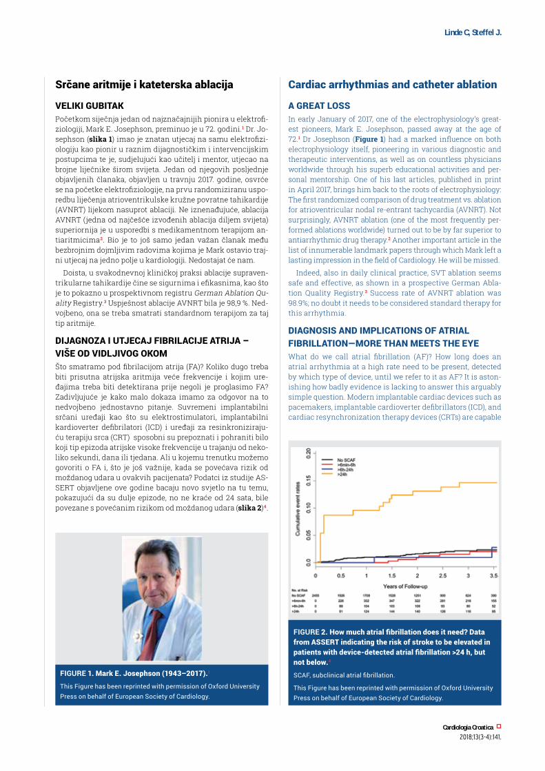
dijAgNOzA i UtjecAj FibRilAcije AtRijA – više Od vidljivOg OkOM Što smatramo pod fibrilacijom atrija (FA)? Koliko dugo treba biti prisutna atrijska aritmija veće frekvencije i kojim ure-đajima treba biti detektirana prije negoli je proglasimo FA? Zadivljujuće je kako malo dokaza imamo za odgovor na to nedvojbeno jednostavno pitanje. Suvremeni implantabilni srčani uređaji kao što su elektrostimulatori, implantabilni kardioverter defibrilatori (ICD) i uređaji za resinkroniziraju-ću terapiju srca (CRT) sposobni su prepoznati i pohraniti bilo koji tip epizoda atrijske visoke frekvencije u trajanju od neko-liko sekundi, dana ili tjedana. Ali u kojemu trenutku možemo govoriti o FA i, što je još važnije, kada se povećava rizik od moždanog udara u ovakvih pacijenata? Podatci iz studije AS-SERT objavljene ove godine bacaju novo svjetlo na tu temu, pokazujući da su dulje epizode, no ne kraće od 24 sata, bile povezane s povećanim rizikom od moždanog udara (slika 2)4.
Figure 1. Mark e. josephson (1943–2017).1
This Figure has been reprinted with permission of Oxford University Press on behalf of European Society of Cardiology.
Figure 2. How much atrial fibrillation does it need? Data from AsseRt indicating the risk of stroke to be elevated in patients with device-detected atrial fibrillation >24 h, but not below.4
SCAF, subclinical atrial fibrillation.
This Figure has been reprinted with permission of Oxford University Press on behalf of European Society of Cardiology.
Cardiologia Croatica
2018;13(3-4):142.
The year in cardiology 2017: arrhythmias and cardiac devices
of detecting and storing any type of atrial high rate episodes from few seconds to days and weeks. But from which time point on do we refer to it as AF and, more importantly, when does stroke risk increase in these patients? Data from the AS-SERT trial published this year shed some new light on this topic, indicating that episodes longer, but not shorter than 24 h were associated with an increased risk of stroke (Figure 2).4
The REVEAL-AF trial (presented at HRS 2017) investigated the prevalence of AF in 385 patients screened with an insertable loop recorder for a median of 22.5 months. The rate of AF de-tection was 6.2% at 30 days, increasing to 33.6% by 24 months, similar to the figures observed in the CRYSTAL-AF trial of pa-tients post-cryptogenic stroke.5 Conversely, however, if both patients with and without previous stroke show a similar rate of such short episodes, these findings again raise the ques-tion of the importance of short duration ‘AF’ as a predictor of stroke and, consequently, the need for anticoagulation. What to do hence with patients of shorter duration ‘AF’? Currently, the best answer in a device patient would be to enrol them in any of the ongoing studies investigating exactly this ques-tion—the ARTESiA or the NOAH trial.6,7 These studies focus on device-detected subclinical atrial fibrillation (SCAF) of short duration and studies if a non-vitamin K antagonists oral anti-coagulant (NOAC) (apixaban in ARTESiA, edoxaban in NOAH) will be superior in reducing stroke and thrombo-embolic risk compared to control therapy. Until the results of these studies are available, initiation of anticoagulation remains without strong evidence base in such patients.
In addition to the duration of AF, the overall risk of the pa-tients as indicated by the CHA2DS2VASc-Score8 as well as certain biomarkers9,10 will likely play a role in identifying pa-tients at increased risk of events and, ultimately, eligibility for anticoagulation. Also here, prospective randomized studies are required to answer this question at the required highest level of evidence.
hOw tO stAy iN siNUs RhythM—is UpstReAM tHerapy tHe clue?Life style modification is about to become a cornerstone in atrial fibrillation therapy. The open studies from Australia—LEGACY11 and CARDIO FIT12—showed that rigorous exercise and weight loss programs on top of risk factor management reduced re-occurrence of atrial fibrillation in overweight [body mass index (BMI) > 27 kg/m2] patients with paroxysmal or persistent atrial fibrillation patients whether on antiar-rhythmic drugs or post-AF ablation.
The RACE 3 investigators (van Gelder et al. presented at ESC 2017) took this concept further and focused on patients with symptomatic early persistent atrial fibrillation and early heart failure diagnosed <3 months. The main hypothesis was that early and intense or ‘upstream therapy’ would prevent or delay atrial remodelling and thereby prevent reoccurrence of atrial fibrillation compared to conventional therapy. Exclusion cri-teria were patients already on mineralocorticoid receptor an-tagonists (MRA) and a left atrium > 50 mm in diameter, NYHA IV and LVEF < 25%. From 2009 to 2015, 119 patients were in-cluded in the upstream arm and 126 in the conventional arm. Upstream rhythm control included angiotensin converting enzyme inhibitors and/or angiotensin receptor blockers, MRA, statins, cardiac rehabilitation therapy, and intensive counsel-ling on dietary restrictions, exercise maintenance, and drug adherence. The control arm of conventional rhythm control
Studija REVEAL-AF (prikazana na HRS-u 2017.) istraživala je prevalanciju FA u 385 bolesnika prateći ih implantabilnim loop snimačem tijekom medijana od 22,5 mjeseci. Učestalost otkrivanja FA bio je 6,2 % nakon 30 dana, a nakon 24-mjese-čnog praćenja se povećala na 33,6 %, slično podatcima utvr-đenima u studiji CRYSTAL-AF u bolesnika nakon moždanog udara neutvrđene etiologije.5 Obrnuto, u objema skupinama pacijenata, s prethodnim moždanim udarom i bez njega, po-stoji slična zastupljenost takvih kratkih epizoda pa ovi za-ključci ponovno nameću pitanje važnosti kratkih epizoda FA kao prediktora moždanog udara i, posljedično, potrebu za antikoagulantnom terapijom. Što učiniti s pacijentima s kra-ćim trajanjem FA? Trenutačno, najbolji odgovor u pacijenata s ugrađenim uređajima bio bi uključiti ih u neku od studija koje detaljno istražuju to pitanje – ispitivanja ARTESiA ili NOAH.6,7 Te su studije usmjerene na pomoću uređaja otkrivenu supkli-ničku FA (SCAF) koja je kratkog trajanja i studije hoće li novi antikoagulantni lijekovi (apiksaban u ARTESia, edoksaban u NOAH) biti superiorniji u smanjenju učestalosti moždanog udara i tromboembolijskog rizika s obzirom na kontrolnu te-rapiju. Toliko dugo dok rezultati tih studija ne budu dostupni, uvođenje antikoagulantne terapije ostaje bez snažnih dokaza u takvih pacijenata.
Uz trajanje FA, dodatno je indicirana procjena rizika u paci-jenata s pomoću CHA2DS2VASc-Scorea8 ili određenih biomar-kera9,10, što će vjerojatno imati ulogu u identifikaciji pacijena-ta s povećanim rizikom od događaja i u konačnici pomoći u određivanju podobnosti za antikaogulantnu terapiju. I ovdje su potrebne prospektivne randomizirane studije kako bi odgo-vorile na to pitanje na zadovoljavajuće visokoj razini dokaza.
KaKo oStati u SinuSnom ritmu – je li Ključ U OsNOvNOj bOlesti kOjA stiMUliRA FibRi-laciju atrija?Modifikacija životnoga stila postaje kamen temeljac u terapiji FA. Otvorene studije iz Australije, LEGACY11 i CARDIO FIT12, po-kazale su da rigorozna vježba i program gubitka tjelesne težine povrh liječenja čimbenika rizika smanjuje ponovno pojavljiva-nje FA u bolesnika s prekomjernom tjelesnom težinom [indeks tjelesne mase (BMI) >27 kg/m2] i s paroksizmalnom ili perzi-stentnom FA koji bi bili liječeni antiaritmicima ili ablacijom.
Istraživači studije RACE 3 (van Gelder i sur., prikazano na Kongresu ESC 2017.) učinili su korak dalje te su se usmjerili na simptomatske bolesnike i dijagnosticiranom perzistentnom FA i ranim srčanim zatajivanjem unutar tri mjeseca. Glavna je hipoteza bila da će rano i intenzivno liječenje bolesti koja stimulira pojavu FA spriječiti i odgoditi atrijsko remodeliranje i posljedično spriječiti ponavljanje FA u usporedbi s uobiča-jenom terapijom. Isključni su kriteriji bili bolesnici već lije-čeni antagonistima mineralokortikoidnih receptora (MRA) i promjer lijevog atrija >50 mm, NYHA IV, stupanj i ejekcijska frakcija lijeve klijetke (LVEF) <25 %. Od 2009. do 2015. godine 119 pacijenata bilo je uključeno u rano i intenzivno liječenje osnovne bolesti, a 126 pacijenata u konvencionalno liječenje. Rano i intenzivno liječenje uključivalo je inhibitore angioten-zin konvertirajućih enzima i/ili blokatore angiotenzinskih re-ceptora, MRA, statine, kardiološku rehabilitaciju i intenzivnu terapijsku edukaciju o dijetetskim ograničenjima, provođenju tjelovježbe i pridržavanje uzimanja lijekova. U kontrolnoj skupini konvencionalna kontrola ritma sastojala se od terapi-je za kontrolu ritma bez provođenja kardiološke rehabilitacije
Cardiologia Croatica
2018;13(3-4):143.
Linde C, Steffel J.
consisted of rhythm control therapy without cardiac rehabili-tation therapy and intensive counselling. Following 3 weeks in each arm patients underwent cardioversion. After 1 year, 75% in the upstream arm and 63% in the conventional study arm were still in sinus rhythm (P = 0.02) with a benefit from upstream therapy across all sub-groups. A significant drop in blood pressure, NT pro-BNP and LDL was also seen in the study arm whereas LVEF or LA-volume did not change. With only a 1 year follow-up, it is not surprising that the composite of CV morbidity/mortality was low and did not differ between the upstream group = 16% and the conventional = 17% groups.
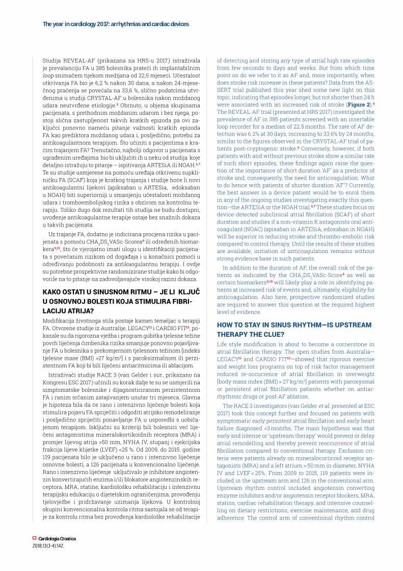
AtRiAl FibRillAtiON AblAtiON ‘dOwN the RAbbit hOle’The prevalence of AF increases with age and many patients are severely symptomatic. Pharmacologic therapy, is prob-lematic, as again evidenced by the possibly detrimental effect of drugs previously used in large scale in AF such as digoxin in a sub study of the ARISTOTLE trial (Lopes et al., presented at ACC 2017). As such, AF ablation has long been hailed as the solution of the problem. According to ESC EHRA guidelines, the indication for AF ablation is to improve quality of life.8 The European AF ablation registry reported an improved EHRA score following AF ablation, with good success of the proce-dure even in long-standing persistent AF (Figure 3).13
In the landmark MANTRA-PAF trial,14 patients were ran-domized to antiarrhythmic drug therapy vs. radiofrequency catheter ablation (RFA) as the first therapeutic intervention for atrial fibrillation. The pre-specified long-term results dem-onstrated after 5 years a lower occurrence and burden of any AF and symptomatic AF in the RFA compared to the AAD group.15 Using 7 days of Holter recordings, 86% of patients in the RFA group were free from AF. Also, quality of life scores were higher in the former compared to the latter group, a sig-nal that was present after 2 years and that persisted during the years thereafter. But also in the control arm a high propor-tion of patients were in sinus rhythm at the end of follow-up
i intenzivne terapijske edukacije. Sljedeća 3 tjedna u svakoj skupini bolesnici su bili podvrgnuti kardioverziji. Nakon jed-ne godine praćenja 75 % bolesnika iz rane i intenzivne skupi-ni i 63 % iz konvencionalne skupine liječenja bili su u sinus-ritmu (P = 0,02), a pozitivni učinak rane i intenzivne terapije registriran je u svim podskupinama. Također je registrirano znatno sniženje arterijskoga tlaka, vrijednosti NT pro-BNP-a i LDL-a, a nije bilo promjene u LVEF-u i volumenu LA. S ob-zirom na samo jednogodišnje praćenje, nije iznenađujuće da su kombinirani ishodi mjereni kardiovaskularnim pobolom/smrtnošću bili rijetki i nisu se razlikovali među skupinama (16 % prema 17 %).
AblAcijA FibRilAcije AtRijA – „kOMpleksNO pUtOvANje”Prevalencija FA raste sa životnom dobi i mnogi bolesnici ima-ju značajne simptome. Farmakološka terapija može biti pove-zana s problemima, poput mogućega štetnog učinka lijekova koji su prije rutinski primjenjivani u bolesnika s FA, primjerice digoksina u podskupini studije ARISTOTEL (Lopes i sur., pri-kazano na ACC 2017.). Kao takva, ablacija FA dugo se proma-trala kao rješenje problema. Prema Smjernicama ESC EHRA, indikacija za ablaciju FA jest poboljšanje kvalitete života.8 Eu-ropski registar ablacije FA objavio je poboljšanje rezultata na EHRA bodovnoj ljestvici nakon ablacije FA, s dobrim rezulta-tom procedure i kod dugotrajne perzistentne FA (slika 3).13
U važnoj studiji MANRA-PAF14 bolesnici su bili randomizi-rani u skupine s antiaritmijskom terapijom (AAD) nasuprot radiofrekventnoj kateterskoj ablaciji (RFA) kao prvoj interven-ciji kod liječenje FA.. Prethodno navedeni ciljni ishod poka-zao je nisku učestalost recidiva FA i pojave simptomatske FA u RFA skupini u usporedbi s AAD-om tijekom praćenja od 5 godina.15 Koristeći se sedmodnevnim holter EKG-om, u 86 % bolesnika u skupini liječenoj s pomoću RFA nije registrirana FA. Usto, i kvaliteta života bila je bolja u prijašnjoj prvoj u us-poredbi s kasnije spomenutom skupinom i to je bilo prisutno nakon dvije godine i perzistiralo godinama poslije. Međutim, i u kontrolnoj skupini velik je broj pacijenata bio u sinusnom ritmu na kraju razdoblja praćenja, što je upućivalo na ranu in-tervenciju. Ovakvi rezultati mogu postaviti pitanje rane pro-cedure RF ablacije i favoriziranje modifikacije životnoga stila te liječenja čimbenika rizika.
Međutim, kvaliteta života nije bila proučavana u randomi-ziranim kontroliranim studijama – niti ima jasno definirane ishode16. To se promijenilo kada su na kongresu ESC-a 2017. predstavljene dugo očekivane studije CAPTAF (Blomström i sur.) i CASTLE AF (Marrouche i sur.). U studiji CAPTAF pro-cjena opće kvalitete života bila je proučavana upitnikom SF 36 nakon 12 mjeseci u 79 pacijenata randomiziranih za RF ablaciju i 76 randomiziranih na antiaritmičnu terapiju. Svi su bolesnici imali implantabilni loop snimač koji je omogućio usporedbu opterećenja s FA. Rezultati pokazuju poboljšanje kvalitete života u objema skupinama, ali s mnogo većim po-boljšanjem u skupini na ablaciji. Opće zdravstveno stanje (di-menzija upitnika SF 36 zbog koje je studija snažna) poboljšana je za 10,5 jedinica ili 15 % u skupini na ablaciji. Procjena EHRA bodovnim sustavom (koja varira između I. i IV.) poboljšana je za prosječno 0,5 (P < 0,01). Ozbiljne nuspojave zabilježene su u 11 % bolesnika u ablacijskoj skupini i 23 % u kontrolnoj sku-pini koja je uključivala i potrebu za implantacijom elektrosti-mulatora srca. Iako nije bilo statističkih razlika u opterećenju
Figure 3. arrhythmia-free survival by type of atrial fibrilla-tion in the eSc-eHra atrial fibrillation ablation long-term registry.13
This Figure has been reprinted with permission of Oxford University Press on behalf of European Society of Cardiology.
Cardiologia Croatica
2018;13(3-4):144.
The year in cardiology 2017: arrhythmias and cardiac devices
reflecting the early intervention. These results may question very early RF ablation procedures and favour life style modi-fication and risk factor treatment.
However, quality of life had not been studied in a rand-omized controlled study—nor have hard endpoints.16 This changed when at ESC 2017 the long-awaited CAPTAF (Blom-ström et al., presented at ESC 2017) and CASTLE AF trial (Mar-rouche et al., presented at ESC 2017) were presented.
In the CAPTAF, general quality of life assessed by SF 36 was studied after 12 months in 79 patients randomized to RF ablation and 76 to antiarrhythmic drug therapy. All patients had implantable loop recorders enabling comparison of AF burden prior to and post-study start. The results indicate an improvement in quality of life in both groups but with a sig-nificantly greater improvement in the ablation group. Specifi-cally, general health—the dimension of the SF 36 for which the study was powered—was improved by 10.5 units or 15% in the ablation group. EHRA score (which ranges between I and IV) improved by on the average 0.5 (P < 0.01). Serious adverse events were reported in 11% in the ablation arm and 23% in the control arm which included need for pacemaker-implan-tation. While there was no statistical difference in AF burden, ablated patients did show only half the AF burden compared to the non-ablated group. It is reasonable to believe that the superior improvement in quality of life in ablated patients was related to absence of AA drugs but also the reduction in AF burden may have had a positive impact. Yet, the discrep-ancy between maintenance of sinus rhythm and symptom relief remains. Furthermore, the results indicate that OAC in-dication remains beyond 1 year also in ablated patients.
Atrial fibrillation often accompanies heart failure with a greater proportion with increasing HF severity. Many car-diologists may have felt reluctant to refer such patients for AF ablation for fear of less success rate and clinical benefit. Such clinical practice may however, change following the results of the CASTLE-AF study. In this study a total of 363 patients were randomly assigned to either undergo AF abla-tion or receive conventional care. To be included patients had to have persistent or paroxysmal AF and LVEF ≤ 35%. All pa-tients had an implantable CRT, ICD, or CRTD enabling moni-toring of atrial fibrillation. Of the 3013 screened individuals, 397 were enrolled and randomized 5 weeks later: 179 to the AF ablation group and 184 to the conventional therapy group. The primary composite endpoint of worsening heart failure or all-cause death was reduced by 38% in the ablation com-pared to the conventional group (HR 0.62, 95% CI 0.43–0.87; P = 0.007). This was driven by a reduction in both components of the combined endpoint, i.e. worsening heart failure [HR 0.56 (95% CI 0.37–0.83); P = 0.004] which occurred instantly and all-cause mortality [HR 0.53 (95% CI 0.32–0.86); P = 0.011] which was evident after a few years. Cardiovascular hospital admissions (HR 0.72, 95% CI 0.52–0.99; P = 0.041) and cardio-vascular mortality (HR 0.49, 95% CI 0.29–0.84; P = 0.009) was also significantly reduced in the ablation compared to the conventional therapy arm. At the same time, ejection fraction improved by 7% after 12 months in the ablation-group com-pared to the conventional treatment group, hence offering a potential mechanism through which these impressive effects were obtained. Over the 5 year duration of the trial, ablated pa-tients were twice as much free from AF as non-ablated. As expected, RF ablation of AF was not free from complications with 3.9% strokes/transient ischemic attack (TIA), 1.7% severe acute bleeding, and 1.7% pericardial effusion. In the conven-
pojavom FA, bolesnici u kojih je provedena ablacija imali su upola manje opterećenje s FA u usporedbi s neablacijskom skupinom. Razumno je vjerovati da je superiorno poboljšanje u kvaliteti života u bolesnika liječenih ablacijom povezano s neuzimanjem antiaritmičnih lijekova, ali i smanjenjem op-terećenja zbog pojave FA. Ipak, ostaje nedosljednost između održavanja sinusnog ritma i pojavnosti simptoma. Nadalje, rezultati upućuju na to da indikacija za OAC ostaje i nakon jedne godine i u bolesnika u kojih je učinjena ablacija.
Fibrilacija atrija često prati zatajivanje srca, a veća učesta-lost FA povezana je s pogoršanjem težine zatajivanja srca. Mnogi kardiolozi možda nerado upućuju takve bolesnike na ablaciju FA zbog straha od manje uspjeha i kliničke koristi. Takva će se klinička praksa možda mijenjati nakon rezultata istraživanja CASTLE-AF. U toj su studiji ukupno 363 pacijenta bila randomizirana u grupu za ablaciju FA ili za liječenje stan-dardnom terapijom. Uključni kriteriji bili su perzistentna ili paroksizmalna fibrilacija atrija i LVEF ≤ 35 %. Svi su pacijenti imali implantirani CRT, ICD ili CRTD s mogućnošću monitori-ranja FA. Od 3013 pregledanih osoba, 397 njih bilo je uključeno i randomizirano pet tjedana poslije: 179 u skupini s ablacijom FA i 184 u skupini na standardnom liječenju. Primarni zajed-nički cilj u obliku pogoršanja zatajivanja srca i ukupne smrt-nosti smanjio se za 38 % u ablacijskoj u usporedbi s konvencio-nalnom skupinom [HR 0,62, 95 % CI 0,43 – 0,87; P = 0,007]. Ovo je bilo potaknuto smanjenjem obiju komponenata kombiniranog ishoda, tj. pogoršanja zatajivanja srca [HR 0,56 (95 % CI 0,37 – 0,83); P = 0,004] koji su se dogodili odmah i uzrokovali smrtni ishod [HR 0,53 (95 % CI 0,32 – 0,86); P = 0,011], što je postalo očito nakon nekoliko godina. Kardiovaskularne hospitalizaci-je [HR 0,72, 95 % CI 0,52 – 0,99; P = 0,041] i kardiovaskularna smrtnost [HR 0,49, 95 % CI 0,29 – 0,84; P = 0,009] također su bile znatno reducirane u ablacijskoj u usporedbi sa skupinom na konvencionalnom liječenju. Istodobno, vrijednosti LVEF-a bile su poboljšane za 7 % nakon 12 mjeseci u ablacijskoj u uspored-bi s drugom skupinom, čime su ponuđeni potencijalni meha-nizmi kojima su ovi impresivni učinci bili postignuti. Tijekom 5 godina trajanja studije u skupini bolesnika randomiziranih na ablaciju registrirano je dvostruko manje FA nego u drugoj skupinu. Kao što se i očekivalo, RF ablacija FA nije bila bez komplikacija. Registrirano je 3,9 % moždanih udara / prolazne ishemijske atake (TIA), 1,7 % teških akutnih krvarenja i 1,7 % perikardijalnih izljeva. U skupini na konvencionalnom liječe-nju moždani udar / TIA bili su registrirani u 6,9 % pacijenata. Hoće li ovi rezultati promijeniti način na koji vidimo ablaciju FA u bolesnika sa zatajivanjem srca? Hoće li promijeniti način na koji liječimo takve pacijente? Uvijek je teško uvesti promje-ne u svakodnevnu praksu na temelju relativno male studije kao što je to CASTLE AF, sa svojim specifičnim uključnim i isključnim kriterijima, kao i ovisnošću o nekoliko događaja i rizika od pogreške tipa I. Unatoč tomu, studija CASTLE AF važno je istraživanje koje je doista prvi dokaz da ablacija FA nije samo postupak kojom se smanjuju simptomi već može utjecati i na pokazatelje kliničkih ishoda te kao rezultat može imati uzročnu ulogu radije nego da bude „neugodna smetnja” u patofiziologiji bolesti. Očekujemo da će kontrolirane randomi-zirane studije sa sličnim fokusom koje su u tijeku – CABANA, RAFT-AF, kao i EAST-AFNET 417 – dodati nove dokaze.
Na pitanje kako najbolje antikoagulirati pacijente tijekom i nakon ablacije FA već je odgovoreno. Kao i kod antagonista vi-tamina K (VKA), neprekinuta antikoagulantna terapija NOAC-ima također je i sigurna i učinkovita. Nakon prvog randomizi-
Cardiologia Croatica
2018;13(3-4):145.
Linde C, Steffel J.
tional arm, stroke/TIA was reported in 6.9%. Will these results change the way we see AF ablation in heart failure patients? Will it change the way we treat these patients? It is always difficult to infer a change in daily practice from a compara-tively (!) small trial such as CASTLE AF, with its specific in-clusion, and exclusion criteria as well as the dependence on few events and subsequent risk of type I error. This notwith-standing, CASTLE AF does represent a landmark trial in that it indeed represents the first evidence that AF ablation may not simply be a symptomatic procedure but may affect hard clinical outcomes in our patients—and, as a result, may in fact play a causal role rather than that of a ‘nuisance bystander’ in the pathophysiology of the disease process. Hopefully, the pending randomized controlled trials (RCTs) with similar fo-cus such as the CABANA and RAFT-AF study, as well as the EAST-AFNET 4 trial17 will add more evidence.
The question on how to best anticoagulate patients at and around AF ablation on the other hand seems answered. Like for Vitamin K antagonists (VKA), uninterrupted anticoagula-tion turned out to be both safe and effective also with NOACs. After the first randomized trial using rivaroxaban in this in-dication had shown no difference in the rate of events (VEN-TURE-AF)18, the larger Re-CIRCUIT study confirmed these results in 635 patients undergoing AF ablation randomized to either uninterrupted dabigatran or warfarin. Major bleeding events post-ablation, although overall low, occurred even sig-nificantly less with Dabigatran compared to Warfarin (1.6% vs. 6.9%; P < 0.001) with no difference in ischaemic events. Final-ly, also apixaban performed well in the AEIOU trial, in which 300 patients were randomly assigned to apixaban uninter-rupted or to the morning dose withheld prior to catheter abla-tion (presented at HRS 2017). When matched to a retrospec-tive uninterrupted warfarin cohort, major bleeding events were similar in both groups and overall occurred in <2% in both arms. The ongoing AXAFA-AFNET 5 study is comparing apixaban to uninterrupted VKA and will be reporting in early 2018.19 For edoxaban, a recent subanalysis of the ENGAGE AF-TIMI 48 trial demonstrated a similar risk of ischaemic and bleeding events in 193 catheter ablation procedures, although only a minority of patients were left on study drug for the pro-cedure.20 A dedicated study, ELIMINATE-AF is underway in-vestigating the efficacy and safety of uninterrupted edoxaban peri-ablation. Overall, the message seems to be emerging in a rather clear fashion that neither withholding (for more than the morning dose) nor bridging seems to be warranted and that a strategy of uninterrupted anticoagulation is the treat-ment of choice also for NOACs in the peri-AF ablation setting.
In a similar way, it is currently unclear how long before or-dinary surgical procedures NOACs need to be discontinued. Recent data from a French multicentre registry indicate that 3 days cessation of therapy predicted NOAC concentrations <30 ng/mL with 91% specificity.21 However, plasma levels are surrogate endpoints; and these data do not deliver proof that stopping NOACs for 72 h is required for all procedures.22 Simi-lar to the perioperative management in the VKA era, bridg-ing with LMWH was performed for years before, ultimately, evidence accumulated that this practice not only does not protect patients from events but may in fact lead to a higher bleeding propensity than uninterrupted warfarin.23,24 Very re-cent evidence from the BRUISE-CONTROL 2 study (Birnie et al., presented at AHA 2017) go in a similar direction: In 662 patients with a CHA2-DS2-VASc score ≥ 2 randomized to either
ranog ispitivanja s rivaroksabanom u ovoj indikaciji, koje nije pokazalo razliku u učestalosti događaja (VENTURE-AF)18, veća studija Re-CIRCUIT potvrdila je ovakve rezultate u 635 bolesni-ka podvrgnutih ablaciji FA koji su randomizirani u neprekinu-to uzimanje dabigatrana ili varfarina. Velika krvarenja nakon ablacije bila su rijetka, no bila su mnogo rjeđa s dabigatranom u usporedbi s varfarinom (1,6 % prema 6,9 %; P <0,001), a nije registrirana razlika u učestalosti ishemijskih događaja. I apik-saban je dobro prošao u studiji AEIOU u kojoj je 300 pacijenata nasumično uključeno u neprekinuto uzimanje apiksabana ili izostavljanje jutarnje doze apiksabana na dan planirane kate-terske ablacije (prikazano na HRS-u 2017.). Kada se usporede sa skupinom na neprekinutoj primjeni varfarina, učestalost velikih krvarenja bila je slična u objema skupinama te su se ukupno pojavila u <2 % bolesnika u objema skupinama. Studija AXAFA-AFNET 5 je u tijeku. Ona uspoređuje apiksaban s ne-prekinutim uzimanjem VKA trebala bi objaviti rezultate počet-kom 2018. godine19. Za edoksaban nedavna subanaliza studije ENGAGE AF-TIMI 48 pokazala je sličan rizik od ishemijskih i krvarećih događaja u 193 kateterske ablacijske procedure, iako je samo manji broj bolesnika ostao na studijskom lijeku tije-kom procedure.20 Studija ELIMINATE-AF je u tijeku i posveće-na je efikasnosti i sigurnosti neprekinutog uzimanja edoksa-bana periablacijski. Ukupno, čini se da je poruka usmjerena na jasan način te da nisu poželjni ni prekid (više od jutarnje doze) ni premoštenje i da je strategija neprekinutog uzimanja antiko-agulantih lijekova izbor i za NOAC u razdoblju oko ablacije FA.
Na sličan način, trenutačno je nejasno koliko ranije bi tre-balo isključiti NOAC prije kirurške procedure. Nedavni podat-ci iz Francuskoga multicentričnog registra pokazali su da trodnevni prekid terapije osigurava koncentraciju NOAC <30 ng/mL s 91 %-tnom specifičnošću.21 Također, još uvijek nije sasvim jasno koliko prije operacijskog zahvata treba preki-nuti terapiju NOAC-a. Novi podatci iz francuskoga multicen-tričnog registra pokazuju, s 91 %-tnom specifičnošću,21 da 3 dana nakon prekidanja terapije, koncentracija NOAC-a pada na <30 ng/mL. Međutim, razine u plazmi nisu primarni ishodi i ovi podatci ne pružaju dokaze da je potrebno zaustavljanje NOAC-a 72 sata prije svih postupaka.22 Slično dugogodišnjem perioperativnom postupku kod primjene varfarina premo-šćivanje niskomolekularnim heparinom zorno pokazuje da ovakva praksa ne samo da ne štiti pacijente od događaja već zapravo može uzrokovati veće krvarenje nego neprekinuta primjena varfarina.23,24 Nedavni dokazi iz studije BRUISE-CONTROL 2 (Birnie i sur., predstavljena na AHA 2017) idu u sličnome smjeru: 662 bolesnika s vrijednostima na bodovnoj ljestvici CHA2DS2-VASc ≥2 bili su randomizirani na nastavak primjene NOAC-a (zadnja primjena lijeka uvečer prije postup-ka) ili prekid najmanje 2 dana. Krvarenje, kao i drugi ishodi (uključujući smrtni ishod i moždani udar) bilo je rijetko i poja-vilo se u istoj mjeri u objema skupinama. Dok su druge studije u tijeku, a bave se sličnim pitanjima (npr. istraživanje PAU-SE, NCT02228798), podatci prvi put upućuju na činjenicu da nastavak NOAC-a (ili barem ograničavanje trajanja prekida) može biti siguran za neke intervencije.
prevencija možDanoG uDara KoD FibRilAcije AtRijASmjernice ESC-a iz 2016. jasno predstavljaju antikoagulan-tnu terapiju NOAC-ima kao preferiranu terapiju za prevenciju moždanog udara u AF8. Može li poboljšanje terapije varfari-nom, kao što je genotipom usmjereno doziranje, promijeniti
Cardiologia Croatica
2018;13(3-4):146.
The year in cardiology 2017: arrhythmias and cardiac devices
continuing NOAC therapy (last intake the evening before the procedure) or interruption for at least 2 days, bleeding as well as other endpoints (including mortality and stroke) was rare and occurred to the same extent in both groups. While other studies are underway assessing a similar question in other surgical settings (e.g. the PAUSE trial; NCT02228798), these data for the first time indicate that continuing NOACs (or at least limiting the time of interruption) may be a safe way to proceed for some interventions.
stROke pReveNtiON iN AtRiAl FibRillAtiONThe 2016 ESC guidelines clearly put anticoagulation with NOACs as the preferred therapy for stroke prevention in AF.8 Could improvements in warfarin therapy such as genotype-guided dosing tip this balance?25,26 So far, the evidence is con-flicting. In contrast, however, evidence is accumulating that even patients with well controlled INRs are not at zero risk of events. On the contrary, a recent sub-analysis from ARISTO-TLE indicated that the vast majority of intracranial haemor-rhages (78.5%) occurred at a therapeutic INR (<3.0).27 As such, NOACs remain the standard due to the consistent results ob-served in the four landmark randomized clinical trials with apixaban, dabigatran, edoxaban, and rivaroxaban in a total of >70 000 patients. There are, however, certain differences be-tween the four NOACs that we are only in the process of un-derstanding. Meticulous analyses of existing RCTs as well as new studies shed new light on these differences and improve individualization of NOAC therapy.
One remaining problem is that of inappropriate use of the ‘reduced’ dose of NOACs. Data from insurance claims analy-ses indicate a rate of up to 40% and more of ‘reduced dose’ use, particularly of apixaban, which does not compare to the 4.7% of patients receiving 2 × 2.5 mg of apixaban in the ARISTO-TLE trial.28 Importantly, the effect of using the reduced dose of apixaban or rivaroxaban in patients without the respective dose-reduction criteria leads to completely unpredictable re-sults as this has never been properly studies in a randomized controlled fashion and can hence not be recommended. In contrast, a ‘lower dose’ regimen was studied specifically in the Re-LY as well as in the ENGAGE AF-TIMI 48 trial with da-bigatran and edoxaban, respectively.29,30 Assessment of the proportion of patients taking the lower dose and/or reduced dose of NOACs in daily clinical practice is one strength of in-surance claims database research; indeed, the results serve to remind us to keep up and increase our educational efforts to alert physicians and patients that reproduction of the positive RCT results will only be possibly by using the investigated dosing regimens. In contrast, the assessment of clinical out-comes in the so-called ‘Real World’ research, particularly with insurance claims databases, needs to be viewed with great caution. Independent of statistical methods for adjustment, residual confounding is substantial, severely limits any in-terpretation of outcomes, and essentially makes assessment of any causal effect impossible, particularly in questions that have never been assessed in an RCT.31
The same is true for the use of other modalities for stroke prevention in AF, particularly percutaneous as well as surgi-cal left atrial appendage occlusion. Several registry data sur-faced in 2017, including the 1-year outcomes of the EWOLU-TION registry which demonstrated a low-stroke rate in over 1000 patients undergoing implantation with the Watchman device (Boersma et al., presented at Europace 2017). However,
aktualna stajališta?25,26 Dosadašnji dokazi nisu bili jednoznač-ni. Nasuprot tomu, međutim, akumuliraju se dokazi da čak i u pacijenata s dobro kontroliranim INR-om, rizik od neželjenih događaja nije nula. Naprotiv, nedavna analiza podskupina istraživanja ARISTOTLE pokazala je da se velika većina in-trakranijalnih krvarenja (78,5 %) dogodila u terapijskom INR-u (<3).27 Kao takvi, NOAC-i ostaju standardna terapija zbog ujednačenih rezultata u četirima značajnim randomizira-nim kliničkim ispitivanjima s apiksabanom, dabigatranom, edoksabanom i rivaroksabanom u ukupno >70 000 bolesnika. Postoje, međutim, određene razlike između četiriju NOAC-a koje su tek u procesu istraživanja. Iscrpne analize postojećih RCT-a, kao i novih istraživanja, osvjetljavaju te razlike i po-boljšavaju individualizaciju terapije NOAC-ima.
Dodatni je problem neadekvatna uporaba smanjene doze NOAC-a. Podatci iz analiza zdravstvenih osiguranja upućuju na uporabu smanjenih doza NOAC-a od 40 % i više, posebno, posebno apiksabana, čija se takva uporaba ne može usporedi-ti s 4,7 % bolesnika na 2 x 2,5mg iz istraživanja ARISTOTLE.28 Važno je da učinak primjene niže doze apiksabana ili rivarok-sabana u bolesnika bez prisutnih odgovarajućih kriterija za smanjenje doze dovodi do potpuno nepredvidljivih rezultata jer takva uporaba nije bila istražena u randomiziranom istra-živanju i stoga se ne preporučuje. Nasuprot tomu, režim „niže doze“ posebno je proučavan u istraživanju Re-LY, kao i u stu-diji ENGAGE AF-TIMI 48 s dabigatranom i edoksabanom.29,30 Procjena udjela pacijenata koji uzimaju nižu dozu i/ili sma-njenu dozu NOAC-a u dnevnoj kliničkoj praksi jedna je od snaga analize baza podataka potraživanja od zdravstvenih osiguranja; štoviše, rezultati služe kao podsjetnik na to da i dalje trebamo povećati naše edukativne napore kako bismo upozorili liječnike i pacijente da će reprodukcija pozitivnih rezultata iz kliničkih istraživanja biti moguća samo adekvat-nom primjenom režima doziranja. Nasuprot tomu moramo biti vrlo oprezni u interpretaciji procjena kliničkih ishoda u takozvanim istraživanjima iz kliničke prakse, osobito s ba-zama podataka potraživanja od zdravstvenih osiguranja. Ne-ovisno o primijenjenim statističkim metodama prilagodbe, preostala je nedorečenost znatna, ozbiljno ograničava svako tumačenje ishoda i u osnovi čini nemogućim procjenu bilo kakvog uzročnog učinka, osobito u pitanjima koja nikada nisu ocijenjena u randomiziranom istraživanju.31
Isto vrijedi i za uporabu drugih modaliteta za prevenciju moždanog udara kod AF-a, osobito perkutane, kao i kirurške okluzije aurikule lijevog atrija. Neki podatci iz registara poja-vili su se tijekom 2017. godine, uključujući jednogodišnje re-zultate registra EWOLUTION koji su pokazali nisku učestalost moždanog udara u više od 1000 pacijenata s implantiranim uređajem Watchman (Boersma i sur., predstavljeno na Euro-pace 2017.). Međutim, na istom sastanku podatci iz jednoga francuskog registra upozorili su na visoku prevalenciju (6,1 %) tromboze okludera u 377 uzastopnih pacijenata s različi-tim ugrađenim sustavima za okluziju aurikule lijevog atrija (Fauchier i sur., predstavljeno na Europace 2017.). Iako je od objavljivanja studije PROTECT-AF prošlo više od 8 godina, još uvijek nije sa sigurnošću utvrđena klinička važnost oklu-dera aurikule lijevog atrija. S obzirom na dostupne dokaze, trenutačne smjernice iz 2016. odgovarajuće dodjeljuju prepo-ruku klase IIb za okluziju LAA u prevenciji moždanog udara u AF-u.8 Daljnji registri vjerojatno neće mijenjati ovu razinu preporuke – to će biti moguće samo s novim rezultatima do-bro dizajniranih randomiziranih studija. Neka istraživanja
Cardiologia Croatica
2018;13(3-4):147.
Linde C, Steffel J.
at the same meeting, data from a French registry indicated a high prevalence (6.1%) of device occluder thrombi in 377 con-secutive patients implanted with various LAA occluder sys-tems (Fauchier et al., presented at Europace 2017). At the end of the day, the place of the LAA occluder still remains to be determined, even >8 years since publication of the PROTECT-AF study. In view of the available evidence, the current 2016 guidelines appropriately assign a Class IIb recommendation to LAA occlusion for stroke prevention in AF.8 Further regis-tries are unlikely to change this level of recommendation—this will only be possible with new results from well-designed RCTs. Some trials (CLOSURE-AF, ASAP-TOO) in high-risk pa-tients are now underway; others, particularly comparing LAA occlusion to the current (!) standard of therapy, i.e. NOACs, are urgently required. Similarly, a strategy of combining LAA oc-clusion with low-level NOAC anticoagulation has never been properly explored but has the potential to strike the golden bridge between the seemingly ‘opposing’, but in fact comple-mentary concepts of anticoagulation and LAA occlusion. Un-fortunately, so far, interest and motivation from the industry to sponsor such a trial has been limited.
veNtRicUlAR tAchycARdiA AblAtiONAblation of ventricular tachycardias (VT) has so far been pri-marily a domain of idiopathic VTs (particularly outflow tract, fascicular VT) and tachycardias with known structural abnor-malities (ischaemic VT, post-myocarditis etc.). In 2017, Pappone et al.32 reported of the largest series of patients with Brugada syndrome who successfully underwent ablation of an epicar-dial arrythmogenic substrate in the RVOT—hence in a chan-nelopathy population previously not deemed amenable for ab-lation. During a median follow-up of 10 months after ablation, elimination of the Brugada ECG phenotype was achieved in 133 of 135 patients undergoing ablation. Will ablation hence be-come standard therapy for Brugada patients? Will all patients with Brugada syndrome, possibly even ‘only’ with Brugada pattern benefit? What is the natural course of the disease after successful ablation? Many questions remain open, but these results certainly open the door to yet another frontier for abla-tion therapy in previously believed to be unsuitable patients.
Indeed, the RVOT harbours not only ‘idiopathic’ VT, but has been recognized in other entities including Brugada (as mentioned above) as well as early manifestation of ARVC as well as certain forms of exercise-induced arrhythmogenic remodelling.33 In 57 consecutive patients with scar-related right ventricular VT, the group of Dr Zeppenfeld identified an isolated subepicardial right ventricular outflow tract scar in high-level endurance athletes which was successfully treated by ablation. Furthermore, the scar pattern observed in this exercise-induced arrhythmogenic remodelling dem-onstrated significant differences compared to that in ARVC and post-inflammatory cardiomyopathy.33 As with the abla-tion approach suggested for Brugada, the approach appears attractive, but confirmation in larger series as well as long-term outcomes are eagerly awaited.
Indeed, even in ‘typical’ VT ablation patients—those with a ‘structural’ VT—success is far from 100%. In a large cohort, Tzou et al.34 compared patients undergoing a repeat procedure to those with a first VT ablation. Not surprisingly, the former indi-viduals more frequently presented with non-ischaemic VT, ICD shocks, and amiodarone treatment. Even though the procedural success was similar between the two groups (93% vs. 92%), com-
(CLOSURE-AF, ASAP-TOO) u visokorizičnih bolesnika sada su u tijeku; druga, posebno ona koja uspoređuju okluziju LAA s trenutačnom (!) standardnom terapijom, tj. NOAC-i su hitno potrebna. Slično tomu, strategija kombiniranja okluzije LAA s niskom dozom NOAC-a nikada nije bila pravilno istražena, ali ima potencijal da pogodi zlatnu sredinu između naizgled ’suprotnih’, ali zapravo komplementarnih koncepata antiko-agulantne terapije i okluzije LAA. Nažalost, farmaceutska je industrija do sada pokazala ograničen interes i motivaciju da sponzorira takvo istraživanje.
AblAcijA veNtRikUlskih tAhikARdijAAblacija ventrikulskih tahikardija do sada se provodila ve-ćinom kod idiopatskih VT (izgonski trakt, fascikulska VT) i kod tahikardija s poznatim strukturnim abnormalnostima (ishemijska VT, postmiokarditična itd.). Tijekom 2017. godine Pappone i sur.32 izvijestili su o najvećoj seriji bolesnika s Bru-gadinim sindromom koji su uspješno podvrgnuti ablaciji epi-kardijalnog aritmogenog supstrata u izlaznom dijelu desne klijetke, dakle u bolesnika sa kanalopatijom, u populaciji koja se prethodno nije smatrala prihvatljivom za ablaciju. Tijekom medijana praćenja od 10 mjeseci nakon ablacije uklanjanje Brugadina fenotipa u EKG-u postignuto je u 133 od 135 bole-snika koji su podvrgnuti ablaciji. Hoće li ablacija postati stan-dardna terapija za pacijente s Brugadinim sindromom? Hoće li svi bolesnici s Brugadinim sindromom, možda čak i oni sa „samo“ EKG Brugadinim fenotipom, imati korist od ablacije? Koji je prirodan tijek bolesti nakon uspješne ablacije? Mno-ga pitanja ostaju otvorena, ali ti rezultati, zasigurno, otvara-ju vrata za ablacijsku terapiju još jednoj skupini bolesnika za koje se prije smatralo da su neprikladni za takvu terapiju.
Doista, u izlaznom dijelu desne klijetke ne krije se samo „idiopatska“ VT već je prepoznato nekoliko drugih entiteta, uključujući Brugadu (kao što je prije spomenuto), kao i ranu manifestaciju aritmogenom kardiomiopatijom desne klijet-ke (ARVC), kao i određene oblike vježbanjem induciranoga aritmogenog preoblikovanja.33 U 57 uzastopnih bolesnika s VT-om iz ožiljnog područja desne klijetke, Zeppenfeld i sur. identificirali su izolirani subepikardijalni ožiljak u izgonsko-me traktu desne klijetke u sportaša s visokom razinom iz-držljivosti koji je uspješno liječen ablacijom. Nadalje, uzorak ožiljka uočen u tom vježbanjem induciranom aritmogenom preoblikovanju pokazao je znatne razlike u usporedbi s onima u ARVC-u i poslijeupalnoj kardiomiopatiji.33 Kao i kod abla-cijskog pristupa koji se predlaže za Brugadu, pristup se čini atraktivnim, ali se očekuju potvrda rezultata u većoj seriji, kao i dugoročni ishodi.
Doista, čak i kod uobičajenih ablacija (bolesnici sa „struk-turnom“ VT) uspjeh je daleko od 100 %. U velikoj kohorti Tzou i sur.34 uspoređivali su bolesnike koji su podvrgnuti ponov-ljenom postupku s onima s prvim ablacijama zbog VT-a. Nije iznenađujuće da su u bolesnika s više postupaka češće bili prisutni neishemijska VT, ICD šokovi i primjena liječenja ami-odaronom. Iako je uspjeh postupka bio sličan u objema skupi-nama (93 % prema 92 %), komplikacije su bile veće (pogotovo perikardijalni izljev i venska tromboza), a preživljenje je bilo lošije (67 % prema 78 %, P = 0,003) u bolesnika s više postu-paka. Kao i kod praktički svih elektrofizioloških postupaka, a zapravo pri gotovo svim postupcima u kardiologiji, takve sofisticirane intervencije trebaju se obavljati u specijalizira-nim centrima kako bi se omogućile maksimalna učinkovitost i sigurnost.
Cardiologia Croatica
2018;13(3-4):148.
The year in cardiology 2017: arrhythmias and cardiac devices
plications trended to be higher (especially for pericardial effu-sion and venous thrombosis) and survival was worse (67% vs 78%, P = 0.003). As with virtually all EP procedures—and, as a matter of fact, virtually all procedures in Cardiology—such high-end interventions need to be concentrated at specialized cen-tres to allow for maximum efficacy and safety of the procedure.
sUddeN cARdiAc deAth—Risk pRedictiON ANd pReveNtiONIn 2016, the DANISH trial demonstrated no overall benefit of primary prophylactic ICD implantation in 556 patients with non-ischaemic heart disease.35 Few studies on cardiac de-vices have been debated as intensely over the last decade. In a recent meta-analysis of 8567 patients of 11c RCTs (including 3128 patients without ischemic heart disease (IHD)), primary prevention ICD implantation reduced the occurrence of all-cause mortality both in patients with (n = 5439) as well as in those without ischaemic heart disease (n = 3128) by 24%.36 Is the question answered then? By far not. As elegantly eluted to in an accompanying editorial by Lars Kober (at the same time the principal investigator of the DANISH trial) to the aforemen-tioned meta-analysis: ‘ICDs work—now it is time to find out who needs them’.37 Indeed, as in the DANISH trial, the ques-tion is not as black or white as sometimes presented; what is the role of concomitant CRT? What is the use of CRT in elderly patients and in those with relevant comorbidities (including severe heart failure)? Does the impact of defibrillators on sur-vival become less over time? Indeed, these questions are not only valid for ICD in patients with non-ischaemic cardiomyo-pathies, which were included in DANISH. Therefore, the aim of the EHRA initiated ‘RESET-SCD’ trial is to test primary pro-phylactic ICD implantation in patients with ischaemic heart disease and compromised ejection fraction and will deliver urgently needed new data for this important population.
And, on another level: Are we at the best that we can do re-garding risk stratification of patients at risk of SCD? Indeed, left ventricular ejection fraction—in spite of being the best documented method for primary prevention ICD eligibil-ity—has important shortcomings. Accumulating evidence indicate that imaging, particularly by MRI, may be helpful. In 399 patients with late gadolinium enhancement (LGE) and an EF ≥ 40% had an over nine-fold increased risk of SCD or abort-ed SCD than those without LGE.38 The incremental value of using multiple ECG parameters in SCD prediction was tested in the community-based Oregon Sudden Unexpected Death Study.39 When heart rate, LV hypertrophy, QRS transition zone, QRS-T angle, QTc, and Tpeak-to-Tend interval were added to tra-ditional risk factors, the c-statistics improved significantly from 0.625 to 0.753 (P < 0.001). This was externally validated in the Atherosclerosis Risk in Communities (ARIC) Study. In the accompanying editorial, Bob Myerburg rightfully states that although encouraging, the long-term predictive value of these ECG markers will require assessment in a carefully designed randomized clinical trial.40
iMplANtAble cARdiAc electRONic devices—MOviNg FURtheR AwAy FROM iNtRAvAscUlAR leAds ANd OtheR ‘UNshAkAble’ pARAdigMsBoth permanent pacemakers as well as ICDs and CRT devices have time after time revolutionized the way that brady- and tachyarrhythmic disorders can be treated. This notwith-
preDviđanje i prevencija iznenaDne Srčane SmrtiTijekom 2016. godine istraživanje DANISH nije dokazalo do-brobit od implantacije ICD uređaja u primarnoj prevenciji u 556 bolesnika s neishemijskom kardiomiopatijom.35 Nekoli-ko studija sa srčanim uređajima bilo je predmet intenzivne rasprave. U nedavnoj metaanalizi 8567 bolesnika iz 11 ran-domiziranih studija (uključujući njih 3128 bez ishemijske bo-lesti srca /IHD/), profilaktična implantacija ICD-a smanjila je ukupnu smrtnost i u onih (n = 5439) s IHD-om, kao i kod onih bez IHD-a (n = 3128) za 24 %.36 Je li sada na pitanje odgovo-reno? Ni približno. Kao što je to u svojemu uvodnom članku sročio Lars Kober (istodobno glavni istražitelj DANISH studije) u osvrtu na gore navedenu metaanalizu: „ICD-ovi rade – sada je vrijeme da saznamo komu ih treba ugraditi.“ Doista, kao i u DANISH istraživanju, pitanje nije toliko crno ili bijelo kao što je pokatkad predstavljeno; koja je uloga istodobnog CRT-a? Koja je uloga CRT-a u starijih pacijenata i u onih s relevan-tnim komorbiditetima (uključujući uznapredovalo zatajivanje srca)? Smanjuje li se utjecaj defibrilatora na preživljavanje ti-jekom vremena? Doista, ova pitanja ne vrijede samo za ICD u bolesnika s neishemičnim kardiomiopatijama, koji su bili uključeni u studiju DANISH. Stoga je cilj EHRA iniciranog istraživanja „RESET-SCD“ ispitivanje primarne profilaktične implantacije ICD-a u bolesnika s IHD-om i smanjenom ejek-cijskom frakcijom te će pružiti hitno potrebne nove podatke za ovu važnu populaciju.
Također, jesmo li postigli sve što smo trebali što se tiče stra-tifikacije rizika u bolesnika kojima prijeti iznenadna srčana smrt (SCD)? Ejekcijska frakcija lijeve klijetke – unatoč tomu što je najbolje dokumentirana metoda za procjenu potrebe pri-marne prevencije ICD-om – ima važne nedostatke. Sve više dokaza upućuje na to da druge slikovne metode, osobito MRI, mogu biti korisne. U 399 bolesnika s kasnijim gadolinijskim pojačavanjem (LGE) i EF ≥40 % registrirano je više od devet puta veći rizik od SCD-a ili zaustavljene SCD nego u bolesnika bez LGE-a.38 Povećana vrijednost primjene višestrukih EKG varijabli u predviđanju SCD-a bila je testirana u kohortnom istraživanju Oregon Sudden Unexpected Death Study.39 Kada su frekvencija srca, hipertrofija lijeve klijetke, zona tranzicije QRS-a, kut QRS-T, QTc i interval od vrha do kraja T-vala doda-ni tradicionalnim čimbenicima rizika, rezultat c-statistike se znatno poboljšao od 0,625 do 0,753 (P <0,001). Ovo je potvrđeno u studiji ARIC. U popratnome članku Bob Myerburg s pravom kaže da će, iako ohrabruje, dugoročna prediktivna vrijednost tih EKG markera zahtijevati procjenu u pažljivo osmišljenom randomiziranom kliničkom ispitivanju.40
implantabilni Srčani eleKtroničKi uređaji – OdMicANje Od iNtRAvAskUlARNih elek-tROdA i dRUgih ’NepOkOlebljivih’ pARAdigMiI trajni elektrostimulatori, kao i ICD i CRT uređaji s vremenom su promijenili način na koji se liječe bradiaritmije i tahiari-tmije. Primjenom ovih sustava uz veću životnu dob nastu-paju komplikacije uglavnom zbog prisutnosti intravaskular-nih elektroda. Prijelomi elektroda i napose njihove infekcije, mogu dovesti do nužnosti ekstrakcije koja je sama po sebi povezana sa znatnim pobolom i smrtnošću – što je nedavno dokazano u prospektivnom registru ELECTRa koje provodi EHRA.41 U 3510 pacijenata koji su bili podvrgnuti ekstrakciji elektroda u 73 centra u 19 europskih zemalja od studenoga
Cardiologia Croatica
2018;13(3-4):149.
Linde C, Steffel J.
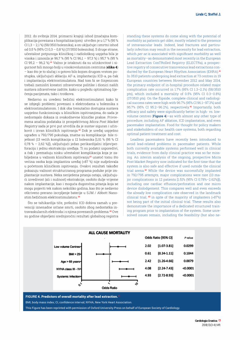
standing these systems do come along with the potential of morbidity as patients get older, mostly related to the presence of intravascular leads. Indeed, lead fractures and particu-larly infection may result in the necessity for lead extraction, which per se is associated with significant morbidity as well as mortality—as demonstrated most recently in the European Lead Extraction ConTRolled Registry (ELECTRa), a prospec-tive registry of consecutive transvenous lead extractions con-ducted by the European Heart Rhythm Association (EHRA).41 In 3510 patients undergoing lead extraction at 73 centres in 19 European countries between November 2012 and May 2014, the primary endpoint of in-hospital procedure-related major complication rate occurred in 1.7% (95% CI 1.3–2.1%) (58/3510 pts), which included a mortality of 0.5% (95% CI 0.3–0.8%) (17/3510 pts). On the flipside, complete clinical and radiologi-cal success rates were high with 96.7% (95% CI 96.1–97.3%) and 95.7% (95% CI 95.2–96.2%), respectively.41 Importantly, both efficacy and safety were significantly better in high- vs. low-volume centres (Figure 4)—as with almost any other type of procedure, including AF ablation, ICD implantation, and even pacemaker implantation. Food for thought for policy makers and stakeholders of our health care systems, both regarding optimal patient treatment and cost.
Leadless pacemakers have recently been introduced to avoid lead-related problems in pacemaker patients. While both currently available systems performed well in clinical trials, evidence from daily clinical practice was so far miss-ing. An interim analysis of the ongoing, prospective Micra Post Market Registry now indicated for the first time that the system is also safe and effective if used outside the clinical trial arena.42 While the device was successfully implanted in 792/795 attempts, major complications were rare {13 ma-jor complications in 12 patients [1.51% (95% CI 0.78%–2.62%)]}, including one cardiac effusion/perforation and one micro device dislodgement. This compares well and even exceeds the already low complication rate observed in the landmark clinical trial, 43 in spite of the majority of implanters (>87%) not being part of the initial clinical trial. These results also demonstrate the importance of a dedicated structured train-ing program prior to implantation of the system. Some unre-solved issues remain, including the feasibility (but also ne-
2012. do svibnja 2014. primarni krajnji ishod (značajna kom-plikacija povezana s hospitalizacijom) utvrđen je u 1,7 % (95 % CI 1,3 – 2,1 %) (58/3510 bolesnika), a on uključuje i smrtni ishod od 0,5 % (95% CI 0,3 – 0,8 %) (17/3510 bolesnika). S druge strane, učestalost potpunoga kliničkog i radiološkog uspjeha bila je visoka i iznosila je 96,7 % (95 % CI 96,1 – 97,3 %) i 95,7 % (95 % CI 95,2 – 96,2 %).41 Važno je istaknuti da su učinkovitost i si-gurnost bili mnogo bolji u visokovolumnim centrima (slika 4) – kao što je to slučaj i s gotovo bilo kojom drugom vrstom po-stupka, uključujući ablaciju AF-a, implantaciju ICD-a, pa čak i implantaciju elektrostimulatora. Nad tom bi se činjenicom trebali zamisliti kreatori zdravstvene politike i dionici naših sustava zdravstvene zaštite, kako u pogledu optimalnog lije-čenja pacijenata, tako i troškova.
Nedavno su uvedeni bežični elektrostimulatori kako bi se izbjegli problemi povezani s elektrodama u bolesnika s elektrostimulatorom. I dok oba trenutačno dostupna sustava uspješno funkcioniraju u kliničkim ispitivanjima, do sada je nedostajalo dokaza iz svakodnevne kliničke prakse. Privre-mena analiza podataka iz prospektivnog Micra Post Market Registry sada je prvi put utvrdila da je sustav siguran i učin-kovit i izvan kliničkih ispitivanja.42 Dok je uređaj uspješno ugrađen u 792/795 pokušaja, znatne su komplikacije bile ri-jetkost (13 većih komplikacija u 12 bolesnika [1,51 % (95 % CI 0,78 % – 2,62 %)]), uključujući jedan perikardijalni izljev/per-foraciju i jednu ekstrakciju uređaja. Ti su podatci usporedivi, a čak i premašuju nisku učestalost komplikacija koja je za-bilježena u važnom kliničkom ispitivanju43 unatoč tomu što većina osoba koja implantira uređaj (>87 %) nije sudjelovala u početnom kliničkom ispitivanju. Ovakvi rezultati također pokazuju važnost strukturiranog programa poduke prije im-plantacije sustava. Neka neriješena pitanja ostaju, uključuju-ći izvedivost (ali i nužnost) ekstrakcije, osobito dulje vrijeme nakon implantacije, kao i moguća dugoročna pitanja koja se mogu pojaviti tek nakon nekoliko godina, kao što je nedavno otkriveno prerano iscrpljenje baterije u SJM / Abbott Nano-stim bežičnom elektrostimulatoru.44
Što se tahikardija tiče, potkožni ICD dobiva zamah u pre-venciji iznenadne srčane smrti, osobito zbog nedostatka in-travaskularnih elektroda i s njima povezanih problema.45 Ove su godine objavljeni srednjoročni rezultati globalnog registra
Figure 4. predictors of overall mortality after lead extraction.41
BMI, body mass index; CI, confidence interval; NYHA, New York Heart Association.
This Figure has been reprinted with permission of Oxford University Press on behalf of European Society of Cardiology.
Cardiologia Croatica
2018;13(3-4):150.
The year in cardiology 2017: arrhythmias and cardiac devices
cessity) of extraction, particularly after years of implantation; as well as possible long-term issues that may only surface after years such as the recently discovered premature battery depletion in the SJM/Abbott Nanostim leadless pacemaker.44
On the tachycardia side, the subcutaneous ICD is gaining momentum for the prevention of sudden cardiac death par-ticularly due to the lack of an intravascular electrode and the associated problems.45 This year, the mid-term results of the global Evaluation oF FactORs ImpacTing CLinical Outcome and Cost EffectiveneSS of the S-ICD (EFFORTLESS S-ICD) registry were published indicating not only fulfilment of the pre-defined endpoints for efficacy and safety but also a low rate of system extraction due to need for antitachycardia pacing, brady pacing, or CRT.46 Prospective studies includ-ing PRAETORIAN and UNTOUCHED are currently ongoing and will need to confirm these positive results. However, given the likely reduced morbidity compared to conventional transvenous systems, treatment of patients at lower risk of SCD than ‘conventional’ ICD recipients appears to be an at-tractive option. To this end, MADIT-SICD was launched last year, investigating the efficacy and safety of the S-ICD (com-pared to the current standard of best medical therapy) in post-myocardial infarction diabetes patients ≥65 years with an LVEF 36–50%.47 In addition to improving our ways and means of risk stratification for SCD, reducing the morbidity of systems protecting patients from SCD seems to be a logi-cal step to tackle the challenges of the Myerburg-Paradox.45 While both leadless pacing as well as the S-ICD hence likely represent a glimpse of what the device field will be moving towards in the future, comparative analyses with existing systems (as indicated) are mostly still ongoing. In addition, the higher cost of these systems may be an obstacle in some health care settings preventing the larger volume use of these devices—which, however, is likely to change over the coming years as with every newly introduced therapy.
One other concern about cardiac devices seems to be less-ened latest since last year, that is the ‘risk’ of MRI in non-MRI-conditional devices (at least in none high-risk patients un-dergoing 1.5 T MRI). Using a specific standardized protocol for patient selection, programming, observation during MRI and reprogramming, the investigators of the MAGNA-SAFE regis-try demonstrated no deaths, lead failures, losses of capture, or ventricular arrhythmias during MRI in 1000 pacemakers and 500 ICDs.48 Whether this is also true for higher risk patients (e.g. pacemaker dependent ICD recipients) remains to be de-termined. Preliminary data for one such high-risk subgroups appears encouraging: two studies presented at HRS 2017 (Pad-manabhan et al. and Brunker et al.) indicate that MRI seems to be safe and feasible in patients with abandoned leads, i.e. patients previously thought to be absolutely contraindicated to undergo MRI scanning. Further studies are required to substan-tiate these findings, but given the totality of recently provided data, several paradigms seem to be tumbling in this previously uncharted area of MRI scanning in implantable devices.
cARdiAc ResyNchRONizAtiON theRApy—be-tweeN gUideliNes, ReAlity, ANd AlteRNAtivesAlthough standard therapy in heart failure, CRT remains un-evenly implemented in ESC countries according to the 2016 EHRA Whitebook.49 The ESC EHRA HFA CRT Survey II in-cluded data on 10 088 new CRT implantations across 42 ESC
EFFORTLESS S-ICD, koji pokazuju ne samo ispunjavanje una-prijed definiranih ishoda za djelotvornost i sigurnost nego i nisku učestalost ekstrakcije sustava zbog potrebe za antita-hikardijskom stimulacijuom, stimulacijuom zbog bradikar-dije ili potrebe za CRT-om.46 Prospektivne studije, uključujući PRAETORIAN i UNTOUCHED, trenutačno su u tijeku i morat će potvrditi ove, pozitivne rezultate. S obzirom na smanjen pobol u usporedbi s konvencionalnim transvenskim susta-vima, čini se da je potkožni ICD atraktivna alternativa za liječenje bolesnika s nižim rizikom od SCD-a u usporedbi s konvencionalnim ICD uređajima. U tu svrhu pokrenuta je prošle godine studija MADIT-SICD koja istražuje djelotvornost i sigurnost S-ICD-a (u usporedbi s trenutačnom najboljom standardnom terapijom) u dijabetičara koji su preboljeli infar-kt miokarda, a stariji su od 65 godina i imaju LVEF 36 – 50 %.47 Osim poboljšanja načina i sredstava za stratifikaciju ri-zika za SCD, smanjenje pobola sustavima koji štite pacijente od SCD-a čini se logičnim korakom u rješavanju Myerburgova paradoksa.45 Premda i bežični elektrostimulatori, kao i S-ICD, vjerojatno daje uvid u ono u što će se polje uređaja kretati pre-ma budućnosti, usporedne analize s postojećim sustavima (prema indikaciji) uglavnom su još u tijeku. Osim toga, veća cijena tih sustava može biti prepreka u nekim zdravstvenim sustavima, koja sprječava veću uporabu takvih uređaja, što će se vjerojatno promijeniti tijekom idućih godina kao što se to događa i sa svakom novouvedenom terapijom.
Čini se da se jedna zabrinutost oko srčanih uređaja tijekom prošle godine smanjila, tj. ’rizik’ od oslikavanja s uređajima koji nisu MRI kompatabilni (barem u niskorizičnih bolesnika koji su bili podvrgnuti oslikavanju MRI-jem s 1,5 T). Uporabom specifičnoga standardiziranog protokola za odabir bolesnika, programiranja, promatranja tijekom postupka MRI-ja i repro-gramiranja, istraživači registra MAGNA-SAFE pokazali su da nije bilo smrti, kvara elektroda, gubitaka odgovora na sti-mulaciju ili ventrikulskih aritmija tijekom oslikavanja MRI-jem u 1000 bolesnika s elektrostimulatorom i 500 oboljelih s ICD-om.48 Još uvijek treba odrediti vrijedi li to i za bolesnike s većim rizikom (npr. bolesnici ovisni o elektrostimulatoru). Preliminarni podatci za jednu takvu podskupinu visokog ri-zika ohrabrujući su: dvije studije prezentirane na HRS-u 2017. (Padmanabhan i sur. te Brunker i sur.) upućuju na to da se MRI čini sigurnim i izvedivim u bolesnika s napuštenim elektro-dama, odnosno u bolesnika za koje se prije pretpostavljlo da je MRI apsolutno kontraindiciran. Potrebne su daljnje studije kako bi potkrijepila ta otkrića, ali, s obzirom na ukupnost ne-davno dobivenih podataka, čini se da će se nekoliko paradi-gmi u ovom, prethodno neistraženom području MRI skenira-nja u bolesnika s implantabilnim uređajima promijeniti.
terapija Srčane reSinKronizacije – izme-đu Smjernica, StvarnoSti i alternativa Iako je to danas standardna terapija u zatajivanju srca, CRT ostaje neujednačeno primijenjen u zemljama ESC-a prema EHRA-inoj „bijeloj knjizi“ iz 2016. godine.49 Istraživanje ESC EHRA HFA CRT II obuhvatilo je podatke o 10 088 novih CRT implantacija u 42 zemlje ESC-a, prikupljenih od listopada 2015. do prosinca 2016. (Normand i sur., kongres ESC-a 2017.). Rezultati pokazuju da, kao u prethodnom istraživanju,50 liječ-nici zaobilaze smjernice51 kada su u pitanju indikacije za im-plantaciju CRT-a. Najčešće odstupanje bilo je primijeniti CRT u LVEF >35 % (u 12 % slučajeva), uski QRS <120 ms (u 8 % sluča-jeva) i u NYHA klasi I (u 3 % slučajeva). Od implantacija 43 %
Cardiologia Croatica
2018;13(3-4):151.
Linde C, Steffel J.
countries collected between October 2015 and December 2016 (Normand et al., presented at ESC 2017). The results indicate that like in the previous survey50 doctors go beyond guide-lines51 recommendations when selecting patients for CRT. The most common deviation was to give CRT in LVEF > 35% in 12%, narrow QRS < 120 ms in 8% and NYHA class I in 3%. Of implantations 43% were in patients with a Class I indication according to guidelines, Class II in 21% and Class III meaning implantation is contraindicated in 8%. The results also imply important differences in between countries and centres. The present CRT Survey II is sufficiently big to permit meaningful benchmarking between countries.
His Bundle pacing has resurrected over the last years as a possible alternative to CRT in some settings.52,53 In a study of 95 patients with an indication for CRT, His bundle pacing was used as a rescue strategy in for failed LV lead or non-response to conventional biventricular pacing (Group I) or as an alterna-tive to the latter for individuals with AV block, bundle branch block, or high ventricular pacing burden. Both groups demon-strated a significant reduction in QRS width, increase in LVEF [30 ± 10% to 43 ± 13% (P = 0.0001)] and improvement in NYHA class.52 Still, many questions remain. Will this be safe and ef-fective also outside specialized centres with great expertise in this technique? Will this also work in patients requiring ICD therapy? And, most importantly, will it turn out to be as effective in reducing hard clinical endpoints (morbidity and mortality) as conventional CRT has been demonstrated to be. Again, randomized clinical trials assessing these open ques-tions will be required, some of which are already ongoing.
Conflict of interest: C.L. has consultant and/or speaker fees from Medtronic, Biotronik, St Jude Medical, Novartis and Vifor. She has received grant support through her institution from Astra Zeneca, Stockholm city council and Heart Lung foundation. J.S. has received consultant and/or speaker fees from Amgen, Astra-Zeneca, Atricure, Bayer, Biosense Webster, Biotronik, Boehringer-Ingelheim, Boston Sci-entific, Bristol-Myers Squibb, Cook Medical, Daiichi Sankyo, Medtronic, Novartis, Pfizer, Sanofi-Aventis, Sorin, St. Jude Medical/Abbott and Zoll. He reports ownership for CorXL. He has received grant support through his institution from Bayer Healthcare, Biosense Webster, Biotronik, Bos-ton Scientific, Daiichi Sankyo, Medtronic, und St. Jude Medical/Abbott.
je bilo u bolesnika s indikacijom klase 1 prema smjernicama, klase 2 u 21 % slučajeva i klasa 3, što znači da je implantacija kontraindicirana u 8 % slučajeva. Rezultati također upozora-vaju na znatne razlike između zemalja i centara. Sadašnje istraživanje CRT Survey II dovoljno je veliko istraživanje da bi se omogućilo uspoređivanje između zemalja.
Elektrostimulacija Hisova snopa posljednjih je godina uskrsnula kao moguća alternativa CRT-u u nekim situacija-ma.52,53 U istraživanju od 95 bolesnika s indikacijom za CRT, elektrostimulacija Hisova snopa iskorištena je kao strategija spašavanja pri neuspjelom plasiranju elektrode u koronarni sinus ili pri nedostatku odgovora na konvencionalnu biven-trikulsku stimulaciju (grupa 1) ili kao alternativa potonjem u osoba s AV-blokom, blokom grane ili učestalom stimulacijom ventrikula. Obje skupine pokazale su znatno smanjenje širine QRS-a, povećanje LVEF-a [30 ± 10 % do 43 ± 13 % (P = 0,0001)] i poboljšanje u NYHA klasi.52 Ipak, ostaju mnoga otvorena pita-nja. Hoće li to biti zahvat koji je siguran i učinkovit i izvan spe-cijaliziranih centara s velikim iskustvom u ovoj tehnici? Hoće li to funkcionirati i u bolesnika koji zahtijevaju ICD terapiju? I, što je najvažnije, hoće li se pokazati kao jednako učinkovit u smanjenju značajnih kliničkih ishoda (pobol i smrtnost) kao što je dokazano kod konvencionalnog CRT-a. Ponovno će se zahtijevati randomizirana klinička ispitivanja koja odgovara-ju na ova otvorena pitanja, od kojih su neka već u tijeku.
liteRAtURe1. Shenasa M. Mark E Josephson MD. Eur Heart J. 2017;38:1355-6. https://doi.org/10.1093/eurheartj/ehx202
2. Katritsis DG, Zografos T, Katritsis GD, Giazitzoglou E, Vachliotis V, Paxinos G, et al. Catheter ablation vs. antiarrhythmic drug therapy in patients with symptomatic atrioventricular nodal re-entrant tachycardia: a randomized, controlled trial. Europace. 2017;19:602-6. https://doi.org/10.1093/europace/euw064
3. Brachmann J, Lewalter T, Kuck KH, Andresen D, Willems S, Spitzer SG, et al. Long-term symptom improvement and patient satisfaction following catheter ablation of supraventricular tachycardia: insights from the German ablation registry. Eur Heart J. 2017;38:1317-26. https://doi.org/10.1093/eurheartj/ehx101
4. Van Gelder IC, Healey JS, Crijns H, Wang J, Hohnloser SH, Gold MR, et al. Duration of device-detected subclinical atrial fibrillation and occurrence of stroke in ASSERT. Eur Heart J. 2017;38:1339-44. https://doi.org/10.1093/eurheartj/ehx042
5. Sanna T, Diener HC, Passman RS, Di Lazzaro V, Bernstein RA, Morillo CA, et al; CRYSTAL AF Investigators. Cryptogenic stroke and underlying atrial fibrillation. N Engl J Med. 2014;370:2478-86. https://doi.org/10.1056/NEJMoa1313600
6. Lopes RD, Alings M, Connolly SJ, Beresh H, Granger CB, Mazuecos JB, et al. Rationale and design of the apixaban for the reduction of thrombo-embolism in patients with device-detected sub-clinical atrial fibrillation (ARTESiA) trial. Am Heart J. 2017;189:137-45. https://doi.org/10.1016/j.ahj.2017.04.008
7. Kirchhof P, Blank BF, Calvert M, Camm AJ, Chlouverakis G, Diener HC, et al. Probing oral anticoagulation in patients with atrial high rate episodes: rationale and design of the non-vitamin K antagonist oral anticoagulants in patients with atrial high rate episodes (NOAH-AFNET 6) trial. Am Heart J. 2017;190:12-8. https://doi.org/10.1016/j.ahj.2017.04.015
8. Kirchhof P, Benussi S, Kotecha D, Ahlsson A, Atar D, Casadei B, et al. 2016 ESC guidelines for the management of atrial fibrillation developed in collaboration with EACTS. Eur Heart J. 2016;37:2893-62. https://doi.org/10.1093/eurheartj/ehw210
9. Hijazi Z, Oldgren J, Lindback J, Siegbahn A, Wallentin L. The ABC risk score for patients with atrial fibrillation—authors’ reply. Lancet. 2016;388:1980-1. https://doi.org/10.1016/S0140-6736(16)31462-3
10. Hijazi Z, Oldgren J, Lindback J, Alexander JH, Connolly SJ, Eikelboom JW, et al; ARISTOTLE and RE-LY Investigators. A biomarker-based risk score to predict death in patients with atrial fibrillation: the ABC (age, biomarkers, clinical history) death risk score. Eur Heart J. 2018 Feb 7;39(6):477-485. https://doi.org/10.1093/eurheartj/ehx584
Cardiologia Croatica
2018;13(3-4):152.
The year in cardiology 2017: arrhythmias and cardiac devices
11. Pathak RK, Middeldorp ME, Meredith M, Mehta AB, Mahajan R, Wong CX, et al. Long-term effect of goal-directed weight management in an atrial fibrillation cohort: a long-term follow-up study (LEGACY). J Am Coll Cardiol. 2015;65:2159-69. https://doi.org/10.1016/j.jacc.2015.03.002
12. Pathak RK, Elliott A, Middeldorp ME, Meredith M, Mehta AB, Mahajan R, et al. Impact of cardiorespiratory fitness on arrhythmia recurrence in obese individuals with atrial fibrillation: the CARDIO-FIT study. J Am Coll Cardiol. 2015;66:985-96. https://doi.org/10.1016/j.jacc.2015.06.488
13. Arbelo E, Brugada J, Blomstrom-Lundqvist C, Laroche C, Kautzner J, Pokushalov E, et al; on the behalf of the ESC-EHRA Atrial Fibrillation Ablation Long-term Registry Investigators. Contemporary management of patients undergoing atrial fibrillation ablation: in-hospital and 1-year follow-up findings from the ESC-EHRA atrial fibrillation ablation long-term regis-try. Eur Heart J. 2017;38:1303-16. https://doi.org/10.1093/eurheartj/ehw564
14. Cosedis Nielsen J, Johannessen A, Raatikainen P, Hindricks G, Walfridsson H, Kongstad O, et al. Radiofrequency ablation as initial therapy in paroxysmal atrial fibrillation. N Engl J Med. 2012;367:1587-95. https://doi.org/10.1056/NEJMoa1113566
15. Nielsen JC, Johannessen A, Raatikainen P, Hindricks G, Walfridsson H, Pehrson SM, et al; MANTRA-PAF Investigators. Long-term efficacy of catheter ablation as first-line therapy for paroxysmal atrial fibrillation: 5-year outcome in a randomised clinical trial. Heart. 2017;103:368-76. https://doi.org/10.1136/heartjnl-2016-309781
16. Zakeri R, Van Wagoner DR, Calkins H, Wong T, Ross HM, Heist EK, et al. The burden of proof: the current state of atrial fibrillation prevention and treatment trials. Heart Rhythm. 2017;14:763-82. https://doi.org/10.1016/j.hrthm.2017.01.032
17. Kirchhof P, Breithardt G, Camm AJ, Crijns HJ, Kuck KH, Vardas P, et al. Improving outcomes in patients with atrial fibrillation: rationale and design of the early treatment of atrial fibril-lation for stroke prevention trial. Am Heart J. 2013;166:442-8. https://doi.org/10.1016/j.ahj.2013.05.015
18. Cappato R, Marchlinski FE, Hohnloser SH, Naccarelli GV, Xiang J, Wilber DJ, et al; VENTURE-AF Investigators. Uninterrupted rivaroxaban vs. uninterrupted vitamin K antagonists for catheter ablation in non-valvular atrial fibrillation. Eur Heart J. 2015;36:1805-11. https://doi.org/10.1093/eurheartj/ehv177
19. Di Biase L, Callans D, Hæusler KG, Hindricks G, Al-Khalidi H, Mont L, et al. Rationale and design of AXAFA-AFNET 5: an investigator-initiated, randomized, open, blinded outcome assess-ment, multi-centre trial to comparing continuous apixaban to vitamin K antagonists in patients undergoing atrial fibrillation catheter ablation. Europace. 2017;19:132-8. https://doi.org/10.1093/europace/euw368
20. Steffel J, Ruff CT, Hamershock RA, Murphy SA, Senior R, Roy D, et al. First experience with edoxaban and atrial fibrillation ablation—insights from the ENGAGE AF-TIMI 48 trial. Int J Cardiol. 2017;244:192-5. https://doi.org/10.1016/j.ijcard.2017.05.098
21. Godier A, Dincq AS, Martin AC, Radu A, Leblanc I, Antona M, et al. Predictors of pre-procedural concentrations of direct oral anticoagulants: a prospective multicentre study. Eur Heart J. 2017;38:2431-9. https://doi.org/10.1093/eurheartj/ehx403
22. Auer J, Huber K, Granger CB. Interruption of non-vitamin K antagonist anticoagulants in patients undergoing planned invasive procedures: how long is long enough? Eur Heart J. 2017;38:2440-3. https://doi.org/10.1093/eurheartj/ehx416
23. Douketis JD, Spyropoulos AC, Kaatz S, Becker RC, Caprini JA, Dunn AS, et al; for the Bridge Investigators. Perioperative bridging anticoagulation in patients with atrial fibrillation. N Engl J Med. 2015;373:823-33. https://doi.org/10.1056/NEJMoa1501035
24. Birnie DH, Healey JS, Wells GA, Verma A, Tang AS, Krahn AD, et al; BRUISE CONTROL Investigators. Pacemaker or defibrillator surgery without interruption of anticoagulation. N Engl J Med. 2013;368:2084-93. https://doi.org/10.1056/NEJMoa1302946
25. Gage BF, Bass AR, Lin H, Woller SC, Stevens SM, Al-Hammadi N, et al. Effect of genotype-guided warfarin dosing on clinical events and anticoagulation control among patients undergo-ing hip or knee arthroplasty: the gift randomized clinical trial. JAMA. 2017;318:1115-24. https://doi.org/10.1001/jama.2017.11469
26. Kimmel SE, French B, Kasner SE, Johnson JA, Anderson JL, Gage BF, et al; COAG Investigators. A pharmacogenetic versus a clinical algorithm for warfarin dosing. N Engl J Med. 2013;369:2283-93. https://doi.org/10.1056/NEJMoa1310669
27. Lopes RD, Guimaraes PO, Kolls BJ, Wojdyla DM, Bushnell CD, Hanna M, et al. Intracranial hemorrhage in patients with atrial fibrillation receiving anticoagulation therapy. Blood. 2017;129:2980-7. https://doi.org/10.1182/blood-2016-08-731638
28. Granger CB, Alexander JH, McMurray JJ, Lopes RD, Hylek EM, Hanna M, et al. Apixaban versus warfarin in patients with atrial fibrillation. N Engl J Med. 2011;365:981-92. https://doi.org/10.1056/NEJMoa1107039
29. Connolly SJ, Ezekowitz MD, Yusuf S, Eikelboom J, Oldgren J, Parekh A, et al. Dabigatran versus warfarin in patients with atrial fibrillation. N Engl J Med. 2009;361:1139-51. https://doi.org/10.1056/NEJMoa0905561
30. Giugliano RP, Ruff CT, Braunwald E, Murphy SA, Wiviott SD, Halperin JL, et al; ENGAGE AF-TIMI 48 Investigators. Edoxaban versus warfarin in patients with atrial fibrillation. N Engl J Med. 2013;369:2093-104. https://doi.org/10.1056/NEJMoa1310907
31. Rosendaal FR, Reitsma PH. Brave real world. J Thromb Haemost. 2016 Nov;14(11):2091. https://doi.org/10.1111/jth.13557
32. Pappone C, Brugada J, Vicedomini G, Ciconte G, Manguso F, Saviano M, et al. Electrical substrate elimination in 135 consecutive patients with Brugada syndrome. Circ Arrhythm Electro-physiol. 2017 May;10(5):e005053. https://doi.org/10.1161/CIRCEP.117.005053
33. Venlet J, Piers SR, Jongbloed JD, Androulakis AF, Naruse Y, den Uijl DW, et al. Isolated subepicardial right ventricular outflow tract scar in athletes with ventricular tachycardia. J Am Coll Cardiol. 2017;69:497-507. https://doi.org/10.1016/j.jacc.2016.11.041
34. Tzou WS, Tung R, Frankel DS, Di Biase L, Santangeli P, Vaseghi M, et al. Outcomes after repeat ablation of ventricular tachycardia in structural heart disease: an analysis from the International VT Ablation Center Collaborative Group. Heart Rhythm. 2017;14:991-7. https://doi.org/10.1016/j.hrthm.2017.03.008
35. Køber L, Thune JJ, Nielsen JC, Haarbo J, Videbæk L, Korup E, et al; DANISH Investigators. Defibrillator implantation in patients with nonischemic systolic heart failure. N Engl J Med. 2016;375:1221-30. https://doi.org/10.1056/NEJMoa1608029
36. Shun-Shin MJ, Zheng SL, Cole GD, Howard JP, Whinnett ZI, Francis DP. Implantable cardioverter defibrillators for primary prevention of death in left ventricular dysfunction with and without ischaemic heart disease: a meta-analysis of 8567 patients in the 11 trials. Eur Heart J. 2017;38:1738-46. https://doi.org/10.1093/eurheartj/ehx028
37. Thune JJ, Kober L. Defibrillators work: now it is time to find out who needs them. Eur Heart J. 2017;38:1747-8. https://doi.org/10.1093/eurheartj/ehx096
38. Halliday BP, Gulati A, Ali A, Guha K, Newsome S, Arzanauskaite M, et al. Association between midwall late gadolinium enhancement and sudden cardiac death in patients with dilated cardiomyopathy and mild and moderate left ventricular systolic dysfunction. Circulation. 2017;135:2106-15. https://doi.org/10.1161/CIRCULATIONAHA.116.026910
39. Aro AL, Reinier K, Rusinaru C, Uy-Evanado A, Darouian N, Phan D, et al. Electrical risk score beyond the left ventricular ejection fraction: prediction of sudden cardiac death in the Oregon sudden unexpected death study and the atherosclerosis risk in communities study. Eur Heart J. 2017;38:3017-25. https://doi.org/10.1093/eurheartj/ehx331
40. Myerburg RJ. Signalling the risk of sudden cardiac death: the ECG and beyond. Eur Heart J. 2017;38:3026-8. https://doi.org/10.1093/eurheartj/ehx420
41. Bongiorni MG, Kennergren C, Butter C, Deharo JC, Kutarski A, Rinaldi CA, et al; Investigators oboE. The European lead extraction controlled (ELECTRa) study: a European Heart Rhythm Association (EHRA) registry of transvenous lead extraction outcomes. Eur Heart J. 2017;38:2995-3005. https://doi.org/10.1093/eurheartj/ehx080
42. Roberts PR, Clementy N, Al Samadi F, Garweg C, Martinez-Sande JL, Iacopino S, et al. A leadless pacemaker in the real-world setting: the micra transcatheter pacing system post-approval registry. Heart Rhythm. 2017;14:1375-9. https://doi.org/10.1016/j.hrthm.2017.05.017
43. Reynolds D, Duray GZ, Omar R, Soejima K, Neuzil P, Zhang S, et al; Micra Transcatheter Pacing Study Group. A leadless intracardiac transcatheter pacing system. N Engl J Med. 2016;374:533-41. https://doi.org/10.1056/NEJMoa1511643
Cardiologia Croatica
2018;13(3-4):153.
Linde C, Steffel J.
44. Lakkireddy D, Knops R, Atwater B, Neuzil P, Ip J, Gonzalez E, et al. A worldwide experience of the management of battery failures and chronic device retrieval of the nanostim leadless pacemaker. Heart Rhythm. 2017;14:1756-63. https://doi.org/10.1016/j.hrthm.2017.07.004
45. Steffel J. The subcutaneous implantable cardioverter defibrillator. Eur Heart J. 2017;38:226-8. https://doi.org/10.1093/eurheartj/ehw666
46. Boersma L, Barr C, Knops R, Theuns D, Eckardt L, Neuzil P, et al. Implant and midterm outcomes of the subcutaneous implantable cardioverter-defibrillator registry: the EFFORTLESS study. J Am Coll Cardiol. 2017;70:830-41. https://doi.org/10.1016/j.jacc.2017.06.040
47. Kutyifa V, Beck C, Brown MW, Cannom D, Daubert J, Estes M, et al; Committee MSIE. Multicenter automatic defibrillator implantation trial-subcutaneous implantable cardioverter defibrillator (MADIT S-ICD): design and clinical protocol. Am Heart J. 2017;189:158-66. https://doi.org/10.1016/j.ahj.2017.04.014
48. Russo RJ, Costa HS, Silva PD, Anderson JL, Arshad A, Biederman RW, et al. Assessing the risks associated with MRI in patients with a pacemaker or defibrillator. N Engl J Med. 2017;376:755-64. https://doi.org/10.1056/NEJMoa1603265
49. Raatikainen MJ, Arnar DO, Merkely B, Camm AJ, Hindricks G. Access to and clinical use of cardiac implantable electronic devices and interventional electrophysiological procedures in the European Society of Cardiology Countries: 2016 report from the European Heart Rhythm Association. Europace. 2016 Aug;18 Suppl 3:iii1-iii79. https://doi.org/10.1093/europace/euw244
50. Bogale N, Priori S, Cleland JG, Brugada J, Linde C, Auricchio A, et al; Scientific Committee, National Coordinates, and Investigators. The European CRT survey: 1 year (9-15 months) follow-up results. Eur J Heart Fail. 2012;14:61-73. https://doi.org/10.1093/eurjhf/hfr158
51. Ponikowski P, Voors AA, Anker SD, Bueno H, Cleland JG, Coats AJ, et al, Members ATF. 2016 ESC guidelines for the diagnosis and treatment of acute and chronic heart failure: the task force for the diagnosis and treatment of acute and chronic heart failure of the European Society of Cardiology (ESC) developed with the special contribution of the Heart Failure As-sociation (HFA) of the ESC. Eur Heart J. 2016;37:2129-200. https://doi.org/10.1093/eurheartj/ehw128
52. Sharma PS, Dandamudi G, Herweg B, Wilson D, Singh R, Naperkowski A, et al. Permanent His-bundle pacing as an alternative to biventricular pacing for cardiac resynchronization therapy: a multi-center experience. Heart Rhythm. 2018 Mar;15(3):413-420. https://doi.org/10.1016/j.hrthm.2017.10.014
53. Ajijola OA, Upadhyay GA, Macias C, Shivkumar K, Tung R. Permanent his-bundle pacing for cardiac resynchronization therapy: initial feasibility study in lieu of left ventricular lead. Heart Rhythm 2017;14:1353-61. https://doi.org/10.1016/j.hrthm.2017.04.003
elektROkARdiOgRAFske MetOde U svAkOdNevNOj pRAksi
Hrvoje Pintarić i suradniciMedicinska naklada, 2018.ISBN: 978-953-176-814-6164 str.17 x 24 cm
ELEKTROKARDIOGRAFSKEMETODE
u svakodnevnoj praksi
ELEKTROKARDIOGRAFSKEMETODEu svakodnevnoj praksi
ELEK
TROK
ARDI
OGRA
FSKE
MET
ODE
u sv
akod
nevn
oj p
raks
i
Hrvoje Pintarići suradnici
Hrvoje Pintarići suradnici
Hrvo
je P
inta
rić i
sura
dnici
ISBN 978-953-176-814-6
000,
00 k
n
9 789531 768146
Prikaz knjige Book Review