Embed Size (px)
Citation preview
PERSPECTIVE www.rsc.org/obc | Organic & Biomolecular Chemistry
Glycosidase inhibition: assessing mimicry of the transition state
Tracey M. Gloster*a,b and Gideon J. Davies*a
Received 5th August 2009, Accepted 30th September 2009First published as an Advance Article on the web 5th November 2009DOI: 10.1039/b915870g
Glycoside hydrolases, the enzymes responsible for hydrolysis of the glycosidic bond in di-, oligo- andpolysaccharides, and glycoconjugates, are ubiquitous in Nature and fundamental to existence. Theextreme stability of the glycosidic bond has meant these enzymes have evolved into highly proficientcatalysts, with an estimated 1017 fold rate enhancement over the uncatalysed reaction. Such rateenhancements mean that enzymes bind the substrate at the transition state with extraordinary affinity;the dissociation constant for the transition state is predicted to be 10-22 M. Inhibition of glycosidehydrolases has widespread application in the treatment of viral infections, such as influenza and HIV,lysosomal storage disorders, cancer and diabetes. If inhibitors are designed to mimic the transitionstate, it should be possible to harness some of the transition state affinity, resulting in highly potent andspecific drugs. Here we examine a number of glycosidase inhibitors which have been developed over thepast half century, either by Nature or synthetically by man. A number of criteria have been proposed toascertain which of these inhibitors are true transition state mimics, but these features have only becritically investigated in a very few cases.
Introduction
Glycosidases, the enzymes responsible for the breakdown of di-,oligo- and polysaccharides, and glyconjugates, are ubiquitousthrough all kingdoms of life. Carbohydrate processing enzymes,including glycosidases and glycosyltransferases (the enzymeswhich transfer saccharides to other saccharide moieties, small
aYork Structural Biology Laboratory, Department of Chemistry,University of York, Heslington, York, YO10 5YW, UK. E-mail:[email protected], [email protected]; Fax: +44 1904 328266;Tel: +44 1904 328260bDepartment of Chemistry, Simon Fraser University, 8888 University Drive,Burnaby, BC, V5A 1S6, Canada
Tracey M. Gloster
Tracey graduated with a B.Sc.in Biochemistry from the Univer-sity of Warwick (2002), whichwas followed by a Ph.D. at theUniversity of York under the su-pervision of Prof. Gideon Davies(2006). This work involved thestructural, kinetic and thermody-namic characterisation of a panelof transition state analogues withglycoside hydrolases. Following ashort period as a post-doctoralfellow in the same laboratory,Tracey was awarded a Sir Henry
Wellcome post-doctoral fellowship by the Wellcome Trust, whichstarted in 2008. Tracey is currently working with David Vocadloat Simon Fraser University investigating the modulation of theO-GlcNAc post-translational modification.
Gideon J. Davies
Gideon Davies followed his B.Sc.in Biochemistry from the Uni-versity of Bristol with a Ph.D.in 1990. Gideon subsequentlymoved to the EMBL HamburgOutstation to work with KeithWilson. Later in the same year,Gideon moved to York to workwith Dale Wigley and GuyDodson on the structure of DNAgyrase, and continued in Yorkwhere he started working oncarbohydrate-active enzymes. In1996 Gideon was awarded a
Royal Society University Research Fellowship and made a Professorat the University of York in 2001. Gideon won the Roy L. Whistlerprize of the International Carbohydrate Organization in 2006.
molecules, lipids or proteins), constitute between 1 and 3% ofthe genome of most organisms.1 The task facing these enzymeswith respect to maintaining efficient and highly specific catalysisis no mean feat; it has been calculated that there are 1.05 ¥ 1012
possible linear and branched forms of a hexasaccharide2 and thatcarbohydrates account for around 75% of the biomass on Earth.The extreme stability of the glycosidic bond and the catalytic ratesglycosidases achieve mean they are among the most proficientof all enzymes.3 Although glycosidases and glycosyltransferasesact on a huge range of differing substrates, individual enzymesmust display specificity related to their function. Indeed, theroles of these enzymes are numerous and diverse ranging fromglycosylation of proteins in the Golgi apparatus to plant cell wall
This journal is © The Royal Society of Chemistry 2010 Org. Biomol. Chem., 2010, 8, 305–320 | 305
Ope
n A
cces
s A
rtic
le. P
ublis
hed
on 0
5 N
ovem
ber
2009
. Dow
nloa
ded
on 1
2/24
/202
1 6:
06:2
4 PM
. View Article Online / Journal Homepage / Table of Contents for this issue
biosynthesis, from breakdown of ingested material in the gut todefence mechanisms against microbial infection.
Great efforts have been made in recent years to design andsynthesize inhibitors of glycosidases. Given their multitude ofroles in vivo, inhibition of these enzymes in a number of differentprocesses is extremely attractive with potential in the treatmentof lysosomal storage diseases,4–7 diabetes8,9 and viral infections10
including influenza11,12 and HIV;13 indeed there are a number ofdrugs currently on the market which are used in the treatment ofsome of these diseases. Acarbose14 and Miglitol15 target intestinaltract a-glucosidases and a-amylases in the treatment of type IIdiabetes. These enzymes are responsible for the digestion of dietarycarbohydrates to monosaccharides that can be absorbed throughthe intestinal wall into the blood stream; their inhibition helps tocontrol uptake of monosaccharides into the blood and suppressan excessive rise in blood glucose. Relenza16,17 and Tamiflu18,19
target a neuraminidase on the influenza virus surface, whichduring infection cleaves terminal sialic acid residues to destroythe receptors recognised by haemagglutinin, and thus plays animportant role in spreading the virus and preventing aggregationof viral particles. Inhibition of the neuraminidase causes thespread of the progeny virus to new cells to be slowed or halted.As with any new drug, the key to producing clinically relevantglycosidase inhibitors is not only high potency, but also specificityover other enzymes which may be encountered. In additionto potential therapeutic applications, glycosidase inhibitors areextremely useful probes for dissecting the subtle differences inmechanism employed by different glycosidase families (see below)and also as tools for chemical biology. In such a regard selectiveinhibitors can be invaluable for modulating effects in cells or in vivo(for example see Ref. 20, 21).
CAZy families
Carbohydrate processing enzymes are classified by primarysequence similarity into ‘families’, which are listed in theCarbohydrate Active enZyme (CAZy) database22 (available athttp://www.cazy.org); at present there are 115 sequence-distinctfamilies of glycosidases. A feature of most CAZy families isthat as the primary sequence dictates structure, and structuredetermines function, the catalytic mechanism is conserved withina family.23 There are, however, some exceptions such as theNAD+-dependent enzymes in GH424 and GH10925 (discussedfurther below), GH97 enzymes have recently been shown tocontain two sub-families which act with inversion and retentionof configuration,26,27 and the GH23 enzymes. Family GH23contains goose type lysozymes, which hydrolyse with inversionof stereochemistry,28 and peptidoglycan lytic transglycosylases,which use an intramolecular rearrangement, with retention ofconfiguration, to form an 1,6-anhydrosugar product;29 the reactionmechanisms involved, however, remain unclear.
Glycosidase mechanisms
Hydrolysis of the glycosidic bond proceeds with either netretention or inversion of anomeric configuration. The ‘classical’mechanisms for glycoside hydrolysis were first proposed byKoshland in 195330 and, now over 50 years later, have stood the testof time and a vast amount of biochemical investigation and remain
largely unchanged (for reviews see Ref. 31–33). Traditionally(although there are exceptions) classical glycosidases possesstwo carboxylate-containing residues which are responsible forhydrolysis. Inversion of stereochemistry is a single step mechanism(Fig. 1a), which allows both substrate and a water molecule tobe bound simultaneously. One of the catalytic residues acts asa general acid and the other as a general base. Protonation ofthe glycosidic oxygen by the general acid and departure of theleaving group is accompanied by concomitant nucleophilic attackby a water molecule that has been deprotonated by the generalbase.34,35 Retention of stereochemistry is a double displacementmechanism, consisting of two inverting steps (Fig. 1b); one of thecatalytic residues acts as the acid/base residue and the other as anucleophile. During the first (glycosylation) step of the reaction theacid/base protonates the glycosidic oxygen to aid leaving groupdeparture, which is concomitant with attack of the nucleophileat the anomeric carbon, and leads to formation of a covalentglycosyl-enzyme intermediate. In the second (deglycosylation)step the acid/base residue deprotonates a water molecule whichprovides a nucleophilic species to attack at the anomeric carbonand displace the glycoside.34,35
As structural and mechanistic studies have become more sophis-ticated, other mechanisms have been proposed for small subsetsof glycoside hydrolases, and this information aids inhibitor designfor these enzymes. A number of families containing glycosidaseswhich hydrolyse substrates containing N-acetylhexosamine withretention of configuration (which are classified into familiesGH18,36 GH20,37 GH56,38 GH84,39,40 GH85,41,42 and is likely forGH2543) have been shown to lack a conventional catalytic nucle-ophile, but instead use a substrate-assisted catalytic mechanism(Fig. 1c). The acetamido group at the C2 position of the substrateacts as a nucleophile to attack the anomeric carbon to create anenzyme-stabilised oxazoline intermediate. The intermediate is hy-drolysed by a water molecule, which is activated by a residue actingas a general base. In most cases, a second carboxylate-containingresidue orients and polarizes the 2-acetamido group to increaseits nucleophilicity. Some sialidases and neuraminidases (such asthose classified into families GH33 and GH34) catalyse hydrolysisof sialic acid-containing substrates (in an exo fashion, where onlythe terminal residue is cleaved), with retention of configuration,using a tyrosine residue as the catalytic nucleophile.44,45 Thisdiffers from the ‘classical’ mechanism where traditionally a residueharbouring a carboxylate group is responsible for nucleophilicattack, but a similar mechanism is employed (Fig. 1d). Thetyrosine residue relays charge from a nearby glutamate residueto provide a nucleophilic oxygen atom carrying some negativecharge; it is proposed a glutamate residue in this position wouldcause electrostatic repulsions with the carboxylate group of thesialic acid. Other sialidases and neuraminidases (such as thoseclassified in families GH5846 and GH8347), which hydrolyse sialicacid-containing substrates in an endo fashion (where cleavageoccurs within a polysaccharide), have been observed to lack thetyrosine residue which acts as a nucleophile in families GH33and GH34, nor possess a carboxylate-containing residue thatcould also fulfil this role. Very recently it has been shown forthe GH83 enzymes at least, that these endo-sialidases hydrolyzewith inversion of configuration.47 As there is only one suitablypositioned carboxylate-containing residue in the active site, ithas been proposed this residue acts as the general acid, and the
306 | Org. Biomol. Chem., 2010, 8, 305–320 This journal is © The Royal Society of Chemistry 2010
Ope
n A
cces
s A
rtic
le. P
ublis
hed
on 0
5 N
ovem
ber
2009
. Dow
nloa
ded
on 1
2/24
/202
1 6:
06:2
4 PM
. View Article Online
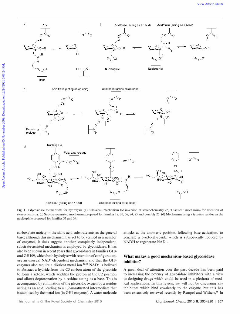
Fig. 1 Glycosidase mechanisms for hydrolysis. (a) ‘Classical’ mechanism for inversion of stereochemistry. (b) ‘Classical’ mechanism for retention ofstereochemistry. (c) Substrate-assisted mechanism proposed for families 18, 20, 56, 84, 85 and possibly 25. (d) Mechanism using a tyrosine residue as thenucleophile proposed for families 33 and 34.
carboxylate moiety in the sialic acid substrate acts as the generalbase; although this mechanism has yet to be verified in a numberof enzymes, it does suggest another, completely independent,substrate-assisted mechanism is employed by glycosidases. It hasalso been shown in recent years that glycosidases in families GH4and GH109, which both hydrolyse with retention of configuration,use an unusual NAD+-dependent mechanism and that the GH4enzymes also require a divalent metal ion.24,25 NAD+ is believedto abstract a hydride from the C3 carbon atom of the glycosideto form a ketone, which acidifies the proton at the C2 positionand allows deprotonation by a residue acting as a base. This isaccompanied by elimination of the glycosidic oxygen by a residueacting as an acid, leading to a 1,2-unsaturated intermediate thatis stabilised by the metal ion (in GH4 enzymes). A water molecule
attacks at the anomeric position, following base activation, togenerate a 3-keto-glycoside, which is subsequently reduced byNADH to regenerate NAD+.
What makes a good mechanism-based glycosidaseinhibitor?
A great deal of attention over the past decade has been paidto increasing the potency of glycosidase inhibitors with a viewto designing drugs which could be used in a plethora of med-ical applications. In this review, we will not be discussing anyinhibitors which bind covalently to the enzyme, but this hasbeen extensively reviewed recently by Rempel and Withers.48 In
This journal is © The Royal Society of Chemistry 2010 Org. Biomol. Chem., 2010, 8, 305–320 | 307
Ope
n A
cces
s A
rtic
le. P
ublis
hed
on 0
5 N
ovem
ber
2009
. Dow
nloa
ded
on 1
2/24
/202
1 6:
06:2
4 PM
. View Article Online
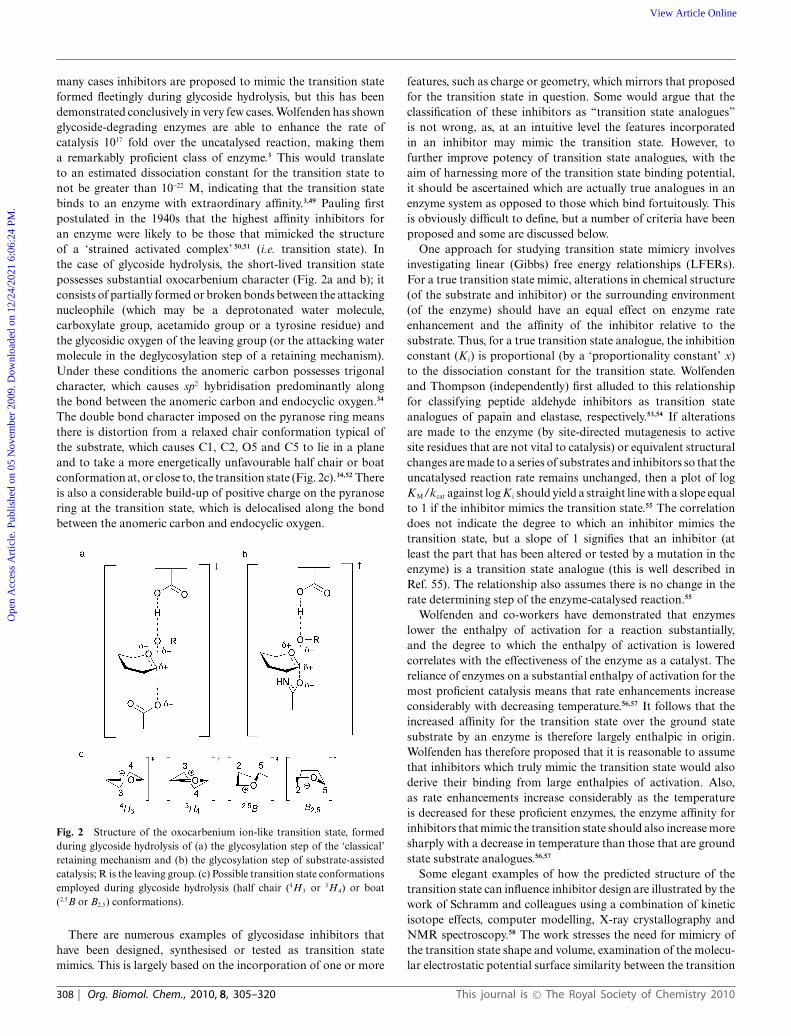
many cases inhibitors are proposed to mimic the transition stateformed fleetingly during glycoside hydrolysis, but this has beendemonstrated conclusively in very few cases. Wolfenden has shownglycoside-degrading enzymes are able to enhance the rate ofcatalysis 1017 fold over the uncatalysed reaction, making thema remarkably proficient class of enzyme.3 This would translateto an estimated dissociation constant for the transition state tonot be greater than 10-22 M, indicating that the transition statebinds to an enzyme with extraordinary affinity.3,49 Pauling firstpostulated in the 1940s that the highest affinity inhibitors foran enzyme were likely to be those that mimicked the structureof a ‘strained activated complex’ 50,51 (i.e. transition state). Inthe case of glycoside hydrolysis, the short-lived transition statepossesses substantial oxocarbenium character (Fig. 2a and b); itconsists of partially formed or broken bonds between the attackingnucleophile (which may be a deprotonated water molecule,carboxylate group, acetamido group or a tyrosine residue) andthe glycosidic oxygen of the leaving group (or the attacking watermolecule in the deglycosylation step of a retaining mechanism).Under these conditions the anomeric carbon possesses trigonalcharacter, which causes sp2 hybridisation predominantly alongthe bond between the anomeric carbon and endocyclic oxygen.34
The double bond character imposed on the pyranose ring meansthere is distortion from a relaxed chair conformation typical ofthe substrate, which causes C1, C2, O5 and C5 to lie in a planeand to take a more energetically unfavourable half chair or boatconformation at, or close to, the transition state (Fig. 2c).34,52 Thereis also a considerable build-up of positive charge on the pyranosering at the transition state, which is delocalised along the bondbetween the anomeric carbon and endocyclic oxygen.
Fig. 2 Structure of the oxocarbenium ion-like transition state, formedduring glycoside hydrolysis of (a) the glycosylation step of the ‘classical’retaining mechanism and (b) the glycosylation step of substrate-assistedcatalysis; R is the leaving group. (c) Possible transition state conformationsemployed during glycoside hydrolysis (half chair (4H3 or 3H4) or boat(2,5B or B2,5) conformations).
There are numerous examples of glycosidase inhibitors thathave been designed, synthesised or tested as transition statemimics. This is largely based on the incorporation of one or more
features, such as charge or geometry, which mirrors that proposedfor the transition state in question. Some would argue that theclassification of these inhibitors as “transition state analogues”is not wrong, as, at an intuitive level the features incorporatedin an inhibitor may mimic the transition state. However, tofurther improve potency of transition state analogues, with theaim of harnessing more of the transition state binding potential,it should be ascertained which are actually true analogues in anenzyme system as opposed to those which bind fortuitously. Thisis obviously difficult to define, but a number of criteria have beenproposed and some are discussed below.
One approach for studying transition state mimicry involvesinvestigating linear (Gibbs) free energy relationships (LFERs).For a true transition state mimic, alterations in chemical structure(of the substrate and inhibitor) or the surrounding environment(of the enzyme) should have an equal effect on enzyme rateenhancement and the affinity of the inhibitor relative to thesubstrate. Thus, for a true transition state analogue, the inhibitionconstant (K i) is proportional (by a ‘proportionality constant’ x)to the dissociation constant for the transition state. Wolfendenand Thompson (independently) first alluded to this relationshipfor classifying peptide aldehyde inhibitors as transition stateanalogues of papain and elastase, respectively.53,54 If alterationsare made to the enzyme (by site-directed mutagenesis to activesite residues that are not vital to catalysis) or equivalent structuralchanges are made to a series of substrates and inhibitors so that theuncatalysed reaction rate remains unchanged, then a plot of logKM/kcat against log K i should yield a straight line with a slope equalto 1 if the inhibitor mimics the transition state.55 The correlationdoes not indicate the degree to which an inhibitor mimics thetransition state, but a slope of 1 signifies that an inhibitor (atleast the part that has been altered or tested by a mutation in theenzyme) is a transition state analogue (this is well described inRef. 55). The relationship also assumes there is no change in therate determining step of the enzyme-catalysed reaction.55
Wolfenden and co-workers have demonstrated that enzymeslower the enthalpy of activation for a reaction substantially,and the degree to which the enthalpy of activation is loweredcorrelates with the effectiveness of the enzyme as a catalyst. Thereliance of enzymes on a substantial enthalpy of activation for themost proficient catalysis means that rate enhancements increaseconsiderably with decreasing temperature.56,57 It follows that theincreased affinity for the transition state over the ground statesubstrate by an enzyme is therefore largely enthalpic in origin.Wolfenden has therefore proposed that it is reasonable to assumethat inhibitors which truly mimic the transition state would alsoderive their binding from large enthalpies of activation. Also,as rate enhancements increase considerably as the temperatureis decreased for these proficient enzymes, the enzyme affinity forinhibitors that mimic the transition state should also increase moresharply with a decrease in temperature than those that are groundstate substrate analogues.56,57
Some elegant examples of how the predicted structure of thetransition state can influence inhibitor design are illustrated by thework of Schramm and colleagues using a combination of kineticisotope effects, computer modelling, X-ray crystallography andNMR spectroscopy.58 The work stresses the need for mimicry ofthe transition state shape and volume, examination of the molecu-lar electrostatic potential surface similarity between the transition
308 | Org. Biomol. Chem., 2010, 8, 305–320 This journal is © The Royal Society of Chemistry 2010
Ope
n A
cces
s A
rtic
le. P
ublis
hed
on 0
5 N
ovem
ber
2009
. Dow
nloa
ded
on 1
2/24
/202
1 6:
06:2
4 PM
. View Article Online
state and putative analogues, and consideration of the energythat every functional group in the transition state contributes tobinding with the enzyme. These methods have been employedin the design of extremely potent inhibitors for human purinenucleoside phosphorylase,59,60 the inhibition of which has greattherapeutic potential; indeed, these compounds are currently inclinical trials for the treatment of T-cell cancers and autoimmunedisorders.61 Similar techniques were used in the inhibitor design ofa 5¢-methylthioadenosine/S-adenosylhomocysteine nucleosidase,which yielded inhibitors in the low femtomolar range.62,63 It isinteresting to note that despite significant homology betweenenzymes from different species, the transition state “poise” variedsubtly which aided the design for both potency and specificity, andhas ramifications for inhibitor design that may be isozyme, tissueor species specific.58
Glycosidase inhibitors
The number of putative glycosidase inhibitors is continuallygrowing, and it is outside the scope of this review to mentionthem all in detail. Instead, we will describe the general featuresof representative glycosidase inhibitors from different classes(concentrating on those that possess six-membered rings andlargely omitting the body of work that has been done on thepyrrolidines and pyrrolizidines), which will be used to put thelater discussion about the properties of transition state mimics incontext; more comprehensive and detailed reviews from the lastdecade about the inhibitors themselves can be found in Ref. 64–70.By far the most popular way of designing glycosidase inhibitorsis to incorporate a nitrogen atom in the saccharide ring andin many cases Nature beat man to the synthesis. Nojirimycin 1(Fig. 3) was the first natural saccharide mimic to be isolated froma Streptomyces strain in 1966,71 and was originally proposed tocontain antibiotic properties. Although capable of inhibiting botha- and b-glycosidases, the hydroxyl group at C1 of the iminosugar is unstable.72 The synthesis of deoxynojirimycin 2, whichlacks this hydroxyl group, was accomplished the following yearby Paulsen and colleagues,73 and also later shown to occurnaturally.74,75 Deoxynojirimycin served as a template for thesubsequent synthesis of different glycoside derivatives (some ofwhich also occur in Nature), including deoxymannojirimycin anddeoxygalactonojirimycin, and all tend to be more potent inhibitorsof a-glycosidases than b-glycosidases.76 1,2-Dideoxynojirimycin,or fagomine, was also isolated,77 but was a less potent inhibitor.78
The drug Miglitol 3,15 used in the treatment of type II diabetes(described earlier), builds on the deoxynojirimycin scaffold with ahydroxyethyl group at the nitrogen atom and Zavesca/Miglustat4,79,80 used to combat the symptoms of Gaucher disease (byinhibition of the glycosyltransferase biosynthesizing the gluco-sylceramide substrate which accumulates during the disease),possesses an N-butyl group.
Many ideas for the synthesis of indolizidines, where a five anda six membered ring are fused together, also came following thediscovery of natural products. Castanospermine 5, first isolatedfrom the legume Castanospermum australe81 and later chemicallysynthesised,82 can inhibit both a-glucosidases and b-glucosidases,and may play a role in preventing spread of dengue virusinfection by inhibiting an endoplasmic reticulum a-glucosidase.83
Similarly swainsonine 6, isolated from Swainsona canescens84 and
later synthesised,85 and kifunensine 7, which was isolated fromKitasatosporia kifunense86 and also chemically synthesised,87 areboth potent a-mannosidase inhibitors;88,89 the latter has shownpromise as an anticancer agent.90 Calystegine compounds, whichpossess a nortropane ring system were first discovered in the early1990s as natural products in the roots of Calystegia sepium91
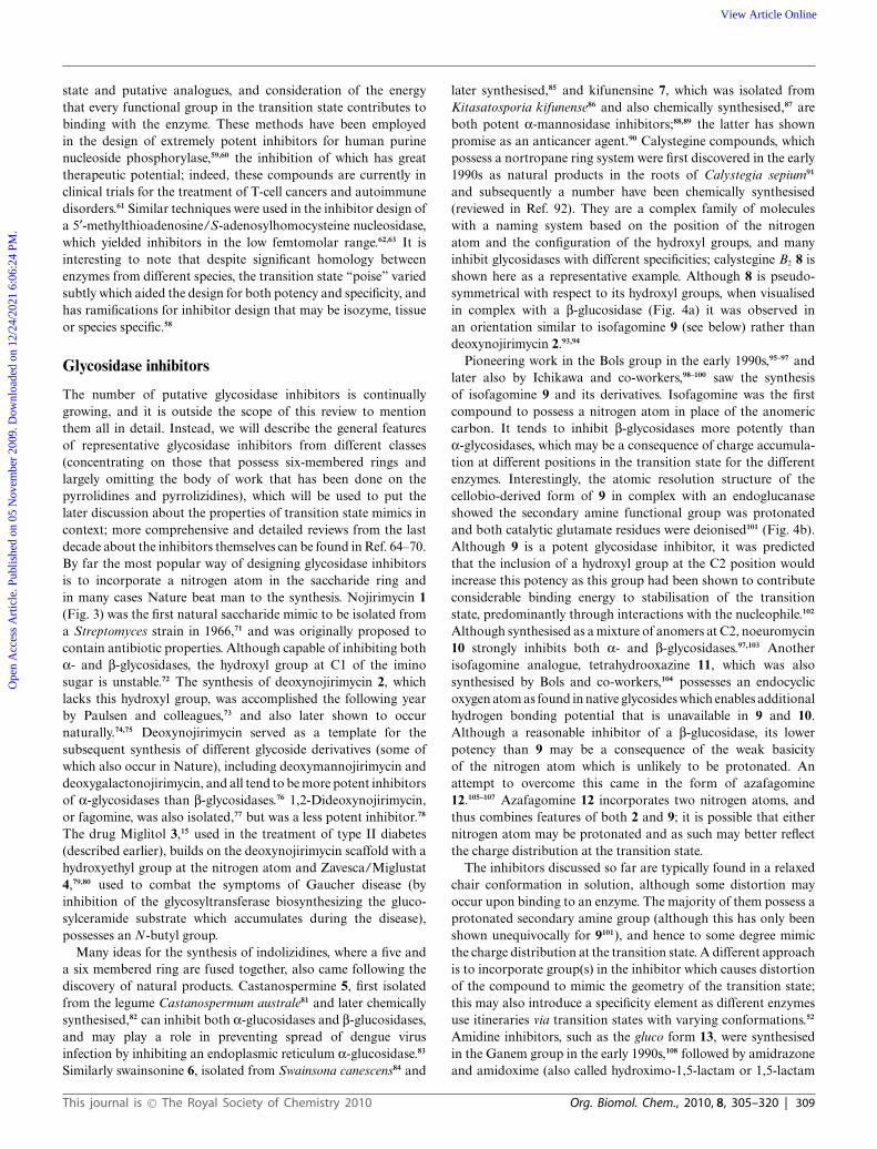
and subsequently a number have been chemically synthesised(reviewed in Ref. 92). They are a complex family of moleculeswith a naming system based on the position of the nitrogenatom and the configuration of the hydroxyl groups, and manyinhibit glycosidases with different specificities; calystegine B2 8 isshown here as a representative example. Although 8 is pseudo-symmetrical with respect to its hydroxyl groups, when visualisedin complex with a b-glucosidase (Fig. 4a) it was observed inan orientation similar to isofagomine 9 (see below) rather thandeoxynojirimycin 2.93,94
Pioneering work in the Bols group in the early 1990s,95–97 andlater also by Ichikawa and co-workers,98–100 saw the synthesisof isofagomine 9 and its derivatives. Isofagomine was the firstcompound to possess a nitrogen atom in place of the anomericcarbon. It tends to inhibit b-glycosidases more potently thana-glycosidases, which may be a consequence of charge accumula-tion at different positions in the transition state for the differentenzymes. Interestingly, the atomic resolution structure of thecellobio-derived form of 9 in complex with an endoglucanaseshowed the secondary amine functional group was protonatedand both catalytic glutamate residues were deionised101 (Fig. 4b).Although 9 is a potent glycosidase inhibitor, it was predictedthat the inclusion of a hydroxyl group at the C2 position wouldincrease this potency as this group had been shown to contributeconsiderable binding energy to stabilisation of the transitionstate, predominantly through interactions with the nucleophile.102
Although synthesised as a mixture of anomers at C2, noeuromycin10 strongly inhibits both a- and b-glycosidases.97,103 Anotherisofagomine analogue, tetrahydrooxazine 11, which was alsosynthesised by Bols and co-workers,104 possesses an endocyclicoxygen atom as found in native glycosides which enables additionalhydrogen bonding potential that is unavailable in 9 and 10.Although a reasonable inhibitor of a b-glucosidase, its lowerpotency than 9 may be a consequence of the weak basicityof the nitrogen atom which is unlikely to be protonated. Anattempt to overcome this came in the form of azafagomine12.105–107 Azafagomine 12 incorporates two nitrogen atoms, andthus combines features of both 2 and 9; it is possible that eithernitrogen atom may be protonated and as such may better reflectthe charge distribution at the transition state.
The inhibitors discussed so far are typically found in a relaxedchair conformation in solution, although some distortion mayoccur upon binding to an enzyme. The majority of them possess aprotonated secondary amine group (although this has only beenshown unequivocally for 9101), and hence to some degree mimicthe charge distribution at the transition state. A different approachis to incorporate group(s) in the inhibitor which causes distortionof the compound to mimic the geometry of the transition state;this may also introduce a specificity element as different enzymesuse itineraries via transition states with varying conformations.52
Amidine inhibitors, such as the gluco form 13, were synthesisedin the Ganem group in the early 1990s,108 followed by amidrazoneand amidoxime (also called hydroximo-1,5-lactam or 1,5-lactam
This journal is © The Royal Society of Chemistry 2010 Org. Biomol. Chem., 2010, 8, 305–320 | 309
Ope
n A
cces
s A
rtic
le. P
ublis
hed
on 0
5 N
ovem
ber
2009
. Dow
nloa
ded
on 1
2/24
/202
1 6:
06:2
4 PM
. View Article Online

Fig. 3 Structure of nojirimycin (1), deoxynojirimycin (2), Miglitol (3), N-butyl deoxynojirimycin (4), castanospermine (5), swainsonine (6), kifunensine(7), calystegine B2 (8), isofagomine (9), noeuromycin (10), tetrahydrooxazine (11), azafagomine (12), gluco-amidine (13), gluco-hydroximolactam(14), glucotetrazole (15), unsubstituted glucoimidazole (16), phenethyl-substituted glucoimidazole (17), isofagomine lactam (18), valienamine (19),validoxylamine A (20), acarbose (21), gluconolactone (22), PUGNAc (23), NAG-thiazoline (24), gluco-nagstatin (25), GlcNAcstatin C (26),2-deoxy-2,3-didehydro-N-acetylneuraminic acid (27), Relenza (28), and Tamiflu (29).
310 | Org. Biomol. Chem., 2010, 8, 305–320 This journal is © The Royal Society of Chemistry 2010
Ope
n A
cces
s A
rtic
le. P
ublis
hed
on 0
5 N
ovem
ber
2009
. Dow
nloa
ded
on 1
2/24
/202
1 6:
06:2
4 PM
. View Article Online
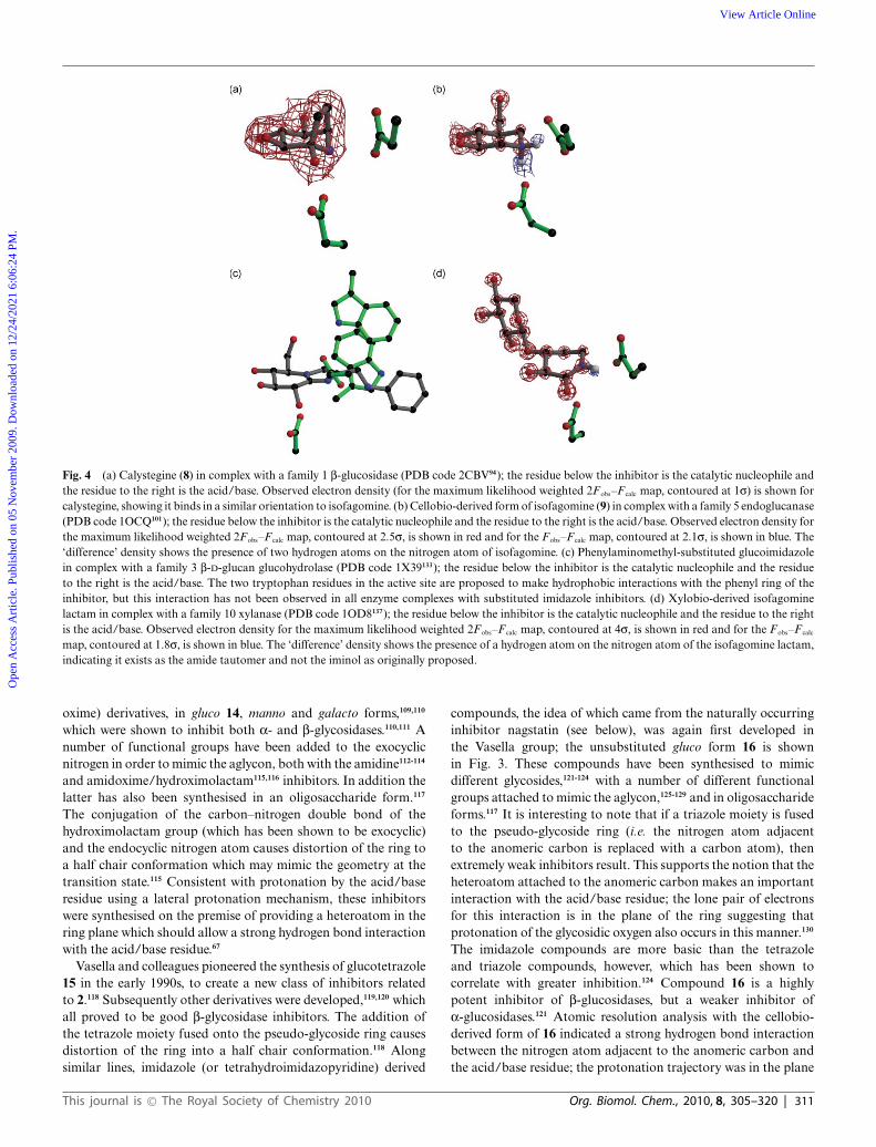
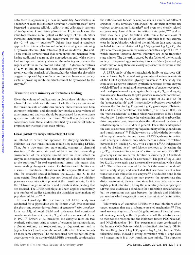
Fig. 4 (a) Calystegine (8) in complex with a family 1 b-glucosidase (PDB code 2CBV94); the residue below the inhibitor is the catalytic nucleophile andthe residue to the right is the acid/base. Observed electron density (for the maximum likelihood weighted 2Fobs–F calc map, contoured at 1s) is shown forcalystegine, showing it binds in a similar orientation to isofagomine. (b) Cellobio-derived form of isofagomine (9) in complex with a family 5 endoglucanase(PDB code 1OCQ101); the residue below the inhibitor is the catalytic nucleophile and the residue to the right is the acid/base. Observed electron density forthe maximum likelihood weighted 2Fobs–F calc map, contoured at 2.5s, is shown in red and for the Fobs–F calc map, contoured at 2.1s, is shown in blue. The‘difference’ density shows the presence of two hydrogen atoms on the nitrogen atom of isofagomine. (c) Phenylaminomethyl-substituted glucoimidazolein complex with a family 3 b-D-glucan glucohydrolase (PDB code 1X39133); the residue below the inhibitor is the catalytic nucleophile and the residueto the right is the acid/base. The two tryptophan residues in the active site are proposed to make hydrophobic interactions with the phenyl ring of theinhibitor, but this interaction has not been observed in all enzyme complexes with substituted imidazole inhibitors. (d) Xylobio-derived isofagominelactam in complex with a family 10 xylanase (PDB code 1OD8137); the residue below the inhibitor is the catalytic nucleophile and the residue to the rightis the acid/base. Observed electron density for the maximum likelihood weighted 2Fobs–F calc map, contoured at 4s, is shown in red and for the Fobs–F calc
map, contoured at 1.8s, is shown in blue. The ‘difference’ density shows the presence of a hydrogen atom on the nitrogen atom of the isofagomine lactam,indicating it exists as the amide tautomer and not the iminol as originally proposed.
oxime) derivatives, in gluco 14, manno and galacto forms,109,110
which were shown to inhibit both a- and b-glycosidases.110,111 Anumber of functional groups have been added to the exocyclicnitrogen in order to mimic the aglycon, both with the amidine112-114
and amidoxime/hydroximolactam115,116 inhibitors. In addition thelatter has also been synthesised in an oligosaccharide form.117
The conjugation of the carbon–nitrogen double bond of thehydroximolactam group (which has been shown to be exocyclic)and the endocyclic nitrogen atom causes distortion of the ring toa half chair conformation which may mimic the geometry at thetransition state.115 Consistent with protonation by the acid/baseresidue using a lateral protonation mechanism, these inhibitorswere synthesised on the premise of providing a heteroatom in thering plane which should allow a strong hydrogen bond interactionwith the acid/base residue.67
Vasella and colleagues pioneered the synthesis of glucotetrazole15 in the early 1990s, to create a new class of inhibitors relatedto 2.118 Subsequently other derivatives were developed,119,120 whichall proved to be good b-glycosidase inhibitors. The addition ofthe tetrazole moiety fused onto the pseudo-glycoside ring causesdistortion of the ring into a half chair conformation.118 Alongsimilar lines, imidazole (or tetrahydroimidazopyridine) derived
compounds, the idea of which came from the naturally occurringinhibitor nagstatin (see below), was again first developed inthe Vasella group; the unsubstituted gluco form 16 is shownin Fig. 3. These compounds have been synthesised to mimicdifferent glycosides,121-124 with a number of different functionalgroups attached to mimic the aglycon,125-129 and in oligosaccharideforms.117 It is interesting to note that if a triazole moiety is fusedto the pseudo-glycoside ring (i.e. the nitrogen atom adjacentto the anomeric carbon is replaced with a carbon atom), thenextremely weak inhibitors result. This supports the notion that theheteroatom attached to the anomeric carbon makes an importantinteraction with the acid/base residue; the lone pair of electronsfor this interaction is in the plane of the ring suggesting thatprotonation of the glycosidic oxygen also occurs in this manner.130
The imidazole compounds are more basic than the tetrazoleand triazole compounds, however, which has been shown tocorrelate with greater inhibition.124 Compound 16 is a highlypotent inhibitor of b-glucosidases, but a weaker inhibitor ofa-glucosidases.121 Atomic resolution analysis with the cellobio-derived form of 16 indicated a strong hydrogen bond interactionbetween the nitrogen atom adjacent to the anomeric carbon andthe acid/base residue; the protonation trajectory was in the plane
This journal is © The Royal Society of Chemistry 2010 Org. Biomol. Chem., 2010, 8, 305–320 | 311
Ope
n A
cces
s A
rtic
le. P
ublis
hed
on 0
5 N
ovem
ber
2009
. Dow
nloa
ded
on 1
2/24
/202
1 6:
06:2
4 PM
. View Article Online
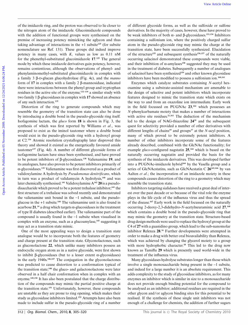
of the imidazole ring, and the proton was observed to lie closer tothe nitrogen atom of the imidazole. Glucoimidazole compoundswith the addition of functional groups were synthesised on thepremise of increasing potency mimicking the aglycon and thustaking advantage of interactions in the +1 subsite125 (for subsitenomenclature see Ref. 131). These groups did indeed improvepotency in many cases, with K i values as low as 0.11 nMfor the phenethyl-substituted glucoimidazole 17.125 The generalmode by which these imidazole derivatives gain potency, however,remains contentious; although the observations of phenyl- andphenylaminomethyl-substituted glucoimidazole in complex witha family 3 b-D-glucan glucohydrolase (Fig. 4c), and the mannoform of 17 in complex with a family 2 b-mannosidase, indicatedthere were interactions between the phenyl group and tryptophanresidues in the active site of the enzyme,132–134 a similar study withtwo family 1 b-glucosidases in complex with 17 showed an absenceof any such interaction.135
Distortion of the ring to generate compounds which mayresemble the geometry of the transition state can also be doneby introducing a double bond in the pseudo-glycoside ring itself.Isofagomine lactam, the gluco form 18 is shown in Fig. 3, thesynthesis of which was first reported by Williams et al., wasproposed to exist as the iminol tautomer where a double bondwould exist in the pseudo-glycoside ring with a hydroxyl groupat C2.136 Atomic resolution crystallography later disproved thistheory and showed it existed as the energetically favoured amidetautomer137 (Fig. 4d). A number of different glycoside forms ofisofagomine lactam have since been synthesised, and have provedto be potent inhibitors of b-glycosidases.138 Valienamine 19, andits analogues, have also proven to be potent inhibitors primarily ofa-glycosidases.139 Valienamine was first discovered as a product ofvalidoxylamine A hydrolysis by Pseudomonas denitrificans, whichin turn was a product of validamycin A hydrolysis,140 and waslater chemically synthesised.141 Validoxylamine A142 20 is a pseudo-disaccharide which proved to be a potent trehalase inhibitor;143 thefirst structure of a trehalase was solved recently and demonstratedthe valienamine unit bound in the -1 subsite, and the pseudo-glucose in the +1 subsite.144 The valienamine unit is also found inacarbose 21,14 a drug which targets a-glucosidases in the treatmentof type II diabetes (described earlier). The valienamine part of thecompound is usually found in the -1 subsite when visualised incomplex with an enzyme, such as a glucoamylase,145 suggesting itmay act as a transition state mimic.
One of the most appealing ways to design a transition stateanalogue would be to incorporate both the features of geometryand charge present at the transition state. Glyconolactones, suchas gluconolactone 22, which unlike many inhibitors possess anendocyclic oxygen atom as in a native glycoside, were first shownto inhibit b-glycosidases (but to a lesser extent a-glycosidases)in the early 1940s.146,147 The conjugation in the glyconolactoneswas predicted to cause distortion to a conformation typical ofthe transition state;148 the gluco- and galactonolactone were laterobserved in a half chair conformation when in complex with anenyzme.149,150 It has also been postulated that the charge distribu-tion of the compounds may mimic the partial positive charge atthe transition state.151 Unfortunately, however, these compoundsare unstable as they are prone to ring opening, which makes theirstudy as glycosidase inhibitors limited.152 Attempts have also beenmade to include sulfur in the pseudo-glycoside ring of a number
of different glycoside forms, as well as the sulfoxide or sulfonederivatives. In the majority of cases, however, these have proved tobe weak inhibitors of both a- and b-glycosidases.153,154 Inhibitorscontaining a sulfonium ion, where the positively charged sulfuratom in the pseudo-glycoside ring may mimic the charge at thetransition state, have been successfully synthesized. Elucidationof the structure155 and subsequent synthesis156,157 of the naturallyoccurring salacinol demonstrated these compounds were viable,and their inhibition of a-amylases158 suggested they may be usedin the treatment of diabetes. Subsequently a number of analoguesof salacinol have been synthesized159 and other known glycosidaseinhibitors have been modified to possess a sulfonium ion.160,161
Enzymes which catalyze substrates containing N-acetyl hex-osamine using a substrate-assisted mechanism are amenable tothe design of selective and potent inhibitors which incorporatefeatures designed to mimic the transition state which exists onthe way to and from an oxazoline ion intermediate. Early workin the field focussed on PUGNAc 23,162 which possesses anN-phenylcarbamate moiety that makes a number of interactionswith active site residues.21,163 The deduction of the mechanismled to the design of NAG-thiazoline 2439 and the subsequentquest for selectivity provided a number of other derivatives withdifferent lengths of chains19 and groups37 at the N-acyl position,many of which proved to be extremely potent inhibitors. Anumber of other inhibitors incorporate some of the featuresalready described, combined with the GlcNAc functionality; forexample gluco-configured nagstatin 25,164 which is based on thenaturally occurring nagstatin and provided the basis for thesynthesis of the imidazole derivatives. This was developed furtherinto a PUGNAc-imidazole hybrid164 by the Vasella group and aseries of GlcNAcstatins, such as GlcNAcstatin C 26165,166 by vanAalten et al.; the incorporation of an imidazole moiety in thesecompounds causes distortion of the ring to a geometry which mayresemble the transition state.
Inhibitors targeting sialidases have received a great deal of inter-est over the past decade or so because of the vital role the enzymeplays in the life cycle of the influenza virus and thus the spreadof the disease.167 Early work in the field focussed on the naturallyoccurring 2-deoxy-2,3-didehydro-N-acetylneuraminic acid 27,168
which contains a double bond in the pseudo-glycoside ring thatmay mimic the geometry at the transition state. Structure-baseddrug design methods were used to replace the hydroxyl group at theC4 of 27 with a guanidino group, which lead to the sub-nanomolarinhibitor Relenza 28.16 Further developments were attempted inorder to make a drug with better oral bioavailability than Relenza,which was achieved by changing the glycerol moiety to a groupwith more hydrophobic character.18 This led to the drug nowknown as Tamiflu 29 which is presently used world-wide in thetreatment of the influenza virus.
Many glycosidases hydrolyse substrates longer than those whichinvolve a single monosaccharide being present in the -1 subsite,and indeed for a large number it is an absolute requirement. Thisadds complexity to the study of glycosidase inhibitors, as for manyenzymes an inhibitor which is similar in size to a monosaccharidedoes not provide enough binding potential for the compound tobe analysed as an inhibitor; additional residues are required in theother glycon and/or aglycon binding sites for this potential to berealised. If the synthesis of these single unit inhibitors was notenough of a challenge for chemists, the addition of further sugars
312 | Org. Biomol. Chem., 2010, 8, 305–320 This journal is © The Royal Society of Chemistry 2010
Ope
n A
cces
s A
rtic
le. P
ublis
hed
on 0
5 N
ovem
ber
2009
. Dow
nloa
ded
on 1
2/24
/202
1 6:
06:2
4 PM
. View Article Online
onto them is approaching a near impossibility. Nevertheless, ina number of cases this has been achieved. Glycosynthases169 havebeen used to generate cellobio-, cellotrio- and cellotetrao- versionsof isofagomine 9 and tetrahydrooxazine 11; in each case theinhibitors became more potent as the length of the inhibitorsincreased demonstrating the requirement for the binding inthe -2, -3 and -4 subsites.170 Others have used a syntheticapproach to obtain cellobio- and cellotrio- analogues containinga hydroximolactam (14), tetrazole (15) or imidazole (16) unit.These studies demonstrated that some inhibitors benefited fromhaving additional sugars at the non-reducing end, while othershad an improved potency when on the reducing end (where thesugars would lie in the product subsites).117 Xylobio- derivativesof 2, 9, 14 and 16 have also been chemically synthesised.171 Inrecent years the synthesis of oligosaccharides where the glycosidicoxygen is replaced by a sulfur atom has also become extremelyuseful at providing inhibitors which cannot be hydrolysed by theenzyme.172
Transition state mimicry or fortuitous binding
Given the volume of publications on glycosidase inhibitors, onlya handful have addressed the issue of whether they are mimics ofthe transition state or fortuitous binders. These studies have beenextremely insightful, and although they involve a large volume ofexperiments and analysis, should be encouraged for other enzymesystems and inhibitors in the future. We will now describe theconclusions from these studies, classified by a number of criteriaproposed for true transition state mimics.
Linear (Gibbs) free energy relationships (LFERs)
As alluded to earlier, one approach for studying whether aninhibitor is a true transition state mimic is by measuring LFERs.Thus, for a true transition state mimic, changes in chemicalstructure of the substrate and inhibitor or the surroundingenvironment of the enzyme should have an equal effect onenzyme rate enhancement and the affinity of the inhibitor relativeto the substrate.55 In real experimental terms, this means thatcorresponding changes in series of substrates and inhibitors ora series of mutational alterations in the enzyme (that are notvital for catalysis) should influence the KM/kcat and K i to thesame extent. Note that this does not demand that the inhibitorpossesses every interaction present at the transition state, for it isthe relative changes in inhibitor and transition state binding thatare assessed. The LFER technique has been applied successfullyto a number of studies examining the transition state analogy ofpeptidase inhibitors.173–176
To our knowledge the first time a full LFER study wasconducted for a glycosidase was by Ermert et al. who examinedthe gluco- and manno-derived forms of tetrazole119 (for the gluco-form see 15), although Field and colleagues had consideredcorrelations between K i and KM/kcat, albeit in a more crude form,in 1991.177 Ermert et al. measured the catalytic rates on twosynthetic substrates using a range of enzymes (a-mannosidases,a b-mannosidase, an a-glucosidase, a b-glucosidase and ab-galactosidase) and the inhibition of both tetrazole compoundson these same enzymes. The methods used here are not totally inaccordance with the way in which LFERs are usually conducted as
the authors chose to test the compounds in a number of differentenzymes. It has, however, been shown that different enzymes usevarious conformation itineraries52 and even very closely relatedenzymes may have different transition state poise,60,178 and sowhat may be a good transition state mimic for one class ofenzymes may not be so for others. Although it is not entirelyclear in the publication which of the inhibition data reported areincluded in the correlation of log 1/K i against log kcat/KM, theplot nevertheless gives a linear correlation with a slope of 1.1,119,179
which suggests tetrazole-derived inhibitors are good transitionstate mimics. The distortion caused by the fusion of the tetrazolemoiety to the pseudo-glycoside ring into a half chair (or envelope)conformation may therefore closely represent the structure at thetransition state.
A LFER study of the tetrasaccharide inhibitor acarbose (21)was performed by Mosi et al. using a number of active site mutantsof the GH13 cyclodextrin glycosyltransferase.180 The study wasconducted using two different synthetic substrates for the enzyme(which differed in length and hence number of subsites occupied),and the dependence of log K i against both log KM/kcat and log KM
was assessed. In each case the plot yielded a reasonable correlation;the log K i vs. log KM/kcat plot gave slopes of 2.2 and 1.6 forthe ‘monosaccharide’ and ‘trisaccharide’ substrates, respectively,whereas the plot for log K i against log KM gave slopes of between0.4 and 0.5. The correlation was better (r2 of 0.96 vs. 0.81) forthe ‘monosaccharide’ substrate, which was assumed to be a bettertest for the -1 subsite where the valienamine unit of acarbose lies;this comparison does, however, show the influence of the choice ofsubstrate upon LFER studies in general. The authors interpretedthe data as acarbose displaying ‘equal mimicry of the ground stateand transition state’.180 This, however, is at odds with the derivationof the equation underpinning the use of LFERs, where a transitionstate analogue is defined only when there is a direct correlationbetween log K i and log KM/kcat with a slope of 1.55 An independentstudy by Berland et al. used kinetic methods to determine thekcat/KM parameters for a series of active site mutants of a GH15glucoamylase and isothermal titration calorimetry (ITC) methodsto measure the Kd values for acarbose.181 The plot of log Kd andlog KM/kcat once again gave a reasonable correlation, with a slopeof 2. The authors accounted for the fact the correlation shouldhave a unity slope, and concluded that acarbose is not a truetransition state mimic for this enzyme.181 The double bond in thevalienamine unit of acarbose may prevent the appropriate ringdistortion to mimic the transition state, but nevertheless remains ahighly potent inhibitor. During the same study deoxynojirimycin(2) was also studied as a candidate for a transition state analogue,but no correlation was seen between the kinetic and inhibitionparameters which suggests it is not a true mimic of the transitionstate.181
Whitworth et al. examined LFERs with two inhibitors whichtarget enzymes that use a substrate-assisted mechanism.182 Theyused an elegant system of modifying the volume of the alkyl groupof the N-acyl moiety at the C2 position in both the substrates usedto monitor the reaction and the inhibitors tested: PUGNAc (23)and NAG-thiazoline (24). The experiments were conducted onthe human O-GlcNAcase, which is classified into family GH84.The resulting plots of log 1/K i against log kcat/KM for the NAG-thiazoline series showed a strong correlation with a slope closeto 1 suggesting it is a true transition state mimic. The opposite,
This journal is © The Royal Society of Chemistry 2010 Org. Biomol. Chem., 2010, 8, 305–320 | 313
Ope
n A
cces
s A
rtic
le. P
ublis
hed
on 0
5 N
ovem
ber
2009
. Dow
nloa
ded
on 1
2/24
/202
1 6:
06:2
4 PM
. View Article Online
however, was true for the PUGNAc series, which show a weakercorrelation suggesting this compound is less likely to mimic thetransition state.182 The results were perhaps surprising given the sp2
anomeric centre in PUGNAc, which may have been predicted toresemble the transition state more than the equivalent sp3 centre inNAG-thiazoline, which may have been expected to better representthe oxazoline intermediate (see Fig. 1). The authors postulatedthat these unexpected results may be attributable to the longerC–S bond length present in NAG-thiazoline in comparison tothe C–O bond in the oxazoline intermediate.182 The longer bondlength may mimic the partial, but significant, bond order betweenthe nucleophilic carbonyl oxygen atom and anomeric carbon of alate transition state.
It is intriguing that the general conclusions in terms of sp2- andsp3-centred inhibitors drawn from a LFER study by Wicki et al.were almost opposite to those described for PUGNAc and NAG-thiazoline (although they were tested against different enzymeswhich may have significantly different transition states). Wickiet al. used a series of active site mutants (both ‘first sphere’and ‘second sphere’) of a xylanase to assess the transition stateanalogy for the xylobio-derived versions of deoxynojirimycin(2), isofagomine (9), lactam oxime (14), imidazole (16) andisofagomine lactam (18).183 The log kcat/KM against log 1/K i
plot for each inhibitor showed a slope of close to 1, but thecorrelation coefficients for the sp3-centred inhibitors (those whichwere more likely to resemble the charge at the transition staterather than the geometry) deoxynojirimycin and isofagomine werepoor (r values of 0.89 and 0.77, respectively, which equates to r2
values of 0.79 and 0.59 to allow direct comparison with valuesdescribed below). However, the sp2-centred inhibitors (which weremore likely to resemble the geometry at the transition state) allshowed a correlation coefficient very close to 1, suggesting theywere transition state mimics.183 These data are in agreement withthe earlier study by Ermert et al. on the tetrazole inhibitors,and suggest that those inhibitors which can distort the pseudo-glycoside ring into a half chair or envelope conformation maygenerally be the best mimics of the transition state.
A detailed investigation into the LFERs of 8 mannosidaseinhibitors was performed by Tailford et al. with a series ofactive site mutants of a family 2 mannosidase.134 In particularthis study examined a number of manno-derived imidazoles (see16 for gluco unsubstituted version) and amidines (see 13 forgluco unsubstituted version) with a number of functional groupsattached which may mimic the aglycon; all compounds wereobserved in a B2,5 conformation in the active site of the enzyme.The substituted manno-imidazole compounds (the three testedhad phenethyl (see 17 for the gluco version), phenylaminomethyland phenyloxymethyl functional groups) all showed a strongcorrelation in the plot of log KM/kcat against log K i with a slopeclose to 1 (Fig. 5a) suggesting they were transition state analogues.The plot for the unsubstituted manno-imidazole also had a slopeclose to 1, but the r2 correlation was lower (at 0.78).134 The studyby Wicki et al. showed the LFER plot for the equivalent xylobio-derived compound had a high correlation coefficient suggestingit was a good transition state analogue;183 this may, however,reflect the differences in the structure of the transition state fordifferent enzyme families. This observation also suggests that, forthis enzyme, the presence of the functional group in the aglyconwas required to properly mimic the transition state. The two
substituted amidines tested, isoquinuclidine and noeuromycin 10,all showed slopes of less than one and poor correlation coefficients,suggesting none of them were good mimics of the transition state.
In contrast to definitions of transition state mimicry whichdepend solely on the intuition or prejudice of an individualscientist, LFERs produce a quantifiable measurement that otherscan repeat. This does not mean, however, that LFER analysesare without flaws. Firstly, if the goal of the study is to simplyobtain tight-binding enzyme inhibition, then a LFER correlationis irrelevant; it would be a mistake to be discouraged by a poorLFER performance if one is primarily only interested in K i values.The second issue, especially where site-directed mutagenesis is theperturbation of choice in the LFER study, is the selection of themutations made. Some would argue, for example, that chargedinhibitors do comparatively poorly in LFER analysis (discussed inRef. 134) because the most energetically significant interactions ofthese charged compounds are with the nucleophile and acid/baseresidues; the two residues which cannot be mutated in theanalysis without compromising the enzyme activity completely.One can imagine that, were it possible, an LFER plot whichincluded nucleophile and acid/base variants would show massivedisruption to catalysis and a similar reduction in inhibitor binding.This raises the issue of how to address the compounds that do“poorly” in LFER tests. Do they mimic something other thanthe transition state, are they fortuitous or adventitious binders,or has the technique itself legislated against certain classes ofcompounds?
Thermodynamics of inhibitor binding
As described earlier, Wolfenden proposed that the increasedaffinity for the transition state over the ground state by an enzymeis primarily derived from enthalpy, and therefore it could be arguedthat inhibitors which truly mimic the transition state should alsobind with large enthalpic contributions.56,57 The issue of whichglycosidase inhibitors were likely to be transition state mimicsbased on their binding thermodynamics was tackled using a panelof 18 inhibitors with a family 1 b-glucosidase.184 Measurementswere made using ITC, which gives a direct read-out on the affinityof the inhibitor, stoichiometry of binding between inhibitor andenzyme and the enthalpic contribution to binding, from whichthe Gibbs free energy and entropy can be calculated (for a recentreview on ITC see Ref. 185). All of the inhibitors examined inthis study (deoxynojirimycin 2, castanospermine 5, calystegineB2 8, isofagomine 9, noeuromycin 10, tetrahydrooxazine 11,azafagomine 12, gluco- 14 and galacto-hydroximolactam, glucote-trazole 15, unsubstituted glucoimidazole 16, glucoimidazole sub-stituted with a phenethyl group 17 and a range of other functionalgroups, and isofagomine lactam 18) displayed a negative andtherefore favourable enthalpic contribution to binding.184 Someof the inhibitors, however, harnessed a significant portion of theirbinding energy from entropy, which questions their ability to betransition state mimics. These include the phenethyl-substitutedglucoimidazole 17 (which incidentally was the most potent of theinhibitors examined in the study), isofagomine 2, and calystegineB2 8. The data show there is no correlation between the inhibitoraffinity and the enthalpic contribution to binding, and nor does thechemistry of the inhibitor appear to influence the thermodynamics,at least not on this system.184
314 | Org. Biomol. Chem., 2010, 8, 305–320 This journal is © The Royal Society of Chemistry 2010
Ope
n A
cces
s A
rtic
le. P
ublis
hed
on 0
5 N
ovem
ber
2009
. Dow
nloa
ded
on 1
2/24
/202
1 6:
06:2
4 PM
. View Article Online
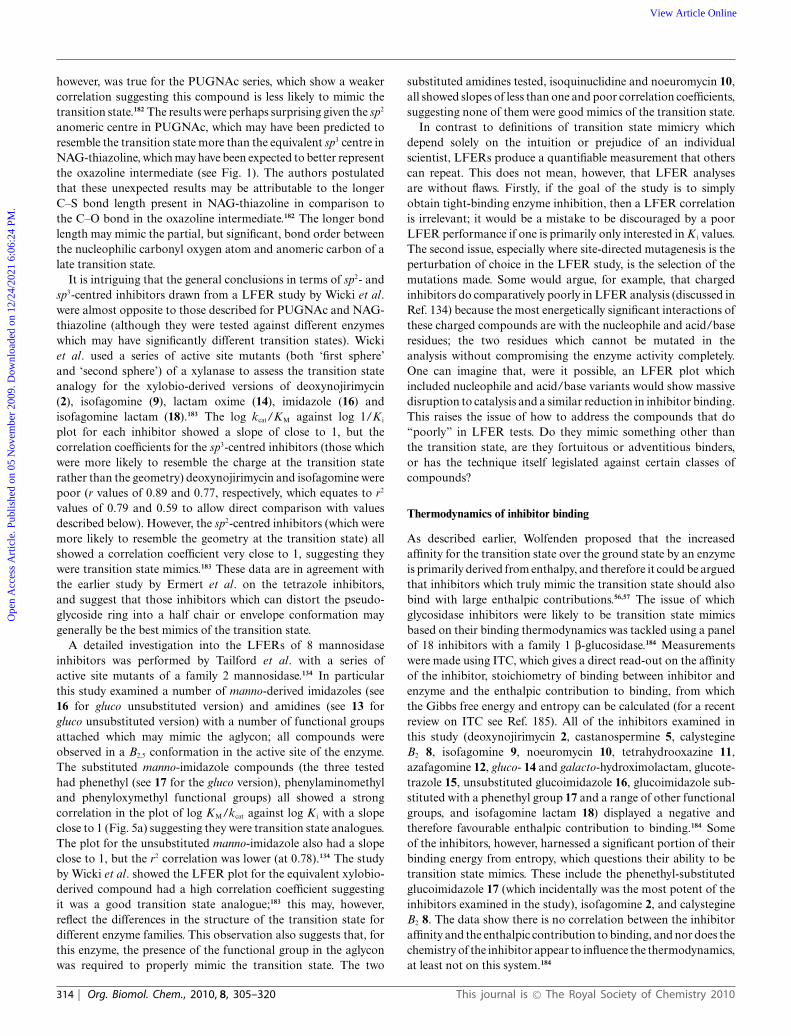
Fig. 5 (a) Linear free energy plot of log K i against log KM/kcat for the phenethyl-substituted mannoimidazole with the family 2 b-mannosidase (datataken from Ref. 134). The best fit line through the points has a slope of 1.09 and a correlation r2 of 0.94, which strongly suggests the compound is agood mimic of the transition state. (b) Enthalpy–entropy compensation plot for inhibitors with a b-glucosidase; the line of best fit has a slope of 0.93 andcorrelation of 0.91. Points in circles represent data from the initial study of 18 compounds (data taken from Ref. 184); filled circles represent those whichmimic the charge at the transition state, open circles those which mimic the geometry. The points in squares represent data from subsequent studies (datataken from Ref. 194 and 195) and demonstrate that even when a compound is enthalpically unfavourable, it still falls on the line. (c) Plot of gain of product(as indicated by an increase in absorbance at 400 nm by production of 2,4-dinitrophenolate from hydrolysis of 2,4-dinitrophenyl b-D-glucopyranoside bya b-glucosidase) against time to illustrate slow onset inhibition. Slow onset inhibition is characterised by an initial rapid catalytic rate (here for 200–300 s),followed by a slower steady state rate. (d) pH dependence of kcat/KM for a b-glucosidase (circles) and 1/K i for 14 (squares); fits to bell-shaped ionisationprofiles are shown (data taken from Ref. 184). In this case the pH dependence for inhibition mirrors that of catalysis, but this is not the case for allinhibitors.
A popular way of assessing thermodynamic data for a seriesof related compounds is using enthalpy–entropy compensationplots. The enthalpy–entropy compensation phenomenon appearsto occur in many, if not all, binding studies and is demonstratedby a linear correlation between the enthalpic and entropiccontributions to binding for a series of perturbations or changes inexperimental variable, such as temperature, series of homologouscompounds or mutants.186 Many dispute the significance ofenthalpy–entropy compensation, claiming it demonstrates theknown thermodynamic laws and the limitations of the valuesmeasured.186,187 Compensation between enthalpy and entropyterms is also likely to be evident where the range of DG◦
a valuesmeasured is relatively small; this limited range is likely to occurboth from the range of values measurable using techniques suchas ITC and the values imposed by biology.186,188–190 In additionthe intrinsic properties of water are also likely to be important inthe compensation. Upon ligand binding a favourable enthalpiccontribution suggests solvent molecules are involved in stronghydrogen bonding interactions, but this is likely to be associatedwith an entropic cost due to the small configurational freedom in
the system, whereas conversely the loss of bound water moleculescontributes favourably to entropy and the disorder of the system,but unfavourably to the enthalpy term as hydrogen bondinginteractions are lost.187,191–193
An enthalpy–entropy plot for the thermodynamic data deter-mined for the 18 inhibitors with a b-glucosidase shows a line ofbest fit with a slope of 0.93 and correlation of 0.91 (Fig. 5b).184
No related inhibitors, however, such as those which may mimicthe charge at the transition state or those which may mimic thegeometry, are closely grouped in the compensation plot. In factvery closely related compounds which only differ by one atom typewere observed at opposite ends of the trend. The ITC data for threeother inhibitors tested with the b-glucosidase since the originalpublication, two 1-deoxy-6-oxa-N-(thiocarbamoyl)calystegines194
and a 3-imino-2-thia-(+)-castanospermine analogue195 (shown inFig. 5b), also lie on the best fit line through the points. Thecastanospermine analogue was shown to differ from the othercompounds in that it bound with an unfavourable enthalpiccontribution which was more than compensated by a largeentropy; despite this different thermodynamic signature, this point
This journal is © The Royal Society of Chemistry 2010 Org. Biomol. Chem., 2010, 8, 305–320 | 315
Ope
n A
cces
s A
rtic
le. P
ublis
hed
on 0
5 N
ovem
ber
2009
. Dow
nloa
ded
on 1
2/24
/202
1 6:
06:2
4 PM
. View Article Online
remarkably still lies on the line. It is interesting to note that a studyinvestigating putative transition state analogues with the humanpurine nucleoside phosphorylase, some of which have picomolaraffinity, also show strong enthalpy–entropy compensation. In thisplot, however, related compounds did tend to cluster in one regionof the plot.196
The differences in the (closely) related inhibitors studied aremost likely attributed to the effects of solvation and desolvation.Whilst ITC studies can quantify the thermodynamics of inhibitorbinding to protein and the subsequent displacement of watermolecules during this process, nothing is known about thesolvation state of either the protein or inhibitor alone. Homans andco-workers have demonstrated that desolvation of the inhibitorupon binding is likely to contribute significantly;197 it may,therefore, only be when more sophisticated methods are availableto measure or model these important contributions that the truethermodynamics of inhibitor binding are revealed.
Slow onset inhibition
Slow onset inhibition, a phenomenon characterised by an initialrapid catalytic rate by an enzyme in the presence of an inhibitor,followed by a decrease to a slower steady state rate after anamount of time (Fig. 5c), has been observed with a number ofglycosidase inhibitors, including nojirimycin 1,198,199 deoxynojir-imycin 2,199 acarbose 21,199 isofagomine 9 and analogues,101,184,200,201
castanospermine 5,76,184 noeuromycin 10,184 azafagomine 12,184
hydroximolactam 14,184 glucotetrazole 15,184 glucoimidazole 16184
and derivatives such as 17125,184 and isofagomine lactam 18.184
Various reasons have been offered as to why inhibitors may displayslow onset. The initial rapid reaction rate may represent formationof the enzyme-inhibitor complex, after which isomerisation55,202–204
or conformational changes76,203,205,206 of the inhibitor or proteincauses formation of a more potent inhibitory species. It hasalso been suggested the lag phase for inhibition may allow atime-dependent protonation of the enzyme or inhibitor.203,206,207
Slow onset inhibition may also be a consequence of a slowinteraction (less than the rate of diffusion) between the inhibitorand enzyme.202,203,208
Schramm and co-workers have suggested that the presenceof slow onset inhibition is a criterion for an inhibitor to be atrue transition state mimic (although not exclusively).178,209 Theybelieve that slow onset is evident among transition state mimickinginhibitors because they bind to an enzyme that is conformationallyoptimised to bind the substrate in the ground state. The lagphase before the true inhibitory potential of an inhibitor becomesapparent is therefore attributed to conformational changes thatmust occur in the active site, which will be less efficient with atransition state analogue than with the ground state substratefor which it optimised.55,178,209 Although evidence of slow onsetinhibition is a useful guide when studying inhibitors that areputative transition state mimics, it is highly likely that inhibitorswhich do not comply also display the phenomenon; this isstrongly suggested merely by the number of glycosidase inhibitorswhich have been reported to show it, as detailed in the previousparagraph. It may also be possible that all inhibitors show slowonset inhibition to some degree, but is more apparent whenmeasuring high affinity inhibition constants at low inhibitorconcentration.55
pH dependence of inhibition
Another proposed criterion for analysing which inhibitors arelikely to be true transition state mimics is the comparison ofthe profiles for pH dependence of inhibition and catalysis.210
A difference in the pH dependence for catalysis vs. inhibition,where, for example, an inhibitor inhibits most effectively when theenzyme is largely inactive, suggests it cannot be a true transitionstate mimic, as, by definition, a catalytically inactive enzymecannot bind the transition state.211,212 A study which examinedthe pH profiles for both catalysis and inhibition using 18 putativetransition state analogues indicated that in very few cases didthe profile for inhibition mirror that for catalysis.184 This wasonly apparent for tetrahydrooxazine (11), the hydroximolactamanalogues (14) (Fig. 5d) and glucotetrazole (15); each of thesecompounds has a low pKa value (3.6 for 11,104 4.8 for 14111
and -4.0 for 15124) and so the titration is likely to reflect thecatalytic residues rather than the inhibitor itself. In fact, it hasbeen suggested the pH profile for inhibition will only mirrorcatalysis when the pKa of the inhibitor is below that of the acidiccatalytic residue of the enzyme,213 but if this was the case it wouldseem intuitively incorrect to assume these were transition stateanalogues. A counter-argument can be made here though, as atthe transition state both the nucleophile and acid/base residuesmay both be partially deionised, and thus tight-binding displayedby inhibitors at high pH where the enzyme is doubly deionisedmay actually better reflect the real situation.213 If this was thecase then deoxynojirimycin 2, castanospermine 5, calystegine B2
8, isofagomine 9, noeuromycin 10, glucoimidazole 16 and someof it derivatives, such as 17, would all be candidates as transitionstate analogues.
It should be acknowledged, however, that analysis of pHprofiles for catalysis and inhibition are extremely circumspect.McIntosh et al. have described how the pKa values of the catalyticresidues of a xylanase ‘cycle’ during catalysis, with changes ofup to 2.5 pH units.214 Although it is assumed in most casesthat the acidic limb of pH dependence for catalysis demonstratestitration of the nucleophile residue and the basic limb titrationof the acid/base residue, and that at the pH optimum forcatalysis, the acid/base will be protonated and the nucleophiledeprotonated, it is impossible to assign this unequivocally in eachcase. Measurement of the microscopic pKa values of the acid/baseand nucleophile demonstrate that they are heavily reliant on theionisation state of the other.214 If the pKa values of the catalyticresidues change by as much as 2.5 pH units during hydrolysis,the question has to be raised as to their likely values at thetransition state, but this can never be determined. It does havewide implications, however, for the likely pKa values of the catalyticresidues when bound to inhibitors that may mimic the transitionstate. The presence of inhibitors complicate the matter even furtheras they themselves may be protonated, or indeed their protonationstate may change upon binding to the enzyme. Atomic resolutionX-ray crystallography has indicated that upon inhibitor bindingthe ionisation state of the catalytic residues can easily change(although they themselves may be influenced by the crystallisationconditions); for example, the xylobio-derived forms of 2215 and18137 bound to an enzyme where the nucleophile and acid/basehad reversed protonation states than required for catalysis, andthe cellobio-derived form of 9 bound to an enzyme where both
316 | Org. Biomol. Chem., 2010, 8, 305–320 This journal is © The Royal Society of Chemistry 2010
Ope
n A
cces
s A
rtic
le. P
ublis
hed
on 0
5 N
ovem
ber
2009
. Dow
nloa
ded
on 1
2/24
/202
1 6:
06:2
4 PM
. View Article Online
residues were deionised.101 Catalytic residues and inhibitors mayalso be subject to pKa changes upon binding, meaning the pKa
values determined for each in the free state are meaningless.213
Both Knowles216 and Brocklehurst217 have warned about the risksinvolved with analysing pH dependence data, both for catalysisand inhibition. The pH profiles for inhibition are likely to reflectcomposites of the free enzyme, free inhibitor and the enzyme-inhibitor complex.216
Conclusions
This article, and a number of other reviews in the glycosidaseinhibition field (including Ref. 64–70), have demonstrated thatthe quest for potent and selective inhibitors is extremely activeat present. The involvement of glycosidases in lysosomal storagedisorders, cancer, viral infections and diabetes demonstrates theneed to continue strong and fruitful research in this area. It is,however, perhaps a little alarming to acknowledge the small gainsin potency made in these inhibitors over the past 50 years orso. As Wolfenden has demonstrated the binding potential of thetransition state to be approximately 10-22 M,56 it has been proposedthat if features of the transition state could be included in thedesign of inhibitors, then higher potency should result. In manycases this has been achieved on an intuitive level, where aspectsof the transition state charge or geometry have been introducedinto inhibitors. However, the exact nature of the transition stateis unknown, and indeed will differ between individual enzymes,and so more quantitative methods are required to gain confidencein the transition state analogy. A number of criteria for transitionstate mimicry have been proposed, and have been discussed inrelation to inhibitors where they have been tested. Of these, itis perhaps the LFERs that use the most quantitative means totest the likelihood of an inhibitor mimicking the transition state,although still gives a qualitative answer. It is interesting also to notethat different conclusions were drawn from the same ‘classes’ ofinhibitors with the different enzymes described here which mayreflect the subtleties of the transition state in each case. Thispoint has been highlighted previously by Schramm and colleaguesby demonstrating that the bovine, human and Mycobacteriumspecies forms of an enzyme utilised either an early (more likethe substrate) or late (more like the product) transition state,and that by incorporating features into inhibitors to mimic eachof these structures, different compounds inhibited each of theenzymes optimally.60,178 Indeed, it has also been suggested thatthe structure of the transition state may change with the evolutionof an enzyme.218 It is, of course, not possible for any compound tomimic the transition state in its entirety as it would have to includepartially formed and broken bonds, a partial positive charge and atrigonal centre which is chemically challenging; harnessing the fullbinding potential of the transition state is therefore never going tobe realised.
It can, of course, be argued that highly effective inhibitors donot have to mimic the transition state and there are a plethora ofpotent and specific drugs in clinical use as examples. A favourablecombination of the necessary hydrogen bond, electrostatic andhydrophobic interactions can lead to inhibitors that bear lit-tle resemblance to the transition state. A picomolar inhibitor(methotrexate) of dihydrofolate reductase was observed usingX-ray crystallography to actually bind upside down when com-
pared to the substrate, even though very similar interactions weremade, demonstrating that Nature can be unpredictable.219 Indeedan array of glycosidase inhibitors have been identified which notonly lack features of the transition state, but any resemblanceof a carbohydrate moiety; these include inhibitory proteins,220,221
cyclopeptides,222 amines (which is why Tris buffer is often observedto inhibit glycosidases)211 and phenyl-imidazole compounds.223
Although the potency of glycosidase inhibitors has been slowto improve over the past half century, it is perhaps only in thepast decade or so that the detailed understanding of reactionmechanism and transition state structure, chemical syntheticmethods to allow inhibitors incorporating different features tobe synthesised and computer modelling to aid inhibitor design,in addition to techniques such as kinetics for measurements ofLFERs and ITC for determination of thermodynamic signatures,have developed sufficiently to realise their full potential in inhibitordesign. Schramm and colleagues have elegantly demonstrated thepower of combining techniques such as X-ray crystallography,computer modelling and kinetic isotope effects to deduce thestructure of the transition state and design analogues,62,63,178,209
which has led to the production of picomolar or femtomolarinhibitors of enzymes with a number of medical applications.Their studies have highlighted some of the subtleties of transitionstate mimic design, which, in conjunction with the enhancedmethodologies should now be used to catalyse the advancementof potency for glycosidase inhibitors. Given the extremely tightbinding of the transition state in glycoside hydrolysis, the designof inhibitors that mimic its structure must be one of the mostefficient ways of achieving highly potent drugs in the quest forinhibiting glycosidases in the future.
Acknowledgements
T.M.G is a Sir Henry Wellcome postdoctoral fellow and a MichaelSmith for Health Research trainee award holder, and G.J.D is aRoyal Society Wolfson Research Merit Award recipient.
Notes and references
1 G. J. Davies, T. M. Gloster and B. Henrissat, Curr. Opin. Struct. Biol.,2005, 15, 637–645.
2 R. A. Laine, Glycobiology, 1994, 4, 759–767.3 R. Wolfenden, X. Lu and G. Young, J. Am. Chem. Soc., 1998, 120,
6814–6815.4 Y. Suzuki, S. Ichinomiya, M. Kurosawa, M. Ohkubo, H. Watanabe,
H. Iwasaki, J. Matsuda, Y. Noguchi, K. Takimoto, M. Itoh, M. Tabe,M. Iida, T. Kubo, S. Ogawa, E. Nanbe, K. Higaki, K. Ohno and R. O.Brady, Ann. Neurol., 2007, 62, 671–675.
5 A. R. Sawkar, W.-C. Cheng, E. Beutler, C.-H. Wong, W. E. Balch andJ. W. Kelly, Proc. Natl. Acad. Sci. U. S. A., 2002, 99, 15428–15433.
6 G. H.-F. Yam, N. Bosshard, C. Zuber, B. Steinmann and J. Roth,Am. J. Physiol.: Cell Physiol., 2006, 290, C1076–C1082.
7 J.-Q. Fan, S. Ishii, N. Asano and Y. Suzuki, Nat. Med., 1999, 5, 112–115.
8 A. J. Krentz and C. J. Bailey, Drugs, 2005, 65, 385–411.9 B. Goke and C. Herrmann-Rinke, Diabetes Metab. Rev., 1998, 14,
S31–S38.10 A. Mehta, N. Zitzmann, P. M. Rudd, T. M. Block and R. A. Dwek,
FEBS Lett., 1998, 430, 17–22.11 M. von Itzstein, Nat. Rev. Drug Discovery, 2007, 6, 967–974.12 J. Zhang and W. Xu, Mini-Rev. Med. Chem., 2006, 6, 428–448.13 M.-J. Papandreou, R. Barbouche, R. Guieu, M. P. Kieny and E.
Fenouillet, Mol. Pharmacol., 2002, 61, 186–193.14 D. Sailer and G. Roder, Arzneimittelforschung, 1980, 30, 2182–2185.
This journal is © The Royal Society of Chemistry 2010 Org. Biomol. Chem., 2010, 8, 305–320 | 317
Ope
n A
cces
s A
rtic
le. P
ublis
hed
on 0
5 N
ovem
ber
2009
. Dow
nloa
ded
on 1
2/24
/202
1 6:
06:2
4 PM
. View Article Online

15 N. Katsilambros, P. Philippides, T. Toskas, J. Protopapas, D. Frangaki,M. Marangos, P. Siskoudis, K. Anastasopoulou, H. Xefteri and I.Hillebrand, Arzneimittelforschung, 1986, 36, 1136–1138.
16 M. von Itzstein, W.-Y. Wu, G. B. Kok, M. S. Pegg, J. C. Dyason, B. Jin,T. V. Phan, M. L. Smythe, H. F. White, S. W. Oliver, P. M. Colman,J. N. Varghese, D. M. Ryan, J. M. Woods, R. C. Bethell, V. J. Hotham,J. M. Cameron and C. R. Penn, Nature, 1993, 363, 418–423.
17 F. G. Hayden, J. J. Treanor, R. F. Betts, M. Lobo, J. D. Esinhart andE. K. Hussey, J. Am. Med. Assoc., 1996, 275, 295–299.
18 C. U. Kim, W. Lew, M. A. Williams, H. Liu, L. Zhang, S. Swami-nathan, N. Bischofberger, M. S. Chen, D. B. Mendel, C. Y. Tai, W. G.Laver and R. C. Stevens, J. Am. Chem. Soc., 1997, 119, 681–690.
19 F. G. Hayden, R. L. Atmar, M. Schilling, C. Johnson, D. Poretz, D.Paar, L. Huson, P. Ward and R. G. Mills, N. Engl. J. Med., 1999, 341,1336–1343.
20 S. A. Yuzwa, M. S. Macauley, J. E. Heinonen, X. Shan, R. J. Dennis,Y. He, G. E. Whitworth, K. A. Stubbs, E. J. McEachern, G. J. Daviesand D. J. Vocadlo, Nat. Chem. Biol., 2008, 4, 483–490.
21 M. S. Macauley, A. K. Bubb, C. Martinez-Fleites, G. J. Davies andD. J. Vocadlo, J. Biol. Chem., 2008, 283, 34687–34695.
22 B. L. Cantarel, P. M. Coutinho, C. Rancurel, T. Bernard, V. Lombardand B. Henrissat, Nucleic Acids Res., 2008, 37, D233–238.
23 B. Henrissat and G. Davies, Curr. Opin. Struct. Biol., 1997, 7, 637–644.24 V. L. Y. Yip, A. Varrot, G. J. Davies, S. S. Rajan, X. Yang, J. Thompson,
W. F. Anderson and S. G. Withers, J. Am. Chem. Soc., 2004, 126, 8354–8355.
25 Q. P. Liu, G. Sulzenbacher, H. Yuan, E. P. Bennett, G. Pietz, K.Saunders, J. Spence, E. Nudelman, S. B. Levery, T. White, J. M. Neveu,W. S. Lane, Y. Bourne, M. L. Olsson, B. Henrissat and H. Clausen,Nat. Biotechnol., 2007, 25, 454–464.
26 T. M. Gloster, J. P. Turkenburg, J. R. Potts, B. Henrissat and G. J.Davies, Chem. Biol., 2008, 15, 1058–1067.
27 M. Kitamura, M. Okuyama, F. Tanzawa, H. Mori, Y. Kitago, H.Watanabe, A. Kimura, I. Tanaka and M. Yao, J. Biol. Chem., 2008,283, 36328–36337.
28 R. Kuroki, L. H. Weaver and B. W. Matthews, Proc. Natl. Acad. Sci.U. S. A., 1999, 96, 8949–8954.
29 A. M. Thunnissen, H. J. Rozeboom, K. H. Kalk and B. W. Dijkstra,Biochemistry, 1995, 34, 12729–12737.
30 D. E. Koshland, Jr, Biol. Rev., 1953, 28, 416–436.31 A. Vasella, G. J. Davies and M. Bohm, Curr. Opin. Chem. Biol., 2002,
6, 619–629.32 D. J. Vocadlo and G. J. Davies, Curr. Opin. Chem. Biol., 2008, 12,
539–555.33 C. S. Rye and S. G. Withers, Curr. Opin. Chem. Biol., 2000, 4, 573–580.34 G. J. Davies, M. L. Sinnott, and S. G. Withers, in Comprehensive
biological catalysis. A mechanistic reference, ed. M. L. Sinnott,Academic Press, London, 1998, Volume I, pp. 119–209.
35 M. L. Sinnott, Chem. Rev., 1990, 90, 1171–1202.36 A. C. Terwisscha van Scheltinga, S. Armand, K. H. Kalk, A. Isogai,
B. Henrissat and B. Dijkstra, Biochemistry, 1995, 34, 15619–15623.37 I. Tews, A. Perrakis, A. Oppenheim, Z. Dauter, K. S. Wilson and C. E.
Vorgias, Nat. Struct. Biol., 1996, 3, 638–648.38 Z. Markovic-Housley, G. Miglierini, L. Soldatova, P. J. Rizkallah, U.
Muller and T. Schirmer, Structure, 2000, 8, 1025–1035.39 S. Knapp, D. Vocadlo, Z. Gao, B. Kirk, J. Lou and S. G. Withers,
J. Am. Chem. Soc., 1996, 118, 6804–6805.40 M. S. Macauley, G. E. Whitworth, A. W. Debowski, D. Chin and D. J.
Vocadlo, J. Biol. Chem., 2005, 280, 25313–25322.41 M. Umekawa, W. Huang, B. Li, K. Fujita, H. Ashida, L. Wang and
K. Yamamoto, J. Biol. Chem., 2008, 283, 4469–4479.42 D. W. Abbott, M. S. Macauley, D. J. Vocadlo and A. B. Boraston,
J. Biol. Chem., 2009, 284, 11676–11689.43 C. Martinez-Fleites, J. E. Korczynska, G. J. Davies, M. J. Cope, J. P.
Turkenburg and E. J. Taylor, Carbohydr. Res., 2009, 344, 1753–1757.44 A. G. Watts, I. Damager, M. L. Amaya, A. Buschiazzo, P. Alzari, A. C.
Frasch and S. G. Withers, J. Am. Chem. Soc., 2003, 125, 7532–7533.45 I. Damager, S. Buchini, M. F. Amaya, A. Buschiazzo, P. Alzari, A. C.
Frasch, A. G. Watts and S. G. Withers, Biochemistry, 2008, 47, 3507–3512.
46 K. Stummeyer, A. Dickmanns, M. Muhlenhoff, R. Gerardy-Schahnand R. Ficner, Nat. Struct. Mol. Biol., 2005, 12, 90–96.
47 T. J. Morley, L. M. Willis, C. Whitfield, W. W. Wakarchuk and S. G.Withers, J. Biol. Chem., 2009, 284, 17404–17410.
48 B. P. Rempel and S. G. Withers, Glycobiology, 2008, 18, 570–586.
49 R. Wolfenden and M. J. Snider, Acc. Chem. Res., 2001, 34, 938–945.50 L. Pauling, Chem. Eng. News, 1946, 24, 1375–1377.51 L. Pauling, Nature, 1948, 161, 707–709.52 G. J. Davies, V. M.-A. Ducros, A. Varrot and D. L. Zechel, Biochem.
Soc. Trans., 2003, 31, 523–527.53 J. O’Connor Westerik and R. Wolfenden, J. Biol. Chem., 1972, 247,
8195–8197.54 R. C. Thompson, Biochemistry, 1973, 12, 47–51.55 M. M. Mader and P. A. Bartlett, Chem. Rev., 1997, 97, 1281–1301.56 R. Wolfenden, M. Snider, C. Ridgway and B. Miller, J. Am. Chem.
Soc., 1999, 121, 7419–7420.57 M. Snider, S. Gaunitz, C. Ridgway, S. A. Short and R. Wolfenden,
Biochemistry, 2000, 39, 9746–9753.58 V. L. Schramm, J. Biol. Chem., 2007, 282, 28297–28300.59 G. A. Kicska, L. Long, H. Horig, C. Fairchild, P. C. Tyler, R. H.
Furneaux, V. L. Schramm and H. L. Kaufman, Proc. Natl. Acad. Sci.U. S. A., 2001, 98, 4593–4598.
60 A. Lewandowicz, E. A. Ringia, L. M. Ting, K. Kim, P. C. Tyler,G. B. Evans, O. V. Zubkova, S. Mee, G. F. Painter, D. H. Lenz, R. H.Furneaux and V. L. Schramm, J. Biol. Chem., 2005, 280, 30320–30328.
61 K. Balakrishnan, R. Nimmanapalli, F. Ravandi, M. J. Keating and V.Gandhi, Blood, 2006, 108, 2392–2398.
62 J. E. Lee, V. Singh, G. B. Evans, P. C. Tyler, R. H. Furneaux, K. A.Cornell, M. K. Riscoe, V. L. Schramm and P. L. Howell, J. Biol. Chem.,2005, 280, 18274–18282.
63 V. Singh, G. B. Evans, D. H. Lenz, J. M. Mason, K. Clinch, S. Mee,G. F. Painter, P. C. Tyler, R. H. Furneaux, J. E. Lee, P. L. Howell andV. L. Schramm, J. Biol. Chem., 2005, 280, 18265–18273.
64 V. H. Lillelund, H. H. Jensen, X. Liang and M. Bols, Chem. Rev.,2002, 102, 515–553.
65 A. E. Stutz, ed., Iminosugars as glycosidase inhibitors. Nojirimycin andbeyond, Wiley-VCH, Weinheim, 1999.
66 N. Asano, R. J. Nash, R. J. Molyneux and G. W. J. Fleet, Tetrahedron:Asymmetry, 2000, 11, 1645–1680.
67 T. D. Heightman and A. T. Vasella, Angew. Chem., Int. Ed., 1999, 38,750–770.
68 A. A. Watson, G. W. J. Fleet, N. Asano, R. J. Molyneux and R. J.Nash, Phytochemistry, 2001, 56, 265–295.
69 N. Asano, Glycobiology, 2003, 13, 93R–104R.70 N. Asano, Cell Mol. Life Sci., 2009, 66, 1479–1492.71 S. Inouye, T. Tsuroka and T. Nida, J. Antibiot., 1966, 19, 288–292.72 S. Inouye, T. Tsuruoka, T. Ito and T. Niida, Tetrahedron, 1968, 24,
2124–2144.73 H. Paulsen, I. Sangster and K. Heyns, Chem. Ber., 1967, 100, 802–815.74 M. Yagi, T. Kouno, Y. Aoyagi and H. Murai, Nippon Nogei Kagaku
Kaishi, 1976, 50, 571–572.75 D. D. Schmidt, W. Frommer, L. Muller and E. Truscheit, Naturwis-
senschaften, 1979, 66, 584–585.76 G. Legler, Adv. Carbohydr. Chem. Biochem., 1990, 48, 319–384.77 M. Koyama and S. Sakamura, Agric. Biol. Chem., 1974, 38, 1111–
1112.78 A. Kato, N. Asano, H. Kizu, K. Matsui, A. A. Watson and R. J. Nash,
J. Nat. Prod., 1997, 60, 312–314.79 F. M. Platt, G. R. Neises, R. A. Dwek and T. D. Butters, J. Biol.
Chem., 1994, 269, 8362–8365.80 A. Zimran and D. Elstein, Philos. Trans. R. Soc. London, Ser. B, 2003,
358, 961–966.81 L. D. Hohenschutz, E. A. Bell, P. J. Jewess, D. P. Leworthy, R. J. Pryce,
E. Arnold and J. Clardy, Phytochemistry, 1981, 20, 811–814.82 J.-L. Reymond, A. A. Pinkerton and P. Vogel, J. Org. Chem., 1991, 56,
2128–2135.83 K. Whitby, T. C. Pierson, B. Geiss, K. Lane, M. Engle, Y. Zhou, R. W.
Doms and M. S. Diamond, J. Virol., 2005, 79, 8698–8706.84 S. M. Colegate, P. R. Dorling and C. R. Huxtable, Aust. J. Chem.,
1979, 32, 2257–2264.85 G. W. J. Fleet, M. J. Gough and P. W. Smith, Tetrahedron Lett., 1984,
25, 1853–1856.86 H. Kayakiri, S. Takase, T. Shibata, M. Okamoto, H. Terano and M.
Hashimoto, J. Org. Chem., 1989, 54, 4015–4016.87 H. Kayakiri, C. Kasahara, T. Oku and M. Hashimoto, Tetrahedron
Lett., 1990, 31, 225–226.88 A. D. Elbein, J. E. Tropea, M. Mitchell and G. P. Kaushal, J. Biol.
Chem., 1990, 265, 15599–15605.89 A. D. Elbein, R. Solf, P. R. Dorling and K. Vosbeck, Proc. Natl. Acad.
Sci. U. S. A., 1981, 78, 7393–7397.
318 | Org. Biomol. Chem., 2010, 8, 305–320 This journal is © The Royal Society of Chemistry 2010
Ope
n A
cces
s A
rtic
le. P
ublis
hed
on 0
5 N
ovem
ber
2009
. Dow
nloa
ded
on 1
2/24
/202
1 6:
06:2
4 PM
. View Article Online
90 P. E. Goss, M. A. Baker, J. P. Carver and J. W. Dennis, Clin. CancerRes., 1995, 1, 935–944.
91 D. Tepfer, A. Goldmann, N. Pamboukdjian, M. Maille, A. Lepingle,D. Chevalier, J. Denarie and C. Rosenberg, J. Bacteriol., 1988, 170,1153–1161.
92 B. Drager, Nat. Prod. Rep., 2004, 21, 211–223.93 P. R. Skaanderup and R. Madsen, J. Org. Chem., 2003, 68, 2115–2122.94 T. M. Gloster, R. Madsen and G. J. Davies, ChemBioChem, 2006, 7,
738–742.95 T. M. Jespersen and M. Bols, Tetrahedron, 1994, 50, 13449–13460.96 T. M. Jespersen, W. Dong, M. R. Sierks, T. Skrydstrup, I. Lundt and
M. Bols, Angew. Chem., Int. Ed. Engl., 1994, 33, 1778–1779.97 J. Andersch and M. Bols, Chem.–Eur. J., 2001, 7, 3744–3747.98 Y. Ichikawa and Y. Igarashi, Tetrahedron Lett., 1995, 36, 4585–4586.99 Y. Ichikawa, Y. Igarashi, M. Ichikawa and Y. Suhara, J. Am. Chem.
Soc., 1998, 120, 3007–3018.100 Y. J. Kim, M. Ichikawa and Y. Ichikawa, J. Org. Chem., 2000, 65,
2599–2602.101 A. Varrot, C. A. Tarling, J. M. Macdonald, R. V. Stick, D. L. Zechel,
S. G. Withers and G. J. Davies, J. Am. Chem. Soc., 2003, 125, 7496–7497.
102 M. N. Namchuk, J. D. McCarter, A. Becalski, T. Andrews and S. G.Withers, J. Am. Chem. Soc., 2000, 122, 1270–1277.
103 H. Z. Liu, X. F. Liang, H. Søhoel, A. Bulow and M. Bols, J. Am.Chem. Soc., 2001, 123, 5116–5117.
104 P. Bach and M. Bols, Tetrahedron Lett., 1999, 40, 3461–3464.105 M. Bols, R. G. Hazell and I. B. Thomsen, Chem.–Eur. J., 1997, 3,
940–947.106 X. Liang and M. Bols, J. Org. Chem., 1999, 64, 8485–8488.107 B. V. Ernholt, I. B. Thomsen, A. Lohse, I. W. Plesner, K. B. Jensen,
R. G. Hazell, X. Liang, A. Jakobsen and M. Bols, Chem.–Eur. J.,2000, 6, 278–287.
108 M. K. Tong, G. Papandreou and B. Ganem, J. Am. Chem. Soc., 1990,112, 6137–6139.
109 B. Ganem and G. Papandreou, J. Am. Chem. Soc., 1991, 113, 8984–8985.
110 G. Papandreou, M. K. Tong and B. Ganem, J. Am. Chem. Soc., 1993,115, 11682–11690.
111 R. Hoos, A. Vasella, K. Rupitz and S. G. Withers, Carbohydr. Res.,1997, 298, 291–298.
112 Y. Bleriot, A. Genre-Grandpierre and C. Tellier, Tetrahedron Lett.,1994, 35, 1867–1870.
113 Y. Bleriot, T. Dintinger, A. Genre-Grandpierre, M. Padrines and C.Tellier, Bioorg. Med. Chem. Lett., 1995, 5, 2655–2660.
114 M.-P. Heck, S. P. Vincent, B. W. Murray, F. Bellamy, C.-H. Wong andC. Mioskowski, J. Am. Chem. Soc., 2004, 126, 1971–1979.
115 R. Hoos, A. B. Naughton, W. Thiel, A. Vasella, W. Weber, K. Rupitzand S. G. Withers, Helv. Chim. Acta, 1993, 76, 2666–2686.
116 S. Vonhoff, T. D. Heightman and A. Vasella, Helv. Chim. Acta, 1998,81, 1710–1725.
117 S. Vonhoff, K. Piens, M. Pipelier, C. Braet, M. Claeyssens and A.Vasella, Helv. Chim. Acta, 1999, 82, 963–980.
118 P. Ermert and A. Vasella, Helv. Chim. Acta, 1991, 74, 2043–2053.119 P. Ermert, A. Vasella, M. Weber, K. Rupitz and S. G. Withers,
Carbohydr. Res., 1993, 250, 113–128.120 T. D. Heightman, P. Ermert, D. Klein and A. Vasella, Helv. Chim.
Acta, 1995, 78, 514–532.121 T. Granier, N. Panday and A. Vasella, Helv. Chim. Acta, 1997, 80,
979–987.122 J. Pabba, N. Mohal and A. Vasella, Helv. Chim. Acta, 2006, 89, 1373–
1386.123 N. Panday, T. Granier and A. Vasella, Helv. Chim. Acta, 1998, 81,
475–490.124 N. Panday and A. Vasella, Synthesis, 1999, SI, 1459–1468.125 N. Panday, Y. Canac and A. Vasella, Helv. Chim. Acta, 2000, 83,
58–79.126 B. Shanmugasundaram and A. Vasella, Helv. Chim. Acta, 2005, 88,
2593–2602.127 M. Terinek and A. Vasella, Helv. Chim. Acta, 2004, 87, 3035–3049.128 M. Terinek and A. Vasella, Helv. Chim. Acta, 2005, 88, 10–22.129 M. Terinek and A. Vasella, Helv. Chim. Acta, 2003, 86, 3482–3509.130 T. D. Heightman, M. Locatelli and A. Vasella, Helv. Chim. Acta, 1996,
79, 2190–2200.131 G. J. Davies, K. S. Wilson and B. Henrissat, Biochem. J., 1997, 321,
557–559.
132 M. Hrmova, R. de Gori, B. J. Smith, A. Vasella, J. N. Vargese andG. B. Fincher, J. Biol. Chem., 2004, 279, 4970–4980.
133 M. Hrmova, V. A. Streltsov, B. J. Smith, A. Vasella, J. N. Vargese andG. B. Fincher, Biochemistry, 2005, 44, 16529–16539.
134 L. E. Tailford, W. A. Offen, N. L. Smith, C. Dumon, C. Morland, J.Gratien, M. P. Heck, R. V. Stick, Y. Bleriot, A. Vasella, H. J. Gilbertand G. J. Davies, Nat. Chem. Biol., 2008, 4, 306–312.
135 T. M. Gloster, S. Roberts, G. Perugino, M. Rossi, M. Moracci, N.Panday, M. Terinek, A. Vasella and G. J. Davies, Biochemistry, 2006,45, 11879–11884.
136 S. J. Williams, V. Notenboom, J. Wicki, D. R. Rose and S. G. Withers,J. Am. Chem. Soc., 2000, 122, 4229–4230.
137 T. Gloster, S. J. Williams, C. A. Tarling, S. Roberts, C. Dupont, P.Jodoin, F. Shareck, S. G. Withers and G. J. Davies, Chem. Commun.,2003, 944–945.
138 V. H. Lillelund, H. Liu, X. Liang, H. Søhoel and M. Bols, Org. Biomol.Chem., 2003, 1, 282–287.
139 X. Chen, Y. Fan, Y. Zheng and Y. Shen, Chem. Rev., 2003, 103, 1955–1977.
140 Y. Kameda and S. Horii, J. Chem. Soc., Chem. Commun., 1972, 746–747.
141 H. Paulsen and F. R. Heiker, Angew. Chem., Int. Ed., 1980, 18, 904–905.
142 S. Ogawa, T. Ogawa, Y. Iasawa, T. Toyokuni, N. Chida and T. Suami,J. Org. Chem., 1984, 49, 2594–2599.
143 Y. Kameda, N. Asano, T. Yamaguchi and K. Matsui, J. Antibiot.,1987, 40, 563–565.
144 R. P. Gibson, T. M. Gloster, S. Roberts, R. A. Warren, I. Storch deGracia, A. Garcia, J. L. Chiara and G. J. Davies, Angew. Chem., Int.Ed., 2007, 46, 4115–4119.
145 A. E. Aleshin, B. Stoffer, L. M. Firsov, B. Svensson and R. B.Honzatko, Biochemistry, 1996, 35, 8319–8328.
146 S. Ezaki, J. Biochem., 1940, 32, 87–90.147 Hobikoshi, J. Biochem., 1942, 35, 39–59.148 D. H. Leaback, Biochem. Biophys. Res. Commun., 1968, 32, 1025–
1030.149 W. P. Burmeister, S. Cottaz, P. Rollin, A. Vasella and B. Henrissat,
J. Biol. Chem., 2000, 275, 39385–39393.150 D. H. Juers, T. D. Heightman, A. Vasella, J. D. McCarter, L.
Mackenzie, S. G. Withers and B. W. Matthews, Biochemistry, 2001,40, 14781–14794.
151 E. T. Reese, F. W. Parrish and M. Ettlinger, Carbohydr. Res., 1971, 18,381–388.
152 C. L. Combes and G. G. Birch, Food Chem., 1988, 27, 283–298.153 T. Kajimoto, K. K.-C. Liu, R. L. Pederson, Z. Zhong, Y. Ichikawa,
J. A. Porco and C.-H. Wong, J. Am. Chem. Soc., 1991, 113, 6187–6196.154 Y. Le Merrer, M. Fuzier, I. Dosbaa, M.-J. Foglietti and J. C. Depezay,
Tetrahedron, 1997, 53, 16731–16746.155 M. Yoshikawa, T. Murakami, H. Shimada, H. Matsuda, J. Yamahara,
G. Tanabe and O. Muraoka, Tetrahedron Lett., 1997, 38, 8367–8370.156 H. Yuasa, J. Takada and H. Hashimoto, Tetrahedron Lett., 2000, 41,
6615–6618.157 A. Ghavami, B. D. Johnston and B. M. Pinto, J. Org. Chem., 2001, 66,
2312–2317.158 A. Ghavami, B. D. Johnston, M. T. Jensen, B. Svensson and B. M.
Pinto, J. Am. Chem. Soc., 2001, 123, 6268–6271.159 S. Mohan and B. M. Pinto, Carbohydr. Res., 2007, 342, 1551–1580.160 L. Svansson, B. D. Johnston, J. H. Gu, B. Patrick and B. M. Pinto,
J. Am. Chem. Soc., 2000, 122, 10769–10775.161 N. S. Kumar and B. M. Pinto, J. Org. Chem., 2006, 71, 1262–1264.162 M. Horsch, L. Hoesch, A. Vasella and D. M. Rast, Eur. J. Biochem.,
1991, 197, 815–818.163 F. V. Rao, H. C. Dorfmueller, F. Villa, M. Allwood, I. M. Eggleston
and D. M. van Aalten, EMBO J., 2006, 25, 1569–1578.164 B. Shanmugasundaram, A. W. Debowski, R. J. Dennis, G. J. Davies,
D. J. Vocadlo and A. Vasella, Chem. Commun., 2006, 4372–4374.165 H. C. Dorfmueller, V. S. Borodkin, M. Schimpl, S. M. Shepherd,
N. A. Shpiro and D. M. van Aalten, J. Am. Chem. Soc., 2006, 128,16484–16485.
166 H. C. Dorfmueller, V. S. Borodkin, M. Schimpl and D. M. van Aalten,Biochem. J., 2009.
167 M. von Itzstein and R. Thomson, Handb. Exp. Pharmacol., 2009, 189,111–154.
168 P. Miendl and H. Tuppy, Hoppe-Seyler’s Z. Physiol. Chem., 1969, 350,1088–1092.
This journal is © The Royal Society of Chemistry 2010 Org. Biomol. Chem., 2010, 8, 305–320 | 319
Ope
n A
cces
s A
rtic
le. P
ublis
hed
on 0
5 N
ovem
ber
2009
. Dow
nloa
ded
on 1
2/24
/202
1 6:
06:2
4 PM
. View Article Online
169 F. A. Shaikh and S. G. Withers, Biochem. Cell Biol., 2008, 86, 169–177.170 J. M. Macdonald, R. V. Stick, D. M. G. Tilbrook and S. G. Withers,
Aust. J. Chem., 2002, 55, 747–752.171 S. J. Williams, R. Hoos and S. G. Withers, J. Am. Chem. Soc., 2000,
122, 2223–2235.172 H. Driguez, ChemBioChem, 2001, 2, 311–318.173 J. E. Hanson, A. P. Kaplan and P. A. Bartlett, Biochemistry, 1989, 28,
6294–6305.174 M. A. Phillips, A. P. Kaplan, W. J. Rutter and P. A. Bartlett,
Biochemistry, 1992, 31, 959–963.175 P. A. Bartlett and C. K. Marlowe, Biochemistry, 1983, 22, 4618–
4624.176 P. A. Bartlett and M. A. Giangiordano, J. Org. Chem., 1996, 61, 3433–
3438.177 R. A. Field, A. H. Haines and E. J. T. Chrystal, Bioorg. Med. Chem.
Lett., 1991, 1, 667–672.178 V. L. Schramm, Arch. Biochem. Biophys., 2005, 433, 13–26.179 S. G. Withers, M. Namchuk and R. Mosi, in Iminosugars as glycosidase
inhibitors. Nojirimycin and beyond, ed. A. E. Stutz, Wiley-VCH,Weinheim, 1999, pp. 188–206.
180 R. Mosi, H. Sham, J. C. M. Uitdehaag, R. Ruiterkamp, B. W. Dijkstraand S. G. Withers, Biochemistry, 1998, 37, 17192–17198.
181 C. R. Berland, B. W. Sigurskjold, B. Stoffer, T. P. Frandsen and B.Svensson, Biochemistry, 1995, 34, 10153–10161.
182 G. E. Whitworth, M. S. Macauley, K. A. Stubbs, R. J. Dennis, E. J.Taylor, G. J. Davies, I. R. Greig and D. J. Vocadlo, J. Am. Chem. Soc.,2007, 129, 635–644.
183 J. Wicki, S. J. Williams and S. G. Withers, J. Am. Chem. Soc., 2007,129, 4530–4531.
184 T. M. Gloster, P. Meloncelli, R. V. Stick, D. L. Zechel, A. Vasella andG. J. Davies, J. Am. Chem. Soc., 2007, 129, 2345–2354.
185 M. W. Freyer and E. A. Lewis, Methods Cell Biol., 2008, 84, 79–113.186 K. Sharp, Protein Sci., 2001, 10, 661–667.187 J. D. Dunitz, Chem. Biol., 1995, 2, 709–712.188 A. Cornish-Bowden, J. Biosci., 2002, 27, 121–126.189 A. Cooper, C. M. Johnson, J. H. Lakey and M. Nollmann, Biophys.
Chem., 2001, 93, 215–230.190 A. Cooper, Curr. Opin. Chem. Biol., 1999, 3, 557–563.191 R. U. Lemieux, Acc. Chem. Res., 1996, 29, 373–380.192 E. Grunwald and C. Steel, J. Am. Chem. Soc., 1995, 117, 5687–5692.193 E. Gallicchio, M. M. Kubo and R. M. Levy, J. Am. Chem. Soc., 1998,
120, 4526–4527.194 M. Aguilar, T. M. Gloster, I. Garcıa-Moreno, C. Ortiz Mellet, G. J.
Davies, A. Llebaria, J. Casas, M. Egido-Gabas and J. M. GarcıaFernandez, ChemBioChem, 2008, 9, 2612–2618.
195 M. Aguilar-Moncayo, T. M. Gloster, J. P. Turkenburg, M. I. Garcıa-Moreno, C. Ortiz Mellet, G. J. Davies and G. Fernadez, Org. Biomol.Chem., 2009, 7, 2738–2747.
196 A. A. Edwards, J. M. Mason, K. Clinch, P. C. Tyler, G. B. Evans andV. L. Schramm, Biochemistry, 2009, 48, 5226–5238.
197 A. H. Daranas, H. Shimizu and S. W. Homans, J. Am. Chem. Soc.,2004, 126, 11870–11876.
198 A. K. Grover and R. J. Cushley, Biochim. Biophys. Acta, 1977, 482,109–124.
199 G. Hanozet, H.-P. Pircher, P. Vanni, B. Oesch and G. Semenza, J. Biol.Chem., 1981, 256, 3703–3711.
200 A. Bulow, I. W. Plesner and M. Bols, Biochim. Biophys. Acta, 2001,1545, 207–215.
201 A. Lohse, T. Hardeli, A. Jensen, I. W. Plesner and M. Bols, Biochem. J.,2000, 349, 211–215.
202 J. F. Morrison, Trends Biochem. Sci., 1982, 7, 102–105.203 J. F. Morrison and C. T. Walsh, Adv. Enzymol. Relat. Areas Mol. Biol.,
1988, 61, 201–301.204 W. Liu, C. J. Rogers, A. J. Fisher and M. D. Toney, Biochemistry, 2002,
41, 12320–12328.205 D. J. Merkler, M. Brenowitz and V. L. Schramm, Biochemistry, 1990,
29, 8358–8364.206 J. V. Schloss, Acc. Chem. Res., 1988, 21, 348–353.207 D. W. Parkin and V. L. Schramm, Biochemistry, 1995, 34, 13961–
13966.208 M. J. Sculley, J. F. Morrison and W. W. Cleland, Biochim. Biophys.
Acta, 1996, 1298, 78–86.209 V. L. Schramm, Annu. Rev. Biochem., 1998, 67, 693–720.210 G. Legler, in Iminosugars as glycosidase inhibitors. Nojirimycin and
beyond, ed. A. E. Stutz, Wiley-VCH, Weinheim, 1999, pp. 31–67.211 M. P. Dale, H. E. Ensley, K. Kern, K. A. R. Sastry and L. D. Byers,
Biochemistry, 1985, 24, 3530–3539.212 G. Legler and M.-T. Finken, Carbohydr. Res., 1996, 292, 103–115.213 M. T. H. Axamawaty, G. W. J. Fleet, K. A. Hannah, S. K. Namgoong
and M. L. Sinnott, Biochem. J., 1990, 266, 245–249.214 L. P. McIntosh, G. Hand, P. E. Johnson, M. D. Joshi, M. Korner, L. A.
Plesniak, L. Ziser, W. W. Wakarchuk and S. G. Withers, Biochemistry,1996, 35, 9958–9966.
215 T. M. Gloster, S. J. Williams, S. Roberts, C. A. Tarling, J. Wicki, S. G.Withers and G. J. Davies, Chem. Commun., 2004, 1794–1795.
216 J. R. Knowles, Crit. Rev. Biochem. Mol. Biol., 1976, 4, 165–173.217 K. Brocklehurst, Protein Eng., Des. Sel., 1994, 7, 291–299.218 S. K. Konstantinidis, M. L. Sinnott and B. G. Hall, Biochem. J., 1993,
291, 15–17.219 J. T. Bolin, D. J. Filman, D. A. Matthews, R. C. Hamlin and J. Kraut,
J. Biol. Chem., 1982, 257, 13650–13662.220 F. Payan, P. Leone, S. Porciero, C. Furniss, T. Tahir, G. Williamson, A.
Durand, P. Manzanares, H. J. Gilbert, N. Juge and A. Roussel, J. Biol.Chem., 2004, 279, 36029–36037.
221 S. Sansen, C. J. De Ranter, K. Gebruers, K. Brij, C. M. Courtin, J. A.Delcour and A. Rabijns, J. Biol. Chem., 2004, 279, 36022–36028.
222 D. R. Houston, K. Shiomi, N. Arai, S. Omura, M. G. Peter, A. Turberg,B. Synstad, V. G. H. Eijsink and D. M. F. van Aalten, Proc. Natl. Acad.Sci. U. S. A., 2002, 99, 9127–9132.
223 Y.-K. Li and L. D. Byers, Biochim. Biophys. Acta, 1989, 999, 227–232.
320 | Org. Biomol. Chem., 2010, 8, 305–320 This journal is © The Royal Society of Chemistry 2010
Ope
n A
cces
s A
rtic
le. P
ublis
hed
on 0
5 N
ovem
ber
2009
. Dow
nloa
ded
on 1
2/24
/202
1 6:
06:2
4 PM
. View Article Online