Embed Size (px)
Citation preview
FORTHERECORD
Glycoprotein production for structureanalysis with stable, glycosylationmutant CHO cell lines established byfluorescence-activated cell sorting
Sonja Wilke,1 Joern Krausze,1 Manfred Gossen,2,3 Lothar Groebe,4
Volker Jager,1 Ermanno Gherardi,5 Joop van den Heuvel,1 and Konrad Bussow1*
1Division of Structural Biology, Helmholtz Centre for Infection Research, 38124 Braunschweig, Germany2Max Delbruck Center for Molecular Medicine (MDC), 13125 Berlin, Germany3Berlin-Brandenburg Centre for Regenerative Therapies (BCRT), 13353 Berlin, Germany4Department of Experimental Immunology, Helmholtz Centre for Infection Research, 38124 Braunschweig, Germany5MRC Centre and Laboratory of Molecular Biology, Cambridge CB2 2QH, United Kingdom
Received 26 January 2010; Accepted 16 March 2010DOI: 10.1002/pro.390Published online 29 March 2010 proteinscience.org
Abstract: Stable mammalian cell lines are excellent tools for the expression of secreted and membrane
glycoproteins. However, structural analysis of these molecules is generally hampered by the
complexity of N-linked carbohydrate side chains. Cell lines with mutations are available that result inshorter and more homogenous carbohydrate chains. Here, we use preparative fluorescence-activated
cell sorting (FACS) and site-specific gene excision to establish high-yield glycoprotein expression for
structural studies with stable clones derived from the well-established Lec3.2.8.1 glycosylation mutantof the Chinese hamster ovary (CHO) cell line. We exemplify the strategy by describing novel clones
expressing single-chain hepatocyte growth factor/scatter factor (HGF/SF, a secreted glycoprotein) and
a domain of lysosome-associated membrane protein 3 (LAMP3d). In both cases, stable GFP-expressingcell lines were established by transfection with a genetic construct including a GFP marker and two
rounds of cell sorting after 1 and 2 weeks. The GFP marker was subsequently removed by heterologous
expression of Flp recombinase. Production of HGF/SF and LAMP3d was stable over several months.1.2 mg HGF/SF and 0.9 mg LAMP3d were purified per litre of culture, respectively. Homogenous
glycoprotein preparations were amenable to enzymatic deglycosylation under native conditions.
Purified and deglycosylated LAMP3d protein was readily crystallized. The combination of FACS andgene excision described here constitutes a robust and fast procedure for maximizing the yield of
glycoproteins for structural analysis from glycosylation mutant cell lines.
Keywords: glycoprotein; crystallization; CHO; Chinese hamster ovary cells; LAMP-3; DC-LAMP;
CD208; HGF; hepatocyte growth factor; scatter factor
Additional Supporting Information may be found in the online version of this article.
Grant sponsor: Helmholtz Association of German Research Centres (Protein Sample Production Facility).
*Correspondence to: Konrad Bussow, Helmholtz Centre for Infection Research, Inhoffenstr. 7, 38124 Braunschweig, Germany.E-mail: [email protected]
1264 PROTEIN SCIENCE 2010 VOL 19:1264—1271 Published by Wiley-Blackwell. VC 2010 The Protein Society
IntroductionThe crystallization of glycoproteins is challenging,
because glycan moieties are flexible, often heteroge-
neous and generally do not contribute to crystal con-
tacts. Large glycans may mask possible crystal con-
tacts on the protein surface. Some glycosylation sites
can be removed by mutagenesis. However, many
proteins require glycosylation for folding and trans-
port through the secretory pathway.
Mutant CHO cell lines that synthesize glycopro-
teins with truncated carbohydrates have enabled the
crystallization of many glycoproteins. The CHO
Lec3.2.8.1 cell line does not process N-linked glycans
beyond the high-mannose type.1 Its protein products
are, therefore, more homogenous and more likely to
crystallize.2 Moreover, high-mannose N-linked gly-
cans can be truncated efficiently to a single N-ace-
tylglucosamine (GlcNAc) by endoglycosidases.
To circumvent establishing stable cell lines,
transient transfection of HEK293 cells is used to
produce glycoproteins for crystallization.3,4 Complex
glycosylation of HEK293-produced proteins is pre-
vented by addition of glycosylation inhibitors such
as kifunensine to the growth medium. In addition, a
mutant HEK293 cell line has been established lack-
ing N-acetylglucosaminyl-transferase I (GnTI) activ-
ity.5 This defect leads to the production of proteins
with homogenous N-linked high-mannose sugars.
Good stable cell lines, although difficult to obtain,
have the advantage that protein production can be
scaled up easily and repeated indefinitely. Industrial
production of therapeutic antibodies and cytokines
relies on stable cell lines and considerable optimiza-
tion has been invested into this system. Usually, sta-
ble cell lines are established by transfection with plas-
mid vectors carrying the gene of interest and a
selection marker. Large numbers of the resulting anti-
biotic resistant cells have to be screened to identify
the rare clones that provide high-product yield and
genetic stability. Removing antibiotic selection pres-
sure during cell propagation frequently leads to
mosaic gene silencing and diminishing protein yields.6
Preparative FACS represents an advantageous
alternative to antibiotic selection in stable cell line
development.7 Cell sorting can isolate events of trans-
gene integration into the rare favorable genomic loci
where high-level expression is possible and silencing
does not occur.8,9 The cell sorting approach requires a
fluorescent readout that is correlated to the expres-
sion of the recombinant product of interest. Internal
ribosomal entry sites (IRES) can be used to couple
the expression of the gene of interest to a marker
that can be readily detected by FACS, such as GFP,
or a cell surface antigen.10,11 Alternatively, the
secreted product itself can be captured on the cell
surface by a polymer matrix or low temperature.12
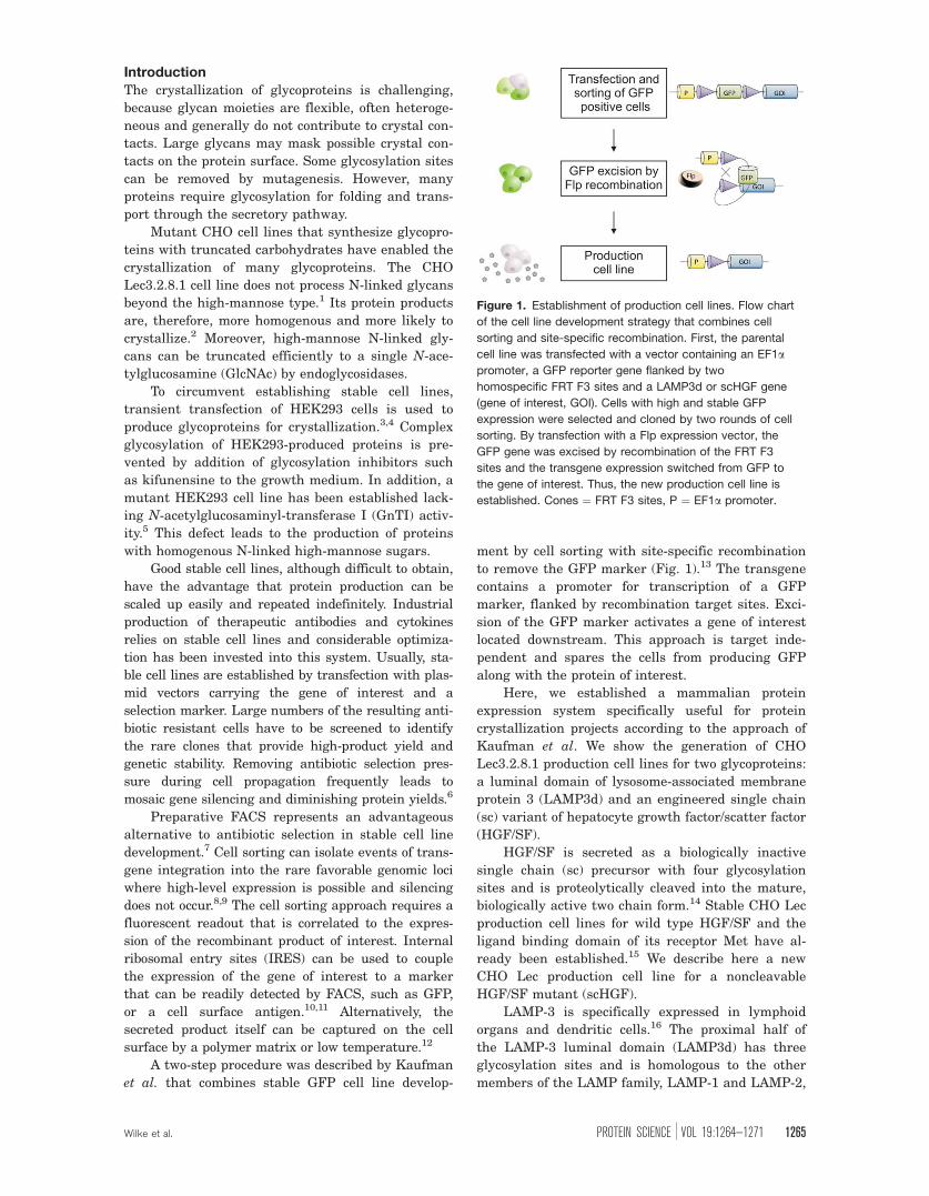
A two-step procedure was described by Kaufman
et al. that combines stable GFP cell line develop-
ment by cell sorting with site-specific recombination
to remove the GFP marker (Fig. 1).13 The transgene
contains a promoter for transcription of a GFP
marker, flanked by recombination target sites. Exci-
sion of the GFP marker activates a gene of interest
located downstream. This approach is target inde-
pendent and spares the cells from producing GFP
along with the protein of interest.
Here, we established a mammalian protein
expression system specifically useful for protein
crystallization projects according to the approach of
Kaufman et al. We show the generation of CHO
Lec3.2.8.1 production cell lines for two glycoproteins:
a luminal domain of lysosome-associated membrane
protein 3 (LAMP3d) and an engineered single chain
(sc) variant of hepatocyte growth factor/scatter factor
(HGF/SF).
HGF/SF is secreted as a biologically inactive
single chain (sc) precursor with four glycosylation
sites and is proteolytically cleaved into the mature,
biologically active two chain form.14 Stable CHO Lec
production cell lines for wild type HGF/SF and the
ligand binding domain of its receptor Met have al-
ready been established.15 We describe here a new
CHO Lec production cell line for a noncleavable
HGF/SF mutant (scHGF).
LAMP-3 is specifically expressed in lymphoid
organs and dendritic cells.16 The proximal half of
the LAMP-3 luminal domain (LAMP3d) has three
glycosylation sites and is homologous to the other
members of the LAMP family, LAMP-1 and LAMP-2,
Figure 1. Establishment of production cell lines. Flow chart
of the cell line development strategy that combines cell
sorting and site-specific recombination. First, the parental
cell line was transfected with a vector containing an EF1apromoter, a GFP reporter gene flanked by two
homospecific FRT F3 sites and a LAMP3d or scHGF gene
(gene of interest, GOI). Cells with high and stable GFP
expression were selected and cloned by two rounds of cell
sorting. By transfection with a Flp expression vector, the
GFP gene was excised by recombination of the FRT F3
sites and the transgene expression switched from GFP to
the gene of interest. Thus, the new production cell line is
established. Cones ¼ FRT F3 sites, P ¼ EF1a promoter.
Wilke et al. PROTEIN SCIENCE VOL 19:1264—1271 1265
which are involved in lysosomal biogenesis, lysoso-
mal fusion of phagosomes and autophagy.17,18
Both proteins were produced and purified in
milligrams per liter of culture medium with the new
cell lines, followed by enzymatic deglycosylation and
crystallization of LAMP3d.
Results
Establishment of GFP master cell linesCHO Lec3.2.8.1 production cell lines were estab-
lished in two steps: stable clonal GFP ‘‘master’’ cell
lines were generated, followed by excision of the
GFP reporter gene by the site-specific recombinase
Flp.19 The latter step placed LAMP3d and scHGF
under control of the vector’s promoter.
The pEFF3EGFPF3mcs vector contains a human
elongation factor 1a (EF1a) promoter and an
enhanced GFP reporter gene, flanked by the syn-
thetic variant F3 of the Flp recombination target
(FRT) site (Supporting Information Fig. S1).13
LAMP3d and scHGF were cloned downstream of
GFP (Fig. 1). CHO Lec3.2.8.1 cells were transfected
with the resulting vectors pEFF3EGFPF3scHGF and
pEFF3EGFPF3LAMP3d. GFP positive master cell
clones that integrated the transgene into favorable
chromosomal loci were selected by two rounds of
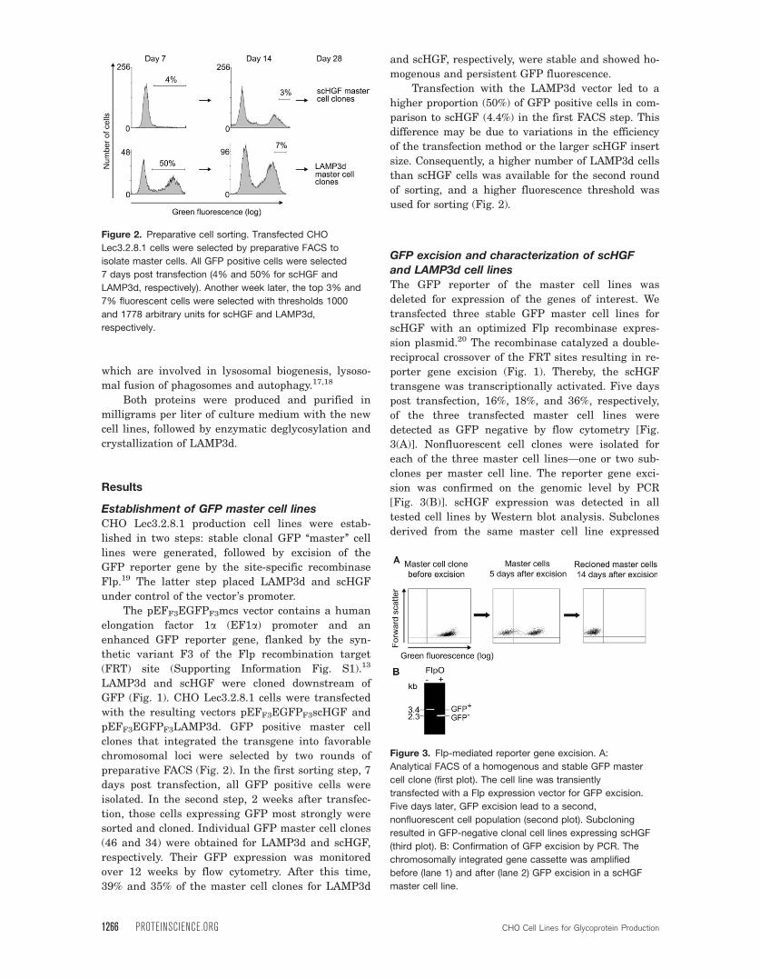
preparative FACS (Fig. 2). In the first sorting step, 7
days post transfection, all GFP positive cells were
isolated. In the second step, 2 weeks after transfec-
tion, those cells expressing GFP most strongly were
sorted and cloned. Individual GFP master cell clones
(46 and 34) were obtained for LAMP3d and scHGF,
respectively. Their GFP expression was monitored
over 12 weeks by flow cytometry. After this time,
39% and 35% of the master cell clones for LAMP3d
and scHGF, respectively, were stable and showed ho-
mogenous and persistent GFP fluorescence.
Transfection with the LAMP3d vector led to a
higher proportion (50%) of GFP positive cells in com-
parison to scHGF (4.4%) in the first FACS step. This
difference may be due to variations in the efficiency
of the transfection method or the larger scHGF insert
size. Consequently, a higher number of LAMP3d cells
than scHGF cells was available for the second round
of sorting, and a higher fluorescence threshold was
used for sorting (Fig. 2).
GFP excision and characterization of scHGF
and LAMP3d cell linesThe GFP reporter of the master cell lines was
deleted for expression of the genes of interest. We
transfected three stable GFP master cell lines for
scHGF with an optimized Flp recombinase expres-
sion plasmid.20 The recombinase catalyzed a double-
reciprocal crossover of the FRT sites resulting in re-
porter gene excision (Fig. 1). Thereby, the scHGF
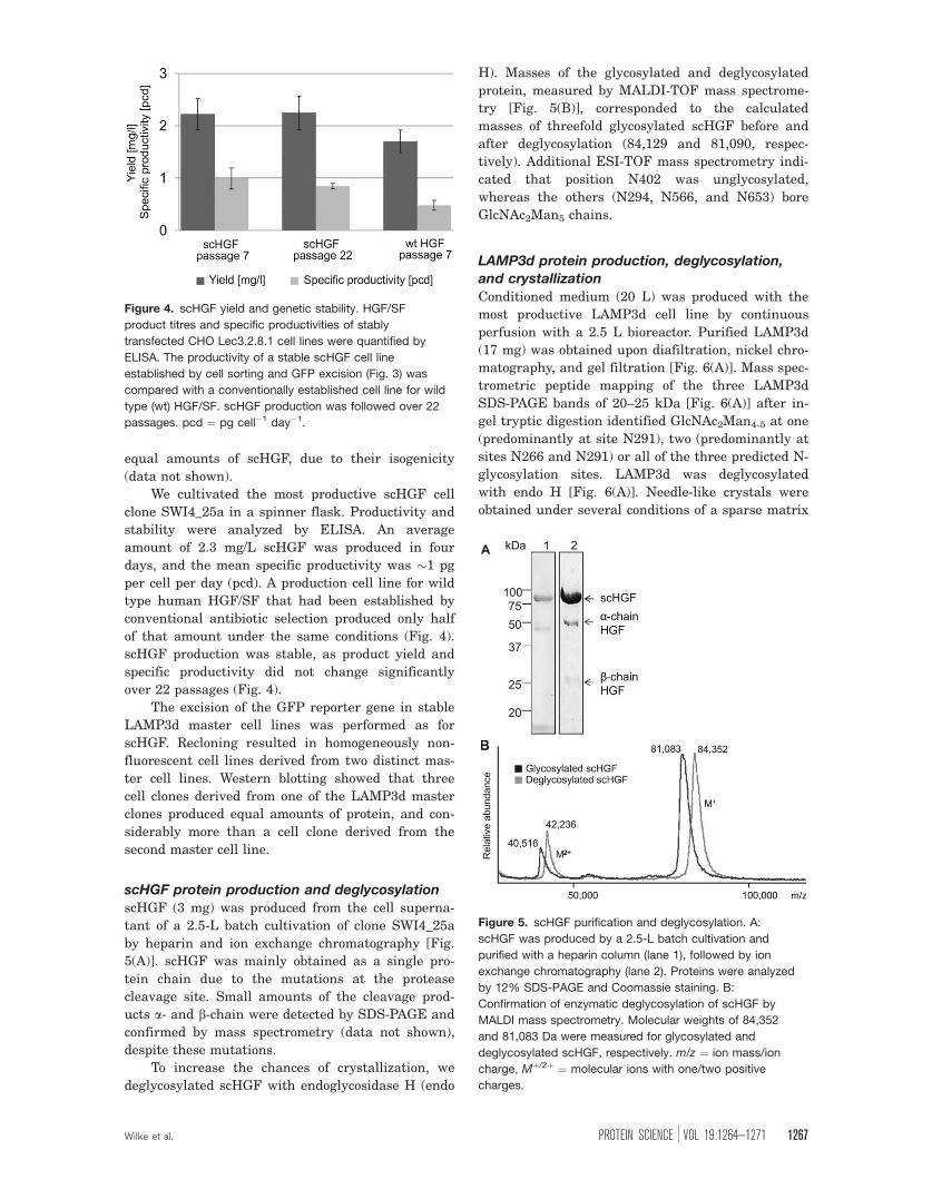
transgene was transcriptionally activated. Five days
post transfection, 16%, 18%, and 36%, respectively,
of the three transfected master cell lines were
detected as GFP negative by flow cytometry [Fig.
3(A)]. Nonfluorescent cell clones were isolated for
each of the three master cell lines—one or two sub-
clones per master cell line. The reporter gene exci-
sion was confirmed on the genomic level by PCR
[Fig. 3(B)]. scHGF expression was detected in all
tested cell lines by Western blot analysis. Subclones
derived from the same master cell line expressed
Figure 2. Preparative cell sorting. Transfected CHO
Lec3.2.8.1 cells were selected by preparative FACS to
isolate master cells. All GFP positive cells were selected
7 days post transfection (4% and 50% for scHGF and
LAMP3d, respectively). Another week later, the top 3% and
7% fluorescent cells were selected with thresholds 1000
and 1778 arbitrary units for scHGF and LAMP3d,
respectively.
Figure 3. Flp-mediated reporter gene excision. A:
Analytical FACS of a homogenous and stable GFP master
cell clone (first plot). The cell line was transiently
transfected with a Flp expression vector for GFP excision.
Five days later, GFP excision lead to a second,
nonfluorescent cell population (second plot). Subcloning
resulted in GFP-negative clonal cell lines expressing scHGF
(third plot). B: Confirmation of GFP excision by PCR. The
chromosomally integrated gene cassette was amplified
before (lane 1) and after (lane 2) GFP excision in a scHGF
master cell line.
1266 PROTEINSCIENCE.ORG CHO Cell Lines for Glycoprotein Production
equal amounts of scHGF, due to their isogenicity
(data not shown).
We cultivated the most productive scHGF cell
clone SWI4_25a in a spinner flask. Productivity and
stability were analyzed by ELISA. An average
amount of 2.3 mg/L scHGF was produced in four
days, and the mean specific productivity was �1 pg
per cell per day (pcd). A production cell line for wild
type human HGF/SF that had been established by
conventional antibiotic selection produced only half
of that amount under the same conditions (Fig. 4).
scHGF production was stable, as product yield and
specific productivity did not change significantly
over 22 passages (Fig. 4).
The excision of the GFP reporter gene in stable
LAMP3d master cell lines was performed as for
scHGF. Recloning resulted in homogeneously non-
fluorescent cell lines derived from two distinct mas-
ter cell lines. Western blotting showed that three
cell clones derived from one of the LAMP3d master
clones produced equal amounts of protein, and con-
siderably more than a cell clone derived from the
second master cell line.
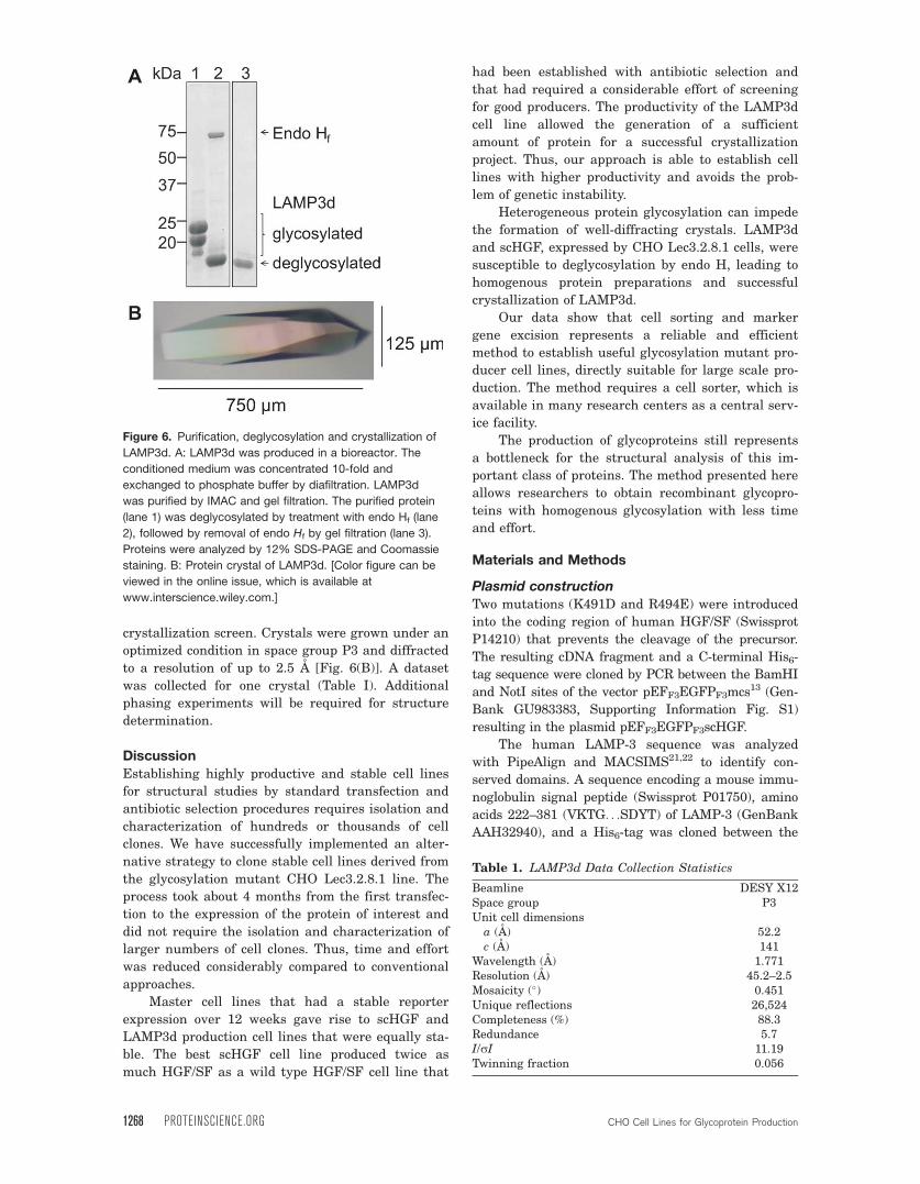
scHGF protein production and deglycosylationscHGF (3 mg) was produced from the cell superna-
tant of a 2.5-L batch cultivation of clone SWI4_25a
by heparin and ion exchange chromatography [Fig.
5(A)]. scHGF was mainly obtained as a single pro-
tein chain due to the mutations at the protease
cleavage site. Small amounts of the cleavage prod-
ucts a- and b-chain were detected by SDS-PAGE and
confirmed by mass spectrometry (data not shown),
despite these mutations.
To increase the chances of crystallization, we
deglycosylated scHGF with endoglycosidase H (endo
H). Masses of the glycosylated and deglycosylated
protein, measured by MALDI-TOF mass spectrome-
try [Fig. 5(B)], corresponded to the calculated
masses of threefold glycosylated scHGF before and
after deglycosylation (84,129 and 81,090, respec-
tively). Additional ESI-TOF mass spectrometry indi-
cated that position N402 was unglycosylated,
whereas the others (N294, N566, and N653) bore
GlcNAc2Man5 chains.
LAMP3d protein production, deglycosylation,
and crystallization
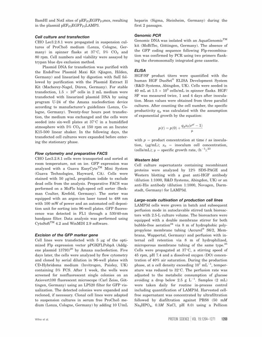
Conditioned medium (20 L) was produced with the
most productive LAMP3d cell line by continuous
perfusion with a 2.5 L bioreactor. Purified LAMP3d
(17 mg) was obtained upon diafiltration, nickel chro-
matography, and gel filtration [Fig. 6(A)]. Mass spec-
trometric peptide mapping of the three LAMP3d
SDS-PAGE bands of 20–25 kDa [Fig. 6(A)] after in-
gel tryptic digestion identified GlcNAc2Man4-5 at one
(predominantly at site N291), two (predominantly at
sites N266 and N291) or all of the three predicted N-
glycosylation sites. LAMP3d was deglycosylated
with endo H [Fig. 6(A)]. Needle-like crystals were
obtained under several conditions of a sparse matrix
Figure 4. scHGF yield and genetic stability. HGF/SF
product titres and specific productivities of stably
transfected CHO Lec3.2.8.1 cell lines were quantified by
ELISA. The productivity of a stable scHGF cell line
established by cell sorting and GFP excision (Fig. 3) was
compared with a conventionally established cell line for wild
type (wt) HGF/SF. scHGF production was followed over 22
passages. pcd ¼ pg cell�1 day�1.
Figure 5. scHGF purification and deglycosylation. A:
scHGF was produced by a 2.5-L batch cultivation and
purified with a heparin column (lane 1), followed by ion
exchange chromatography (lane 2). Proteins were analyzed
by 12% SDS-PAGE and Coomassie staining. B:
Confirmation of enzymatic deglycosylation of scHGF by
MALDI mass spectrometry. Molecular weights of 84,352
and 81,083 Da were measured for glycosylated and
deglycosylated scHGF, respectively. m/z ¼ ion mass/ion
charge, Mþ/2þ ¼ molecular ions with one/two positive
charges.
Wilke et al. PROTEIN SCIENCE VOL 19:1264—1271 1267
crystallization screen. Crystals were grown under an
optimized condition in space group P3 and diffracted
to a resolution of up to 2.5 A [Fig. 6(B)]. A dataset
was collected for one crystal (Table I). Additional
phasing experiments will be required for structure
determination.
Discussion
Establishing highly productive and stable cell lines
for structural studies by standard transfection and
antibiotic selection procedures requires isolation and
characterization of hundreds or thousands of cell
clones. We have successfully implemented an alter-
native strategy to clone stable cell lines derived from
the glycosylation mutant CHO Lec3.2.8.1 line. The
process took about 4 months from the first transfec-
tion to the expression of the protein of interest and
did not require the isolation and characterization of
larger numbers of cell clones. Thus, time and effort
was reduced considerably compared to conventional
approaches.
Master cell lines that had a stable reporter
expression over 12 weeks gave rise to scHGF and
LAMP3d production cell lines that were equally sta-
ble. The best scHGF cell line produced twice as
much HGF/SF as a wild type HGF/SF cell line that
had been established with antibiotic selection and
that had required a considerable effort of screening
for good producers. The productivity of the LAMP3d
cell line allowed the generation of a sufficient
amount of protein for a successful crystallization
project. Thus, our approach is able to establish cell
lines with higher productivity and avoids the prob-
lem of genetic instability.
Heterogeneous protein glycosylation can impede
the formation of well-diffracting crystals. LAMP3d
and scHGF, expressed by CHO Lec3.2.8.1 cells, were
susceptible to deglycosylation by endo H, leading to
homogenous protein preparations and successful
crystallization of LAMP3d.
Our data show that cell sorting and marker
gene excision represents a reliable and efficient
method to establish useful glycosylation mutant pro-
ducer cell lines, directly suitable for large scale pro-
duction. The method requires a cell sorter, which is
available in many research centers as a central serv-
ice facility.
The production of glycoproteins still represents
a bottleneck for the structural analysis of this im-
portant class of proteins. The method presented here
allows researchers to obtain recombinant glycopro-
teins with homogenous glycosylation with less time
and effort.
Materials and Methods
Plasmid construction
Two mutations (K491D and R494E) were introduced
into the coding region of human HGF/SF (Swissprot
P14210) that prevents the cleavage of the precursor.
The resulting cDNA fragment and a C-terminal His6-
tag sequence were cloned by PCR between the BamHI
and NotI sites of the vector pEFF3EGFPF3mcs13 (Gen-
Bank GU983383, Supporting Information Fig. S1)
resulting in the plasmid pEFF3EGFPF3scHGF.
The human LAMP-3 sequence was analyzed
with PipeAlign and MACSIMS21,22 to identify con-
served domains. A sequence encoding a mouse immu-
noglobulin signal peptide (Swissprot P01750), amino
acids 222–381 (VKTG. . .SDYT) of LAMP-3 (GenBank
AAH32940), and a His6-tag was cloned between the
Figure 6. Purification, deglycosylation and crystallization of
LAMP3d. A: LAMP3d was produced in a bioreactor. The
conditioned medium was concentrated 10-fold and
exchanged to phosphate buffer by diafiltration. LAMP3d
was purified by IMAC and gel filtration. The purified protein
(lane 1) was deglycosylated by treatment with endo Hf (lane
2), followed by removal of endo Hf by gel filtration (lane 3).
Proteins were analyzed by 12% SDS-PAGE and Coomassie
staining. B: Protein crystal of LAMP3d. [Color figure can be
viewed in the online issue, which is available at
www.interscience.wiley.com.]
Table 1. LAMP3d Data Collection Statistics
Beamline DESY X12Space group P3Unit cell dimensionsa (A) 52.2c (A) 141
Wavelength (A) 1.771Resolution (A) 45.2–2.5Mosaicity (�) 0.451Unique reflections 26,524Completeness (%) 88.3Redundance 5.7I/rI 11.19Twinning fraction 0.056
1268 PROTEINSCIENCE.ORG CHO Cell Lines for Glycoprotein Production
BamHI and NotI sites of pEFF3EGFPF3mcs, resulting
in the plasmid pEFF3EGFPF3LAMP3.
Cell culture and transfection
CHO Lec3.2.8.1 were propagated in suspension cul-
tures of ProCho5 medium (Lonza, Cologne, Ger-
many) in spinner flasks at 37�C, 5% CO2 and
80 rpm. Cell numbers and viability were assayed by
trypan blue dye exclusion method.
Plasmid DNA for transfection was purified with
the EndoFree Plasmid Maxi Kit (Qiagen, Hilden,
Germany) and linearized by digestion with SalI fol-
lowed by purification with the Plasmid Extract II
Kit (Macherey-Nagel, Duren, Germany). For stable
transfection, 1.5 � 106 cells in 2 mL medium were
transfected with linearized plasmid DNA by using
program U-24 of the Amaxa nucleofection device
according to manufacturer’s guidelines (Lonza, Co-
logne, Germany). Twenty-four hours post transfec-
tion, the medium was exchanged and the cells were
seeded into six-well plates at 37�C in a humidified
atmosphere with 5% CO2 at 150 rpm on an Incutec
K15-500 linear shaker. In the following days, the
transfected cell cultures were expanded before enter-
ing the stationary phase.
Flow cytometry and preparative FACS
CHO Lec3.2.8.1 cells were transported and sorted at
room temperature, not on ice. GFP expression was
analyzed with a Guava EasyCyteTM Mini System
(Guava Technologies, Hayward, CA). Cells were
stained with 50 lg/mL propidium iodide to exclude
dead cells from the analysis. Preparative FACS was
performed on a MoFlo high-speed cell sorter (Beck-
man Coulter, Krefeld, Germany). The sorter was
equipped with an argon-ion laser tuned to 488 nm
with 100 mW of power and an automated cell deposi-
tion unit for sorting into 96-well plates. GFP fluores-
cence was detected in FL1 through a 530/40-nm
bandpass filter. Data analysis was performed using
CytoSoftTM 4.2 and WinMDI 2.9 software.
Excision of the GFP marker geneCell lines were transfected with 5 lg of the opti-
mized Flp expression vector pPGKFLPobpA (Addg-
ene plasmid 13793)20 by Amaxa nucleofection. Five
days later, the cells were analyzed by flow cytometry
and cloned by serial dilution in 96-well plates with
CD-Hybridoma medium (Invitrogen, Paisley, UK)
containing 5% FCS. After 1 week, the wells were
screened for nonfluorescent single colonies on an
Axiovert100 fluorescent microscope (Carl Zeiss, Got-
tingen, Germany) using an LP520 filter for GFP vis-
ualization. The detected colonies were expanded and
recloned, if necessary. Clonal cell lines were adapted
to suspension cultures in serum free ProCho5 me-
dium (Lonza, Cologne, Germany) by adding 10 U/mL
heparin (Sigma, Steinheim, Germany) during the
first 2 passages.
Genomic PCRGenomic DNA was isolated with an AquaGenomicTM
kit (MoBiTec, Gottingen, Germany). The absence of
the GFP coding sequence following Flp-recombina-
tion was confirmed by PCR using two primers flank-
ing the chromosomally integrated gene cassette.
ELISA
HGF/SF product titers were quantified with the
human HGF DuoSetVR
ELISA Development System
(R&D Systems, Abingdon, UK). Cells were seeded in
40 mL at 1.5 � 105 cells/mL in spinner flasks. HGF/
SF was measured twice, 1 and 4 days after inocula-
tion. Mean values were obtained from three parallel
cultures. After counting the cell number, the specific
productivity qp was calculated with the assumption
of exponential growth by the equation:
pðtÞ ¼ pð0Þ þ qpx0ðelt � 1Þl
with p ¼ product concentration at time t as inocula-
tion, (lg/mL); x0 ¼ inoculum cell concentration,
(cells/mL); l ¼ specific growth rate, (h�1).23
Western blotCell culture supernatants containing recombinant
proteins were analyzed by 12% SDS-PAGE and
Western blotting with a goat anti-HGF antibody
(dilution 1:1000, R&D Systems, Abingdon, UK) or an
anti-His antibody (dilution 1:1000, Novagen, Darm-
stadt, Germany) for LAMP3d.
Large-scale cultivation of production cell lines
LAMP3d cells were grown in batch and subsequent
perfusion mode in autoclavable stirred tank bioreac-
tors with 2.5-L culture volume. The bioreactors were
equipped with a double membrane stirrer for both
bubble-free aeration24 via 8 m of hydrophobic poly-
propylene membrane tubing (AccurelVR
S6/2, Mem-
brana, Wuppertal, Germany) and perfusion with in-
ternal cell retention via 8 m of hydrophilized,
microporous membrane tubing of the same type.25
Cells were propagated at 37�C, a stirring speed of
45 rpm, pH 7.4 and a dissolved oxygen (DO) concen-
tration of 40% air saturation. During the production
phase, at a cell density exceeding 107 mL�1, temper-
ature was reduced to 32�C. The perfusion rate was
adjusted to the metabolic consumption of glucose
avoiding a drop below 2.5 g L�1. Samples (2 mL)
were taken daily for routine in-process control
including quantification of LAMP3d. Harvested cell-
free supernatant was concentrated by ultrafiltration
followed by diafiltration against PBS8 (50 mM
Na2HPO4, 0.3M NaCl, pH 8.0) using a Pellicon
Wilke et al. PROTEIN SCIENCE VOL 19:1264—1271 1269
2 tangential flow system equipped with two 10-kDa
cut-off cartridges (Millipore, Billerica MA). scHGF
was produced by a similar process in a 2.5-L culture
volume, but without perfusion or diafiltration.
Purification, deglycosylation,
and crystallization of LAMP3d
The human LAMP3d was purified by immobilized
metal ion affinity chromatography (IMAC) on a 50 mL
Ni-NTA superflow (Qiagen) column using PBS8
and an imidazole gradient for elution. The protein
was further purified by gel filtration on a 320 mL
Superdex 75 pg XK 26/60 column (GE Lifesciences)
in GF buffer (10 mM HEPES, pH 7.4, 150 mM
NaCl). LAMP3d was deglycosylated at 1 mg/mL over
night at 37�C by adding sodium acetate to 100 mM
and endoglycosidase Hf to 10,000 U/mL (Endo Hf,
NEB). Endo Hf is a fusion protein of endo H and malt-
ose binding protein. Endo Hf and contaminants were
removed by gel filtration in GF buffer on a 16/60 size
exclusion column of Superdex 75 (GE Healthcare).
The protein was concentrated to 22 mg/mL with a
10,000 MWCO Vivaspin membrane concentrator
(Sartorius Stedim Biotech, Gottingen, Germany).
Crystallization screens were set up in 96-well
format with a mosquito nanolitre pipetting machine
(TTP LabTech, Melbourn, UK) and various screening
suites (Qiagen). Upon optimization, single crystals
were obtained in droplets composed of 1 lL 22 mg/mL
protein and 1 lL of reservoir buffer (0.1M citric acid
pH 5.0, 5% PEG 6000).
Crystals were transferred into reservoir supple-
mented with 30% PEG 6000 and immediately flash
frozen in liquid nitrogen. Diffraction data were
acquired by an X-ray home source (Rikagu, Seve-
noak, UK) and at beamline X12 at the EMBL Out-
station (Hamburg, Germany).
Purification and deglycosylation of scHGF
scHGF was purified from cell supernatant by hepa-
rin and ion exchange chromatography. Cell superna-
tant (2.5 L) were directly loaded on a 25 mL heparin
sepharose column. The column was washed with
200 mM Tris-HCl, pH 8.0, and 250 mM NaCl. scHGF
was eluted with a linear gradient of 250–1000 mM
NaCl. The pooled scHGF-containing fractions were
diluted with 200 mM Tris-HCl, pH 8.0 to 250 mM
NaCl. The protein was immediately loaded on a
7.9 mL Mono S cation exchange column and, upon
washing, was eluted with a linear gradient of 250–
1000 mM NaCl. Purified scHGF was deglycosylated
in 0.1M sodium acetate by 30 U Endo Hf (NEB) per
lg protein over night at 37�C.
Mass spectrometry
LAMP3d protein bands were excised from a Coomas-
sie-stained SDS-PAGE gel, tryptically digested and
desalted (Ziptip) using standard protocols. The
(glyco-)peptides then were subjected to MALDI/TOF
(Bruker, Ultraflex) and ESI (Micromass QTOF2)
mass spectrometric analyses. The identity of relevant
(glyco)-peptides was verified by MS/MS analyses.
To prepare desalted samples of scHGF, 50 lL
Ni-NTA superflow beads (Qiagen) were added to
samples of His6-tag scHGF. scHGF was bound by
shaking for 1 h and unbound material was removed
upon centrifugation. Beads were washed thrice in 5
mM Tris-HCl, pH 8.0 and scHGF was eluted thrice
with 100-lL aliquots of 35% acetonitrile, 0.1% TFA
and shaking for 10 min. Eluates were subjected to
MALDI-TOF-MS analysis.
Acknowledgments
The authors are greatly indebted to Prof. Pamela Stan-
ley for the CHO Lec3.2.8.1 cells, which made this study
possible. The authors thank Sarah-Maria Tokarski for
excellent technical assistance, Nadine Konisch for
operation of bioprocessors, and Uwe Wengler and Jens
de Groot for help with protein purification and crystal-
lization. They also thank Dr. Manfred Weiss, Dr. Bjorn
Klink, and Dr. Joachim Reichelt for supporting diffrac-
tion data collection as well as Dr. Manfred Nimtz for
mass spectrometric measurements.
References
1. Stanley P (1989) Chinese hamster ovary cell mutantswith multiple glycosylation defects for production ofglycoproteins with minimal carbohydrate heterogeneity.Mol Cell Biol 9:377–383.
2. Davis SJ, Puklavec MJ, Ashford DA, Harlos K, JonesEY, Stuart DI, Williams AF (1993) Expression of solu-ble recombinant glycoproteins with predefined glycosy-lation: application to the crystallization of the T-cellglycoprotein CD2. Protein Eng 6:229–232.
3. Chang VT, Crispin M, Aricescu AR, Harvey DJ, Nettle-ship JE, Fennelly JA, Yu C, Boles KS, Evans EJ,Stuart DI, Dwek RA, Jones EY, Owens RJ, Davis SJ.(2007) Glycoprotein structural genomics: solving theglycosylation problem. Structure 15:267–273.
4. Aricescu AR, Lu W, Jones EY (2006) A time- and cost-efficient system for high-level protein production inmammalian cells. Acta Crystallogr 62:1243–1250.
5. Reeves PJ, Callewaert N, Contreras R, Khorana HG(2002) Structure and function in rhodopsin: high-levelexpression of rhodopsin with restricted and homogene-ous N-glycosylation by a tetracycline-inducible N-ace-tylglucosaminyltransferase I-negative HEK293S stablemammalian cell line. Proc Natl Acad Sci USA 99:13419–13424.
6. Liu W, Xiong Y, Gossen M (2006) Stability and homoge-neity of transgene expression in isogenic cells. J MolMed 84:57–64.
7. Mattanovich D, Borth N (2006) Applications of cellsorting in biotechnology. Microb Cell Fact 5:12.
8. Bestor TH (2000) Gene silencing as a threat to the suc-cess of gene therapy. J Clin Invest 105:409–411.
9. Kito M, Itami S, Fukano Y, Yamana K, Shibui T (2002)Construction of engineered CHO strains for high-levelproduction of recombinant proteins. Appl Microbiol Bio-technol 60:442–448.
1270 PROTEINSCIENCE.ORG CHO Cell Lines for Glycoprotein Production
10. Gaines P, Wojchowski DM (1999) pIRES-CD4t, a dicis-tronic expression vector for MACS- or FACS-basedselection of transfected cells. Biotechniques 26:683–688.
11. Mancia F, Patel SD, Rajala MW, Scherer PE, Nemes A,Schieren I, Hendrickson WA, Shapiro L (2004) Optimiza-tion of protein production in mammalian cells with a coex-pressed fluorescent marker. Structure 12:1355–1360.
12. Pichler J, Hesse F, Wieser M, Kunert R, Galosy SS,Mott JE, Borth N (2009) A study on the temperaturedependency and time course of the cold capture anti-body secretion assay. J Biotechnol 141:80–83.
13. Kaufman WL, Kocman I, Agrawal V, Rahn HP, BesserD, Gossen M (2008) Homogeneity and persistence oftransgene expression by omitting antibiotic selection incell line isolation. Nucleic Acids Res 36:e111.
14. Birchmeier C, Birchmeier W, Gherardi E, Vande WoudeGF (2003) Met, metastasis, motility and more. Nat RevMol Cell Biol 4:915–925.
15. Gherardi E, Sandin S, Petoukhov MV, Finch J, YoulesME, Ofverstedt LG, Miguel RN, Blundell TL, VandeWoude GF, Skoglund U, Svergun DI. (2006) Structuralbasis of hepatocyte growth factor/scatter factor and METsignalling. Proc Natl Acad Sci USA 103:4046–4051.
16. de Saint-Vis B, Vincent J, Vandenabeele S, VanbervlietB, Pin JJ, Ait-Yahia S, Patel S, Mattei MG, Bancher-eau J, Zurawski S, Davoust J, Caux C, Lebecque S.(1998) A novel lysosome-associated membrane glycopro-tein, DC-LAMP, induced upon DC maturation, is tran-siently expressed in MHC class II compartment.Immunity 9:325–336.
17. Eskelinen EL, Saftig P (2009) Autophagy: a lysosomaldegradation pathway with a central role in health anddisease. Biochim Biophys Acta 1793:664–673.
18. Huynh KK, Eskelinen EL, Scott CC, Malevanets A, Saf-tig P, Grinstein S (2007) LAMP proteins are required forfusion of lysosomes with phagosomes. EMBO J 26:313–324.
19. Andrews BJ, Proteau GA, Beatty LG, Sadowski PD(1985) The FLP recombinase of the 2 micron circleDNA of yeast: interaction with its target sequences.Cell 40:795–803.
20. Raymond CS, Soriano P (2007) High-efficiency FLPand PhiC31 site-specific recombination in mammaliancells. PLoS ONE 2:e162.
21. Plewniak F, Bianchetti L, Brelivet Y, Carles A, Chal-mel F, Lecompte O, Mochel T, Moulinier L, Muller A,Muller J, Prigent V, Ripp R, Thierry JC, ThompsonJD, Wicker N, Poch O. (2003) PipeAlign: a new toolkitfor protein family analysis. Nucleic Acids Res 31:3829–3832.
22. Thompson JD, Muller A, Waterhouse A, Procter J, Bar-ton GJ, Plewniak F, Poch O (2006) MACSIMS: multiplealignment of complete sequences information manage-ment system. BMC Bioinformatics 7:318.
23. Pirt SJ (1975) Principles of microbe and cell cultiva-tion. Oxford: Blackwell Scientific Publications Ltd.
24. Lehmann J, Piehl GW, Schulz R (1987) Bubblefree cell culture aeration with porous moving mem-branes. In: Karger S, Ed. Develop Biol Standard66:227–240.
25. Blasey HD, Jager V (1990) Strategies to increase theefficiency of membrane aerated and perfused animalcell bioreactors by an improved medium perfusion. In:Sasaki R, Ikura K, Eds. Animal cell culture and pro-duction of biologicals. Dordrecht, The Netherlands:Kluwer Acad. Pub., pp 61–73.
Wilke et al. PROTEIN SCIENCE VOL 19:1264—1271 1271