Embed Size (px)
Citation preview
Immundiagnostik AG, Stubenwald-Allee 8a, D 64625 Bensheim Tel.: ++49 6251 70190-0 Fax: ++ 49 6251 849430 e.mail: [email protected] www.Immundiagnostik.com
96+ 2 °C
+ 8 °C
Arbeitsanleitung / Manual
Glycin ELISA Kit Zur Bestimmung von Glycin in humanem EDTA-Plasma und Urin
Glycine ELISA Kit For the determination of glycine in human EDTA plasma and urine
Nur zu Forschungszwecken / For research use only
Gültig ab / Valid from 12.08.2011
K 7013

ELISA Glycin
1
Inhaltsverzeichnis / table of contents Seite / page
1. VERWENDUNGSZWECK 3
2. EINLEITUNG 3
3. TESTPRINZIP 4
4. INHALT DER TESTPACKUNG 5
5. ERFORDERLICHE LABORGERÄTE UND HILFSMITTEL 5
6. VORBEREITUNG UND LAGERUNG DER REAGENZIEN 6
7. HINWEISE UND VORSICHTSMASSNAHMEN 7
8. PROBENVORBEREITUNG 7
9. TESTDURCHFÜHRUNG 8
PIPETTIERSCHEMA PROBENVORBEREITUNG 8 PIPETTIERSCHEMA TESTDURCHFÜHRUNG 9
10. AUSWERTUNG DER ERGEBNISSE 10
ERWARTETE ERGEBNISSE 12
11. TESTCHARAKTERISTIKA 12
KREUZREAKTION 12 PRÄZISION UND REPRODUZIERBARKEIT 13 SENSITIVITÄT 13 WIEDERFINDUNG 13 LINEARITÄT 14
12. EINSCHRÄNKUNGEN 14
13. LITERATUR 14
14. ALLGEMEINE HINWEISE ZUM TEST 15

ELISA Glycin
2
1. INTENDED USE 18
2. INTRODUCTION 18
3. PRINCIPLE OF THE TEST 19
4. MATERIAL SUPPLIED 20
5. MATERIAL REQUIRED BUT NOT SUPPLIED 20
6. PREPARATION AND STORAGE OF REAGENTS 21
7. PRECAUTIONS 22
8. SPECIMEN COLLECTION AND PREPARATION 22
9. ASSAY PROCEDURE 23
SAMPLE PREPARATION PROCEDURE 23 TEST PROCEDURE 24
10. EVALUATION OF RESULTS 25
EXPECTED RESULTS 26
11. PERFORMANCE CHARACTERISTICS 27
CROSS REACTIVITY 27 PRECISION AND REPRODUCIBILITY 27 SENSITIVITY 27 RECOVERY 28 LINEARITY 28
12. LIMITATIONS 29
13. REFERENCES 29
14. GENERAL NOTES ON THE TEST AND TEST PROCEDURE 30

ELISA Glycin
3
1. VERWENDUNGSZWECK
Dieser ELISA-Test ist für die Bestimmung von Glycin in humanem EDTA-Plasma und Urin geeignet. Nur zu Forschungszwecken.
2. EINLEITUNG
Glycin ist die kleinste und einfachste α-Aminosäure. Es ist nicht essentiell und ein wichtiger Bestandteil nahezu aller Proteine. Das Kollagen des Bindegewebes enthält besonders viel Glycin. Darüber hinaus ist Glycin ein wichtiger Knotenpunkt im Stoffwechsel. Die Umsetzung von Serin zu Glycin dient neben der Erzeugung von Glycin auch der Synthese von Thymin-Nukleotiden und anderen Bestandteilen der Erbsubstanz (Purine).
Glycin dient zusammen mit Succinyl-CoA der Biosynthese der Porphyrine, welche im menschlichen Stoffwechsel eine zentrale Rolle spielen: als Häm-Gruppe im roten Blutfarbstoff sowie in vielen Enzymen, z.B der Katalase (Wasserstoffperoxid-Entgiftung) und den Enzymen der Atmungskette im Mitochondrium. Auch Kreatin, der Energiespeicher im Muskel, wird aus Glycin aufgebaut.
Als Baustein des Glutathions (neben Cystein und Glutaminsäure) spielt Glycin eine wichtige Rolle beim Schutz vor oxidativem Stress und bei der Biotransformation schädlicher Stoffe.
Die Gabe von Glycin wird deshalb mit diversen positiven Effekten in Verbindung gebracht:
Glycin scheint bei Diabetes eine günstige Rolle bei der Vermeidung von Folgekrankheiten zu spielen, indem es gewebeschädigende und entzündliche Prozesse vermindert: In klinischen Studien waren bei Patienten mit Glycingabe der TNF-Rezptor 1 sowie das HbA1c (Marker für das Fortschreiten von entzündlichen bzw. gewebeschädigenden Prozessen bei Diabetes) signifikant niedriger als in der Placebogruppe.
Außerdem vermindert Glycin die Cytokin-Produktion von Fettzellen, was zur antiinflammatorischen Wirkung von Glycin beiträgt.
Neben diesen Funktionen ist Glycin im Zentralnervensystem als einer der wichtigsten inhibitorischen Neurotransmitter am Glycinrezeptor wirksam. Es moduliert sensorische und motorische Impulse und spielt damit für Sinneswahrnehmung und Bewegung eine bedeutenden Rolle.
Im Rückenmark hemmen glycinfreisetzende Nervenzellen die Motoneurone des Vorderhorns, wodurch es zu einer Herabsetzung der Aktivität der Skelettmuskulatur kommt. Hohe Konzentrationen von Glycin haben einen positiven Einfluss auf die Länge und Intensität des Schlafes.

ELISA Glycin
4
Darüber hinaus wird ein angstlösender Effekt diskutiert, den man sich über die Hemmung der Noradrenalinausschüttung im Locus Coeruleus erklärt.
Im Vorderhirn hingegen wirkt Glycin an einer speziellen Glycin-Bindungsstelle am NMDA-Rezeptor, neben dem hauptsächlichen Agonisten Glutamat, stimulierend. Dort trägt es zur Induktion synaptischer Plastizität bei, d.h. zur „Bahnung“ bestimmter Wege, was ein wesentlicher Prozess des Lernens und der Gedächtnisfunktion ist.
Indikationen:
Bei Patienten mit Zwangsneurosen sowie bei Patienten mit Schizophrenie wurden signifikant erniedrigte Glycin-Blutspiegel gefunden.
3. TESTPRINZIP
Der Test basiert auf der Methode des kompetitiven Enzymimmunoassays. Zur Vorbereitung wird die zu untersuchende Probe mit einem Derivatisierungs-reagenz zur Kopplung des enthaltenen Glycin versetzt. Anschließend wird die derivatisierte Probe mit einem polyklonalen Glycin-Antiserum in einer mit Glycin-Derivat (Tracer) beschichteten ELISA-Platte inkubiert. Während der Inkubation kompetitiert das Zielantigen in der Probe mit dem an die Platte gebundenen Tracer um die Bindung der polyklonalen Antikörper. Hierbei verdrängt das Zielantigen in der Probe den Antikörper aus der Bindung an den Tracer. Daher ist die Konzentration des an den Tracer gebundenen Antikörpers umgekehrt proportional zu der Konzentration des Zielantigens in der Probe. Beim zweiten Inkubationsschritt wird ein Peroxidase-markierter Sekundärantikörper zugegeben, der an die polyklonalen Anti-Glycin-Antikörper bindet. Nach einem Waschschritt zur Entfernung ungebundener Komponenten wird das Peroxidasesubstrat Tetramethylbenzidin (TMB) zugegeben. Die Enzymreaktion wird durch Zugabe von Säure abgestoppt. Dadurch erfolgt ein Farbumschlag von blau nach gelb. Die entstandene chromogene Verbindung wird photometrisch bei 450 nm gemessen. Die Intensität der Farbe ist umgekehrt proportional zur Konzentration des gemessenen Analyten, d.h. mit steigender Glycin-Konzentration in der Probe reduziert sich die Konzentration der an den Tracer gebundenen Antikörper und das Signal nimmt ab. Parallel dazu wird eine Standardkurve – optische Dichte (Absorption bei 450 nm) versus Standardkonzentration – erstellt, aus der die Konzentrationen der Proben ermittelt werden.

ELISA Glycin
5
4. INHALT DER TESTPACKUNG
Artikel Nr. Inhalt Kit Komponenten Menge
K7013MTP PLATE Mikrotiterplatte, vorbeschichtet 12 x 8 Vertiefungen
K7013ST STD Standards (0; 50; 100; 250; 500; 1000 μM), vorverdünnt in Reaktionspuffer (gebrauchsfertig)
6 x 1 Fläschchen
K7013KO CTRL 1 CTRL 2 Kontrollen, vorverdünnt in Reaktionspuffer (gebrauchsfertig)
2 x 1 Fläschchen
K7013WP WASHBUF Waschpufferkonzentrat (10-fach) 2 x 100 ml
K7013AK AB Anti-Glycin-Antikörper (lyophilisiert) 2 x 1 Fläschchen
K7013K 2.AB POD-Antikörper (Konzentrat) 60 μl
K7013CSP 2.ABDIL Konjugatstabilisierungspuffer 12 ml
K7013RP REABUF Reaktionspuffer 45 ml
K7013DR DER Derivatisierungsreagenz 2 x 25,7 mg
K7013LM DMF Dimethylformamid (DMF) 3,5 ml
K7013AP ASYBUF Assaypuffer (10-fach) 2 x 5 ml
K7013TMB SUB TMB-Substrat 25 ml
K7013AC STOP Stopplösung 15 ml
5. ERFORDERLICHE LABORGERÄTE UND HILFSMITTEL
Bidestilliertes Wasser (aqua bidest.) Präzisionspipetten und Einmalpipettenspitzen mit variablen Volumina von
10 - 1000 μl Folie zum Abkleben der Mikrotiterplatte Mikrotiterplattenschüttler Multikanal- bzw. Multipipette Zentrifuge, 10000 x g Vortex-Mixer Laborübliche Glas- oder Plastikröhrchen (Einmalartikel) Mikrotiterplattenphotometer mit Filter 450 nm
(Referenzfilter 620 oder 690 nm)

ELISA Glycin
6
6. VORBEREITUNG UND LAGERUNG DER REAGENZIEN
Bitte achten Sie bei mehrfachem Einsatz der Platte darauf, dass die Reagenzien wie in der Vorschrift beschrieben gelagert und nur die für den jeweiligen Ansatz benötigten Reagenzienmengen frisch angesetzt werden. Der Kit kann so je nach Probenaufkommen bis zu 2x bis zum angegebenen Haltbarkeitsdatum verwendet werden.
Reagenzien mit einem Volumen kleiner 100 μl sollten vor Gebrauch zentrifugiert werden, um Volumenverluste zu vermeiden.
Das Waschpufferkonzentrat (WASHBUF) muss vor Gebrauch 1:10 in bidestilliertem Wasser (aqua bidest.) verdünnt werden (100 ml WASHBUF + 900 ml aqua bidest.); gut mischen. Aufgrund der hohen Salz-konzentration in den Stammlösungen kann es zu Kristallbildungen kommen. Die Kristalle lösen sich bei Raumtemperatur bzw. im Wasserbad bei 37°C auf. Das Pufferkonzentrat kann bei 2-8°C bis zum angegebenen Haltbarkeitsdatum aufbewahrt werden. Die verdünnte Pufferlösung ist bei 2-8°C einen Monat in einem geschlossenen Gefäß haltbar.
Die Standards (STD) und die Kontrollen (CTRL1, CTRL2) sind bereits in Reaktionspuffer (REABUF) verdünnt und werden bei -20°C gelagert. Für den Test werden die Standards und Kontrollen aufgetaut und können bis zu 3x wieder eingefroren werden. Das Wiedereinfrieren der Standards und Kontrollen sollte sofort nach Entnahme erfolgen.
Der Inhalt eines Fläschchens Derivatisierungsreagenz (DER) (25,7 mg) wird in 1,5 ml DMF gelöst und das Fläschchen für 5 min auf einen Horizontalschüttler gelegt. ACHTUNG: DMF ist giftig, bitte Hinweise und Vorsichtsmaßnahmen beachten (siehe Kapitel 6). Nach Gebrauch ist das Restreagenz zu entsorgen. Das DER sollte unmittelbar vor Gebrauch frisch angesetzt werden. Durch die Aufteilung des DER in 2 Gefäße ist der ELISA in zwei Ansätze teilbar. Bitte beachten: DMF greift Plastik an, DMF reagiert nicht mit Polypropylen-Produkten und Glasgefäßen
Der Assaypuffer (ASYBUF) muss vor Gebrauch 1:10 in bidestilliertem Wasser (aqua bidest.) verdünnt werden (5 ml Konzentrat + 45 ml aqua bidest.); gut mischen. Durch die Aufteilung des ASYBUF in 2 Gefäße ist der ELISA in zwei Ansätze teilbar. Verdünnter Assaypuffer (ASYBUF) ist stabil und kann 4 Wochen bei 2-8 °C aufbewahrt werden.
Der Inhalt eines Fläschchens anti-Glycin-Antikörper (AB) wird in 3 ml verdünntem Waschpuffer gelöst. Durch die Aufteilung des AB in 2 Gefäße ist der ELISA in zwei Ansätze teilbar. Verdünnter Glycin-Antikörper (AB) ist stabil und kann 4 Wochen bei 2-8 °C aufbewahrt werden.

ELISA Glycin
7
Der POD-Antikörper (2.AB) wird 1:200 in Konjugatstabilisierungs-puffer (2.ABDIL) verdünnt (z.B. 55 μl 2.AB + 11 ml 2.ABDIL; nur die benötigte Menge ansetzten). Unverdünnter POD-Antikörper (2.AB) ist bei 2-8°C bis zum angegebenen Haltbarkeitsdatum stabil. Verdünnter POD-Antikörper (2.AB) ist bedingt stabil und kann 5 Tage bei 2-8°C aufbewahrt werden.
Alle anderen Testreagenzien sind bei 2-8°C zu lagern und bei entsprechender Lagerung bis zum angegebenen Haltbarkeitsdatum (siehe Etikett) verwendbar.
7. HINWEISE UND VORSICHTSMASSNAHMEN
Nur zu Forschungszwecken.
Standards und Kontrollen sind auf Humanplasma aufgebaut. Sie sind auf HIV und Hepatitis B getestet und für negativ befunden worden. Jedoch sollten die Testkomponenten immer wie potentiell infektiöses Material behandelt werden.
Die Stopplösung besteht aus verdünnter H2SO4. H2SO4 ist eine starke Säure und muss auch im verdünnten Zustand mit Vorsicht verwendet werden. H2SO4 verursacht bei Kontakt mit der Haut Verätzungen. Es sollte daher mit Schutzhandschuhen und Schutzbrille gearbeitet werden. Bei Kontakt mit der Schwefelsäure muss die verätzte Stelle sofort mit viel Wasser gespült werden.
DMF ist giftig. Es kann das Kind im Mutterleib schädigen und ist gesundheitsschädlich beim Einatmen und bei Hautkontakt. Arbeiten mit DMF sollten daher mit Schutzhandschuhen und Schutzbrille unter dem Abzug vorgenommen werden. Bei Haut- oder Augenkontakt sofort mit viel Wasser spülen und Arzt konsultieren
Die Reagenzien dürfen nach Ablauf des angegebenen Haltbarkeitsdatums nicht mehr verwendet werden.
8. PROBENVORBEREITUNG
EDTA-Plasma und Urin
Als Probe eignet sich venöses Nüchternblut und Morgenurin. Die Haltbarkeit der Plasma-Probe beträgt bei 2-8°C eine Woche. Die Urinprobe sollte gekühlt versendet werden, ist aber bis 24 Stunden bei Raumtemperatur stabil. Die Haltbarkeit der Urinprobe beträgt zwei Tage bei 2-8°C. Zur längeren Lagerung sollten die Plasma- und Urin-Proben bei -20°C aufbewahrt werden.
Lipämische und hämolytische Proben beeinflussen das Testergebnis und sollten nicht verwendet werden.

ELISA Glycin
8
EDTA-Plasma- und Urinproben werden für die Derivatisierung vorverdünnt.
Proben mit sichtbaren Mengen an Feststoff (meist Kryoproteine) sollten vor Einsatz mind. 5 min bei 10000 x g zentrifugiert werden. Der resultierende Überstand wird im Test eingesetzt.
Zur weiteren Vorbereitung muss die Probe mit einem DER (Derivati-sierungsreagenz) zur Derivatisierung des enthaltenen Glycins versetzt werden (Details siehe Pipettierschema Probenvorbereitung).
9. TESTDURCHFÜHRUNG
Hinweise
Qualitätskontrollen sollten immer mitgemessen werden.
Inkubationszeit, Temperatur und Pipettiervolumina sind vom Hersteller festgelegt. Jegliche Abweichung der Testvorschrift, die nicht mit dem Hersteller koordiniert wurde, kann zu fehlerhaften Ergebnissen führen. Immundiagnostik AG übernimmt keine Haftung.
Die Bestimmung ist immer nach der im Kit beigefügten Arbeitsanleitung durchzuführen.
Pipettierschema Probenvorbereitung
EDTA-Plasmaproben werden im Faktor 1:20 vorverdünnt. Dafür werden jeweils 25 μl Probe mit 475 μl REABUF (Reaktionspuffer) verdünnt.
Urinproben werden im Faktor 1:100 vorverdünnt. Dafür werden jeweils 10 μl Probe mit 990 μl REABUF (Reaktionspuffer) verdünnt.
Die Derivatisierung der Standards (STD), der Kontrollen (CTRL) und der verdünnten Proben (SAMPLE) wird als Einzelbestimmung in Mikro-reaktionsgefäßen (Volumen mind. 2 ml) durchgeführt.
1. Im Test dürfen nur Reagenzien und Proben verwendet werden, welche Raumtemperatur (18-26°C) aufweisen.
2. Je 200 μl gebrauchsfertiger Standard (STD) bzw. 200 μl gebrauchsfertige Kontrollen (CTRL) bzw. 200 μl vorverdünnte Probe (SAMPLE) in Mikroreaktionsgefäße pipettieren.
3. 50 μl frisch angesetztes Derivatisierungsreagenz (DER) in alle Reaktionsgefäße (Standards, Kontrollen und Proben) pipettieren, gut mischen und sofort auf einem Horizontalschüttler (180-240 rpm) 60 min bei Raumtemperatur (18-26°C) inkubieren.

ELISA Glycin
9
4. Anschließend in alle verwendeten Mikroreaktionsgefäße 1500 μl verdünnten Assaypuffer (ASYBUF) zugeben, gut mischen und auf einem Horizontalschüttler (180-240 rpm) 30 min bei Raumtemperatur (18-26°C) inkubieren.
2 x 50 μl der so vorbereiteten Proben (STD, CTRL, SAMPLE) werden im ELISA als Doppelbestimmung eingesetzt.
Pipettierschema Testdurchführung
5. Positionen für Standard/ Kontrolle/ Probe (STD/CTRL/SAMPLE) in Doppelbestimmung am Protokollblatt markieren.
6. Die benötigten Streifen der Mikrotiterplatte (PLATE) aus dem Kit nehmen. Nicht verwendete Mikrotiterplattenstreifen können abgeklebt bis zum angegebenen Haltbarkeitsdatum bei 2-8°C gelagert werden.
7. Mikrotiterplattenstreifen 5 x mit je 250 μl verdünntem Waschpuffer waschen. Nach dem letzten Waschschritt Reste von Waschpuffer durch mehrmaliges Ausklopfen auf saugfähigem Papier entfernen.
8. 2 x 50 μl der vorbereiteten derivatisierten Proben (STD, CTRL, SAMPLE) werden aus den Mikroreaktionsgefäßen in die Vertiefungen der Mikrotiterplatte (PLATE) pipettiert.
9. 50 μl verdünnter anti-Glycin-Antikörper (AB) pro Vertiefung pipettieren. Streifen luftdicht abdecken.
10. Über Nacht (15-20 Stunden) bei 2-8°C inkubieren.
11. Inhalt der Platte verwerfen und 5 x mit je 250 μl verdünntem Waschpuffer waschen. Nach dem letzten Waschschritt Reste von Waschpuffer durch mehrmaliges Ausklopfen auf saugfähigem Papier entfernen.
12. 100 μl verdünnten POD-AK (2. AB) in alle Vertiefungen pipettieren.
13. Streifen abdecken und 1 Stunde bei Raumtemperatur (18-26°C) unter Schütteln (180-240 rpm) inkubieren.

ELISA Glycin
10
14. Inhalt der Platte verwerfen und 5x mit je 250 μl verdünntem Waschpuffer waschen. Nach dem letzten Waschschritt Reste von WASHBUF durch mehrmaliges Ausklopfen auf saugfähigem Papier entfernen.
15. 100 μl TMB-Substrat (SUB) in alle Vertiefungen pipettieren.
16. 15-20 min bei Raumtemperatur (18-26°C) im Dunkeln inkubieren*
17. 100 μl Stopplösung (STOP) in alle Vertiefungen pipettieren und im Mikrotiterplattenphotometer im Schüttelmodus mischen.
18. Extinktion sofort im Mikrotiterplattenphotometer mit einer Messwellenlänge von 450 nm messen. Sofern die höchste Extinktion der Standards (STD) den Messbereich des Photometers übersteigt, sollte die Messung sofort bei einer Messwellenlänge von 405 nm wiederholt und diese Ergebnisse für eine Auswertung herangezogen werden. Wenn möglich, sollten bei jeder Messung die Extinktionen der Messwellenlänge mit den Extinktionen einer Referenzwellenlänge verglichen werden. Zulässige Referenzwellenlängen sind z.B.: 595 nm, 620 nm, 630 nm, 650 nm und 690 nm
* Die Intensität der Farbentwicklung ist temperaturabhängig. Es wird empfohlen den Farbumschlag während der Inkubationszeit zu beobachten und entsprechend der Farbentwicklung die Reaktion zu stoppen.
10. AUSWERTUNG DER ERGEBNISSE
Bei einer Durchführung des Tests unter strikter Einhaltung der Volumenangaben für Standards, Kontrollen und Probenbehandlung sind Standards, Kontrollen sowie Plasmaproben gleich verdünnt, deshalb wird bei der Auswertung der Ergebnisse für Plasma kein Verdünnungsfaktor mit berechnet.
Bei einer 1:100 Verdünnung der Urinproben muss das Ergebnis mit dem Faktor 5 multipliziert werden.
Auswertungsfunktionen
Die unten beschriebenen mathematischen Modelle können alternativ zur Auswertung benutzt werden. Wir empfehlen die 4-Parameter-Funktion.

ELISA Glycin
11
1. 4-Parameter-Funktion
Für die optische Dichte empfehlen wir eine lineare Ordinate und für die Konzentration eine logarithmische Abszisse (bei einer logarithmischen Abszisse muss für den Standard mit der Konzentration 0 ein Wert kleiner 1 eingegeben werden, wir empfehlen 0,01).
2. Punkt-zu-Punkt-Auswertung
Für die optische Dichte und für die Konzentration empfehlen wir eine lineare Ordinate bzw. Abszisse.
3. Gewichtete Spline-Funktion
Für die optische Dichte empfehlen wir eine lineare Ordinate und für die Konzentration eine logarithmische Abszisse (bei einer logarithmischen Abszisse muss für den Standard mit der Konzentration 0 ein Wert kleiner 1 eingegeben werden, z.B. 0,01).
Vor jeder automatischen Auswertung sollte stets eine Kontrolle der Doppelwerte auf Plausibilität („Ausreißerkontrolle“) durchgeführt werden; falls dies nicht durch das verwendete Programm erfolgt, sollte die Kontrolle manuell durchgeführt werden.
Kontrollen
Zur Überwachung der Qualität der Analyse sollten bei jedem Testansatz Kontrollen mitgeführt werden. Die Ergebnisse der Kontrollen müssen auf Richtigkeit überprüft werden. Liegen ein oder mehrere Werte außerhalb des angegebenen Bereiches, ist es möglich, dass auch die Patientenproben falsch ermittelt wurden.
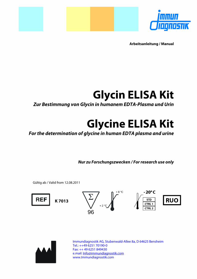
Die Konzentrationen der Kontrollen und Patientenproben können direkt aus der Kalibrierkurve in μmol/l abgelesen werden. Die folgende Abbildung zeigt ein typisches Beispiel einer Standardkurve. Sie darf nicht zur Auswertung der Messwerte benutzt werden.
ELISA Glycin
12
Musterkalibrierkurve
0
0,5
1
1,5
2
2,5
0,1 1 10 100 1000
c in µmol/l
OD
Erwartete Ergebnisse
Anhand einer laborinternen Studie mit Plasma-Proben von augenscheinlich Gesunden (n=75) wurde ein Median von 239,54 μmol/l ermittelt. Der Bereich zwischen 5%- und 95%-Quantile wurde als Normalbereich angenommen:
Normalbereich: 147,55 μmol/l – 438,12 μmol/l
Wir empfehlen jedem Labor, seinen eigenen Normalwerte-Bereich zu erstellen, weil Referenzbereiche stark von der Auswahl des Probanden-kollektivs abhängig sind. Die Angabe des Normalbereichs dient lediglich der Orientierung und kann von anderen publizierten Daten abweichen.
11. TESTCHARAKTERISTIKA
Kreuzreaktion
L-Alanin < 1 % β-Alanin < 0,2 %

ELISA Glycin
13
Präzision und Reproduzierbarkeit
EDTA-Plasma
Intra-Assay (n=9)
Probe Glycin [μmol/l]
Variationskoeffizient (CV) [%]
1 95,4 8,6
2 208,1 11,0
Inter-Assay (n=6)
Probe Glycin [μmol/l]
Variationskoeffizient (CV) [%]
1 124,0 6,6
2 318,6 16,3
Sensitivität
Die Nachweisgrenze wurde als B0 + 2 SD festgelegt. Gemessen wurde 32-mal der Standard Null.
Probe
Glycin Mittelwert
[OD]
2 Standard-abweichungen
(2 x SD)
Nachweis-grenze
[μmol/l]
Standard Null 1,7 0,2 14,3
Wiederfindung
Unterschiedliche Mengen an Glycin wurden zu einer Plasmaprobe gegeben (Spike) und anschließend im ELISA gemessen. Die analytische Wiederfindung von Glycin wurde bei 2 verschiedenen Konzentrationen aus den theoretisch erwarteten und den praktisch gemessenen Werten ermittelt. Die theoretisch erwarteten Werte wurden dabei aus der Summe der gemessenen Konzentration in der Probe ohne Spike und der zugegebenen Menge ermittelt. Die mittlere Wiederfindung für alle Konzentrationen der EDTA-Plasmaprobe betrug 106,1 % (n=10).

ELISA Glycin
14
EDTA-Plasma
Spike [μmol/l]
Glycin erwartet
[μmol/l]
Glycin gemessen
[μmol/l]
Wiederfindung [%]
0 X x = 167,2 100,0
100 167,2+100= 267,2 276,0 103,3
300 167,2+300= 467,2 537,4 115,0
Linearität
Die Linearität des ELISAs wurde durch Verdünnen einer aufgestockten EDTA-Plasmaprobe bestimmt. Die mittlere Linearität betrug 98,3 % (n=10).
Verdünnung Messwert [μmol/l]
Erwartet [μmol/l]
Wiederfindung [%]
original 248,8 248,8 100,0
1+1 118,3 124,4 95,1
1+2 80,9 82,9 97,5
1+3 62,6 62,2 100,7
12. EINSCHRÄNKUNGEN
Stark hämolysierte oder lipämische Proben können zu fehlerhaften Ergebnissen führen. Wir empfehlen, solche Proben nicht zu analysieren.
13. LITERATUR
Hons J, Zirko R, Ulrychova M, Cermakova E, Doubek P, Libiger J. Glycine serum level in schizophrenia: relation to negative symptoms. Psychiatry Res. 2010 Apr 30;176(2-3):103-8. Epub 2010 Jan 22
Ohnuma T, Sakai Y, Maeshima H, Hatano T, Hanzawa R, Abe S, Kida S, Shibata N, Suzuki T, Arai H. Changes in plasma glycine, L-serine, and D-

ELISA Glycin
15
serine levels in patients with schizophrenia as their clinical symptoms improve: results from the Juntendo University Schizophrenia Projects (JUSP). Prog Neuropsychopharmacol Biol Psychiatry. 2008 Dec 12;32(8):1905-12. Epub 2008 Sep 16.
Greenberg WM, Benedict MM, Doerfer J, Perrin M, Panek L, Cleveland WL, Javitt DC. Adjunctive glycine in the treatment of obsessive-compulsive disorder in adults. J Psychiatr Res. 2009 Mar;43(6):664-70. Epub 2008 Nov 30.
Ehrlich S, Franke L, Schneider N, Salbach-Andrae H, Schott R, Craciun EM, Pfeiffer E, Uebelhack R, Lehmkuhl U. Aromatic amino acids in weight-recovered females with anorexia nervosa. Int J Eat Disord. 2009 Mar;42(2):166-72.
Yang CR, Svensson KA. Allosteric modulation of NMDA receptor via elevation of brain glycine and D-serine: the therapeutic potentials for schizophrenia. Pharmacol Ther. 2008 Dec;120(3):317-32. Epub 2008 Aug 27.
Heresco-Levy U, Javitt DC, Ermilov M, Mordel C, Horowitz A, Kelly D. Double-blind, placebo-controlled, crossover trial of glycine adjuvant therapy for treatment-resistant schizophrenia. Br J Psychiatry. 1996 Nov;169(5):610-7.
Cruz M, Maldonado-Bernal C, Mondragón-Gonzalez R, Sanchez-Barrera R, Wacher NH, Carvajal-Sandoval G, Kumate J. Glycine treatment decreases proinflammatory cytokines and increases interferon-gamma in patients with type 2 diabetes. J Endocrinol Invest. 2008 Aug;31(8):694-9.
Garcia-Macedo R, Sanchez-Muñoz F, Almanza-Perez JC, Duran-Reyes G, Alarcon-Aguilar F, Cruz M. Glycine increases mRNA adiponectin and diminishes pro-inflammatory adipokines expression in 3T3-L1 cells. Eur J Pharmacol. 2008 Jun 10;587(1-3):317-21. Epub 2008 Apr 8.
14. ALLGEMEINE HINWEISE ZUM TEST
Reagenzien dieser Testpackung enthalten organische Lösungsmittel. Be-rührungen mit der Haut oder den Schleimhäuten sind zu vermeiden.
Sämtliche in der Testpackung enthaltene Reagenzien dürfen ausschließlich zu Forschungszwecken eingesetzt werden.
Die Reagenzien sollten nach Ablauf des angegebenen Haltbarkeitsdatums nicht mehr verwendet werden (Haltbarkeitsdatum siehe Testpackung).
Einzelkomponenten mit unterschiedlichen Lot-Nummern aus verschiede-nen Testpackungen sollten nicht gemischt oder ausgetauscht werden.
Für die Qualitätskontrolle sind die dafür erstellten Richtlinien für medizi-nische Laboratorien zu beachten.
Die charakteristischen Testdaten wie Pipettiervolumina der verschiedenen Komponenten und der Aufbereitung der Proben wurden firmenintern

ELISA Glycin
16
festgelegt. Nicht mit dem Hersteller abgesprochene Veränderungen in der Testdurchführung können die Resultate beeinflussen. Die Firma Immun-diagnostik AG übernimmt für direkt daraus resultierende Schäden und Folgeschäden keine Haftung.

ELISA Glycine
17
96+ 2 °C
+ 8 °C
Manual
Glycine ELISA Kit
For the determination of glycine in human EDTA plasma and urine
For research use only
Valid from 12.08.2011
K 7013

ELISA Glycine
18
1. INTENDED USE
The Glycine ELISA Kit is intended for the quantitative determination of glycine in human EDTA plasma and urine. It is for research use only.
2. INTRODUCTION
Glycine is the smallest α-amino acid and it is not essential to the human diet. Most proteins incorporate only small quantities of glycine. A notable exception is collagen, which contains about 35% glycine.
Glycine plays a key role in different metabolic pathways. Porphyrins are biosynthesized from glycine and succinyl-CoA. They are part of Hemoglobin and many enzymes as Catalase and the enzymes of the respiratory chain. Moreover, glycine provides the central C2N subunit of all purines (components of the DNA).
Glycine is one of three amino acids of glutathione, which is the major endogenous antioxidant produced by the cells, participating directly in the neutralization of free radicals and the detoxification of many xenobiotics (foreign compounds) and carcinogens.
Treatment with glycine is likely to show diverse beneficial effects: Patients with Type 2 diabetes treated with glycine had a significant decrease in A1C and in proinflammatory cytokines and also an important increase of IFN-gamma. This means that glycine may help to prevent tissue damage caused by chronic inflammation in patients with Type 2 diabetes.
In the central nervous system glycine acts as an inhibitory neurotransmitter, especially in the spinal cord, brainstem, and retina. Inhibitory spinal neurons that release glycine act on alpha motoneurons and decrease activity of skeletal muscles. High concentrations of glycine seem to improve sleep quality.
In the forebrain glycine is a required co-agonist along with glutamate for NMDA receptors. NMDA receptors are excitatory and play a critical role in synaptic plasticity, a cellular mechanism for learning and memory.
Recent study shows that treatment with glycine may help patients with obsessive compulsive disorder.
In patients with schizophrenia glycine serum levels were negatively associated with the intensity of negative symptoms suggesting a possible implication of NMDA receptor dysfunction in pathogenesis of negative symptoms in schizophrenia.

ELISA Glycine
19
Indications:
In patients with obsessive compulsive disorders and in patients with schizophrenia significantly lower serum levels of glycine have been found.
3. PRINCIPLE OF THE TEST
This assay is based on the method of competitive enzyme linked immunoassays. The sample preparation includes the addition of a derivatization reagent for glycine coupling. Afterwards, the treated samples and the polyclonal glycine antiserum are incubated in wells of microplate coated with a glycine-derivative (tracer). During the incubation period, the target glycine in the sample competes with the tracer immobilized on the wall of the microtiter wells for the binding of the polyclonal antibodies. The glycine in the sample displaces the antibodies out of the binding to the tracer. Therefore, the concentration of the tracer-bound antibody is inverse proportional to the glycine concentration in the sample. During the second incubation step, a peroxidase-conjugated antibody is added to each microtiter well to detect the anti-glycine antibodies. After washing away the unbound components tetramethylbenzidine (TMB) is added as a peroxidase substrate. Finally, the enzymatic reaction is terminated by an acidic stop solution. The color changes from blue to yellow and the absorbance is measured in the photometer at 450 nm. The intensity of the yellow color is inverse proportional to the glycine concentration in the sample; this means, high glycine concentration in the sample reduces the concentration of tracer-bound antibodies and lowers the photometric signal.
A dose response curve of absorbance unit (optical density, OD at 450 nm) vs. concentration is generated using the values obtained from the standards. Glycine present in the patient samples is determined directly from this curve.

ELISA Glycine
20
4. MATERIAL SUPPLIED
Catalog No Content Kit Components Quantity
K7013MTP PLATE One holder with precoated strips 12 x 8 wells
K7013ST STD Standards (0, 50, 100, 250, 500, 1000 μM), diluted in reaction buffer (ready to use)
6 x 1 vial
K7013KO CTRL 1 CTRL 2
Controls, diluted in reaction buffer (ready to use)
2 x 1 vial
K7013WP WASHBUF Wash buffer concentrate (10-fold) 2 x 100 ml
K7013AK AB Anti-glycine antibody (lyophilized) 2 x 1 vial
K7013K 2.AB POD antibody (concentrate) 60 μl
K7013CSP 2.ABDIL Conjugate stabilizing buffer 12 ml
K7013RP REABUF Reaction buffer 45 ml
K7013DR DER Derivatization reagent 2 x 25,7 mg
K7013LM DMF Dimethylformamide (DMF) 3,5 ml
K7013AP ASYBUF Assay buffer concentrate (10-fold) 2 x 5 ml
K7013TMB SUB TMB substrate 25 ml
K7013AC STOP Stop solution 15 ml
5. MATERIAL REQUIRED BUT NOT SUPPLIED
Double distilled water (aqua bidest.)
Precision pipettors and disposable tips to deliver 10-1000 μl
Foil to cover the microtiter plate
Horizontal microtiter plate shaker
A multi-channel dispenser or repeating dispenser
Centrifuge capable of 10000 x g
Vortex-Mixer
Standard laboratory glass or plastic vials, cups, etc.
Microtiter plate reader at 450 nm (reference wave length 620 or 690 nm)

ELISA Glycine
21
6. PREPARATION AND STORAGE OF REAGENTS
To run assay more than once ensure that reagents are stored at conditions stated on the label. Prepare only the appropriate amount necessary for each assay. The kit can be used up to 2 times within the expiry date stated on the label.
Reagents with a volume less than 100 μl should be centrifuged before use to avoid loss of volume.
Dilute the wash buffer concentrate (WASHBUF) with aqua bidest. 1:10 before use (100 ml WASHBUF + 900 ml aqua bidest.), mix well. Crystals may occur due to high salt concentration in the stock solution. The crystals must be redissolved at room temperature or at 37°C using a water bath before dilution. The WASHBUF is stable at 2-8°C until the expiry date stated on the label. Diluted buffer solution can be stored in a closed flask at 2-8°C for one month.
Standards (STD) and controls (CTRL1, CTRL2) are already diluted in reaction buffer (REABUF). Store standards and controls frozen at -20°C, thaw before use in the test, and re-freeze immediately after use. Standards and controls can be re-frozen up to 3 times.
Dissolve the content of one vial of derivatization reagent (DER) (25.7 mg) in 1.5 ml DMF. CAUTION: DMF is toxic (see chapter 6 - precautions). Put the vial on a horizontal shaker for 5 min. Dispose of any rest of the reagent after use. DER must be prepared immediately before use. The ELISA kit can be separated into two performances by providing two DER vials. Please note: DMF attacks all plastics but not polypropylene products and laboratory glass.
Dilute the assay buffer (ASYBUF) with aqua bidest. 1:10 before use (5 ml concentrate + 45 ml aqua bidest.), mix well. The ELISA kit can be separated into two performances by providing 2 x 5 ml assay buffer.
Dissolve the anti-glycine antibody (AB) in 3 ml of diluted wash buffer. The ELISA kit can be separated into two performances by providing two AB vials. Diluted anti-glycine antibody is stable over a longer period. It can be stored at 2-8°C for 4 weeks.
Dilute the POD antibody (2.AB) 1:200 with conjugate stabilizing buffer (2.ABDIL) (e.g. 55 μl 2.AB + 11 ml 2.ABDIL, prepare only the required amount). The undiluted POD antibody (2.AB) is stable at 2-8°C until the expiry date stated on the label. Diluted POD antibody (2.AB) is not stable over a longer period. It can be stored at 2-8°C for only 5 days.

ELISA Glycine
22
All other test reagents are ready for use. Test reagents are stable until the expiry date (see label of test package) when stored at 2-8°C.
7. PRECAUTIONS
For research use only.
Human materials used in kit components were tested and found to be negative for HIV and Hepatitis B. However, for safety reasons all kit components should be treated as if potentially infectious.
Stop solution is composed of sulfuric acid, which is a strong acid. Even diluted, it still must be handled with care. It can cause acid burns and should be handled with gloves, eye protection, and appropriate protective clothing. Any spills should be wiped out immediately with copious quantities of water.
DMF is toxic. It may cause harm to the unborn child and is harmful by inhalation and in contact with skin. Work under hood and apply preventive skin protection. In case of skin or eye contact flush with plenty of water and get medical attention
Reagents should not be used beyond the expiration date shown on kit label.
8. SPECIMEN COLLECTION AND PREPARATION
EDTA plasma and urine
Venous fasting blood and urine are suited for this test system. Blood samples are stable for one week at 2-8°C. Urine samples should be sent cooled, but they are stable for 24 h at room temperature. Otherwise they are stable for two days at 2-8°C. For longer storage, blood and urine samples should be frozen at -20°C.
Lipemic or hemolytic samples may give erroneous results and should not be used for analysis.
The EDTA plasma and urine samples are diluted for derivatization.
Samples with visible amounts of precipitates should be centrifuged at least for 5 min at 10000 x g. The resulting supernatant is used in the assay.
For sample preparation, a DER (derivatization reagent) for coupling of glycine is added (details are given in the sample preparation procedure).

ELISA Glycine
23
9. ASSAY PROCEDURE
Procedural notes
Quality control guidelines should be observed.
Incubation time, incubation temperature, and pipetting volumes of the different components are defined by the producer. Any variations of the test procedure that are not coordinated with the producer may influence the test results. Immundiagnostik AG can therefore not be held reliable for any damage resulting from this.
The assay should always be performed according to the enclosed manual.
Sample preparation procedure
Dilute EDTA plasma samples with reaction buffer by factor 1:20, i.e. 25 μl EDTA plasma sample + 475 μl REABUF (reaction buffer).
Dilute urine samples with reaction buffer by factor 1:100, i.e. 10 μl urine sample + 990 μl REABUF (reaction buffer).
Coupling of standards (STD), controls (CTRL) and diluted samples (SAMPLE) is carried out in single analysis (in vials with capacity of at least 2 ml).
1. Bring all reagents and samples to room temperature (18-26°C).
2. Add 200 μl of ready to use standards (STD), 200 μl of ready to use controls (CTRL) and 200 μl of diluted samples (SAMPLE) in the corresponding vial.
3. Add 50 μl of freshly prepared derivatization reagent (DER) into each vial (standards, controls and samples), mix well and incubate for 60 min on a shaker (180-240 rpm) at room temperature (18-26°C).
4. Afterwards add 1500 μl of assay buffer (ASYBUF) into each vial, mix well and incubate for 30 min on a shaker (180-240 rpm) at room temperature (18-26°C).
2 x 50 μl of each treated sample (STD, CTRL, SAMPLE) are used in the ELISA as duplicates.

ELISA Glycine
24
Test procedure
5. Mark the positions of standards (STD)/ controls (CTRL)/ samples (SAMPLE) in duplicate on a protocol sheet.
6. Take as many microtiter strips (PLATE) as needed from kit. Store unused strips covered at 2-8°C. Strips are stable until the expiry date stated on the label.
7. Wash each well 5 times by dispensing 250 μl of diluted wash buffer into each well. After the final washing step, the inverted microtiter plate (PLATE) should be firmly tapped on absorbent paper to remove excess solution.
8. For the analysis in duplicate, take 2 x 50 μl of standards (STD) / controls (CTRL) / samples (SAMPLE) out of the vial and add into the respective wells of the microtiter plate (PLATE).
9. Add 50 μl diluted anti-glycine antibody (AB) into each well. Cover the plate tightly.
10. Incubate overnight (15-20 hours) at 2-8°C.
11. Aspirate the contents of each well. Wash each well 5 times by dispensing 250 μl of diluted wash buffer into each well. After the final washing step, the inverted microtiter plate (PLATE) should be firmly tapped on absorbent paper to remove excess solution.
12. Add 100 μl diluted POD antibody (2. AB) into each well.
13. Cover plate tightly and incubate for 1 hour at room temperature (18-26°C) on a horizontal shaker (180-240 rpm).
14. Aspirate the contents of each well. Wash each well 5 times by dispensing 250 μl of diluted wash buffer into each well. After the final washing step, the inverted microtiter plate (PLATE) should be firmly tapped on absorbent paper to remove excess solution.
15. Add 100 μl of TMB substrate (SUB) into each well.

ELISA Glycine
25
16. Incubate for 15-20 min at room temperature (18-26°C) in the dark*.
17. Add 100 μl of stop solution (STOP) into each well, mix thoroughly.
18. Determine absorption immediately with an ELISA reader at 450 nm. If the highest extinction of the standards (STD) is above the range of the photometer, absorption must be measured immediately at 405 nm and the obtained results used for evaluation. If possible, the extinctions from each measurement should be compared with extinctions obtained at a reference wavelength, e.g. 595 nm, 620 nm, 630 nm, 650 nm and 690 nm can be used.
* The intensity of the color change is temperature sensitive. We recommend to observe the color change and to stop the reaction upon good differentiation.
10. EVALUATION OF RESULTS
If the test is performed in strict compliance with the manufacturer’s instructions (i.e. with the exact volumes for standards, controls, samples, and with correct sample treatment), standards, controls, and samples are equally diluted. Therefore, no dilution factor is required for the calculation of results from serum samples.
For urine samples with 1:100 dilution, the values calculated from the calibration curve have to be multiplicated by a factor of 5 to obtain the true results.
The following algorithms can be used alternatively to calculate the results. We recommend using the "4-parameter-algorithm".
1. 4-parameter-algorithm
It is recommended to use a linear ordinate for optical density and a logarithmic abscissa for concentration. When using a logarithmic abscissa, the zero calibrator must be specified with a value less than 1 (e.g. 0.01).
2. Point-to-point-calculation
We recommend a linear ordinate for optical density and a linear abscissa for concentration.
3. Spline-algorithm
We recommend a linear ordinate for optical density and a logarithmic abscissa for concentration. When using a logarithmic abscissa, the zero calibrator must be specified with a value less than 1 (e.g. 0.01).

ELISA Glycine
26
Plausibility of the measured pairs of values should be examined before automatically evaluating the results. If this option is not available within the used program the pairs of values should be controlled manually.
Controls
Control samples or plasma pools should be analyzed with each run. Results generated from the analysis of control samples should be evaluated for acceptability using appropriate statistical methods. The results for the patient samples may not be valid if within the same assay one or more values of the quality control samples are outside the acceptable limits.
The concentration of controls and patient samples can be determined directly from the calibration curve. In the following, an example of a calibration curve is given, do not use it for the calculation of your results. Example of calibration curve
0
0,5
1
1,5
2
2,5
0,1 1 10 100 1000
c in µmol/l
OD
Expected results
Based on internal studies with plasma samples of evidently healthy persons (n=75) a median of 239,54 μmol/l was calculated. The normal range was set between the 5th and 95th percentile:
Normal range: 147.55 μmol/l – 438.12 μmol/l

ELISA Glycine
27
We recommend each laboratory to develop its own normal range. The values mentioned above are only for orientation and can deviate from other published data.
11. PERFORMANCE CHARACTERISTICS
Cross reactivity
L-Alanine < 1 % β-Alanine < 0.2 %
Precision and reproducibility
EDTA plasma:
Intra-Assay (n=9)
Sample Glycine [μmol/l]
coefficient of variation (CV) [%]
1 95.4 8.6
2 208.1 11.0
Inter-Assay (n=6)
Sample Glycine [μmol/l]
coefficient of variation (CV) [%]
1 124.0 6.6
2 318.6 16.3
Sensitivity
The sensitivity was set as B0 + 2SD. The zero-standard was measured 32 times.
Sample
Glycine mean value
[OD]
2 x Standard deviation (SD)
Detection limit [μmol/l]
zero-standard 1.7 0.2 14.3

ELISA Glycine
28
Recovery
One sample was spiked with different glycine concentrations and measured using this assay. The analytical recovery rate was determined by the expected and measured glycine levels. The expected levels were calculated as the sum of the measured glycine concentration in the original sample and the spiked glycine amount. The mean recovery rate for all concentrations was 106.1 % (n=10).
EDTA plasma:
Spike [μmol/l]
Glycine expected
[μmol/l]
Glycine measured
[μmol/l]
Recovery [%]
0 x X= 167.2 100.0
100 167.2+100= 267.2 276.0 103.3
300 167.2+300= 467.2 537.4 115.0
Linearity
The linearity of the ELISA was determined by the dilution of a spiked patient sample. The mean linearity was 98.3 % (n=10).
EDTA-plasma
Dilution Measured [μmol/l]
Expected [μmol/l] Recovery [%]
Original 248.8 248.8 100.0
1+1 118.3 124.4 95.1
1+2 80.9 82.9 97.5
1+3 62.6 62.2 100.7

ELISA Glycine
29
12. LIMITATIONS
Hemolytic and lipemic samples may give erroneous results. Do not measure hemolytic and lipemic samples.
13. REFERENCES
Hons J, Zirko R, Ulrychova M, Cermakova E, Doubek P, Libiger J. Glycine serum level in schizophrenia: relation to negative symptoms. Psychiatry Res. 2010 Apr 30;176(2-3):103-8. Epub 2010 Jan 22
Ohnuma T, Sakai Y, Maeshima H, Hatano T, Hanzawa R, Abe S, Kida S, Shibata N, Suzuki T, Arai H. Changes in plasma glycine, L-serine, and D-serine levels in patients with schizophrenia as their clinical symptoms improve: results from the Juntendo University Schizophrenia Projects (JUSP). Prog Neuropsychopharmacol Biol Psychiatry. 2008 Dec 12;32(8):1905-12. Epub 2008 Sep 16.
Greenberg WM, Benedict MM, Doerfer J, Perrin M, Panek L, Cleveland WL, Javitt DC. Adjunctive glycine in the treatment of obsessive-compulsive disorder in adults. J Psychiatr Res. 2009 Mar;43(6):664-70. Epub 2008 Nov 30.
Ehrlich S, Franke L, Schneider N, Salbach-Andrae H, Schott R, Craciun EM, Pfeiffer E, Uebelhack R, Lehmkuhl U. Aromatic amino acids in weight-recovered females with anorexia nervosa. Int J Eat Disord. 2009 Mar;42(2):166-72.
Yang CR, Svensson KA. Allosteric modulation of NMDA receptor via elevation of brain glycine and D-serine: the therapeutic potentials for schizophrenia. Pharmacol Ther. 2008 Dec;120(3):317-32. Epub 2008 Aug 27.
Heresco-Levy U, Javitt DC, Ermilov M, Mordel C, Horowitz A, Kelly D. Double-blind, placebo-controlled, crossover trial of glycine adjuvant therapy for treatment-resistant schizophrenia. Br J Psychiatry. 1996 Nov;169(5):610-7.
Cruz M, Maldonado-Bernal C, Mondragón-Gonzalez R, Sanchez-Barrera R, Wacher NH, Carvajal-Sandoval G, Kumate J. Glycine treatment decreases proinflammatory cytokines and increases interferon-gamma in patients with type 2 diabetes. J Endocrinol Invest. 2008 Aug;31(8):694-9.
Garcia-Macedo R, Sanchez-Muñoz F, Almanza-Perez JC, Duran-Reyes G, Alarcon-Aguilar F, Cruz M. Glycine increases mRNA adiponectin and diminishes pro-inflammatory adipokines expression in 3T3-L1 cells. Eur J Pharmacol. 2008 Jun 10;587(1-3):317-21. Epub 2008 Apr 8.
ELISA Glycine
30
14. GENERAL NOTES ON THE TEST AND TEST PROCEDURE
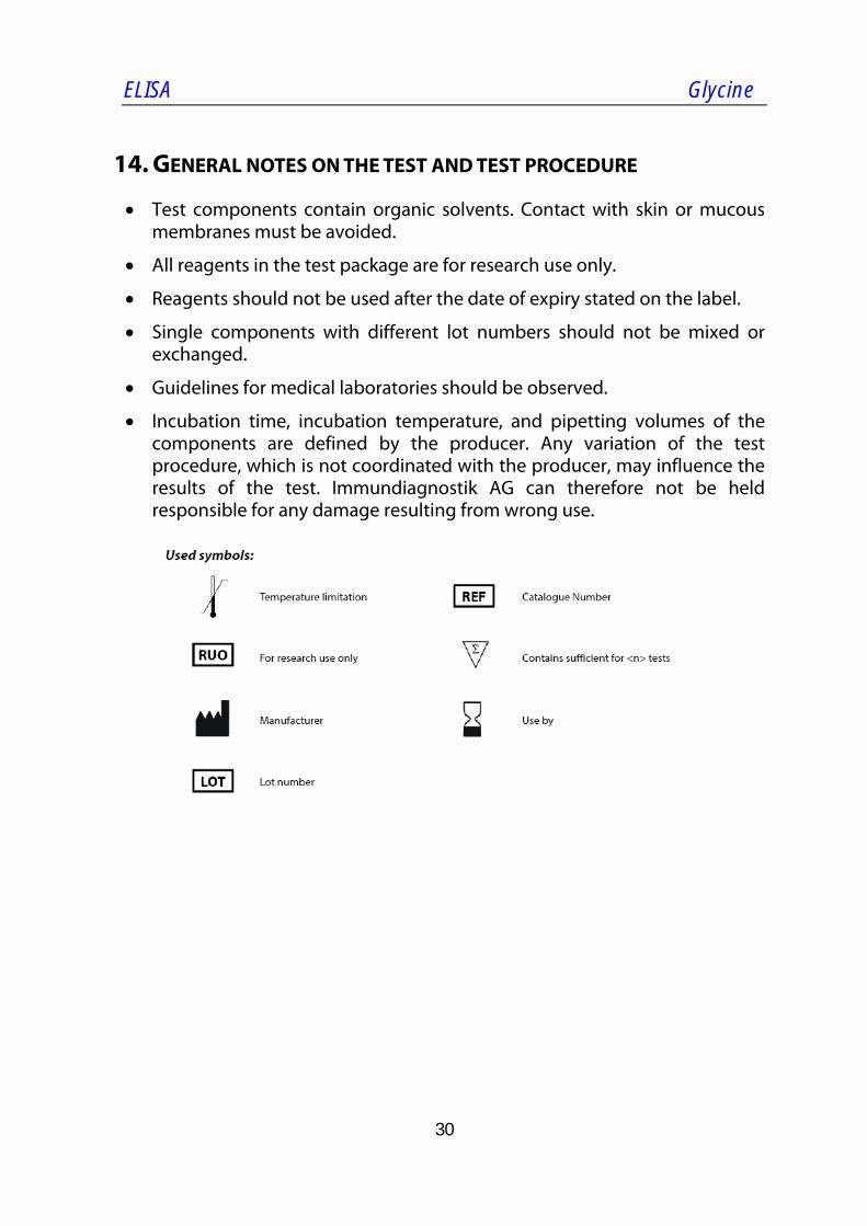
Test components contain organic solvents. Contact with skin or mucous membranes must be avoided.
All reagents in the test package are for research use only.
Reagents should not be used after the date of expiry stated on the label.
Single components with different lot numbers should not be mixed or exchanged.
Guidelines for medical laboratories should be observed.
Incubation time, incubation temperature, and pipetting volumes of the components are defined by the producer. Any variation of the test procedure, which is not coordinated with the producer, may influence the results of the test. Immundiagnostik AG can therefore not be held responsible for any damage resulting from wrong use.