Embed Size (px)
Citation preview

Glutathione Depletion Induced by c-Myc Downregulation TriggersApoptosis on Treatment with Alkylating Agents1
Annamaria Biroccio*, Barbara Benassi*, Francesco Fiorentino y and Gabriella Zupi*
*Experimental Chemotherapy Laboratory, Experimental Research Center, Regina Elena Cancer Institute,Rome 00158, Italy; y ‘‘Genoma’’ Molecular Genetics Laboratory, Rome 00198, Italy
Abstract
Here we investigate the mechanism(s) involved in the
c-Myc–dependent drug response of melanoma cells.
By using three M14-derived c-Myc low-expressing
clones, we demonstrate that alkylating agents, cispla-
tin and melphalan, trigger apoptosis in the c-Myc
antisense transfectants, but not in the parental line. On
the contrary, topoisomerase inhibitors, adriamycin and
camptothecin, induce apoptosis to the same extent
regardless of c-Myc expression. Because we previ-
ously demonstrated that c-Myc downregulation de-
creases glutathione (GSH) content, we evaluated the
role of GSH in the apoptosis induced by the different
drugs. In control cells treated with one of the alkylat-
ing agents or the others, GSH depletion achieved by
L-buthionine-sulfoximine preincubation opens the
apoptotic pathway. The apoptosis proceeded through
early Bax relocalization, cytochrome c release, and
concomitant caspase-9 activation, whereas reactive
oxygen species production and alteration of mitochon-
dria membrane potential were late events. That GSH
was determining in the c-Myc –dependent drug-
induced apoptosis was demonstrated by altering the
intracellular GSH content of the c-Myc low-expressing
cells up to the level of controls. Indeed, GSH ethyl
ester–mediated increase of GSH abrogated apopto-
sis induced by cisplatin and melphalan by inhibition
of Bax/cytochrome c redistribution. The relationship
among c-Myc, GSH content, and the response to al-
kylating agent has been also evaluated in the M14 Myc
overexpressing clones as well as in the melanoma JR8
c-Myc antisense transfectants. All together, these
results demonstrate that GSH plays a key role in
governing c-Myc–dependent drug-induced apoptosis.
Neoplasia (2004) 6, 195–206
Keywords: c-Myc, glutathione, antineoplastic drugs, apoptosis, melanoma.
Introduction
Several papers report that susceptibility to apoptosis can
contribute to the response of tumor cells to most cytotoxic
agents currently used. Thus, alterations that impair suscep-
tibility to apoptosis should produce drug resistance. Muta-
tions affecting the c-myc proto-oncogene are among the most
common genetic lesions found in a variety of human cancers
[1,2]. The role of c-myc oncogene on apoptosis has been
extensively documented. Under appropriate circumstances,
both repression and overexpression of c-Myc can lead to
apoptosis. For example, c-myc antisense or dominant-defec-
tive causes apoptosis in a variety of transformed cell types [3],
whereas c-Myc expression can sensitize cells to a wide range
of different stimuli such as low serum conditions [4], hypoxia
[5], and deprivation of specific growth factors [6,7]. Many
studies have been dedicated to c-Myc–mediated apoptotic
pathways initiated by DNA-damaging agents due to the poten-
tial relevance to cancer chemotherapy. Nevertheless, the role
of c-Myc in susceptibility to drug-induced cell death is still
obscure with results being contradictory up to now.
Deregulated c-Myc expression has been reported not only
to enhance tumor cell sensitivity but also to induce resistance
to antineoplastic agents. In particular, overexpression of c-Myc
has been reported to increase cellular susceptibility to chemo-
therapy-induced apoptosis [8–10]. Conversely, in other exper-
imental models, c-Myc overexpression increases the
resistance to alkylating agents, as well as to adriamycin
(ADR) and etoposide, whereas it does not influence the
response to ionizing radiation [11,12]. Then again, inducible
antisense c-myc gene transfer confers sensitivity to cisplatin
(CDDP) in a drug-resistant human small cell lung carcinoma
line [13]. In this context, we previously demonstrated that
treatment with c-myc antisense oligodeoxynucleotides in-
duces apoptosis and enhances CDDP antitumoral efficacy in
several human melanoma cell lines [14–16]. The role of c-Myc
on the apoptosis and drug sensitivity of melanoma cells has
been evaluated by our group by using stable c-Myc low-
expressing transfectants. We have demonstrated that although
c-Myc downregulation induces apoptosis and sensitizes M14
Abbreviations: ADR, adriamycin; CDDP, cisplatin; L-PAM, melphalan; CPT, camptothecin;
ROS, reactive oxygen species; Dwm, mitochondrial membrane potential; DHE, dihydroethi-
dium; GSH, reduced glutathione; BSO, L-buthionine-sulfoximine; PI, propidium iodide; PIPES,
piperazine-N,N V-bis[2-ethanesulfonic acid]; PBS, phosphate-buffered saline
Address all correspondence to: Dr. Annamaria Biroccio, Experimental Chemotherapy
Laboratory, Regina Elena Cancer Institute, Via delle Messi d’Oro 156, Rome 00158, Italy.
E-mail: [email protected] work was supported by grants from the Italian Association for Cancer Research (AIRC),
Miniestro della Salute, and CNR-MIUR.
Received 26 September 2003; Revised 16 December 2003; Accepted 22 December 2003.
CopyrightD 2004 Neoplasia Press, Inc. All rights reserved 1522-8002/04/$25.00
DOI 10.1593/neo.03370
Neoplasia . Vol. 6, No. 3, May/June 2004, pp. 195 – 206 195
www.neoplasia.com
RESEARCH ARTICLE

melanoma cells to the alkylating agent CDDP, it has no
such effect with DNA topoisomerase inhibitors ADR and
camptothecin (CPT) [17]. Recently, by using inducible
c-Myc antisense transfectants, we also found that down-
regulation of c-Myc triggers apoptosis through glutathione
(GSH) depletion [18]. GSH is an endogenous cysteine-con-
taining tripeptide, playing a key role in drug detoxification
through a number of mechanisms, including antioxidant
activity [19], DNA repair [20,21], conjugation of cellular toxins
[22], and pumping of toxic chemotherapeutics out of cells
through the multidrug resistance–associated proteins [23].
Elevated intracellular GSH can augment cell resistance to
irradiation and chemotherapy, especially to alkylating
agents, whereas diminution of intracellular GSH content
can increase cell response to radiotherapy and chemo-
therapy [19,24]. Drug resistance has been correlated
to increased levels of both messenger RNA and activity of
g-glutamylcysteine synthetase [25,26]—the rate-limiting en-
zyme for GSH biosynthesis. In addition, transfection of
the cDNA encoding for this enzyme into tumor cells leads
to increased activity and drug resistance, along with GSH
levels [27].
Hence, the main objective of this paper was to investigate
the role of GSH on the c-Myc–dependent chemosensitivity
of melanoma cells.
Materials and Methods
Cell Culture
Three stable c-Myc antisense transfectants (MAS51,
MAS53, and MAS69) and a control clone (MN2) were
previously obtained by transfecting the M14 human mela-
noma parental line with an expression vector carrying the
antisense c-myc cDNA and/or the neomycin selection
marker gene [17]. M14 cells were also transfected with
the pCDNAI–c-myc cDNA expression vector and two
c-Myc overexpressing clones (MS41 and MS58) were
selected and employed in the study. Moreover, three stable
c-Myc antisense transfectants (JAS26, JAS28, and JAS43)
and the JN3 control clone were obtained by transfecting the
JR8 melanoma cell line with the pCDNA3–c-myc antisense
expression vector and the empty vector, respectively. All
transfectants were grown at 37jC in completed neomycin-
containing (0.8 mg/ml; Invitrogen, Carlsbad, CA) RPMI
1640 medium (Invitrogen).
Treatments
Clinical-grade CDDP (Pronto Platamine) and ADR (Adri-
blastina) were obtained from Pharmacia (Milan, Italy). CPT
and melphalan (L-PAM; Alkeran) were purchased from Sig-
ma (Milan, Italy) and Glaxo Wellcome (Verona, Italy), re-
spectively. Drug dilutions were freshly prepared before each
experiment.
In particular, cells were seeded in 60-mm Petri (Nunc,
Mascia Brunelli, Milan, Italy) dishes at a density of 2�105
cells/dish. After 24 hours, cells were exposed to increasing
doses of drugs (for clonogenic experiments) or to the IC50
dose of each antineoplastic agent (for all the other assays).
The IC50 doses employed were 6.7 mM CDDP for 2 hours,
15 mM L-PAM for 2 hours, 0.37 mM ADR for 1 hour, and 2 mMCPT for 2 hours.
To evaluate cell colony-forming ability, aliquots of cell
suspension from each sample were seeded into 60-mm
Petri dishes with complete medium and incubated for 10 to
12 days. Colonies were stained with 2% methylene blue in
95% ethanol and counted (one colony z 50 cells). Surviv-
ing fractions were calculated as the ratio of absolute
survival of the treated sample/absolute survival of the
control sample.
In the experiments with GSH ethyl ester (Sigma) or
L-buthionine-sulfoximine (BSO; Sigma), cells were preincu-
bated with 5 mM GSH ethyl ester for 24 hours or with 10 mM
BSO for 6 hours (doses with no toxic effect on cell survival).
Then cells were washed three-fold and treated with the
different antineoplastic agents.
Evaluation of Apoptosis
Apoptosis was detected by flow cytometric (annexin V),
biochemical (caspase-3 activity), and morphological
(Hoechst staining) assays. Annexin V-FITC versus propi-
dium iodide (PI) assay (Vibrant apoptosis assay, V-13242;
Molecular Probes, Eugene, OR) was performed as previ-
ously described [18]. Briefly, adherent cells were harvested,
suspended in the annexin-binding buffer (1�106 cells/ml),
and incubated with annexin V-FITC and PI for 15 minutes, at
room temperature in the dark, then immediately analyzed by
flow cytometry. The data are presented as biparametric dot
plots showing the annexin V-FITC green fluorescence
versus the PI red fluorescence. Analysis of cell death was
performed from 0 to 96 hours after the end of each drug
treatment.
Caspase-3 activity was measured by a colorimetric assay
(K2027; Clontech, Basingstoke, UK) according to the man-
ufacturer’s instruction.
Cytocentrifuge preparations were stained with Hoechst
33258 dye (Sigma) and cover-slipped. Cell morphology was
evaluated by fluorescence microscopy.
GSH Determination
Intracellular GSH content was measured by a colorimetric
assay (Bioxytech GSH-400; Oxis International, Inc., Port-
land, OR) according to the manufacturer’s instruction.
Determination of Cytosolic Proteins
The determination of cytosolic proteins was performed
as previously reported [18]. Cells were harvested and
washed with phosphate-buffered saline (PBS), then collect-
ed by centrifugation at 700�g for 7 minutes at 4jC. Cell
pellet was resuspended in extraction buffer containing 220
mM mannitol, 68 mM sucrose, 50 mM piperazine-N,N V-
bis[2-ethanesulfonic acid] (PIPES)–NaOH (pH 7.4), 50
mM EGTA, 2 mM MgCl2, 1 mM dithiothreitol, and protease
inhibitors. After 30 minutes of incubation on ice, cells were
homogenized with glass Dounce homogenizer. Cell homo-
genates were spun at 14,000�g for 15 minutes at 4jC, and
196 c-Myc –Mediated Sensitivity Depends on GSH Biroccio et al.
Neoplasia . Vol. 6, No. 3, 2004

supernatants were removed and stored at �80jC until
analysis by gel electrophoresis. Twenty micrograms of
cytosolic protein extracts was run on denaturating 12%
sodium dodecyl sulfate polyacrylamide gel electrophore-
sis. Rabbit anti–procaspase-3 (1:500; Upstate Biotech-
nology, New York, NY), mouse anti–Bcl-2 (1:200, clone
124; DAKO SA, Glostrup, Denmark), rabbit anti–Bcl-xL
(1:500, clone S-18; Santa Cruz Biotechnology, Santa
Cruz, CA), rabbit anti-Bax (1:500, clone N-20; Santa
Cruz Biotechnology), mouse anti–cytochrome c (1:500,
clone 7H8.2c12; Pharmingen, San Diego, CA), mouse
anti–caspase-9 (1:500, clone 96-2-2; Upstate Biotechnol-
ogy), and mouse anti–b-actin, (1:1,000, clone AC-40;
Sigma) antibodies were used to detect protein expression
in the extramitochondria compartment. Enhanced Chemi-
luminescence Detection System (ECL) was employed for
chemoluminescence detection.
Mitochondrial Membrane Potential (Dwm)
Dwm was assessed by using JC-1, a cationic dye that
exhibits mitochondria potential–dependent accumulation,
without being affected by plasmalemma potential. JC-1 ac-
cumulates in the cytoplasm, where it produces green
fluorescence and forms red fluorescent J-aggregates in
the mitochondria. Mitochondria depolarization is indicated
by a decrease in the red/green fluorescence intensity ratio.
Adherent cells (about 5�105) were first assayed for viabil-
ity and then loaded with 10 mM JC-1 in RPMI 1640
medium, for 30 minutes at 37jC in the dark. After incuba-
tion, cells were washed twice and resuspended in PBS,
then immediately analyzed by flow cytometry. As positive
control of the assay, M14 cells were treated with increasing
doses of either FCCP (0.1, 1, and 5 mM for 10 minutes) or
valinomycin (00.1, 0.1, and 1 mg/ml for 10 minutes). As
internal negative control, cells were exposed to nigericin
(0.1, 1, and 10 mg/ml for 10 minutes). The data are
presented as biparametric panels with the green J-mono-
mers fluorescence plotted versus the red J-aggregates
fluorescence. Analysis of Dwm was performed from 0 to
72 hours following the end of each drug administration.
Reactive Oxygen Species (ROS) Production
The evaluation of ROS was performed as previously
described [17]. Briefly, adherent cells (about 5 � 105) were
first assayed for viability and then incubated with 4 mMdihydroethidium (DHE; Molecular Probes, Eugene, OR) for
45 minutes at 37jC in PBS. After incubation, cells were
immediately analyzed by flow cytometry. The data are pre-
sented as biparametric panels with the red DHE fluores-
cence intensity plotted versus the forward scatter. Analysis
of ROS generation was performed from 0 to 72 hours
following the end of each drug treatment.
Statistical Analysis
The results are presented as mean ± S.D. Significant
changes were assessed by using Student’s t-test for unpaired
data, and P values less than .05 were considered significant.
Results
Effects of c-Myc Downregulation on Apoptosis Vary
According to Specific Drug Action
We previously demonstrated that the downregulation of
c-Myc increases the susceptibility of M14 melanoma cell line
to the alkylating agent CDDP, but not to DNA topoisomerase
inhibitors ADR and CPT [17]. In particular, the CDDP dose
inhibiting survival by about 50% in control cells (6.7 mM) was
able to reduce the surviving fraction of c-Myc antisense
transfectants by about 90%. By contrast, at 0.37 mM ADR
and 2 mM CPT, the survival of both control and c-Myc
antisense transfectants was about 50%.
With the aim to study the mechanism(s) responsible for
the different drug responses elicited by the reduction of
c-Myc expression, three M14-derived clones, expressing
low levels of c-Myc (MAS51, MAS53, and MAS69) compared
to control cells (M14 parental line and MN2 control clone)
previously obtained and characterized [17], were used.
Figure 1A shows the cytofluorimetric analysis of the annexin
V versus PI staining performed in MN2 control clone and in
the representative MAS69 c-Myc transfectant. Cells were
treated with the IC50 dose of each antineoplastic agent.
Besides ADR (0.37 mM), CPT (2 mM), and CDDP (6.7 mM),
another alkylating agent, L-PAM (15 mM), was included in the
study, and the kinetics of cell death was followed up from 0 to
96 hours after the end of each drug administration. Following
treatments with CDDP and L-PAM, no apoptosis (annexin
V+/PI� region of the dot plot panels) was observed in the
MN2 control clone, whereas an increasing percentage of the
annexin V+/PI+ cells appeared with time after exposure to
both alkylating agents. By contrast, c-Myc low-expressing
cells were much more susceptible to CDDP- and L-PAM–
induced cell death, showing an increasing percentage of
apoptotic cells after treatment with both alkylating agents.
However, apoptosis was triggered in both MN2 control and
c-Myc antisense transfectants by exposure to ADR and also
CPT, with the percentage of the annexin V+/PI� cells in-
creasing from 0 to 96 hours following treatment. Figure 1B
shows the percentage of both annexin V+/PI� and annexin
V+/PI+ cells after each drug administration, reported for all
the controls and the three c-Myc antisense transfectants. As
evident, after CDDP and L-PAM treatments, neither control
line had more than 5% of annexin V+/PI� cells, whereas a
progressive significant increase in the annexin V+/PI+ cells
(from about 5% to 45%) was observed. On the contrary,
c-Myc antisense transfectants showed an increase of both
PI� and PI+ annexin V+ cells following alkylating agent
exposure. Then again, all cells, both control and c-Myc
low-expressing cells, died prevalently from apoptosis fol-
lowing exposure to topoisomerase inhibitors, with the per-
centage of the annexin V+/PI� cells reaching values of
about 40% 96 hours after the end of ADR and CPT
treatment.
Biochemical and morphological evaluation of cell death
was also performed at 96 hours after the end of drug
exposure. Figure 2A shows the analysis of caspase-3 activ-
ity (upper panels) and the expression of its inactive form
c-Myc – Mediated Sensitivity Depends on GSH Biroccio et al. 197
Neoplasia . Vol. 6, No. 3, 2004

(bottom panels) carried out both in control and c-Myc anti-
sense transfectants after exposure to alkylating and top-
oisomerase inhibitor agents. No involvement of caspase-3
was observed in CDDP- and L-PAM–treated control cells,
whereas activation of caspase-3 associated with a reduction
of its inactive form (procaspase) was revealed after treat-
ment with ADR and CPT. On the contrary, activation of
caspase-3 was found in the c-Myc low-expressing cells
regardless of drug employed. Hoechst staining (Figure 2B )
revealed that the morphological features characteristic of
apoptosis were observed in the c-Myc low-expressing cells
following exposure to CDDP and L-PAM, whereas no apo-
ptotic bodies were ever detected in control cells on treatment
with both alkylating agents. By contrast, apoptotic cells were
evident in all the cell lines after ADR and CPT treatment,
regardless of c-Myc expression.
Figure 1. c-Myc downregulation differently affects apoptosis according to the drug mechanism of action. (A) Cytofluorimetric dot plot analysis of the annexin
V-FITC versus PI staining assay performed on adherent cells of MN2 control clone and MAS69 c-Myc low-expressing transfectants from 0 to 96 hours following the
end of the treatments with the IC50 doses of CDDP, L-PAM, ADR, or CPT. (B) Percentage of annexin V+/PI� and annexin V+/PI+ adherent cells evaluated in the
drug-treated M14 and MN2 control cells (white bars) and in the drug-treated MAS51, MAS53, and MAS69 c-Myc transfectants (gray bars) from 0 to 96 hours after
the end of the treatments with the IC50 doses of CDDP, L-PAM, ADR, or CPT. Values of the untreated control cells and c-Myc transfectants are always below 10%;
thus, they have not been included in the histograms. Statistical analysis: P values for the PI� cells are < .05 at 0 to 24 hours, < .01 at 48 to 72 hours, and < .001 at
96 hours after exposure to CDDP and L-PAM for all the three c-Myc antisense transfectants when compared to both control cells, whereas P values are always < .05
for PI+ cells at all the reported times following treatment with both alkylating agents.
198 c-Myc –Mediated Sensitivity Depends on GSH Biroccio et al.
Neoplasia . Vol. 6, No. 3, 2004
GSH Depletion Activates Apoptosis on Treatment with
Alkylating Agents But Not with Topoisomerase Inhibitors
Because we previously demonstrated that c-Myc down-
regulation decreases the intracellular GSH content [18], we
investigated whether it was responsible for the opening of the
apoptotic pathway observed in the c-Myc low-expressing
clones when treated with the alkylating agents. To this aim,
the intracellular GSH concentration was modulated in control
cells by incubation with BSO, a specific inhibitor of GSH
synthesis. The administration of BSO, given before each
drug exposure, depleted intracellular GSH concentration in
the control cells by 50% (Figure 3A). Figure 3B (upper
panels) shows that the depletion of GSH, achieved by
preincubation with BSO, activated apoptosis in M14 and
MN2 control cells following treatment with CDDP and
L-PAM. Apoptosis was already evident as early as 24 hours
and enhanced with increasing time, reaching about 40% at
72 hours, similar to that observed in the c-Myc antisense
transfectants. By contrast, when BSO was administered
before exposure to ADR and CPT, no change in the drug-
mediated apoptosis was observed, with the percentage
of the annexin V+/PI� cells being similar between BSO-
preincubated and unincubated control cells (Figure 3B,
bottom panels).
The specific role of GSH in the c-Myc–dependent drug-
induced apoptosis was demonstrated by increasing the
intracellular GSH content in the c-Myc antisense trans-
fectants. Figure 4 shows the intracellular GSH content and
apoptosis in the three c-Myc low-expressing clones ex-
posed to GSH ethyl ester. The intracellular GSH content
was raised by about two-fold in the c-Myc low-expressing
clones on treatment with GSH ethyl ester (Figure 4A).
CDDP- and L-PAM–triggered apoptosis was completely
abrogated in the c-Myc antisense transfectants when pre-
incubated with GSH ethyl ester because no annexin V+/PI�
cells were observed in the ester-treated c-Myc low-
expressing clones within the 72 hours after treatment
(Figure 4B, upper panels). On the contrary, the increase
in GSH content following ester administration did not pro-
tect the c-Myc low-expressing clones from the ADR- and
CPT-induced apoptosis (Figure 4B, bottom panels).
To strengthen the relationship among c-Myc, GSH, and
drug sensitivity, M14 cells have been transfected with a
c-myc cDNA-containing vector and two selected c-Myc
overexpressing clones have been used. Figure 5A shows
the Western blot analysis of c-Myc protein expression and
the corresponding intracellular GSH content evaluated in
MN2 control clone, MS41, and MS58 c-Myc transfectants.
The amount of c-Myc protein in the two c-Myc overexpress-
ing clones was about two- to three-fold higher than the
MN2 control clone, and GSH content was about 50 and
65 nmol/mg protein in the control and c-Myc transfectants,
respectively. The enhancement of intracellular GSH content
induced by c-Myc overexpression led to an increase in
CDDP resistance well evident at the highest dose employed
(Figure 5B). In fact, the surviving fractions at 16 mM were
about 10% in the control clone and about 50% in the two
c-Myc transfectants. BSO-mediated GSH depletion in
MS41 and MS58 c-Myc transfectants significantly sensitized
them to CDDP treatment, with the survival of the c-Myc
overexpressing clones being about 1% in the BSO-treated
cells at 16 mM drug concentration.
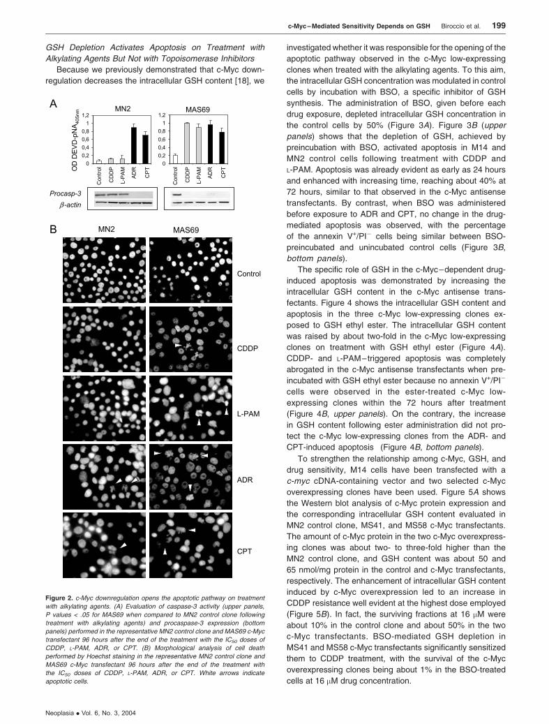
Figure 2. c-Myc downregulation opens the apoptotic pathway on treatment
with alkylating agents. (A) Evaluation of caspase-3 activity (upper panels,
P values < .05 for MAS69 when compared to MN2 control clone following
treatment with alkylating agents) and procaspase-3 expression (bottom
panels) performed in the representative MN2 control clone and MAS69 c-Myc
transfectant 96 hours after the end of the treatment with the IC50 doses of
CDDP, L-PAM, ADR, or CPT. (B) Morphological analysis of cell death
performed by Hoechst staining in the representative MN2 control clone and
MAS69 c-Myc transfectant 96 hours after the end of the treatment with
the IC50 doses of CDDP, L-PAM, ADR, or CPT. White arrows indicate
apoptotic cells.
c-Myc – Mediated Sensitivity Depends on GSH Biroccio et al. 199
Neoplasia . Vol. 6, No. 3, 2004
Drug-Induced Apoptosis Modulated by GSH through Its
Action on Mitochondria Level
To identify the signaling molecules involved in GSH-
modulated apoptosis following treatments with CDDP and
L-PAM, we analyzed the activation of CD95 receptor path-
way, the status of p53, and the role of mitochondria.
GSH-dependent apoptosis did not involve either the
CD95 system or any modulation in the p53 protein expres-
sion (data not shown). In addition, although the sequencing
analysis of the most mutated hot spot regions in the p53-
encoding sequence revealed that they were all wild type, the
p53 oncoprotein was not active in the M14 melanoma cells
(data not shown).
To determine the role of the mitochondria pathway, we
checked the cytosolic Bcl-2, Bcl-xL, and Bax levels; cyto-
chrome c; and the cleavage of procaspase-9. BSO-induced
apoptosis in control cells following treatment with alkylating
agents did not involve changes in the expression of Bcl-2 and
Bcl-xL (Figure 6A). On the contrary, a reduction in the
cytosolic Bax expression and a cytochrome c redistribution
were evident 24 hours after the end of treatment with CDDP
and L-PAM in the BSO-exposed control cells. Consequently,
procaspase-9 was found to be activated by cleavage exclu-
sively in the BSO-exposed control cells after treatment with
both alkylating agents. No modulation of these molecules
was observed in the BSO-unexposed control cells when
treated with CDDP and L-PAM.
The early Bax/cytochrome c translocation observed
in BSO-exposed control cells on treatment with CDDP and
L-PAM occurred independently of the depolarization of the
mitochondria membrane potential. As shown in Figure 6B
(upper panels), a significant reduction in Dwm was observed
only 48 hours after the end of the treatments with both
alkylating agents, whereas no shift in the JC-1 double
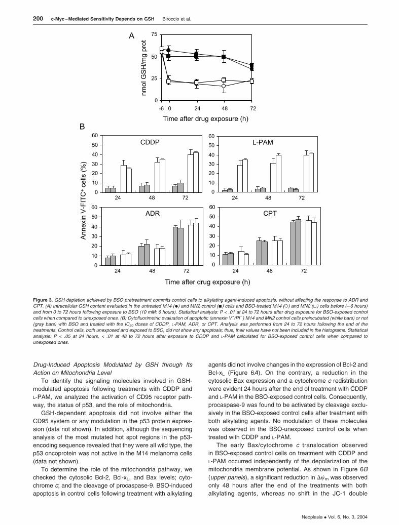
Figure 3. GSH depletion achieved by BSO pretreatment commits control cells to alkylating agent-induced apoptosis, without affecting the response to ADR and
CPT. (A) Intracellular GSH content evaluated in the untreated M14 (.) and MN2 control (n) cells and BSO-treated M14 (o) and MN2 (5) cells before (�6 hours)
and from 0 to 72 hours following exposure to BSO (10 mM; 6 hours). Statistical analysis: P < .01 at 24 to 72 hours after drug exposure for BSO-exposed control
cells when compared to unexposed ones. (B) Cytofluorimetric evaluation of apoptotic (annexin V+/PI�) M14 and MN2 control cells preincubated (white bars) or not
(gray bars) with BSO and treated with the IC50 doses of CDDP, L-PAM, ADR, or CPT. Analysis was performed from 24 to 72 hours following the end of the
treatments. Control cells, both unexposed and exposed to BSO, did not show any apoptosis; thus, their values have not been included in the histograms. Statistical
analysis: P < .05 at 24 hours, < .01 at 48 to 72 hours after exposure to CDDP and L-PAM calculated for BSO-exposed control cells when compared to
unexposed ones.
200 c-Myc –Mediated Sensitivity Depends on GSH Biroccio et al.
Neoplasia . Vol. 6, No. 3, 2004

fluorescence was revealed in the CDDP- and L-PAM–trea-
ted MN2 control clone. Moreover, the alkylator-dependent
alteration of Dwm was concomitant to the generation of ROS.
Figure 6B (bottom panels) shows that CDDP and L-PAM
treatments led to late ROS generation exclusively in the
BSO-exposed control cells starting 48 hours after the end
of the exposure to both alkylating agents, downstream
cytochrome c release, and caspase-9 activation. The ob-
served alteration of Dwm and ROS production enhanced with
increasing time after drug treatment (data not shown).
The ability of GSH to modulate drug-induced apoptosis by
acting at the mitochondria level was also observed when
GSH content was increased in the c-Myc low-expressing
cells after exposure with GSH ethyl ester. As shown in the
Figure 7, the treatment of the c-Myc low-expressing cells
with CDDP and L-PAM activated the same mitochondrial
pathway: early Bax redistribution, release of cytochrome c,
and concomitant caspase-9 activation (Figure 7A), upstream
alteration of mitochondria membrane potential (Figure 7B,
upper panels), and ROS production (Figure 7B, bottom
panels). GSH ester preincubation inhibited the apoptotic
cascade by blocking the early apoptotic events. Indeed,
Bax and cytochrome c redistribution was completely abro-
gated in the GSH ester-exposed c-Myc low-expressing cells
following treatment with both alkylating agents (Figure 7A).
Consequently, the downstream mitochondria membrane
depolarization and ROS generation were inhibited in the
GSH ester-exposed c-Myc antisense transfectant treated
with CDDP and L-PAM (Figure 7B ).
All the same experiments were performed in the M14
parental line and MAS51 and MAS53 c-Myc low-express-
ing clones, giving similar results to those obtained with the
MN2 and MAS69 c-Myc antisense transfectants (data not
shown).
Figure 4. GSH increase following ester administration protects c-Myc transfectants from CDDP- and L-PAM – triggered apoptosis, without altering topoisomerase
inhibitor-induced cell death. (A) Intracellular GSH content evaluated in the untreated MAS51 (E), MAS53 (y), and MAS69 (– ) c-Myc transfectants and GSH ester-
treated MAS51 (D), MAS53 (w), and MAS69 (*) c-Myc transfectants before (�24 hours) and from 0 to 72 hours following exposure to GSH ethyl ester (5 mM;
24 hours). Statistical analysis: P < .01 at 24 to 72 hours after drug exposure for GSH ester-exposed c-Myc antisense transfectants when compared to unexposed
ones. (B) Cytofluorimetric evaluation of apoptotic (annexin V+/PI�) MAS51, MAS53, and MAS69 c-Myc low-expressing clones cells preincubated (white bars) or
not (gray bars) with GSH ester and treated with the IC50 doses of CDDP, L-PAM, ADR, or CPT. Analysis was performed from 24 to 72 hours following the end of the
treatments. Values for c-Myc transfectants, both unexposed and exposed to GSH ester, are always below 10%; thus, they have not been included in the
histograms. Statistical analysis: P < .05 at 24 hours, < .01 at 48 to 72 hours after exposure to CDDP and L-PAM calculated for GSH ester-exposed c-Myc antisense
transfectants when compared to unexposed ones.
c-Myc – Mediated Sensitivity Depends on GSH Biroccio et al. 201
Neoplasia . Vol. 6, No. 3, 2004

Relationship Among c-Myc, Intracellular GSH Content, and
Alkylating Agent–Induced Apoptosis in JR8 Melanoma
Cells
To extend the results obtained, an other human mela-
noma line, displaying a high level of c-Myc protein, was
transfected with an expression vector carrying c-myc cDNA
in antisense orientation. Figure 8A shows the Western blot
analysis of c-Myc protein expression in JR8 parental line,
JN3 control clone, and three c-Myc antisense transfectants
(JAS26, JAS28, and JAS43). The amount of c-Myc protein in
the three c-Myc antisense transfectants was about five times
lower than the JN3 control clone or the parental line. Be-
sides, analysis of intracellular GSH content showed that it
was reduced by about 50% in the JAS26, JAS28, and JAS43
c-Myc antisense transfectants when compared to control
cells (Figure 8B ).
To verify the relationship between the c-Myc–dependent
GSH depletion and drug response, CDDP and ADR were
chosen as representative agents for alkylating and topoiso-
merase inhibitor drugs, respectively. Figure 8C shows the
survival curves of the JR8 parental line, the JN3 control
clone, and the three c-Myc low-expressing clones exposed
to increasing doses of CDDP and ADR. c-Myc low-express-
ing clones clearly displayed a greater sensitivity to CDDP if
compared to control cells. On the contrary, no difference in
sensitivity to ADR among the parental, control, and c-Myc
low-expressing cells was observed. Analysis of apoptosis
performed by using the IC50 dose of each drug (Figure 8D)
demonstrated that CDDP triggered apoptosis in the c-Myc
antisense transfectants but not in control cells, whereas ADR
induced apoptosis in all the lines regardless of c-Myc ex-
pression. GSH ester pretreatment protected c-Myc trans-
fectants from CDDP-induced apoptosis without affecting the
ADR-induced cell death.
Discussion
In this work, we demonstrate that GSH has a crucial role in
the c-Myc–dependent drug-induced apoptosis. This paper
follows our previous results showing that the downregulation
of c-Myc sensitizes M14 melanoma cells to alkylating agent
CDDP but not to DNA topoisomerase inhibitors ADR and
CPT [17]. In an effort to elucidate the mechanism(s) account-
ing for the different drug sensitivity, we evaluated the ability
of drugs to trigger apoptosis, and included another alkylating
agent (L-PAM) in the study. Our results demonstrate that
both CDDP and L-PAM triggered apoptosis in the c-Myc low-
expressing clones but not in control lines. On the contrary,
activation of programmed cell death occurred to the same
extent in control and c-Myc antisense transfectants after
ADR and CPT administration. Thus, alkylating agents acti-
vate a c-Myc–dependent apoptosis, whereas a c-Myc–in-
dependent cell death is induced by topoisomerase inhibitors.
Because we previously demonstrated that c-Myc down-
regulation reduced the intracellular GSH content [18], we
hypothesized that GSH might influence the c-Myc–depen-
dent (rather than c-Myc–independent) drug-induced apopto-
sis. To this aim, intracellular GSH content was normalized
between control and c-Myc low-expressing cells. As a result,
modulation of GSH content did not affect the response to
ADR and CPT, whereas it significantly influenced CDDP-
and L-PAM–induced apoptosis. In particular, GSH ester
administration in the c-Myc low-expressing cells increased
the intracellular GSH up to the levels of control cells and
abrogated apoptosis induced by CDDP and L-PAM. GSH
depletion by BSO preincubation opened the apoptotic path-
way in control cells treated with one alkylating agent or the
others. The opening of the apoptotic pathway by GSH
depletion exclusively occurred following alkylating agents,
thus indicating that it was related to the specific drug action.
The relationship among c-Myc, GSH content, and the
response to alkylating agents has been also evaluated in the
M14 c-Myc overexpressing clones as well as in the mela-
noma JR8 c-Myc antisense transfectants. The results
demonstrated that GSH depletion is responsible for the
c-Myc–dependent (rather than c-Myc–independent) drug-
induced apoptosis.
Unlike many other proposed mechanisms of drug
resistance, elevated GSH content may act through
different pathways to limit the effectiveness of multiple
types of chemotherapeutic agents. GSH has been shown
to protect tumor cells by detoxifying chemotherapeutic
drugs through conjugation reactions catalyzed by GSH
S-transferase. Export of GSH conjugate out of tumor
Figure 5. c-Myc – dependent GSH increase induces resistance to CDDP
treatment. (A) Western blot of c-Myc protein expression and intracellular GSH
content performed in the MN2 control clone and in the MS41 and MS58 c-Myc
overexpressing clones (P < .05 calculated for all c-Myc transfectants when
compared to control cells). (B) Survival curves of MN2 control clone (w), MS41
(n), MS58 (E) c-Myc transfectants, BSO-exposed MS41 (5), and BSO-
exposed MS58 (D) treated with increasing doses of CDDP. Surviving fractions
were calculated as ratio of the absolute survival of the treated sample/absolute
survival of the untreated one ( P < .01 calculated for the two c-Myc trans-
fectants when compared to control clone at 16 �M CDDP, and P < .001 cal-
culated for unexposed when compared to BSO-exposed c-Myc transfectants).
202 c-Myc –Mediated Sensitivity Depends on GSH Biroccio et al.
Neoplasia . Vol. 6, No. 3, 2004
cells by the GS-X pump represents the final elimination
of the overall detoxification system [28,29]. In addition,
GSH can be transported to the mitochondria, where it
plays a protective role as ROS scavenger [30]. The role
of GSH or GSH-related mechanisms has been found to
be particularly important in the case of alkylating agents
[31–33]. In fact, although the GSH-related mechanisms
are the most important events in the modulation of
cytotoxicity to CDDP and L-PAM, the mechanism for
ADR or CPT resistance involves other molecules, such
as the P170 in the case of ADR resistance. Moreover,
although the toxicity of CDDP and L-PAM largely
depends on intracellular GSH levels, modulation of
GSH does not always influence ADR or CPT cytotoxicity,
although this does vary among cell types. Depletion of
GSH, by prolonged incubation with BSO, increases the
lethality of CPT-11 in V79 hamster lung fibroblasts [34],
of etoposide in K562 human erythroleukemia cells [35],
and of ADR in different cell types [36–38]. In U937 human
promonocytic cells, although BSO potentiated the toxicity
of CDDP and L-PAM manifested by the suppression of
apoptosis and the induction of necrosis, it did not affect the
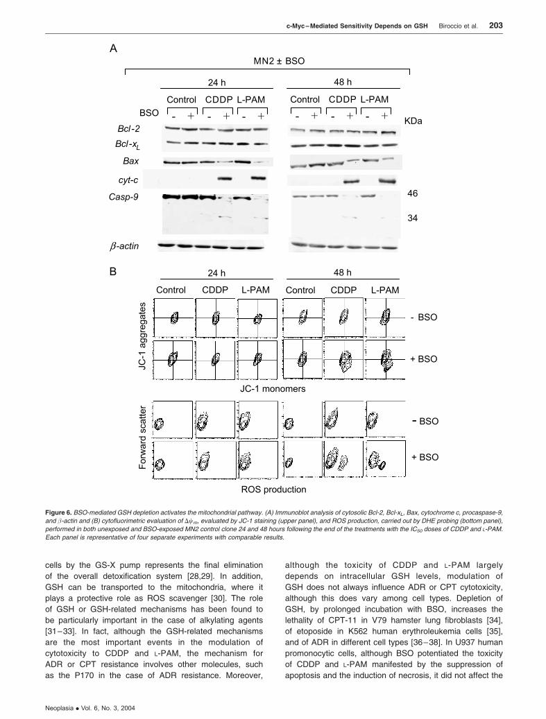
Figure 6. BSO-mediated GSH depletion activates the mitochondrial pathway. (A) Immunoblot analysis of cytosolic Bcl-2, Bcl-xL, Bax, cytochrome c, procaspase-9,
and �-actin and (B) cytofluorimetric evaluation of Dwm, evaluated by JC-1 staining (upper panel), and ROS production, carried out by DHE probing (bottom panel),
performed in both unexposed and BSO-exposed MN2 control clone 24 and 48 hours following the end of the treatments with the IC50 doses of CDDP and L-PAM.
Each panel is representative of four separate experiments with comparable results.
c-Myc – Mediated Sensitivity Depends on GSH Biroccio et al. 203
Neoplasia . Vol. 6, No. 3, 2004

mode nor the extent of death caused by CPT and ADR.
Comprehensively, the literature shows, however, that the
key role of GSH content in the sensitivity/resistance to
alkylating agents was mainly due to its detoxification or
antioxidant properties.
In our experimental models, we can exclude the antiox-
idant mechanism of GSH as the principal determinant factor
in c-Myc–dependent drug sensitivity because we found the
ROS production to be not the primary event in the activation
of GSH-mediated programmed cell death, but it represents a
downstream effector of apoptosis. Here, we show that
GSH influenced the c-Myc–dependent alkylator-induced
apoptosis by its ability to act at the mitochondria level,
without affecting the p53 and CD95 systems. Instead,
BSO-mediated GSH depletion in control cells treated with
CDDP and L-PAM rapidly induced Bax/cytochrome c re-
distribution and caspase-9 activation as early events,
with alteration of mitochondria membrane potential and
ROS production as late events. GSH ester preincubation
inhibited the apoptotic cascade by blocking the early
and, consequently, the late mitochondria-related apoptotic
events. The effect of GSH on the activation of cytochrome
c–dependent apoptotic pathway might be due to the ability of
alkylating agents to bind to GSH [39,40]. This would imply an
Figure 7. Ester-mediated GSH increase inhibits the mitochondrial pathway. (A) Immunoblot analysis of cytosolic Bcl-2, Bcl-xL, Bax, cytochrome c, procaspase-9,
and �-actin and (B) cytofluorimetric evaluation of Dwm, evaluated by JC-1 staining (upper panel), and ROS production, carried out by DHE probing (bottom panel),
performed in both unexposed and GSH ester-exposed MAS69 c-Myc low-expressing transfectant 24 and 48 hours following the end of the treatments with the IC50
doses of CDDP and L-PAM. Each panel is representative of four separate experiments with comparable results.
204 c-Myc –Mediated Sensitivity Depends on GSH Biroccio et al.
Neoplasia . Vol. 6, No. 3, 2004

impoverishment of free GSH from the mitochondrial buffer of
cells, leading to a cytochrome c–dependent apoptosis.
Therefore, because we previously demonstrated that
c-Myc downregulation decreases GSH content by reducing
its synthesis [18], the synergistic effect between c-Myc
downregulation and alkylating agents can be attributable to
cooperation in the GSH depletion.
In summary, we demonstrate that: 1) alkylating agents
trigger a c-Myc–dependent apoptosis, whereas a c-Myc–
independent cell death is induced by topoisomerase
inhibitors; 2) c-Myc–dependent drug-induced apoptosis
is due to GSH depletion; and 3) GSH-mediated drug-
induced apoptosis occurs by cytochrome c release.
All together, these results lend support to the clinical
approach based on c-myc antisense therapy being com-
bined with some antineoplastic agents (i.e., CDDP and
L-PAM), rather than others (ADR and CPT).
Acknowledgement
We thank Adele Petricca for her helpful assistance in typing
the manuscript.
References[1] Boxer LM and Dang CV (2001). Translocations involving c-myc and
c-myc function. Oncogene 20, 5595 – 5610.
[2] Nesbit CE, Tersak JM, and Prochownik EV (1999). Myc oncogenes and
human neoplastic disease. Oncogene 18, 3004 – 3016.
[3] Thompson EB (1998). The many roles of c-Myc in apoptosis. Annu Rev
Physiol 60, 575– 600.
Figure 8. Relationship among c-Myc, intracellular GSH content, and alkylating agent – induced apoptosis in the JR8 melanoma cells. (A) Western blot of c-Myc
protein expression and (B) intracellular GSH content performed in the JR8 melanoma cell line, JN3 control clone, and JAS26, JAS28, and JAS43 c-Myc low-
expressing clones ( P < .05 calculated for all c-Myc antisense transfectants when compared to both control cells). (C) Survival curves of JR8 (.), JN3 control clone
(n), JAS26 (5), JAS28 (w), and JAS43 (D) c-Myc antisense transfectants exposed to increasing doses of either CDDP (left panel, P < .05 at 3.4 and P < .01 at 6.7
and 16 �M, calculated for all c-Myc antisense transfectants when compared to both control cells) or ADR (right panel). Surviving fractions were calculated as
the ratio of the absolute survival of the treated sample/absolute survival of the untreated one. (D) Percentage of apoptotic cells evaluated 48 hours following the
treatment with the IC50 doses of either CDDP (P < .01 calculated for all c-Myc antisense transfectants when compared to both control cells) or ADR, in both the
unexposed (white bars) and GSH ethyl ester– exposed (black bars) JR8 parental line and JN3, JAS26, JAS28, and JAS43 transfectants.
c-Myc – Mediated Sensitivity Depends on GSH Biroccio et al. 205
Neoplasia . Vol. 6, No. 3, 2004

[4] Evan GI, Wyllie AH, Gilbert CS, Littlewood TD, Land H, Brooks M,
Waters CM, Penn LZ, and Hancock DC (1992). Induction of apoptosis
in fibroblasts by c-myc protein. Cell 69, 119 – 128.
[5] Alarcon RM, Rupnow BA, Graeber TG, Knox SJ, and Giaccia AJ
(1996). Modulation of c-Myc activity and apoptosis in vivo. Cancer
Res 56, 4315 – 4319.
[6] Askew DS, Ashmun RA, Simmons BC, and Cleveland JL (1991). Con-
stitutive c-myc expression in an IL-3 – dependent myeloid cell line sup-
presses cell cycle arrest and accelerates apoptosis. Oncogene 6,
1915 – 1922.
[7] Klefstrom J, Vastrik I, Saksela E, Valle J, Eilers M, and Alitalo K (1994).
c-Myc induces cellular susceptibility to the cytotoxic action of TNF-
alpha. EMBO J 13, 5442 – 5450.
[8] Nesbit CE, Fan S, Zhang H, and Prochownik EV (1998a). Distinct
apoptotic responses imparted by c-myc and max. Blood 92,
1003 – 1010.
[9] Nesbit CE, Grove LE, Yin XY, and Prochownik EV (1998b). Differential
apoptotic behaviors of c-myc, N-myc, and L-myc oncoproteins. Cell
Growth Differ 9, 731 – 741.
[10] Arango D, Corner GA, Wadler S, Catalano PJ, and Augenlicht LH
(2001). c-myc/p53 interaction determines sensitivity of human colon
carcinoma cells to 5-fluorouracil in vitro and in vivo. Cancer Res 61,
4910 – 4915.
[11] Sklar MD and Prochownik EV (1991). Modulation of cis-platinum re-
sistance in Friend erythroleukemia cells by c-myc. Cancer Res 51,
2118 – 2123.
[12] Niimi S, Nakagawa K, Yokota J, Tsunokawa Y, Nishio K, Terashima Y,
Shibuya M, Terada M, and Saijo N (1991). Resistance to anticancer
drugs in NIH3T3 cells transfected with c-myc and/or c-H-ras genes. Br J
Cancer 63, 237– 241.
[13] Van Waardenburg RC, Meijer C, Burger H, Nooter K, De Vries EG,
Mulder NH, and De Jong S (1997). Effects of an inducible anti-sense
c-myc gene transfer in a drug-resistant human small-cell-lung-carcino-
ma cell line. Int J Cancer 73, 544 – 550.
[14] Leonetti C, D’Agnano I, Lozupone F, Valentini A, Geiger T, Zon G,
Calabretta B, Citro G, and Zupi G (1996). Antitumor effect of c-myc
antisense phosphorothioate oligodeoxynucleotides on human melano-
ma cells in vitro and in mice. J Natl Cancer Inst 88, 419 –429.
[15] Citro G, D’Agnano I, Leonetti C, Perini R, Bucci B, Zon G, Calabretta B,
and Zupi G (1998). c-myc antisense oligodeoxynucleotides enhance
the efficacy of cisplatin in melanoma chemotherapy in vitro and in nude
mice. Cancer Res 58, 283 – 289.
[16] Leonetti C, Biroccio A, Candiloro A, Citro G, Fornari C, Mottolese M,
Del Bufalo D, and Zupi G (1999). Increase of cisplatin sensitivity by
c-myc antisense oligodeoxynucleotides in a human metastatic melano-
ma inherently resistant to cisplatin. Clin Cancer Res 5, 2588 – 2595.
[17] Biroccio A, Benassi B, Amodei S, Gabellini C, Del Bufalo D, and Zupi G
(2001). c-Myc down-regulation increases susceptibility to cisplatin
through reactive oxygen species-mediated apoptosis in M14 human
melanoma cells. Mol Pharmacol 60, 174 – 182.
[18] Biroccio A, Benassi B, Filomeni G, Amodei S, Marchini S, Chiorino G,
Rotilio G, Zupi G, and Ciriolo MR (2002). Glutathione influences
c-Myc – induced apoptosis in M14 human melanoma cells. J Biol Chem
277, 43763 – 43770.
[19] Zhang K, Mack P, and Wong KP (1998). Glutathione-related mecha-
nisms in cellular resistance to anticancer drugs. Int J Oncol 12,
871– 882.
[20] Chen G and Zeller WJ (1991). Augmentation of cisplatin (DDP) cyto-
toxicity in vivo by DL-buthionine sulfoximine (BSO) in DDP-sensitive and
-resistant rat ovarian tumors and its relation to DNA interstrand cross
links. Anticancer Res 11, 2231 – 2237.
[21] Yen L, Woo A, Christopoulopoulos G, Batist G, Panasci L, Roy R, Mitra
S, and Alaoui-Jamali MA (1995). Enhanced host cell reactivation ca-
pacity and expression of DNA repair genes in human breast cancer
cells resistant to bi-functional alkylating agents. Mutat Res 337,
179– 189.
[22] Gamcsik MP, Millis KK, and Hamill TG (1997). Kinetics of the conjuga-
tion of aniline mustards with glutathione and thiosulfate. Chem Biol
Interact 105, 35 –52.
[23] Barrand MA, Bagrij T, and Neo SY (1997). Multidrug resistance-asso-
ciated protein: a protein distinct from P-glycoprotein involved in cyto-
toxic drug expulsion. Gen Pharmacol 28, 639– 645.
[24] Vukovic L and Osmak M (1999). Reversal of carboplatin resistance in
human laryngeal carcinoma cells. Neoplasm 46, 335 –341.
[25] Bailey HH, Gipp JJ, Ripple M, Wilding G, and Mulcahy RT (1992).
Increase in gamma-glutamylcysteine synthetase activity and steady-
state messenger RNA levels in melphalan-resistant DU-145 human
prostate carcinoma cells expressing elevated glutathione levels.
Cancer Res 52, 5115 – 5118.
[26] Godwin AK, Meister A, O’Dwyer PJ, Huang CS, Hamilton TC, and
Anderson ME (1992). High resistance to cisplatin in human ovarian
cancer cell lines is associated with marked increase of glutathione syn-
thesis. Proc Natl Acad Sci USA 89, 3070 –3074.
[27] Mulcahy RT, Bailey HH, and Gipp JJ (1995). Transfection of comple-
mentary DNAs for the heavy and light subunits of human gamma- glu-
tamylcysteine synthetase results in an elevation of intracellular
glutathione and resistance to melphalan. Cancer Res 55, 4771 – 4775.
[28] Ishikawa T (1992). The ATP-dependent glutathione S-conjugate export
pump. Trends Biochem Sci 17, 463 –468.
[29] Ishikawa T and Ali-Osman F (1993). Glutathione-associated cis-
diamminedichloroplatinum(II) metabolism and ATP-dependent efflux
from leukemia cells. Molecular characterization of glutathione – plati-
num complex and its biological significance. J Biol Chem 268,
20116 – 20125.
[30] Meister A (1991). Glutathione deficiency produced by inhibition of its
synthesis, and its reversal; applications in research and therapy. Phar-
macol Ther 51, 155 – 194.
[31] Meijer C, Mulder NH, Timmer-Bosscha H, Sluiter WJ, Meersma GJ,
and de Vries EG (1992). Relationship of cellular glutathione to the
cytotoxicity and resistance of seven platinum compounds. Cancer
Res 52, 6885 – 6889.
[32] Timmer-Bosscha H, Timmer A, Meijer C, de Vries EG, de Jong B, Oos-
terhuis JW, and Mulder NH (1993). cis-Diamminedichloroplatinum(II)
resistance in vitro and in vivo in human embryonal carcinoma cells.
Cancer Res 53, 5707 – 5713.
[33] Hamilton D, Fotouhi-Ardakani N, and Batist G (2002). The glutathione
system in alkylator resistance. Cancer Treat Res 112, 67 –87.
[34] Sawyer TE and Bonner JA (1996). The interaction of buthionine sul-
phoximide (BSO) and the topoisomerase I inhibitor CPT-11. Br J
Cancer Suppl 27, S109 – S113.
[35] Gantchev TG and Hunting DJ (1997). Enhancement of etoposide
(VP-16) cytotoxicity by enzymatic and photodynamically induced oxida-
tive stress. Anticancer Drugs 8, 164 –173.
[36] Lee FY, Vessey AR, and Siemann DW (1988). Glutathione as a deter-
minant of cellular response to doxorubicin. NCI Monogr 6, 211 –215.
[37] Mans DR, Schuurhuis GJ, Treskes M, Lafleur MV, Retel J, Pinedo HM,
and Lankelma J (1992). Modulation by D,L-buthionine-S,R-sulphoximine
of etoposide cytotoxicity on human non-small cell lung, ovarian and
breast carcinoma cell lines. Eur J Cancer 28A, 1447 –1452.
[38] Crescimanno M, Borsellino N, Leonardi V, Flandina C, Flugy A, Rausa
L, and D’Alessandro N (1994). Effect of buthionine sulfoximine on the
sensitivity to doxorubicin of parent and MDR tumor cell lines. J Chemo-
ther 6, 343 –348.
[39] Dirven HA, Dictus EL, Broeders NL, van Ommen B, and van Bladeren
PJ (1995). The role of human glutathione S-transferase isoenzymes in
the formation of glutathione conjugates of the alkylating cytostatic drug
thiotepa. Cancer Res 55, 1701 – 1706.
[40] David-Cordonnier MH, Laine W, Joubert A, Tardy C, Goossens JF,
Kouach M, Briand G, Thi Mai HD, Michel S, Tillequin F, Koch M,
Leonce S, Pierre A, and Bailly C (2003). Covalent binding to glutathione
of the DNA-alkylating antitumor agent, S23906-1. Eur J Biochem 270,
2848 – 2859.
206 c-Myc –Mediated Sensitivity Depends on GSH Biroccio et al.
Neoplasia . Vol. 6, No. 3, 2004