Embed Size (px)
Citation preview
“fendo-03-00153” — 2012/12/15 — 12:18 — page 1 — #1
REVIEW ARTICLEpublished: 17 December 2012
doi: 10.3389/fendo.2012.00153
Glucose: an energy currency and structural precursor inarticular cartilage and bone with emerging roles as anextracellular signaling molecule and metabolic regulatorAli Mobasheri*
Faculty of Medicine and Health Sciences, School of Veterinary Medicine and Science, University of Nottingham, Nottingham, UK
Edited by:
Alison Gartland, The University ofSheffield, UK
Reviewed by:
Alexandrina Ferreira Mendes,University of Coimbra, PortugalBasem M. Abdallah, University ofSouthern Denmark, Denmark
*Correspondence:
Ali Mobasheri, Faculty of Medicineand Health Sciences, School ofVeterinary Medicine and Science,University of Nottingham, SuttonBonington Campus, SuttonBonington, Nottingham,Leicestershire LE12 5RD, UK.e-mail: [email protected]
In the skeletal system glucose serves as an essential source of energy for the develop-ment, growth, and maintenance of bone and articular cartilage. It is particularly neededfor skeletal morphogenesis during embryonic growth and fetal development. Glucose isvital for osteogenesis and chondrogenesis, and is used as a precursor for the synthesis ofglycosaminoglycans, glycoproteins, and glycolipids. Glucose sensors are present in tissuesand organs that carry out bulk glucose fluxes (i.e., intestine, kidney, and liver).The beta cellsof the pancreatic islets of Langerhans respond to changes in blood glucose concentrationby varying the rate of insulin synthesis and secretion. Neuronal cells in the hypothalamusare also capable of sensing extracellular glucose. Glucosensing neurons use glucose asa signaling molecule to alter their action potential frequency in response to variations inambient glucose levels. Skeletal muscle and adipose tissue can respond to changes in cir-culating glucose but much less is known about glucosensing in bone and cartilage. Recentresearch suggests that bone cells can influence (and be influenced by) systemic glucosemetabolism. This focused review article discusses what we know about glucose transportand metabolism in bone and cartilage and highlights recent studies that have linked glu-cose metabolism, insulin signaling, and osteocalcin activity in bone. These new findings inbone cells raise important questions about nutrient sensing, uptake, storage and process-ing mechanisms and how they might contribute to overall energy homeostasis in healthand disease. The role of glucose in modulating anabolic and catabolic gene expression innormal and osteoarthritic chondrocytes is also discussed. In summary, cartilage and bonecells are sensitive to extracellular glucose and adjust their gene expression and metabolismin response to varying extracellular glucose concentrations.
Keywords: glucose, extracellular signaling, articular cartilage, bone, glucosensing, hexokinase, glucose transport,
osteocalcin
INTRODUCTIONAll living cells must be able to regulate their metabolic activitywhen faced with nutrient fluctuations in the extracellular envi-ronment (Mobasheri et al., 2008). Sensing the abundance and localfluctuations in the concentration of extracellular nutrients is a fun-damental property of all living cells. Indeed, it has been suggestedthat it is an absolute requirement for the ability of living cells toadapt to changes in their environment (Shirazi-Beechey, 2005).
Nutrient-sensing is defined as a living cell’s ability to recognizeand respond to fuel substrates and is essential for the survivalof all prokaryotes and eukaryotes. Studies in plants (Rollandet al., 2002), yeast (Forsberg and Ljungdahl, 2001), and bacteria(Gilmore et al., 2003) have demonstrated that these organisms areable to sense and respond to changes in extracellular carbon andnitrogen metabolites. For example, in plants sugar-sensing allowsphotosynthesis to be switched off when carbohydrates are plenti-ful and turned on again when sugar levels are low (Rolland et al.,2002; Lejay et al., 2003). This adaptation involves hormonal regu-lation of gene expression and expressed protein function allowingthe plant to make efficient and economic use of its energy stores.
In eukaryotic cells the physiological maintenance of nutri-ent and metabolite homeostasis is crucial to many fundamen-tal cellular functions (Rolland et al., 2001). These functionsinclude division, proliferation, differentiation, excitability, secre-tion, senescence (Nemoto et al., 2004), and apoptosis (Martenset al., 2005).
Each type of metabolic fuel used by living cells requires adistinct and carefully regulated uptake, storage, and utilizationpathway involving transport, regulatory, and accessory molecules.In order to conserve valuable resources a cell will only producethe biomolecules that it requires at any one time. These require-ments may change when cells engage in different activities such asdivision, proliferation, differentiation, and apoptosis. The quan-tity and type of nutrients and metabolic fuels that are availableto a cell will also determine the complement of enzymes it needsto express from its genome for efficient utilization of the availablenutrients. The uptake and storage of nutrients can also profoundlyaffect the size and morphology of cells.
Some metabolic fuels are also important structural precur-sors for the synthesis of other biochemicals and biological
www.frontiersin.org December 2012 | Volume 3 | Article 153 | 1
“fendo-03-00153” — 2012/12/15 — 12:18 — page 2 — #2
Mobasheri Glucose as a signaling molecule
macromolecules. Glucose is an example of a metabolic fuel and astructural substrate for the synthesis of glycoproteins and glyco-conjugates. Specific receptors on the cell membrane are activatedin the presence of specific fuel molecules communicate to the cellnucleus by means of biochemical signaling cascades. This mecha-nism allows cells to maintain awareness of the available nutrientsin their environment in order to adjust their metabolism to utilizethe available substrate molecules most efficiently.
GLUCOSE AS A SIGNALING MOLECULE IN YEAST ANDMAMMALIAN CELLSIt is now well established that glucose is an extracellular sig-naling molecule in Saccharomyces cerevisiae (Santangelo, 2006).Yeast cells possess elaborate mechanisms for sensing the availabil-ity and levels of glucose and other sugars. Sugar-sensing allowsthem to adjust cellular metabolism to best utilize the availableresources and respond appropriately during periods of nutrientstress (Thevelein and de Winde, 1999).
Extracellular glucose and other sugars can also function assignaling molecules in mammalian cells, exerting transcriptionalcontrol over many different genes – this has resulted in the estab-lishment of a discipline known as “nutrigenomics” (van Ommenand Stierum, 2002; van Ommen, 2004; Corthesy-Theulaz et al.,2005; Grayson, 2010). Nutrigenomics is an exciting scientific dis-cipline that explores how genes interact with nutrients and hownutrients influence gene expression. It involves the study of theeffects of foods and food constituents on gene expression (vanOmmen and Stierum, 2002). This field of study has emergedbecause of the realization that the health effects of food-derivedsubstances start at the molecular level (van Ommen and Stierum,2002; van Ommen, 2004). However, long before the advent ofnutrigenomics, it was known that nutrients have the capacityto influence gene expression in microorganisms. Cells generallyadapt to alterations in the extracellular concentrations of anygiven nutrient by regulating its transport rate across the plasmamembrane (and subsequently its storage and metabolism). Suchadaptation is essential for numerous subcellular functions andmay involve transcriptional control of transporter genes and cellsurface sensors.
Extracellular glucose and other monosaccharides also altermRNA and protein stability. Accordingly, the cells, tissues, andorgans of living organisms must possess sophisticated molecularmechanisms for sensing extracellular glucose. Invariably, whenthis control is lost, glucose homeostasis is compromised, which isoften followed by metabolic disease.
This focused review article discusses the importance of glucoseas a universal energy currency and the molecular mechanismsinvolved in glucose sensing in the pancreas and the gut beforefocusing on glucose transport and metabolism in bone andcartilage and highlighting areas for future research.
HEXOKINASE: AN INTRACELLULAR GLUCOSE SENSORThe hexokinase glucose sensor concept was introduced over fourdecades ago (Matschinsky and Ellerman, 1968). Hexokinase (alsoknown as glucokinase) is an evolutionarily conserved intracellularglucose sensor (Moore et al., 2003; Olsson et al., 2003; Rolland andSheen, 2005). It functions as a glucose-phosphorylating enzyme,
which is the first enzyme in glycolysis. It is a key regulator ofenergy expenditure in most cells as it catalyzes the first step in themetabolism of glucose but has also been proposed to be involvedin sugar sensing and signaling in yeast and in plants (Moore et al.,2003; Olsson et al., 2003; Rolland and Sheen, 2005). Hexokinaseconverts glucose into glucose-6-phosphate and controls glycolyticflux and glucose oxidation in pancreatic β cells thus acting as anintracellular glucose sensor (Matschinsky, 1996).
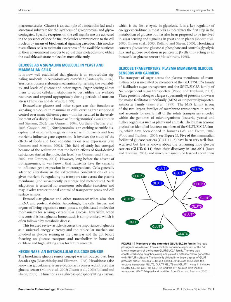
GLUCOSE TRANSPORTERS: PLASMA MEMBRANE GLUCOSESENSORS AND CARRIERSThe transport of sugar across the plasma membrane of mam-malian cells is mediated by members of the GLUT/SLC2A familyof facilitative sugar transporters and the SGLT/SLC5A family ofNa+-dependent sugar transporters (Wood and Trayhurn, 2003).These proteins belong to a larger superfamily of proteins known asthe major facilitator superfamily (MFS) or uniporter-symporter-antiporter family (Saier et al., 1999). The MFS family is oneof the two largest families of membrane transporters in natureand accounts for nearly half of the solute transporters encodedwithin the genomes of microorganisms (bacteria, yeasts) andhigher organisms such as plants and animals. The human genomeproject has identified fourteen members of the GLUT/SLC2A fam-ily, which have been cloned in humans (Wu and Freeze, 2002;Wood and Trayhurn, 2003; see Figure 1). Five of the mammalianfacilitated glucose carriers (GLUTs 1–5) have been very well char-acterized but less is known about the remaining nine glucosecarriers (GLUTs 6–14) since their discovery in late 2001 (Joostand Thorens, 2001) and much remains to be learned about their
FIGURE 1 | Members of the extended GLUT/SLC2A family. The radialphylogram was derived from a multiple sequence alignment of the 14known members of the human GLUT/SLC2A family. The tree wasconstructed using neighbor-joining analysis of a distance matrix generatedwith PHYLIP software. The family is divided into three classes of GLUTproteins; class I includes GLUTs1-4 and GLUT14; class II includes thefructose transporter GLUT5, GLUT7, GLUT9 and GLUT11; class III includesGLUT6, GLUT8, GLUT10, GLUT12, and the H+-coupled myo-inositoltransporter, HMIT. Adapted and modified from Wood and Trayhurn (2003).
Frontiers in Endocrinology | Bone Research December 2012 | Volume 3 | Article 153 | 2
“fendo-03-00153” — 2012/12/15 — 12:18 — page 3 — #3
Mobasheri Glucose as a signaling molecule
expression, tissue distribution, and transport functions (Uldry andThorens, 2004).
FUNCTIONAL ROLES OF THE GLUT PROTEINSGLUT1, GLUT3, and GLUT4 are high-affinity transporterswhereas GLUT2 is a low-affinity transporter; GLUT5 is primarilya fructose carrier (Thorens, 1996). High-affinity transporters arefound in many metabolically active tissues, but their expressionis higher in highly metabolic cells (i.e., hepatocytes, absorptiveintestine epithelial cells, and proximal tubule cells; Tal et al., 1990;Thorens et al., 1990). GLUT1 is expressed in many human tissuesincluding articular cartilage (Richardson et al., 2003; Mobash-eri et al., 2005, 2008; Phillips et al., 2005; Peansukmanee et al.,2009; Rosa et al., 2009) and intervertebral disc (IVD; Richardsonet al., 2008). IVD is anatomically and functionally very similarto cartilage although in contrast to cartilage it develops fromnotocordal cells rather than mesenchymal cells (Richardson et al.,2008). GLUT1 is abundantly expressed in the brain (Flier et al.,1987a), erythrocytes (Mueckler et al., 1985), and the liver, butis present in significantly lower quantities in cardiac and skeletalmuscle which express other glucose transporters including GLUT3(Guillet-Deniau et al., 1994; Hocquette and Abe, 2000; Shepherdet al., 1992) and GLUT4 (James et al., 1988; Charron et al., 1989).Elevated levels of the GLUT1 glucose transporter are induced byras or src oncogenes (Flier et al., 1987b) and a role for this glucosetransporter has been proposed in oncogenic transformation andtumor development ( Nagamatsu et al., 1993; Mellanen et al., 1994;Younes et al., 1996; Burstein et al., 1998; Semenza et al., 2001).GLUT2 is expressed in tissues carrying large glucose fluxes, suchas the pancreas, intestine, kidney, and liver (Thorens, 1996), as wellas in brain where it is involved in maintaining glucose homeosta-sis, and in cells where glucose-sensing is necessary (i.e., pancreaticβ cells and hypothalamic neurons; Waeber et al., 1995). Indeed,in many experimental models of diabetes, GLUT2 gene expres-sion is decreased in pancreatic β cells, which could lead to a lossof glucose-induced insulin secretion. As an adaptive response tovariations in metabolic conditions, the expression of the GLUT1–5 transporters is regulated by glucose and different hormones(Thorens, 1996).
GLUCOSE-SENSING VIA THE GLUT2 GLUCOSETRANSPORTER AND HEXOKINASEEarly studies on glucose-sensing in the central nervous systemsuggested that the brain uses glucose-sensing units that are sim-ilar to the pancreas (Maekawa et al., 2000). Ependymal cellshave been proposed to be putative glucose sensors in the brain.Immunohistochemical studies have shown strong hexokinase-likeimmunoreactivities in the ependymal cells, endothelial cells, andmany serotonergic neurons (Maekawa et al., 2000). Ependymalcells were found to exhibit GLUT2 and GLUT1-like immunoreac-tivities on the cilia in addition to GLUT4-like immunoreactivitydensely in the cytoplasmic area. These results raised the possibilitythat these cells form part of a more sophisticated array of glucosesensors in the brain.
The study of certain neuroendocrine cells outside the pan-creatic islet has led to the suggestion that the intracellularglucose sensor hexokinase may play a broader role in intracellular
glucose-sensing by neuroendocrine cells than was thought previ-ously (Jetton et al., 1994) and more recent work has shown thatelectrogenic sugar entry via SGLT1 and SGLT3 provide a novelmechanism for glucose-sensing by neuroendocrine cells (Gribbleet al., 2003).
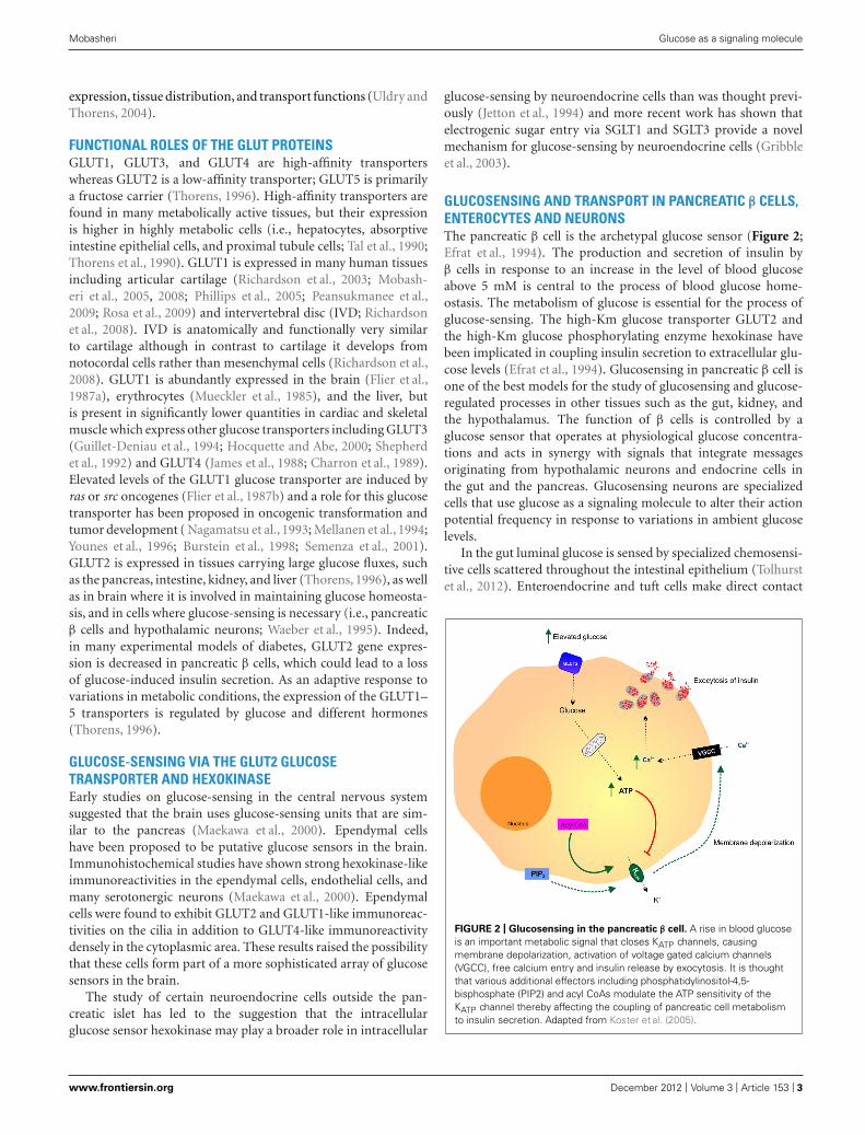
GLUCOSENSING AND TRANSPORT IN PANCREATIC β CELLS,ENTEROCYTES AND NEURONSThe pancreatic β cell is the archetypal glucose sensor (Figure 2;Efrat et al., 1994). The production and secretion of insulin byβ cells in response to an increase in the level of blood glucoseabove 5 mM is central to the process of blood glucose home-ostasis. The metabolism of glucose is essential for the process ofglucose-sensing. The high-Km glucose transporter GLUT2 andthe high-Km glucose phosphorylating enzyme hexokinase havebeen implicated in coupling insulin secretion to extracellular glu-cose levels (Efrat et al., 1994). Glucosensing in pancreatic β cell isone of the best models for the study of glucosensing and glucose-regulated processes in other tissues such as the gut, kidney, andthe hypothalamus. The function of β cells is controlled by aglucose sensor that operates at physiological glucose concentra-tions and acts in synergy with signals that integrate messagesoriginating from hypothalamic neurons and endocrine cells inthe gut and the pancreas. Glucosensing neurons are specializedcells that use glucose as a signaling molecule to alter their actionpotential frequency in response to variations in ambient glucoselevels.
In the gut luminal glucose is sensed by specialized chemosensi-tive cells scattered throughout the intestinal epithelium (Tolhurstet al., 2012). Enteroendocrine and tuft cells make direct contact
FIGURE 2 | Glucosensing in the pancreatic β cell. A rise in blood glucoseis an important metabolic signal that closes KATP channels, causingmembrane depolarization, activation of voltage gated calcium channels(VGCC), free calcium entry and insulin release by exocytosis. It is thoughtthat various additional effectors including phosphatidylinositol-4,5-bisphosphate (PIP2) and acyl CoAs modulate the ATP sensitivity of theKATP channel thereby affecting the coupling of pancreatic cell metabolismto insulin secretion. Adapted from Koster et al. (2005).
www.frontiersin.org December 2012 | Volume 3 | Article 153 | 3
“fendo-03-00153” — 2012/12/15 — 12:18 — page 4 — #4
Mobasheri Glucose as a signaling molecule
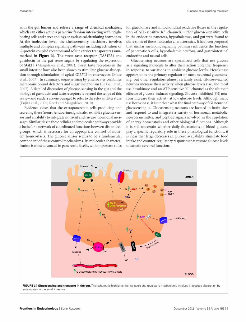
with the gut lumen and release a range of chemical mediators,which can either act in a paracrine fashion interacting with neigh-boring cells and nerve endings or as classical circulating hormones.At the molecular level, the chemosensory machinery involvesmultiple and complex signaling pathways including activation ofG-protein coupled receptors and solute carrier transporters (sum-marized in Figure 3). The sweet taste receptor (TAS1R3) andgustducin in the gut sense sugars by regulating the expressionof SGLT1 (Margolskee et al., 2007). Sweet taste receptors in thesmall intestine have also been shown to stimulate glucose absorp-tion through stimulation of apical GLUT2 in enterocytes (Maceet al., 2007). In summary, sugar-sensing by enterocytes combinesmembrane bound detectors and sugar metabolism (Le Gall et al.,2007). A detailed discussion of glucose-sensing in the gut and thebiology of gustducin and taste receptors is beyond the scope of thisreview and readers are encouraged to refer to the relevant literature(Fujita et al., 2009; Reed and Margolskee, 2010).
Evidence exists that the extrapancreatic cells producing andsecreting these (neuro)endocrine signals also exhibit a glucose sen-sor and an ability to integrate nutrient and (neuro)hormonal mes-sages. Similarities in these cellular and molecular pathways providea basis for a network of coordinated functions between distant cellgroups, which is necessary for an appropriate control of nutri-ent homeostasis. The glucose sensor seems to be a fundamentalcomponent of these control mechanisms. Its molecular character-ization is most advanced in pancreatic β cells, with important roles
for glucokinase and mitochondrial oxidative fluxes in the regula-tion of ATP-sensitive K+ channels. Other glucose-sensitive cellsin the endocrine pancreas, hypothalamus, and gut were found toshare some of these molecular characteristics. It has been proposedthat similar metabolic signaling pathways influence the functionof pancreatic α cells, hypothalamic neurons, and gastrointestinalendocrine and neural cells.
Glucosensing neurons are specialized cells that use glucoseas a signaling molecule to alter their action potential frequencyin response to variations in ambient glucose levels. Hexokinaseappears to be the primary regulator of most neuronal glucosens-ing, but other regulators almost certainly exist. Glucose-excitedneurons increase their activity when glucose levels rise, and mostuse hexokinase and an ATP-sensitive K+ channel as the ultimateeffector of glucose-induced signaling. Glucose-inhibited (GI) neu-rons increase their activity at low glucose levels. Although manyuse hexokinase, it is unclear what the final pathway of GI neuronalglucosensing is. Glucosensing neurons are located in brain sitesand respond to and integrate a variety of hormonal, metabolic,neurotransmitter, and peptide signals involved in the regulationof energy homeostasis and other biological functions. Althoughit is still uncertain whether daily fluctuations in blood glucoseplay a specific regulatory role in these physiological functions, itis clear that large decreases in glucose availability stimulate foodintake and counter-regulatory responses that restore glucose levelsto sustain cerebral function.
FIGURE 3 | Glucosensing and transport in the gut. This schematic highlights the transport and regulatory mechanisms involved in glucose absorption byenterocytes in the small intestine.
Frontiers in Endocrinology | Bone Research December 2012 | Volume 3 | Article 153 | 4
“fendo-03-00153” — 2012/12/15 — 12:18 — page 5 — #5
Mobasheri Glucose as a signaling molecule
It is also suggested that sweet taste signaling functions as ahypothalamic glucose sensor (Ren et al., 2009). The heterodimericG-protein coupled sweet receptor TAS1R2/TAS1R3 has been pro-posed as a candidate membrane-bound brain glucosensor (Renet al., 2009).
Finally, glucosensing is altered in obesity and after recurrentbouts of hypoglycemia, and this altered sensing may contribute tothe adverse outcomes of these conditions. Thus, although a greatdeal is already known, much more remains to be learned about thephysiological function of brain glucosensing neurons. It is possiblethat similar glucosensing mechanisms may operate in other organsystems.
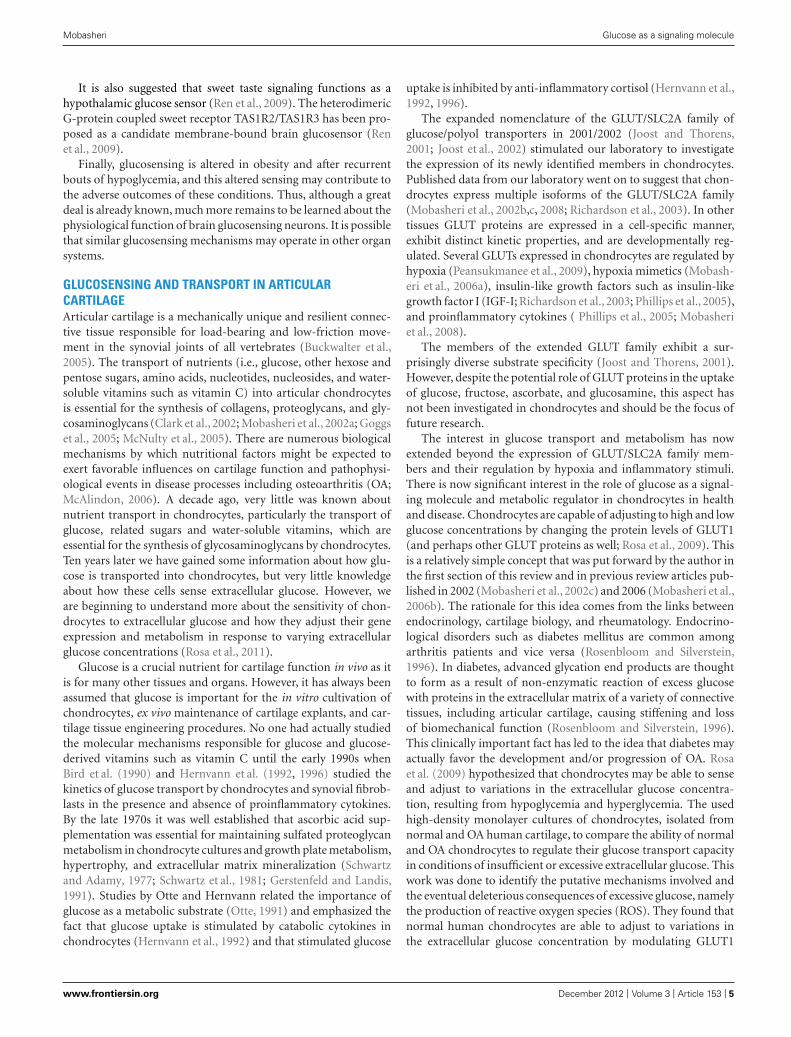
GLUCOSENSING AND TRANSPORT IN ARTICULARCARTILAGEArticular cartilage is a mechanically unique and resilient connec-tive tissue responsible for load-bearing and low-friction move-ment in the synovial joints of all vertebrates (Buckwalter et al.,2005). The transport of nutrients (i.e., glucose, other hexose andpentose sugars, amino acids, nucleotides, nucleosides, and water-soluble vitamins such as vitamin C) into articular chondrocytesis essential for the synthesis of collagens, proteoglycans, and gly-cosaminoglycans (Clark et al., 2002; Mobasheri et al., 2002a; Goggset al., 2005; McNulty et al., 2005). There are numerous biologicalmechanisms by which nutritional factors might be expected toexert favorable influences on cartilage function and pathophysi-ological events in disease processes including osteoarthritis (OA;McAlindon, 2006). A decade ago, very little was known aboutnutrient transport in chondrocytes, particularly the transport ofglucose, related sugars and water-soluble vitamins, which areessential for the synthesis of glycosaminoglycans by chondrocytes.Ten years later we have gained some information about how glu-cose is transported into chondrocytes, but very little knowledgeabout how these cells sense extracellular glucose. However, weare beginning to understand more about the sensitivity of chon-drocytes to extracellular glucose and how they adjust their geneexpression and metabolism in response to varying extracellularglucose concentrations (Rosa et al., 2011).
Glucose is a crucial nutrient for cartilage function in vivo as itis for many other tissues and organs. However, it has always beenassumed that glucose is important for the in vitro cultivation ofchondrocytes, ex vivo maintenance of cartilage explants, and car-tilage tissue engineering procedures. No one had actually studiedthe molecular mechanisms responsible for glucose and glucose-derived vitamins such as vitamin C until the early 1990s whenBird et al. (1990) and Hernvann et al. (1992, 1996) studied thekinetics of glucose transport by chondrocytes and synovial fibrob-lasts in the presence and absence of proinflammatory cytokines.By the late 1970s it was well established that ascorbic acid sup-plementation was essential for maintaining sulfated proteoglycanmetabolism in chondrocyte cultures and growth plate metabolism,hypertrophy, and extracellular matrix mineralization (Schwartzand Adamy, 1977; Schwartz et al., 1981; Gerstenfeld and Landis,1991). Studies by Otte and Hernvann related the importance ofglucose as a metabolic substrate (Otte, 1991) and emphasized thefact that glucose uptake is stimulated by catabolic cytokines inchondrocytes (Hernvann et al., 1992) and that stimulated glucose
uptake is inhibited by anti-inflammatory cortisol (Hernvann et al.,1992, 1996).
The expanded nomenclature of the GLUT/SLC2A family ofglucose/polyol transporters in 2001/2002 (Joost and Thorens,2001; Joost et al., 2002) stimulated our laboratory to investigatethe expression of its newly identified members in chondrocytes.Published data from our laboratory went on to suggest that chon-drocytes express multiple isoforms of the GLUT/SLC2A family(Mobasheri et al., 2002b,c, 2008; Richardson et al., 2003). In othertissues GLUT proteins are expressed in a cell-specific manner,exhibit distinct kinetic properties, and are developmentally reg-ulated. Several GLUTs expressed in chondrocytes are regulated byhypoxia (Peansukmanee et al., 2009), hypoxia mimetics (Mobash-eri et al., 2006a), insulin-like growth factors such as insulin-likegrowth factor I (IGF-I; Richardson et al., 2003; Phillips et al., 2005),and proinflammatory cytokines ( Phillips et al., 2005; Mobasheriet al., 2008).
The members of the extended GLUT family exhibit a sur-prisingly diverse substrate specificity (Joost and Thorens, 2001).However, despite the potential role of GLUT proteins in the uptakeof glucose, fructose, ascorbate, and glucosamine, this aspect hasnot been investigated in chondrocytes and should be the focus offuture research.
The interest in glucose transport and metabolism has nowextended beyond the expression of GLUT/SLC2A family mem-bers and their regulation by hypoxia and inflammatory stimuli.There is now significant interest in the role of glucose as a signal-ing molecule and metabolic regulator in chondrocytes in healthand disease. Chondrocytes are capable of adjusting to high and lowglucose concentrations by changing the protein levels of GLUT1(and perhaps other GLUT proteins as well; Rosa et al., 2009). Thisis a relatively simple concept that was put forward by the author inthe first section of this review and in previous review articles pub-lished in 2002 (Mobasheri et al., 2002c) and 2006 (Mobasheri et al.,2006b). The rationale for this idea comes from the links betweenendocrinology, cartilage biology, and rheumatology. Endocrino-logical disorders such as diabetes mellitus are common amongarthritis patients and vice versa (Rosenbloom and Silverstein,1996). In diabetes, advanced glycation end products are thoughtto form as a result of non-enzymatic reaction of excess glucosewith proteins in the extracellular matrix of a variety of connectivetissues, including articular cartilage, causing stiffening and lossof biomechanical function (Rosenbloom and Silverstein, 1996).This clinically important fact has led to the idea that diabetes mayactually favor the development and/or progression of OA. Rosaet al. (2009) hypothesized that chondrocytes may be able to senseand adjust to variations in the extracellular glucose concentra-tion, resulting from hypoglycemia and hyperglycemia. The usedhigh-density monolayer cultures of chondrocytes, isolated fromnormal and OA human cartilage, to compare the ability of normaland OA chondrocytes to regulate their glucose transport capacityin conditions of insufficient or excessive extracellular glucose. Thiswork was done to identify the putative mechanisms involved andthe eventual deleterious consequences of excessive glucose, namelythe production of reactive oxygen species (ROS). They found thatnormal human chondrocytes are able to adjust to variations inthe extracellular glucose concentration by modulating GLUT1
www.frontiersin.org December 2012 | Volume 3 | Article 153 | 5
“fendo-03-00153” — 2012/12/15 — 12:18 — page 6 — #6
Mobasheri Glucose as a signaling molecule
synthesis and degradation. However, OA chondrocytes exposedto high glucose were unable to down-regulate GLUT1. OA-derived chondrocytes accumulated more glucose and producedmore ROS. The authors concluded that impaired GLUT1 down-regulation might constitute an important pathogenic mechanismby which diabetic conditions characterized by hyperglycemia canpromote degenerative changes, thus facilitating the progressionof OA.
More recent in vitro work by the same group has demonstratedthat extracellular glucose can modulate anabolic and catabolicgene expression in normal and osteoarthritic human chondro-cytes (Rosa et al., 2011). The authors used real time RT-PCR todemonstrate that extracellular glucose concentration can modu-late the expression of genes encoding collagen type I, collagen typeII, and matrix metalloproteinases MMP-1 and MMP-13. Exposureto high glucose (30 mM) increased the mRNA levels of both MMP-1 and MMP-13 in OA-derived chondrocytes, whereas in normalchondrocytes only MMP-1 increased. Incubating chondrocyte cul-tures with transforming growth factor-β (TGF-β), a pro-anabolicand chondrogenic growth factor, down-regulated MMP-13 geneexpression. However, exposure to high glucose for 24 h blockedthe TGF-β-induced down-regulation of MMP-13 gene expression,while the inhibitory effect of TGF-β on MMP-1 expression wasonly partially reduced. Therefore, exposure of human chondro-cytes to high glucose appears to favor catabolic gene expressionand degradative signaling pathways in chondrocytes.
These recent studies highlight the fact that chondrocytes senseand respond to changes in the concentration of extracellularglucose. There are also age-related changes in this sensing andadaptation phenomenon as the ability of chondrocytes to adjustto high glucose concentrations is lost in aging/OA chondrocytes.The fact that this ability is compromised in aged/OA chondro-cytes is an important finding and suggests that these metabolicalterations favor oxidative stress and catabolic gene expression.
GLUCOSENSING AND TRANSPORT IN BONEEvidence for GLUT expression and glucose transport in bone andbone-derived cells is relatively scant. A recent PubMed with thekeywords “glucose transporter, bone and GLUT” yielded a limitednumber of articles. The earliest article was published in 1996. Inthis paper by Thomas et al. (1996), GLUT1 expression (mRNA andprotein) was demonstrated in UMR 106-01 (a clonal rat osteosar-coma cell line that displays many osteogenic and osteoblasticfeatures) by studying the effects of dexamethasone on glucosemetabolism. There is evidence demonstrating the expression ofGLUT1 and GLUT4 in murine models of endochondral bone for-mation as well as their role in the bone formation process (Maorand Karnieli, 1999). GLUT 1–5 expression has been demonstratedindependently by another group in a murine model of endochon-dral bone formation (Ohara et al., 2001). There is also evidencefor GLUT1 isoform expression in human osteosarcoma cell lines(Fan et al., 2010; Cifuentes et al., 2011) and rodent osteoblastic(PyMS) cells in which glucose transport is regulated by parathy-roid hormone and insulin-like growth factor 1 (IGF-1; Zoidis et al.,2011). It is becoming increasingly apparent that glucose transportand metabolism is important for bone cell function and boneitself has the capacity to influence systemic glucose metabolism.
The following sections discuss these emerging concepts in greaterdetail.
THE RELEVANCE OF GLUCOSE METABOLISM IN BONEREMODELINGBone remodeling is a continuous process that involves old boneresorption and new bone formation. Bone remodeling occursnormally, as a physiologically regulated process during growth,development, and adaptation to mechanical load and physicalexercise. Bone remodeling controls the reshaping and replacementof bone following injuries such as fractures but also follow-ing micro-damage, which is known to occur during intensivephysical activity. Remodeling effectively responds to the func-tional demands of mechanical loading, in coordination withendocrine signals. An imbalance in the regulation of boneresorption and bone formation results in metabolic bone dis-eases such as osteoporosis (Rosen, 2000). It can also occur as aconsequence of chronic joint disease; for example subchondralsclerosis is associated with age-related joint degeneration (Burrand Gallant, 2012). Bone remodeling is accomplished by themetabolically active osteoblasts and osteoclasts. Osteoblasts areosteogenic cells of mesenchymal origin and osteoclasts are giantmultinucleated bone-resorbing cells that arise from the fusionof monocytes/macrophages (Teitelbaum, 2000; Del Fattore et al.,2012). Recent studies have highlighted that glucose metabolism isimportant for bone remodeling. The following section discussesthe emerging role of osteocalcin in osteoblasts mediated glucosehomeostasis.
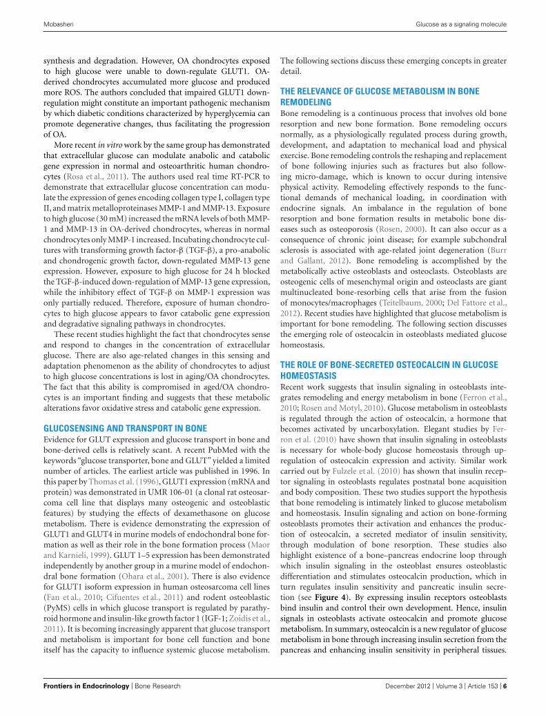
THE ROLE OF BONE-SECRETED OSTEOCALCIN IN GLUCOSEHOMEOSTASISRecent work suggests that insulin signaling in osteoblasts inte-grates remodeling and energy metabolism in bone (Ferron et al.,2010; Rosen and Motyl, 2010). Glucose metabolism in osteoblastsis regulated through the action of osteocalcin, a hormone thatbecomes activated by uncarboxylation. Elegant studies by Fer-ron et al. (2010) have shown that insulin signaling in osteoblastsis necessary for whole-body glucose homeostasis through up-regulation of osteocalcin expression and activity. Similar workcarried out by Fulzele et al. (2010) has shown that insulin recep-tor signaling in osteoblasts regulates postnatal bone acquisitionand body composition. These two studies support the hypothesisthat bone remodeling is intimately linked to glucose metabolismand homeostasis. Insulin signaling and action on bone-formingosteoblasts promotes their activation and enhances the produc-tion of osteocalcin, a secreted mediator of insulin sensitivity,through modulation of bone resorption. These studies alsohighlight existence of a bone–pancreas endocrine loop throughwhich insulin signaling in the osteoblast ensures osteoblasticdifferentiation and stimulates osteocalcin production, which inturn regulates insulin sensitivity and pancreatic insulin secre-tion (see Figure 4). By expressing insulin receptors osteoblastsbind insulin and control their own development. Hence, insulinsignals in osteoblasts activate osteocalcin and promote glucosemetabolism. In summary, osteocalcin is a new regulator of glucosemetabolism in bone through increasing insulin secretion from thepancreas and enhancing insulin sensitivity in peripheral tissues.
Frontiers in Endocrinology | Bone Research December 2012 | Volume 3 | Article 153 | 6
“fendo-03-00153” — 2012/12/15 — 12:18 — page 7 — #7
Mobasheri Glucose as a signaling molecule
FIGURE 4 | Putative mechanism of insulin signaling and osteocalcin
regulation in osteoblasts. Insulin released from pancreatic β cells bindsthe insulin receptor (InsR) on osteoblasts, which increases osteocalcin (OC)synthesis. This feeds back in a forward loop to β cells thus stimulatinginsulin secretion and also regulating glucose homeostasis potentially viaGLUTs expressed in osteoblasts. Adapted from Fulzele and Clemens (2012).
This recent finding reinforces the fact that bone is intimatelyinvolved in regulating glucose and lipid metabolism. Therefore,complex interactions between bone and the pancreas integratebone remodeling and glucose metabolism (Ng, 2011). Insulinand osteocalcin are key molecular links between bone remodelingand energy metabolism. The osteoblast is an important cellulartarget of insulin action and is used to control whole-body glu-cose homeostasis and energy expenditure and bone resorptionis the mechanism regulating osteocalcin activation (Clemens andKarsenty, 2011). Clearly, this is a rapidly expanding field and recentstudies that are beyond the scope of this review suggest that in addi-tion to osteocalcin, other osteoblast-derived hormones such asadiponectin (Bacchetta et al., 2009) may contribute to the emerg-ing function of the skeleton as a regulator of energy metabolism(Yoshikawa et al., 2011).
THE EFFECT OF GLUCOSE ON OSTEOBLASTOGENESIS ANDOSTEOCLASTOGENESISOsteoblastogenesis and osteoclastogenesis refer to the formationof osteoblasts and osteoclasts respectively. These two processesmay be regulated by glucose itself, at the metabolic substratelevel. Hyperglycemia has been implicated in the pathogenesisof diabetic bone disease. To examine the effects of glucose onosteoclastogenesis, Wittrant et al. (2008) studied the effect ofhigh d(+)glucose on RANKL-induced osteoclastogenesis usingRAW264.7 cells and bone marrow macrophages (BMM) as invitro models of bone resorption. The authors showed that highd(+)glucose concentrations inhibit RANKL-induced osteoclas-togenesis. High d(+)glucose inhibits osteoclast formation, ROSproduction, caspase-3 activity, and migration in response to
RANKL through a metabolic pathway. Their findings also suggestthat high d(+)glucose may alter RANKL-induced osteoclast for-mation by inhibiting redox-sensitive NF-kappaB activity throughan anti-oxidative mechanism. This study increases our under-standing of the role of glucose in diabetes-associated bone disease.This data suggest that high glucose levels may alter bone turnoverby decreasing osteoclast differentiation and function in diabetesand provide new insight into the biologic effects of glucose onosteoclastogenesis.
Mature osteoclasts rely on the citric acid cycle and mito-chondrial respiration to generate high levels of ATP productionfor acid secretion and bone resorption. A study by Kim et al.(2007), has reported that glycolysis, oxidative phosphorylation,and lactate production are increased during receptor activator ofnuclear factor-kappaB ligand (RANKL)-induced osteoclastogen-esis in RAW264.7 and bone marrow-derived macrophage cells.This study indicates that glucose metabolism is increased dur-ing osteoclast differentiation resulting in a metabolic shift towardaccelerated glucose metabolism at an early stage of RANKL-stimulated osteoclast differentiation. Increased mitochondrialoxidative phosphorylation will then result in elevated ATP pro-duction and enhanced osteoclast differentiation. Taken together,these studies indicate that there is a link between hyperglycemiaand osteoclastogenesis.
The link between diabetes mellitus and the loss of bone min-eral density and quality has focused research on the effects ofglucose on osteoclastogenesis. Consequently, there has been lessfocus on the regulation of osteoblastogenesis by glucose. Zhen et al.(2010) studied the effects of the oral anti-diabetic drug metforminon rat primary osteoblasts and found that this drug reverses thedeleterious effects of high glucose on osteoblast function. Highconcentrations of glucose reduced cellular proliferation, alka-line phosphatase activity and calcium deposition. In contrasthigh glucose significantly increased ROS and apoptosis in a dose-dependent manner. Metformin reversed the effects of high glucoseby increasing cellular proliferation, alkaline phosphatase activity,and calcium deposition, as well as inhibiting ROS and apoptosis.These findings suggest that glucose is a regulator of osteoblas-togenesis since it affects osteoblast formation and function in adose-dependent manner.
Osteoblastogenesis and osteoclastogenesis contribute to boneremodeling and since both cell types are under the control of mul-tiple hormones, there are also exciting opportunities for studyinghow the processes of bone turnover and remodeling are regu-lated by metabolic hormones such as insulin and leptin. Forexample, bone formation by osteoblasts is negatively regulatedby leptin, which is secreted by adipocytes. Leptin deficiency leadsto increased osteoblast activity and increased bone mass. In con-trast, osteocalcin acts as a regulator of insulin in the pancreasand adiponectin in the adipocyte to modulate energy metabolism.Osteocalcin deficiency in knockout mice leads to decreased insulinand adiponectin secretion, insulin resistance, higher serum glucoselevels, and increased adiposity (Wolf, 2008). These links highlightthe importance of glucose metabolism and insulin action in boneand how insulin signaling in osteoblasts contributes to whole-bodyglucose homeostasis by increasing the expression and activity ofosteocalcin.
www.frontiersin.org December 2012 | Volume 3 | Article 153 | 7
“fendo-03-00153” — 2012/12/15 — 12:18 — page 8 — #8
Mobasheri Glucose as a signaling molecule
CONCLUDING REMARKS AND FUTURE RESEARCHPRIORITIESThe integrity of bone and articular cartilage is maintained viathe finely tuned interaction between the cells in these tissuesand systemic, paracrine and endocrine mediators. Disruption ofhomeostatic processes involved in bone and cartilage turnovercan lead to a variety of diseases including osteoporosis andosteoarthritis. Although various regulatory and signaling sys-tems are known to be involved in maintaining bone and cartilagehealth, our basic understanding of these processes is limited com-pared to other tissues and organs. This review has highlightedthat glucose is a source of energy and a structural precursorfor extracellular matrix production in bone and cartilage. Glu-cose is also an important but neglected signaling molecule inarticular cartilage and bone. Recent studies also point to osteo-calcin as a new regulator of pancreatic insulin production andglucose metabolism. Bone remodeling is now believed to affectenergy metabolism through uncarboxylated osteocalcin secretedby osteoblasts (Ferron et al., 2010; Fulzele et al., 2010; Rosen andMotyl, 2010; Confavreux, 2011). Systemic conditions such as dia-betes and obesity directly influence bone metabolism. Adiposetissue and the adipokines (i.e., leptin) secreted by adipocytesaffect bone mass. Expression of the ESP gene, which encodesa tyrosine phosphatase dephosphorylating the insulin recep-tor, is exclusive to osteoblasts and regulates glucose homeosta-sis and adiposity through controlling the osteoblastic secretionof osteocalcin (Zee et al., 2012). An undercarboxylated formof osteocalcin acts on the pancreas increasing insulin secre-tion, sensitivity, and β-cell proliferation (Schwetz et al., 2012).Interestingly, osteocalcin deficiency in knockout mice leads todecreased insulin and adiponectin secretion, insulin resistance,higher serum glucose levels, and increased adiposity (Wolf, 2008).These studies highlight the importance of bone in the regula-tion of systemic glucose homeostasis. Whether bone has thus farunexplored endocrine roles remains to be determined (Schwetzet al., 2012).
Despite these recent advances, we still know very little aboutglucose-sensing, transport, and metabolism in the cells of artic-ular cartilage and bone compared to other tissues. We also knowvery little about how insulin and GF-1 exert overlapping roles inphysiologic processes in skeletal tissues. Recent studies have iden-tified previously unrecognized skeletal actions of insulin, whichsuggests that insulin-responsive bone cells participate in the regu-lation of global energy homeostasis (Fulzele and Clemens, 2012).Globally, there are increasing numbers of patients with insulinresistance, type 2 diabetes mellitus, and osteoporosis. Obesity,insulin resistance, and diabetes are global health issues and havemajor impact on bone and joint health. Further research is neededto expand our understanding of glucosensing and glucose signal-ing in osteoblasts, osteoclasts, and chondrocytes in health anddisease. We are learning more about glucose metabolism in bonecells but the equivalent information in cartilage is largely lacking.This knowledge is important may reveal new therapeutic targetsand pathways for treating metabolic diseases of articular cartilageand bone. Increasing our knowledge of these fundamental pro-cesses and the links between diabetes and bone diseases is likely toreveal new therapeutic targets for treating bone and joint disorders.
ACKNOWLEDGMENTSI wish to thank Dr. Madura Batuwangala for the illustrations inthis article. I acknowledge the collaboration of the WALTHAMCentre for Pet Nutrition and the financial support of The Well-come Trust, the National Centre for the Replacement, Refinementand Reduction of Animals in Research (NC3Rs; grant number:Mobasheri.A.28102007), the Biotechnology and Biological Sci-ences Research Council (BBSRC; grants BBSRC/S/M/2006/13141and BB/G018030/1), and the Engineering and Physical SciencesResearch Council (EPSRC). The author is the co-ordinator of theD-BOARD Consortium funded by European Commission Frame-work 7 program (EU FP7; HEALTH.2012.2.4.5–2, project number305815, Novel Diagnostics and Biomarkers for Early Identificationof Chronic Inflammatory Joint Diseases).
REFERENCESBacchetta, J., Boutroy, S., Guebre-
Egziabher, F., Juillard, L., Drai, J.,Pelletier, S., et al. (2009). The rela-tionship between adipokines, osteo-calcin and bone quality in chronickidney disease. Nephrol. Dial. Trans-plant. 24, 3120–3125.
Bird, T. A., Davies, A., Baldwin, S.A., and Saklatvala, J. (1990). Inter-leukin 1 stimulates hexose trans-port in fibroblasts by increasing theexpression of glucose transporters. J.Biol. Chem. 265, 13578–13583.
Buckwalter, J. A., Mankin, H. J., andGrodzinsky, A. J. (2005). Articu-lar cartilage and osteoarthritis. Instr.Course Lect. 54, 465–480.
Burr, D. B., and Gallant, M. A. (2012).Bone remodelling in osteoarthritis.Nat. Rev. Rheumatol. 8, 665–673.
Burstein, D. E., Reder, I., Weiser, K.,Tong, T., Pritsker, A., and Haber, R. S.(1998). GLUT1 glucose transporter:
a highly sensitive marker of malig-nancy in body cavity effusions. Mod.Pathol. 11, 392–396.
Charron, M. J., Brosius, F. C., III, Alper,S. L., and Lodish, H. F. (1989). Aglucose transport protein expressedpredominately in insulin-responsivetissues. Proc. Natl. Acad. Sci. U.S.A.86, 2535–2539.
Cifuentes, M., Garcia, M. A., Arra-bal, P. M., Martinez, F., Yanez, M.J., Jara, N., et al. (2011). Insulinregulates GLUT1-mediated glucosetransport in MG-63 human osteosar-coma cells. J. Cell. Physiol. 226, 1425–1432.
Clark, A. G., Rohrbaugh, A. L., Otter-ness, I., and Kraus, V. B. (2002). Theeffects of ascorbic acid on cartilagemetabolism in guinea pig articularcartilage explants. Matrix Biol. 21,175–184.
Clemens, T. L., and Karsenty, G. (2011).The osteoblast: an insulin target cell
controlling glucose homeostasis. J.Bone Miner. Res. 26, 677–680.
Confavreux, C. B. (2011). Bone: froma reservoir of minerals to a regula-tor of energy metabolism. Kidney Int.Suppl. S121, S14–S19.
Corthesy-Theulaz, I., den Dunnen, J. T.,Ferre, P., Geurts, J. M., Muller, M.,van Belzen, N., et al. (2005). Nutrige-nomics: the impact of biomics tech-nology on nutrition research. Ann.Nutr. Metab. 49, 355–365.
Del Fattore, A., Teti, A., and Rucci,N. (2012). Bone cells and the mech-anisms of bone remodelling. Front.Biosci. (Elite Ed.) 4, 2302–2321.
Efrat, S., Tal, M., and Lodish, H. F.(1994). The pancreatic beta-cell glu-cose sensor. Trends Biochem. Sci. 19,535–538.
Fan, J., Zhou, J. Q., Yu, G. R., andLu, D. D. (2010). Glucose trans-porter protein 1-targeted RNA inter-ference inhibits growth and invasion
of the osteosarcoma cell line MG63in vitro. Cancer Biother. Radiopharm.25, 521–527.
Ferron, M., Wei, J., Yoshizawa, T., DelFattore, A., DePinho, R. A., Teti,A., et al. (2010). Insulin signaling inosteoblasts integrates bone remodel-ing and energy metabolism. Cell 142,296–308.
Flier, J. S., Mueckler, M., McCall, A. L.,and Lodish, H. F. (1987a). Distribu-tion of glucose transporter messengerRNA transcripts in tissues of rat andman. J. Clin. Invest. 79, 657–661.
Flier, J. S., Mueckler, M. M., Usher, P.,and Lodish, H. F. (1987b). Elevatedlevels of glucose transport and trans-porter messenger RNA are inducedby ras or src oncogenes. Science 235,1492–1495.
Forsberg, H., and Ljungdahl, P. O.(2001). Sensors of extracellular nutri-ents in Saccharomyces cerevisiae.Curr. Genet. 40, 91–109.
Frontiers in Endocrinology | Bone Research December 2012 | Volume 3 | Article 153 | 8
“fendo-03-00153” — 2012/12/15 — 12:18 — page 9 — #9
Mobasheri Glucose as a signaling molecule
Fujita, Y., Wideman, R. D., Speck,M., Asadi, A., King, D. S., Webber,T. D., et al. (2009). Incretin releasefrom gut is acutely enhanced by sugarbut not by sweeteners in vivo. Am.J. Physiol. Endocrinol. Metab. 296,E473–E479.
Fulzele, K., and Clemens, T. L. (2012).Novel functions for insulin in bone.Bone 50, 452–456.
Fulzele, K., Riddle, R. C., DiGirolamo,D. J., Cao, X., Wan, C., Chen, D., et al.(2010). Insulin receptor signaling inosteoblasts regulates postnatal boneacquisition and body composition.Cell 142, 309–319.
Gerstenfeld, L. C., and Landis, W. J.(1991). Gene expression and extra-cellular matrix ultrastructure of amineralizing chondrocyte cell culturesystem. J. Cell Biol. 112, 501–513.
Gilmore, K. S., Srinivas, P., Akins,D. R., Hatter, K. L., and Gilmore,M. S. (2003). Growth, development,and gene expression in a persis-tent Streptococcus gordonii biofilm.Infect. Immun. 71, 4759–4766.
Goggs, R., Vaughan-Thomas, A., Clegg,P. D., Carter, S. D., Innes, J.F., Mobasheri, A., et al. (2005).Nutraceutical therapies for degener-ative joint diseases: a critical review.Crit. Rev. Food Sci. Nutr. 45, 145–164.
Grayson, M. (2010). Nutrigenomics.Nature 468, S1.
Gribble, F. M., Williams, L., Simp-son, A. K., and Reimann, F. (2003).A novel glucose-sensing mecha-nism contributing to glucagon-likepeptide-1 secretion from the GLUTagcell line. Diabetes 52, 1147–1154.
Guillet-Deniau, I., Leturque, A., andGirard, J. (1994). Expression andcellular localization of glucose trans-porters (GLUT1, GLUT3, GLUT4)during differentiation of myogeniccells isolated from rat foetuses. J. CellSci. 107(Pt 3), 487–496.
Hernvann, A., Aussel, C., Cynober, L.,Moatti, N., and Ekindjian, O. G.(1992). IL-1 beta, a strong media-tor for glucose uptake by rheuma-toid and non-rheumatoid culturedhuman synoviocytes. FEBS Lett. 303,77–80.
Hernvann, A., Jaffray, P., Hilliquin,P., Cazalet, C., Menkes, C. J., andEkindjian, O. G. (1996). Interleukin-1 beta-mediated glucose uptake bychondrocytes. Inhibition by cor-tisol. Osteoarthritis Cartilage 4,139–142.
Hocquette, J. F., and Abe, H. (2000).Facilitative glucose transporters inlivestock species. Reprod. Nutr. Dev.40, 517–533.
James, D. E., Brown, R., Navarro, J., andPilch, P. F. (1988). Insulin-regulatable
tissues express a unique insulin-sensitive glucose transport protein.Nature 333, 183–185.
Jetton, T. L., Liang, Y., Pettepher, C.C., Zimmerman, E. C., Cox, F. G.,Horvath, K., et al. (1994). Analy-sis of upstream glucokinase promoteractivity in transgenic mice and iden-tification of glucokinase in rare neu-roendocrine cells in the brain and gut.J. Biol. Chem. 269, 3641–3654.
Joost, H. G., Bell, G. I., Best, J. D., Birn-baum, M. J., Charron, M. J., Chen, Y.T., et al. (2002). Nomenclature of theGLUT/SLC2A family of sugar/polyoltransport facilitators. Am. J. Physiol.Endocrinol. Metab. 282, E974–E976.
Joost, H. G., and Thorens, B.(2001). The extended GLUT-familyof sugar/polyol transport facilita-tors: nomenclature, sequence char-acteristics, and potential function ofits novel members (review). Mol.Membr. Biol. 18, 247–256.
Kim, J. M., Jeong, D., Kang, H. K.,Jung, S. Y., Kang, S. S., and Min, B.M. (2007). Osteoclast precursors dis-play dynamic metabolic shifts towardaccelerated glucose metabolism atan early stage of RANKL-stimulatedosteoclast differentiation. Cell. Phys-iol. Biochem. 20, 935–946.
Koster, J. C., Permutt, M. A., andNichols, C. G. (2005). Diabetes andinsulin secretion: the ATP-sensitiveK+ channel (K ATP) connection.Diabetes 54, 3065–3072.
Le Gall, M., Tobin, V., Stolarczyk, E.,Dalet, V., Leturque, A., and Brot-Laroche, E. (2007). Sugar sensing byenterocytes combines polarity, mem-brane bound detectors and sugarmetabolism. J. Cell. Physiol. 213,834–843.
Lejay, L., Gansel, X., Cerezo, M., Tillard,P., Muller, C., Krapp, A., et al. (2003).Regulation of root ion transportersby photosynthesis: functional impor-tance and relation with hexokinase.Plant Cell 15, 2218–2232.
Mace, O. J., Affleck, J., Patel, N., and Kel-lett, G. L. (2007). Sweet taste recep-tors in rat small intestine stimulateglucose absorption through apicalGLUT2. J. Physiol. 582, 379–392.
Maekawa, F., Toyoda, Y., Torii, N.,Miwa, I., Thompson, R. C., Fos-ter, D. L., et al. (2000). Localizationof glucokinase-like immunoreactiv-ity in the rat lower brain stem: forpossible location of brain glucose-sensing mechanisms. Endocrinology141, 375–384.
Maor, G., and Karnieli, E. (1999).The insulin-sensitive glucose trans-porter (GLUT4) is involved in earlybone growth in control and diabeticmice, but is regulated through the
insulin-like growth factor I receptor.Endocrinology 140, 1841–1851.
Margolskee, R. F., Dyer, J., Kokrashvili,Z., Salmon, K. S., Ilegems, E., Daly,K., et al. (2007). T1R3 and gustducinin gut sense sugars to regulate expres-sion of Na+-glucose cotransporter 1.Proc. Natl. Acad. Sci. U.S.A. 104,15075–15080.
Martens, G., Cai, Y., Hinke, S.,Stange, G., Van de Casteele, M., andPipeleers, D. (2005). Nutrient sens-ing in pancreatic beta cells suppressesmitochondrial superoxide generationand its contribution to apoptosis.Biochem. Soc. Trans. 33, 300–301.
Matschinsky, F. M. (1996). Banting Lec-ture 1995. A lesson in metabolic reg-ulation inspired by the glucokinaseglucose sensor paradigm. Diabetes 45,223–241.
Matschinsky, F. M., and Ellerman, J. E.(1968). Metabolism of glucose in theislets of Langerhans. J. Biol. Chem.243, 2730–2736.
McAlindon, T. E. (2006). Nutraceuti-cals: do they work and when shouldwe use them? Best Pract. Res. Clin.Rheumatol. 20, 99–115.
McNulty, A. L., Vail, T. P., and Kraus,V. B. (2005). Chondrocyte trans-port and concentration of ascorbicacid is mediated by SVCT2. Biochim.Biophys. Acta 1712, 212–221.
Mellanen, P., Minn, H., Grenman, R.,and Harkonen, P. (1994). Expres-sion of glucose transporters in head-and-neck tumors. Int. J. Cancer 56,622–629.
Mobasheri, A., Bondy, C. A., Moley, K.,Mendes, A. F., Rosa, S. C., Richard-son, S. M., et al. (2008). Facilita-tive glucose transporters in articularchondrocytes. Expression, distribu-tion and functional regulation ofGLUT isoforms by hypoxia, hypoxiamimetics, growth factors and pro-inflammatory cytokines. Adv. Anat.Embryol. Cell Biol. 200, 1 p followingvi, 1–84.
Mobasheri, A., Carter, S. D., Martin-Vasallo, P., and Shakibaei, M. (2002a).Integrins and stretch activated ionchannels; putative components offunctional cell surface mechanore-ceptors in articular chondrocytes.Cell Biol. Int. 26, 1–18.
Mobasheri, A., Neama, G., Bell, S.,Richardson, S., and Carter, S. D.(2002b). Human articular chondro-cytes express three facilitative glu-cose transporter isoforms: GLUT1,GLUT3 and GLUT9. Cell Biol. Int. 26,297–300.
Mobasheri, A., Vannucci, S. J., Bondy,C. A., Carter, S. D., Innes, J. F.,Arteaga, M. F., et al. (2002c). Glu-cose transport and metabolism in
chondrocytes: a key to understand-ing chondrogenesis, skeletal devel-opment and cartilage degradation inosteoarthritis. Histol. Histopathol. 17,1239–1267.
Mobasheri, A., Dobson, H., Mason, S.L., Cullingham, F., Shakibaei, M.,Moley, J. F., et al. (2005). Expressionof the GLUT1 and GLUT9 facilitativeglucose transporters in embryonicchondroblasts and mature chondro-cytes in ovine articular cartilage. CellBiol. Int. 29, 249–260.
Mobasheri, A., Platt, N., Thorpe, C., andShakibaei, M. (2006a). Regulation of2-deoxy-D-glucose transport, lactatemetabolism, and MMP-2 secretion bythe hypoxia mimetic cobalt chloridein articular chondrocytes. Ann. N. Y.Acad. Sci. 1091, 83–93.
Mobasheri, A., Shakibaei, M., Mobash-eri, R., Richardson, S. M., andHoyland, J. A. (2006b). Glucosesensing in chondrocytes via GLUT1and GLUT3: implications for artic-ular cartilage and intervertebral discmetabolism. Curr. Rheumatol. Rev. 2,109–121.
Moore, B., Zhou, L., Rolland, F., Hall,Q., Cheng, W. H., Liu, Y. X., et al.(2003). Role of the Arabidopsis glu-cose sensor HXK1 in nutrient, light,and hormonal signaling. Science 300,332–336.
Mueckler, M., Caruso, C., Baldwin, S.A., Panico, M., Blench, I., Morris, H.R., et al. (1985). Sequence and struc-ture of a human glucose transporter.Science 229, 941–945.
Nagamatsu, S., Sawa, H., Wakizaka,A., and Hoshino, T. (1993). Expres-sion of facilitative glucose transporterisoforms in human brain tumors. J.Neurochem. 61, 2048–2053.
Nemoto, S., Fergusson, M. M., andFinkel, T. (2004). Nutrient avail-ability regulates SIRT1 through aforkhead-dependent pathway. Sci-ence 306, 2105–2108.
Ng, K. W. (2011). Regulation of glucosemetabolism and the skeleton. Clin.Endocrinol. (Oxf.) 75, 147–155.
Ohara, H., Tamayama, T., Maemura,K., Kanbara, K., Hayasaki, H.,Abe, M., et al. (2001). Immunocy-tochemical demonstration of glucosetransporters in epiphyseal growthplate chondrocytes of young ratsin correlation with autoradiographicdistribution of 2-deoxyglucose inchondrocytes of mice. Acta His-tochem. 103, 365–378.
Olsson, T., Thelander, M., and Ronne,H. (2003). A novel type of chloro-plast stromal hexokinase is the majorglucose-phosphorylating enzyme inthe moss Physcomitrella patens. J.Biol. Chem. 278, 44439–44447.
www.frontiersin.org December 2012 | Volume 3 | Article 153 | 9
“fendo-03-00153” — 2012/12/15 — 12:18 — page 10 — #10
Mobasheri Glucose as a signaling molecule
Otte, P. (1991). Basic cell metabolismof articular cartilage. Manometricstudies. Z. Rheumatol. 50, 304–312.
Peansukmanee, S., Vaughan-Thomas,A., Carter, S. D., Clegg, P. D., Taylor,S., Redmond, C., et al. (2009). Effectsof hypoxia on glucose transport inprimary equine chondrocytes in vitroand evidence of reduced GLUT1 geneexpression in pathologic cartilage invivo. J. Orthop. Res. 27, 529–535.
Phillips, T., Ferraz, I., Bell, S., Clegg, P.D., Carter, S. D., and Mobasheri, A.(2005). Differential regulation of theGLUT1 and GLUT3 glucose trans-porters by growth factors and pro-inflammatory cytokines in equinearticular chondrocytes. Vet. J. 169,216–222.
Reed, D. R., and Margolskee, R. F.(2010). Gustation genetics: sweetgustducin! Chem. Senses 35, 549–550.
Ren, X., Zhou, L., Terwilliger, R., New-ton, S. S., and de Araujo, I. E.(2009). Sweet taste signaling func-tions as a hypothalamic glucose sen-sor. Front. Integr. Neurosci. 3:12. doi:10.3389/neuro.07.012.2009
Richardson, S., Neama, G., Phillips,T., Bell, S., Carter, S. D., Moley,K. H., et al. (2003). Molecularcharacterization and partial cDNAcloning of facilitative glucose trans-porters expressed in human artic-ular chondrocytes; stimulation of2-deoxyglucose uptake by IGF-I andelevated MMP-2 secretion by glucosedeprivation. Osteoarthritis Cartilage11, 92–101.
Richardson, S. M., Knowles, R., Tyler,J., Mobasheri, A., and Hoyland,J. A. (2008). Expression of glu-cose transporters GLUT-1, GLUT-3,GLUT-9 and HIF-1alpha in normaland degenerate human interverte-bral disc. Histochem. Cell Biol. 129,503–511.
Rolland, F., Moore, B., and Sheen,J. (2002). Sugar sensing and signal-ing in plants. Plant Cell 14(Suppl.),S185–S205.
Rolland, F., and Sheen, J. (2005). Sugarsensing and signalling networks inplants. Biochem. Soc. Trans. 33,269–271.
Rolland, F., Winderickx, J., andThevelein, J. M. (2001). Glucose-sensing mechanisms in eukaryoticcells. Trends Biochem. Sci. 26,310–317.
Rosa, S. C., Goncalves, J., Judas,F., Mobasheri, A., Lopes, C., andMendes, A. F. (2009). Impairedglucose transporter-1 degradationand increased glucose transportand oxidative stress in response tohigh glucose in chondrocytes fromosteoarthritic versus normal humancartilage. Arthritis Res. Ther. 11, R80.
Rosa, S. C., Rufino, A. T., Judas, F. M.,Tenreiro, C. M., Lopes, M. C., andMendes, A. F. (2011). Role of glu-cose as a modulator of anabolic andcatabolic gene expression in normaland osteoarthritic human chondro-cytes. J. Cell. Biochem. 112, 2813–2824.
Rosen, C. J. (2000). Pathogenesis ofosteoporosis. Baillieres Best. Pract.Res. Clin. Endocrinol. Metab. 14,181–193.
Rosen, C. J., and Motyl, K. J. (2010).No bones about it: insulin modu-lates skeletal remodeling. Cell 142,198–200.
Rosenbloom, A. L., and Silverstein, J.H. (1996). Connective tissue andjoint disease in diabetes mellitus.Endocrinol. Metab. Clin. North Am.25, 473–483.
Saier, M. H. Jr., Beatty, J. T., Goffeau, A.,Harley, K. T., Heijne, W. H., Huang,S. C., et al. (1999). The major facil-itator superfamily. J. Mol. Microbiol.Biotechnol. 1, 257–279.
Santangelo, G. M. (2006). Glucose sig-naling in Saccharomyces cerevisiae.Microbiol. Mol. Biol. Rev. 70,253–282.
Schwartz, E. R., and Adamy, L. (1977).Effect of ascorbic acid on arylsul-fatase activities and sulfated proteo-glycan metabolism in chondrocytecultures. J. Clin. Invest. 60, 96–106.
Schwartz, E. R., Oh, W. H., andLeveille, C. R. (1981). Experimen-tally induced osteoarthritis in guineapigs: metabolic responses in articu-lar cartilage to developing pathology.Arthritis Rheum. 24, 1345–1355.
Schwetz, V., Pieber, T., and Obermayer-Pietsch, B. (2012). The endocrine roleof the skeleton: background and clin-ical evidence. Eur. J. Endocrinol. 166,959–967.
Semenza, G. L., Artemov, D., Bedi, A.,Bhujwalla, Z., Chiles, K., Feldser,D., et al. (2001). ‘The metabolismof tumours’: 70 years later. NovartisFound. Symp. 240, 251–260; discus-sion 260–264.
Shepherd, P. R., Gould, G. W., Colville,C. A., McCoid, S. C., Gibbs, E. M., andKahn, B. B. (1992). Distribution ofGLUT3 glucose transporter proteinin human tissues. Biochem. Biophys.Res. Commun. 188, 149–154.
Shirazi-Beechey, S. P. (2005). “A mat-ter of taste. Nutrient sensing throughthe plasma membrane of eukary-otic cells,” in A Biochemical Soci-ety Focused Meeting, held at theRoyal Agricultural College, Cirences-ter, 25–29 September 2004, Bio-chemist, 35–38.
Tal, M., Schneider, D. L., Thorens, B.,and Lodish, H. F. (1990). Restrictedexpression of the erythroid/brain
glucose transporter isoform toperivenous hepatocytes in rats. Mod-ulation by glucose. J. Clin. Invest. 86,986–992.
Teitelbaum, S. L. (2000). Bone resorp-tion by osteoclasts. Science 289, 1504–1508.
Thevelein, J. M., and de Winde, J. H.(1999). Novel sensing mechanismsand targets for the cAMP-proteinkinase A pathway in the yeast Saccha-romyces cerevisiae. Mol. Microbiol. 33,904–918.
Thomas, D. M., Rogers, S. D., Ng, K.W., and Best, J. D. (1996). Dexam-ethasone modulates insulin receptorexpression and subcellular distribu-tion of the glucose transporter GLUT1 in UMR 106-01, a clonal osteogenicsarcoma cell line. J. Mol. Endocrinol.17, 7–17.
Thorens, B. (1996). Glucose trans-porters in the regulation of intestinal,renal, and liver glucose fluxes. Am. J.Physiol. 270, G541–G553.
Thorens, B., Cheng, Z. Q., Brown, D.,and Lodish, H. F. (1990). Liver glu-cose transporter: a basolateral pro-tein in hepatocytes and intestine andkidney cells. Am. J. Physiol. 259,C279–C285.
Tolhurst, G., Reimann, F., and Grib-ble, F. M. (2012). Intestinal sensingof nutrients. Handb. Exp. Pharmacol.209, 309–335.
Uldry, M., and Thorens, B. (2004).The SLC2 family of facilitated hex-ose and polyol transporters. PflugersArch. 447, 480–489.
van Ommen, B. (2004). Nutrigenomics:exploiting systems biology in thenutrition and health arenas. Nutri-tion 20, 4–8.
van Ommen, B., and Stierum, R. (2002).Nutrigenomics: exploiting systemsbiology in the nutrition and healtharena. Curr. Opin. Biotechnol. 13,517–521.
Waeber, G., Pedrazzini, T., Bonny, O.,Bonny, C., Steinmann, M., Nicod, P.,et al. (1995). A 338-bp proximal frag-ment of the glucose transporter type2 (GLUT2) promoter drives reportergene expression in the pancreaticislets of transgenic mice. Mol. Cell.Endocrinol. 114, 205–215.
Wittrant, Y., Gorin, Y., Woodruff, K.,Horn, D., Abboud, H. E., Mohan, S.,et al. (2008). High d(+)glucose con-centration inhibits RANKL-inducedosteoclastogenesis. Bone 42, 1122–1130.
Wolf, G. (2008). Energy regulation bythe skeleton. Nutr. Rev. 66, 229–233.
Wood, I. S., and Trayhurn, P. (2003).Glucose transporters (GLUT andSGLT): expanded families of sugartransport proteins. Br. J. Nutr. 89,3–9.
Wu, X., and Freeze, H. H. (2002).GLUT14, a duplicon of GLUT3, isspecifically expressed in testis as alter-native splice forms. Genomics 80,553–557.
Yoshikawa, Y., Kode, A., Xu, L.,Mosialou, I., Silva, B. C., Ferron,M., et al. (2011). Genetic evidencepoints to an osteocalcin-independentinfluence of osteoblasts on energymetabolism. J. Bone Miner. Res. 26,2012–2025.
Younes, M., Lechago, L. V., Somoano,J. R., Mosharaf, M., and Lechago,J. (1996). Wide expression of thehuman erythrocyte glucose trans-porter Glut1 in human cancers. Can-cer Res. 56, 1164–1167.
Zee, T., Settembre, C., Levine, R. L., andKarsenty, G. (2012). T-cell proteintyrosine phosphatase regulates boneresorption and whole-body insulinsensitivity through its expression inosteoblasts. Mol. Cell. Biol. 32, 1080–1088.
Zhen, D., Chen, Y., and Tang, X. (2010).Metformin reverses the deleteriouseffects of high glucose on osteoblastfunction. J. Diabetes Complications24, 334–344.
Zoidis, E., Ghirlanda-Keller, C., andSchmid, C. (2011). Stimulation ofglucose transport in osteoblastic cellsby parathyroid hormone and insulin-like growth factor I. Mol. Cell.Biochem. 348, 33–42.
Conflict of Interest Statement: Theauthor declares that this review arti-cle was prepared in the absence of anycommercial or financial relationshipsthat could be construed as a potentialconflict of interest.
Received: 07 May 2012; accepted: 19November 2012; published online: 17December 2012.Citation: Mobasheri A (2012) Glucose:an energy currency and structural pre-cursor in articular cartilage and bonewith emerging roles as an extracellularsignaling molecule and metabolic reg-ulator. Front. Endocrin. 3:153. doi:10.3389/fendo.2012.00153This article was submitted to Frontiers inBone Research, a specialty of Frontiers inEndocrinology.Copyright © 2012 Mobasheri. This is anopen-access article distributed under theterms of the Creative Commons Attribu-tion License, which permits use, distri-bution and reproduction in other forums,provided the original authors and sourceare credited and subject to any copy-right notices concerning any third-partygraphics etc.
Frontiers in Endocrinology | Bone Research December 2012 | Volume 3 | Article 153 | 10