Embed Size (px)
Citation preview
Lepr. Rev. ( I 9 7 5 ) 46 , 29- 3 7
G l o m e ru l o n e p h ri t i s i n Lep rosyA P e rc u ta n e o u s R e n a l B iopsy S t u d y
K. v. JOHNY , * A_ B . A . KA RAT, P. s . s . RAO and
A. DATE
S chieffelin L eprosy Research Sanatorium, Karigiri and
Christian Medicai Col/ege Hospital. Vel/ore, South [ndia
Thirty-five patients with lepromatous or borderline leprosy selected at random were investigated for evidence of renal disease . Renal functional impairment was detected in nearly two-thirds of the patients and histological lesions were present in 46%. Twenty-three per cent of the cases showed a prolifera tive type of glomerulonephritis, mesangial sclerosis without significant hy percellularity was seen in I I %, amyloidosis was p resent in 6%. One patient had interstitial nephritis .
Introduction
Renal involvement in leprosy has been recognized by Japanese workers since the beginning of the century. Mitsuda and Ogawa published their observations in English in 1 93 7 and described "nephritis of all kinds" in leprosy in an autopsy analysis of I S O cases. Similar observations were also made by Kean and Childress ( 1 942) from the Isthmus of Panama. Acute oedema of the hands and feet occasionally associated with proteinuria were later reported to complicate all clinicai types of leprosy during reactive episodes and progressive reaction (Davison , 1 96 1 ; Wheate, 1 962 ; Cochrane, 1 964). Im paired renal function and abnormal urinary sediment have also been observed in patients with leprosy in varying reactional states (Gokhale and Kurkure, 1 95 8 ; Thomas et ai. , 1 97 0 ; Gutman , L u and Durtz, 1 97 3 ).
Amyloidosis of the kidney was the commonest histological abn ormality observed by North American workers, occurring in nearly one-half of the patients with lepromatous leprosy (Powell and Swan, 1 95 5 ; Shuttleworth and Ross, 1 95 6 ; Williams, Cathcart and Calkins, 1 965) . Amyloidosis complicated leprosy i n as many as 80% of cases in Spain (Granels, 1 968)_ In contrast , renal amyloidosis was seen in only 6% of leprosy patients s tudied in Mexico (Williams, Cathcart and Calkins, 1 96 5 ) and a similar low incidence was reported from Japan (Mitsuda and Ogawa, 1 93 7 ) and India (Junnarkar, 1 95 7 ; Desikan and Job, 1 968 ; Sachdev, Puri and Bansal, 1 969). Though amyloidosis complicated reactional leprosy much
• Reprint requests should be addressed to Dr K_ J_ J ohny, Associate Professor and Head, Department of Nephrology , Christian Medicai College Hospital, Vellore-632004, Tamil Nadu, South India .
. Received for publication, 22 July, 1 974.
30 K. V. JOH N Y , A. B. A. K A R AT, P. S. S. R AO A N D A. D A TE
more frequen tly , it was seen in non-reactional types as well (Brusco and Masan ti , 1 963 ).
Renal fai lure secondary to amyloidosis was the leading cause of death in U . S . Sanatoria (Powell a n d Swan, 1 9 5 5 ; Shuttleworth a n d Ross, 1 9 5 6 ; Will iams, Cathcart and Calkins, 1 96 5 ) and even more so in Spain (Granels, 1 968) . In con tras t, in only 4 out of 3 7 cases of leprosy ( 1 0 . 8%) studied at post mortem in South India was death attributable to renal fai lure ( Desikan and Job, 1 968) . Junnarkar fai led to demonstrate any remarkable renal lesion in 20 cases of leprom atous and tuberculoid leprosy at post mortem except for one instan ce of amyloidosis ( Junnarkar, 1 9 5 7 ) .
There have been only a few renal biopsy studies i n patients with leprosy. Durtz and Gutm an reported 2 cases of proli ferative glomerulonephri tis in 8 biopsied cases of leprom atous leprosy with erythema nodosum leprosum from Taiwan ( Durtz and Gutman, 1 972 , 1 9 73) . Another case wi th proli fera tive glomerulonephri tis has been reported by Shwe ( 1 972) . The wide discrepancy in the nature of renal lesions seen in different countries prompted this enquiry into the nature of renal disease in leprosy in I ndia .
Materiais and Methods
Patients admitted to the Schieffelin Leprosy Research Sanatorium with d ifferent cl inicaI types of leprosy with or without ery thema nodosum leprosum and in varying stages of therapy were selected at random. The cases were divided into lepromatous and borderline groups according to the system of Ridley and Jopling, using clinicai, histological and immunological cri teria ( Ridley and Jopling, 1 966) . The bacterial load was estimated after standard skin smears from 8 si tes and was expressed as the bacterial index ( B I ) according to Ridley's scale ( Ridley, 1 964) . Only patien ts with erythema nodosum leprosum were classi fied as having reaction . A 24 h u rine sample was examined for acid fast bacilli .
I m munological studies included estimation of antistreptolysin O (ASO) ti tre (Rantz and Randall , 1 94 5 ) , antinuclear factor, rheumatoid factor and examination of lupus ery thematosus ( LE) phenomenon by the method of M agath and Winkle ( 1 9 5 2) .
R E N A L H ISTOLOGY
Percutaneous renal biopsy was performed with a Franklin m odi fied VimSilvermann needle following the method of Kark and Muechrcke ( 1 954) . Sections from ali specimens were stained with hematoxyiin eosin, periodic acid Schi ff (PAS) , congo red and Ziehl-Neelsen stains and e xamined with the light m icroscope . Sections were independently interpreted by two of us (KVJ and AD). The h istological lesions were graded as follows:
( 1 ) Proliferative. (PGN) Diffuse prolifera tive : showing diffuse hypercellularity of endothelial and mesangial cells with or without polymorphonuclear exudation in the glomerular tuft.
Mesangial prolifera tive : Hypercellularity of varying degree confmed to the axial region .
( 1 1 ) Chronic sc/erosing. Showing glomerular hyalinization , focal tuft adhesions and interstitial scarring.
( I I I ) Mesangial sc/erosis. Glomeruli showing increase in PAS positive material in the axial region without significant hypercellularity.
GLOM ERULONEPH RITIS IN LEPROSY 3 1
( I V ) A myloidosis. Was confirmed by the presence of congo red posi tive material in the renal tissue .
(V ) In ters titial nephritis. With predominant involvement of intersti t ium and tubules consist ing of d i ffuse in tersti tial monon uclear cell i nfi l tration , in terst it ial fibrosis and tub ular atrophy .
R EN A L FU NCTlONAL ASSESSMENT
I nvestigations included routine urine analysis for albumin and sediments, 24 h urine protein excretion (Thomas et al. , 1 970) , endogenous creatinine c1earance ( Bonsnes and Taussky, 1 945 ) and blood urea estimation (Varley, 1 96 7 ) . Addit ional biochemical tests measured serum sodium, phosphorus , potassi um , calcium , sugar, alkaline phosphatase and congo red re tention (Varley, 1 96 7 ) . Serum protein was determined using cellulose acetate strips and Shandon universal electrophoret ic apparatus (K ingsley, 1 942 ; Smith , 1 960) .
ResuI ts
Adequate renal biopsies were obtained in the 35 patients analysed for this report. Thirty-four were males. The mean age of the group was 3 1 . 7 years and ranged from 1 8 to 57 years. Twen ty-nine patien ts had lepromatous leprosy, 2 borderl ine lepromatous, 1 bord erline borderl ine and 3 borderl ine tuberculoid. Mean d uration of disease at the t ime of study based on the history was 9.8 years and ranged from 3 mon ths to 25 years.
Those wi th lepromatous leprosy had a more prolonged illness (mean 1 0 .3 years) than those wi th borderline leprosy (7 . 1 years). Twenty-four patien ts had past history of ery thema nodosum leprosum of which 4 had E N L at the t ime of study . In the rem aining 20 the last reaction had occurred within one week to 1 3 months prior to the study (mean 5 months). Five patients had received no form of an ti-Ieprosy therapy at the t ime of study . Twenty-eight patients had been on therapy with single or multi pie d rugs for varying periods, the commonest drug employed being dapsone . The dura tion of treatment varied from a few months to over 1 0 years . The BI in the group ranged from O to 4 . 25 wi th a mean of 2 . 5 3 . Acid fast bacilli were n ot present in the urine of a n y o f the patients.
No definite s tatistical correlation could be established between any of the factors m entioned above and the presence of abnormal renal histology.
REN A L HISTOLOGY
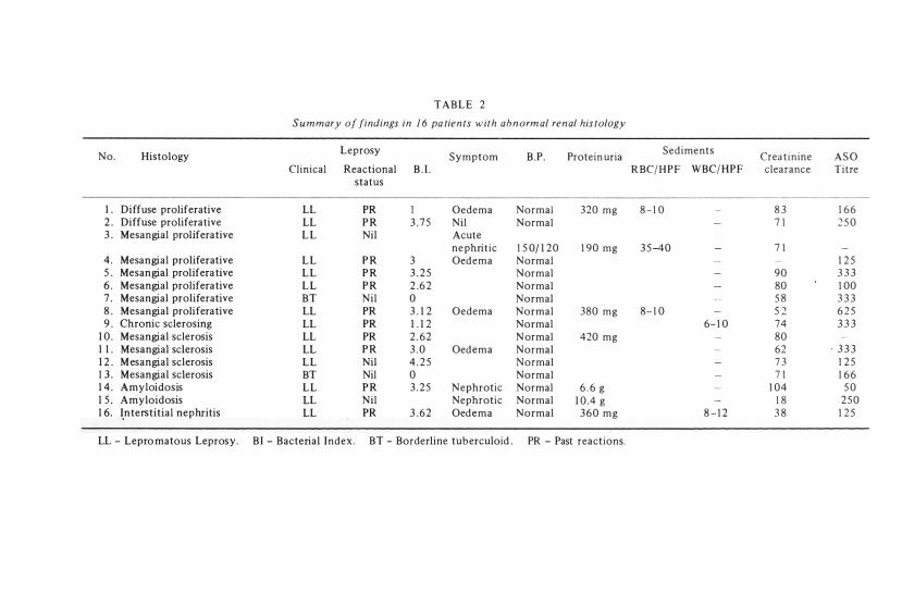
Table 1 summarizes the histological findings in the 35 patients studied . Abnormal histology was seen in 1 6 cases (45%). Hypercellu lari ty of the endothelial and mesangial cells w as the most frequent histological abnormality .
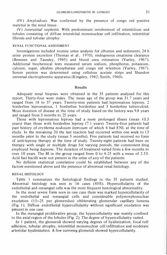
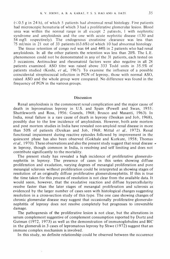
In the most severe form seen in one case there was marked hypercellularity of the endothelial and mesangial cel ls and considerable polymorphonuclear exudation ( 1 5-25 per glomerulus) obliterating glomerular capillary lumena ( Fig. I ) . Diffuse endothelial hypercellularity without significant exudation was present in one case .
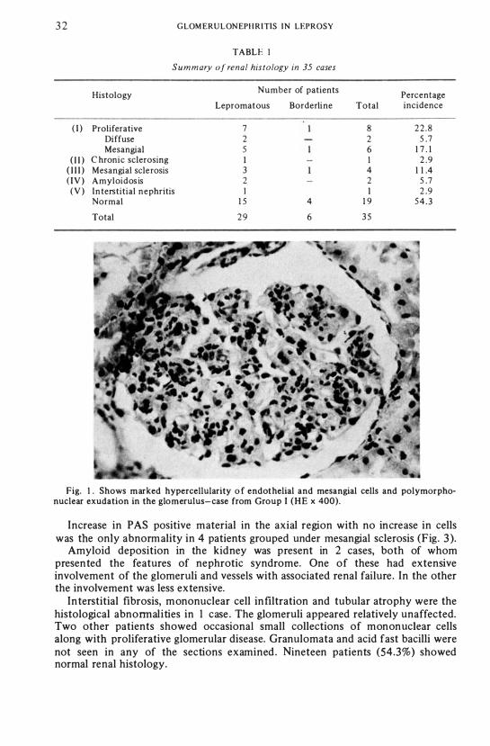
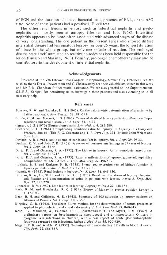
I n the mesangial proliferative group, the hypercellularity was mainly confined to the axial region of the lobules ( Fig. 2 ). The degree of hypercellularity varied.
In I patient , the glomeruli showed varying degrees of hyalinizat ion, focal tuft adhesion, tubular atrophy, interstitial mononuclear cell i nfiltration and moderate arteri olar hyalinization . A few surviving glomeruli showed hypercellularity.
3 2 GLOM ERULON EPH R I TlS I N LEPROSY
TABLE I
S u m m ary of renal histology in 35 cases
Histology Number of patients
Percentage Lepromatous Borderline Total incidence
( I ) Prolifera tive 7 8 2 2 . 8 Diffuse 2 2 5 .7 Mesangial 5 6 1 7 . 1
( 1 1 ) C h ronic sclerosing I I 2 .9 ( 1 1 1 ) Mesangial sclerosis 3 4 1 1 .4 ( I V ) Amyloidos is 2 2 5 .7 ( V ) In terstitial nephritis I I 2 .9
Normal 1 5 4 1 9 5 4 . 3
Total 2 9 6 3 5
Fig. I . Shows marked hypercellularity o f endothelial and mesangial cells and polymorphonuclear exudation in the glomerulus-case from Group I (HE x 400).
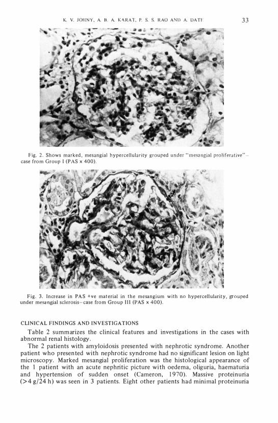
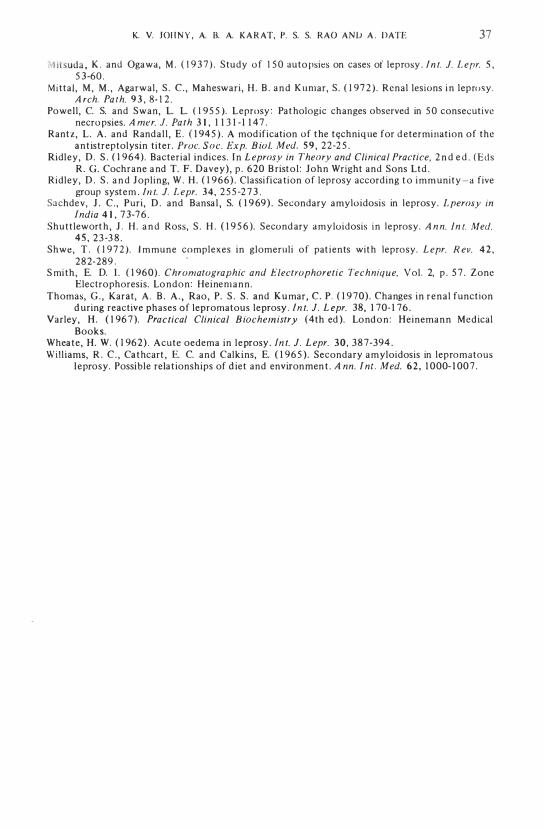
Increase in PAS positive material in the axial region with no increase in cells was the only abnormality in 4 patients grouped under mesangial sclerosis (Fig. 3 ).
Amyloid deposition in the kidney was present in 2 cases, both of whom presented the features of nephrotic syndrome. One of these had extensive involvement of the glomeruli and vessels with associated renal failure. In the other the involvement was less extensive.
Interstitial fibrosis, mononuclear cell infiltration and tubular atrophy were the histological abnormalities in I case . The glomeruli appeared relatively unaffected. Two other patients showed occasional small collections of mononuclear cells along with proliferative glomerular disease. Granulomata and acid fast bacilli were not seen in any of the sections examined . Nineteen patients (54.3%) showed normal renal histology.
K. V. JOH N Y . A. B . A. K A R A T. P. S. S. R AO A N D A. DATE 3 3
Fig. 2 . Shows marked . mesangial hypercellularity grouped under " m esa ngial prol ifera tive" -case fro m Group I (PAS x 400) .
Fig . 3 . Increase in PA S +ve mat erial in the mesangium with no hypercellularity . grouped under mesa ngial sclerosis- case ÍTom Group I I I (PAS x 400) .
CLINICAL FINOINGS ANO INVESTIGATIONS
Table 2 summ arizes the clinical features and investigations in the cases with abnormal renal histology .
The 2 patients with amyloidosis presented with nephrotic syndrome. Another patient who presented with nephrotic syndrome had no significant lesion on light microscopy. Marked mesangial proliferation was the histological appearance of the 1 patient with an acute nephritic picture with oedema, oliguria, haematuria and hypertension of sudden onset (Cameron, 1 970) . Massive proteinuria (> 4 g/24 h ) was seen in 3 patients. Eight other patients had minimal proteinuria
T A B L E 2
S u m mary of findings in 1 6 pa tients with abnorm al renal his tology
No. Histology Leprosy
Symptom B.P. Protein uria Sediments
Crea tinine A S O Clinicai Reactional B . 1 . R BC/ H P F W BC/ HP F clearance Titre
status
I . Diffuse proliferative LL PR I Oedema Normal 320 mg 8 - 1 0 8 3 1 6 6 2 . Diffuse prolifera tive LL P R 3 . 7 5 Nil Normal 7 1 2 5 0 3 . Mesangial prolifera tive L L Nil Acute
ne phritic 1 5 0/ 1 2 0 1 90 mg 3 5 -40 7 1 4 . Mesangial proliferative L L P R 3 Oed ema Normal 1 2 5 5 . Mesangial prolifera tive L L P R 3 . 2 5 N ormal 90 3 3 3 6 . Mesangial prolifera tive L L P R 2 .6 2 Normal 80 1 00 7 . Mesangial proliferative BT Nil O Normal 5 8 3 3 3 8 . Mesangial prolifera tive LL PR 3 . 1 2 Oedema Normal 380 mg 8- 1 0 5 2 6 2 5 9 . Chronic sclerosing LL PR 1 . 1 2 Normal 6- 1 0 74 3 3 3
1 0 . Mesangial sclerosis LL PR 2 . 62 N ormal 420 mg 80 1 1 . Mesangial sclerosis LL P R 3 . 0 Oed ema Normal 62 . 3 3 3 1 2 . M esangial sclerosis L L Nil 4 . 2 5 Normal 7 3 1 2 5 1 3 . Mesangial scJerosis BT Nil O Normal 7 1 1 6 6 1 4 . Amyloidosis L L P R 3 . 2 5 Nephrotic Normal 6 . 6 g 1 04 5 0 1 5 . A myloidosis LL Nil Nephrotic Normal 1 0 .4 g 1 8 2 5 0 1 6. �n terstitial nephritis LL PR 3 . 62 Oed e m a Normal 360 m g 8 - 1 2 3 8 1 2 5
LL - Lepro matous Leprosy. B I - Bacterial Index . BT - Borderline tub erculoid . PR - Past reactions.
K. V . J OH N Y , A. B. A. K A R AT, P. S . S. R AO A N D A . DATE 3 5
« 0 . 5 g i n 2 4 h ) , o f wh ich 5 pa t ien ts had abnorm al renal histo logy . Five pat i ents had microscopic hematuria of which 3 had a pro l i ferative glomerular lesion . Blood urea was wi thi n the normal range in ali e xcept 2 pat ien ts , I with nephrot ic syndrome and amyloidosis and the one wi th acute neph ri t ic disease ( 1 30 and 54 mg% respective ly) . The endogenous creat in ine c1earance was less than 75 ml/min i n 2 1 out of 33 patients (63 .6%) of which 1 0 had abn ormal histology .
The tissue re ten t ion of congo red was 64 and 44% i n 2 patien ts who had ren al amyloidosis. In ali the other patients the re te nt ion was less than 20%. The L . E . phenomenon could n o t be demonstrated in any o f the 3 1 pa t ients , each tested on 3 occasions. An t inuc\ear and rheumatoid fac tors were also nega tive in ali 28 patients e xam ine d . ASa t i tre was raised above 333 Todd un i ts in 3 5 . 5% of pat ients studied ( Koshi , e t aL, 1 96 7 ) . To examine the i n fl u ence of a possible coinciden tal streptococcal i n fect ion i n PGN of le prosy , those wi th normal ASa, raised Asa and the whole group were compare d . No d i fference was found in the freq uency of PGN in the various groups .
Discussion
Renal amyloidosis i s the commonest renal complicat ion and the major cause of death in lepromatous leprosy i n U . S . and Spain (Powell and Swan, 1 9 5 5 ; Shutt leworth and Ross , 1 95 6 ; Granels, 1 968 ; B rusco and Masant i , 1 963) . I n I ndia , renal fai lure is a rare cause o f death in leprosy ( Desikan and J o b , 1 968) , possibly due to the low incidence of am yloidosis. However, both ante mortem and post mortem studies in I nd ia have revealed non-amyloid renal d isease i n more than 5 0% of patien ts (Desikan and J ob , 1 968 ; M i ttal e t ai. , 1 9 72) . Renal functional impa i rment during reactive episodes followed by improvement i n the quiescent phase has also been observed (Gokhale and Kurk ure , 1 95 8 ; Thomas et ai. , 1 970) . These observations and also the presen t study suggest that renal disease ." in leprosy, though common in India , i s resolving and self l imit ing and does not contribute sign ificantly to the mortali ty .
The present s tudy has revealed a high incidence of proliferative glomerulonephritis in leprosy. The presence of cases i n this series showi ng d iffuse proliferation and exudat ion , varying degrees of mesangial proliferat ion and pure mesangial sclerosis wi thout proliferation could be in terpreted as showing stages of resolution of an originally d i ffuse prol iferative glomerulonephri tis. I f this is true the time taken for this process of resolution is not c\ear from the available data. l t would seem, however, that the exudative reaction and d i ffuse hy percellu larity resolve faster than the later stages of mesangial prol i ferat ion and sclerosis as evidenced by the larger number of cases seen wi th histological changes suggesting resolu tion i n a cross-section study of this type . The one case showing changes of chronic glomerular disease may suggest that occasionally proli fe ra tive glomerulonephritis of leprosy does no t resolve completely but progresses to i rreversible damage .
The pathogenesis of the proliferative lesion is not clear, but the alterations in serum complement suggestive of complement consumption reported by Durtz and Gutman ( 1 97 2 , 1 97 3 ) as well a s the demonstration of immunoglobulins and BIC in the glomeruli in 3 cases of lepromatous leprosy by Shwe ( 1 972 ) suggest that an i mmune complex m echanism is involved .
I n this study, no de finite relationship could be observed between the occurence
3 6 GLOM ERU LON EPH RITIS I N LEP ROSY
of PGN and the d urat ion of i l l l l ess, bacte rial load , presence of ENL , or the ASO ti tre . None of these pati ents had a posi tive L .E . cell tes t .
The o ther ren al lesions in le prosy such as i n te rst i t ia l nephri tis and pyelone phri tis are mostly seen at autopsy ( Desikan and J ob , 1 968 ) . I n terstit ial nephri t is appeaIs to be more often associated with advanced stages o f the d isease of very long standing. The one patien t in the present series who showed diffuse in terst i t ia l d isease had lepromatous l eprosy for over 2 5 years, the longes t d u ration of i l lness in the whole group, but only one e pisode of reaction . The p rolonged disease state i tse l f unrelated to reactive e pisodes has been held responsible for t he lesion (B rusco and M asan ti , 1 9 63) . Possibly, prolonged chemotherapy may also be con tributory to the developmen t o f i n te rst i t ial nephri tis .
Acknowledgements
Presented at the Vth I nternational Congress in Nephrology, Mex ico Ci ty , October 1 972 . We wish to thank Drs A. Jeevaratnam and C. Chakravarthy for their va luable assistance in this work and Mr P. K . Ch andran for sec retar ia l assis tance. We are also grateful to the Superintenden t , S . L. R.S . , Karigi r i , for permit t ing us to investigate these pa t ien ts and also extend ing to us ali necessary help .
References
Bonsnes, R . W. and Taussk y , H. H. ( 1 9 4 5 ) . On the calorimetric determination of creatinine by laffee reactio n . J. Bia/. Chem. 1 5 8 , 5 8 1 -5 9 ! .
BTUS�O , C. M . and Masant i , J . G . ( 1 9 6 3 ) . Causes of death of leprosy patients, influence o f lepra reactions and renal d isease . ln t. J. L epr. 3 1 , 1 4-2 5 .
Cameron, 1 . S . ( 1 9 7 0 ) Glo meru lonephritis . Brit. Med. J. 4 , 285 -28 9 . Cochra ne, R. G . ( 1 964) . Complicating conditions d u e t o leprosy . I n L eprasy i n Theary and
Prac tice, 2 nd ed . ( Eds R . G . Cochra ne a nd T. F . Davey). p . 3 3 1 . Bristol : 10hn Wright and Sons Ltd .
Davison , A. R . ( 1 9 6 1 ) . Acute edema of hands and feet in leprosy . lnt. J. L epr. 29, 29-3 3 . Desikan, K . V . and 10b , C . K . ( 1 9 6 8 ) . A review o f postmortem findings i n 3 7 cases o f leprosy.
ln t. J. L epr. 36 , 3 2-44 . Durtz, D. 1 . and Gutman, R. A. ( 1 9 7 2 ) . The kidney in leprosy : An I m munologic target organ .
ln t. J. L epr. 40, 2 1 7-2 1 8 . . )urtz, D. 1 . and Gutman, R. A. ( 1 97 3 ) . Renal manifestations of leprosy : glomerulo nephritis a
com plication off EN L. A mero J. Trap. Med. Hyg. 2 2 , 49 6-5 0 2 . I .okhale, B . B . a n d Kurkure, N . B . ( 1 9 5 8 ). Phenol red excret ion test of kidney function i n
leprosy pat ients. lndial! J . Med. S ei. 1 2 , 3 3 1 -3 3 3 . ! ; ranels , M . ( 1 9 6 8 ). Renal lesions in leprosy . ln t. 1. L epr. 36, 645-6 5 0 . ' ; utman, R . A . , L u , W. H . a n d Durtz , D . J . O 9 7 3 ). Renal inanifestations of leprosy : I mpaired
acid i fication and concentration of urine in pat ients with leprosy . A m ero J. Trap. Med. Hyg. 22 , 2 23 -2 2 8 .
J unnarkar , R. V. ( 1 9 5 7 ) . Late lesions in leprosy. Leprasy in lndia 29 , 1 48- 1 5 4 . Kark , R. M . a n d Muechrcke , R. C. ( 95 4 ) . Biopsy of kidney in pronse position . Laneet I ,
\ 047- 1 049. Kean, B . H . and Childress, M . E. ( 1 94 2 ) . Summary of 1 03 autopsies on l e prosy patients on
Isthmus of Panama . l n t. J . L epr. 1 0 , 5 1 -5 9 . Kingsley, G . R . ( 1 94 2 ) . The direct B iuret method for t h e d etermination of serum proteins as
applied to photoelectric and visual calorimetry . J. L ab. Clin. Med. 27, 8 4 0-845 . Koshi, G . , Mammen, A . , Feld man, D . B . , Bhakthavizia m , C . and Myers, R . M . ( 1 9 6 7 ) . A
preliminary report on beta-haemolytic streptococci and anti-streptolysin O titers in pyogenic skin infect ions in children, with a case report of acute glomeruJonephritis following repeated skin in fections. lndian J. Med. Res. 5 5 , 920-9 2 9 .
Magath , T. B . a'nd Winkle, V . ( 1 9 5 2 ) . Technique of demonstrating LE cells in blood . A mero J. Clin. Pa th. 22, 5 8 6-5 8 7 .
K. V. JOH N Y , A. B. A. K A R AT, P. S. S. RAO AND A . DATE 37
, I i l suda , K . and Oga wa, M . ( 1 9 3 7 ) . Study o f 1 5 0 auto psies on cases of leprosy . ln l. J. L epr. 5 , 5 3-60 .
Mitta l , M , M . , Agarwal , S . C . , Maheswari , H . B . a n d K u mar, S . ( 9 7 2 ) . Renal lesions i n leprosy . A reh. Pa l h. 9 3 , 8- 1 2 .
Po well , C. S. and Swan, L. L. ( 1 9 5 5 ) . Leprosy : Pat hologic changes observed in 5 0 consecutive necro psies. A mer. J. Pa lh 3 1 , 1 1 3 1 - 1 1 47 .
Rant z , L . A . and Rand a l l , E. ( 1 94 5 ) . A mod ification of t he t !}chniq ue for d eterminat ion o f the a n t istreptolysin t i ter . Proe. S o e . Exp. B io l. Med. 5 9 , 2 2 -2 5 .
Ridley , D . S . ( 964) . Bacterial indices . I n L eprosy in Theory and Clinica i Praclice, 2 n d e d . (Eds R . G. Cochrane a nd T. F. Davey) , p . 620 B rist o l : John Wright and Sons Lt d .
Ridley, D . S . a n d J opl ing, W . H . ( 966) . Classi fica t ion o f leprosy according t o i m m unit y -a five group system . ln l. J. L epr. 34 , 2 5 5 -2 7 3 .
Sa chde v , J . c. , Puri , D . and Bansa l , S. ( 1 9 69 ) . Secondary amylo idosis in l epros y . L perosy in /ndia 4 1 , 7 3-7 6 .
Shutt lewort h , J . H . a n d Ross, S . H . ( 1 9 5 6 ). Second ary a m y loidosis in l e prosy . A n n. In l. Med. 4 5 , 2 3 -3 8 .
Shwe, T . ( 9 7 2 ) . I m mune complexes in glomerul i o f pat ients wit h leprosy . L epr. R ev. 4 2 , 282-289 .
'
S mith , E. D. I . ( 960) . Chromalographic and Eleelrophorelic Teehniq u e, Vol . 2, p . 5 7 . Zone Electro phoresis. Lo ndon : Heine m a n n .
Thomas, G . , Karat , A . B . A . , Rao , P. S . S . and K u mar , C . P . ( 9 70) . Changes in r e n a l funct ion d uring reactive phases of lepromatous leprosy . ln l. J. L epr. 38, 1 70- 1 76 .
Varley, H . ( 1 96 7 ). Practical Clinicai B ioehem islry (4th ed ). Lo nd o n : Heinema n n MedicaI Books.
Wheate , H. W. ( 962) . Acute oedema in l eprosy . /n l. J. L epr. 30, 38 7-394 . Wil l iams, R . C . , Ca t hcart , E. C. and Calkins, E. ( 1 96 5 ) . Secondary a m yloidosis in lepromatous
l eprosy. Possible rela t ionships of d iet and environ men t . A nn. In I . Med. 6 2 , 1 000- 1 00 7 .