Embed Size (px)
Citation preview
334
J Bras Patol Med Lab, v. 53, n. 5, p. 334-337, October 2017CaSE rEPort
Glomangiopericytoma: an unusual type of nasosinusal tumor
Glomangiopericitoma: tipo incomum de tumor nasossinusal
Nedio Atolini Jr.1, 2; Luis Fernando Melotti1, 2; Tatiany T. Yamamoto1, 2; Vanessa Lunelli1, 2; Gustavo P. L. Lang1, 2; Daniela A. Silveira1, 2; Maurício G. Silva2
1. Universidade Federal da Fronteira Sul (UFFS), Rio Grande do Sul, Brazil. 2. Hospital São Vicente de Paulo (HSVP), Rio Grande do Sul, Brazil.
First submission on 30/01/17; last submission on 25/08/17; accepted for publication on 20/09/17; published on 20/10/17
aBStraCt
Glomangiopericytomas are soft tissue tumors showing distinct perivascular myoid differentiation in sinonasal region that correspond to less than 0.5% of neoplasms in this region. We report the case of a 39-year-old patient with intranasal tumor of hemangiopericytoid pattern and immunohistochemistry compatible with glomangiopericytoma. We opted for external and endonasal surgical treatment, with preoperative embolization. Glomangiopericytomas are uncommon and are characterized by frequent recurrence, but metastases are rare. Generally painless, they present with unilateral nasal obstruction and/or epistaxis, with a polypoid, reddish and friable mass, and the diagnosis can be confirmed by histopathological and immunohistochemical examination.
Key words: hemangiopericytoma; nose neoplasms; paranasal sinus neoplasms; otorhinolaryngologic diseases.
introDuCtion
Glomangiopericytoma is a sinonasal tumor that presents a myoid perivascular phenotype(1). It accounts for less than 0.5% of neoplasms in the nasal cavity and paranasal sinuses and approximately 5% of all hemangiopericytomas(2, 3). It is usually less aggressive than other hemangiopericytomas, but can present high rates of long-term local recurrence (up to 50%), mainly due to incomplete resections(2). It is classified by the World Health Organization (WHO) as a borderline, or low malignant potential tumor(2, 3). It generally affects individuals aged 40-60 years, without difference between sexes.
We describe the case of a 39-year-old patient who presented with a large intranasal mass causing a lump and coming out through the right nostril, provoking recurrent epistaxis with history of a one year growth.
CaSE rEPort
A 39-year-old male patient, previously healthy, came into our service with an intranasal tumor of polypoid aspect coming
out through his right nostril, provoking swelling of the nasal pyramid on the right and of the septum on the left. The lesion had progressed for more than a year, initially presenting just painless unilateral nasal obstruction, which worsened, until it reached, in the latest month, bilateral nasal obstruction, anosmia, rhinorrhea, and in the last week, recurrent bleeding episodes, with the tumor going some millimeters out of the nasal cavity.
A biopsy of the lesion was performed, which happened with a bleeding of difficult control. Grossly, the tumor presented as a purplish mass, with a gelatinous aspect, friable at biopsy.
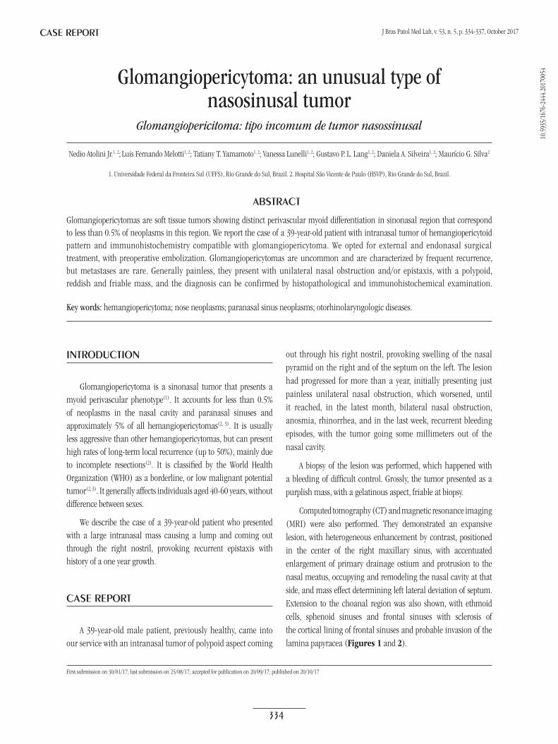
Computed tomography (CT) and magnetic resonance imaging (MRI) were also performed. They demonstrated an expansive lesion, with heterogeneous enhancement by contrast, positioned in the center of the right maxillary sinus, with accentuated enlargement of primary drainage ostium and protrusion to the nasal meatus, occupying and remodeling the nasal cavity at that side, and mass effect determining left lateral deviation of septum. Extension to the choanal region was also shown, with ethmoid cells, sphenoid sinuses and frontal sinuses with sclerosis of the cortical lining of frontal sinuses and probable invasion of the lamina papyracea (Figures 1 and 2).
10.5
935/
1676
-244
4.20
1700
54
335
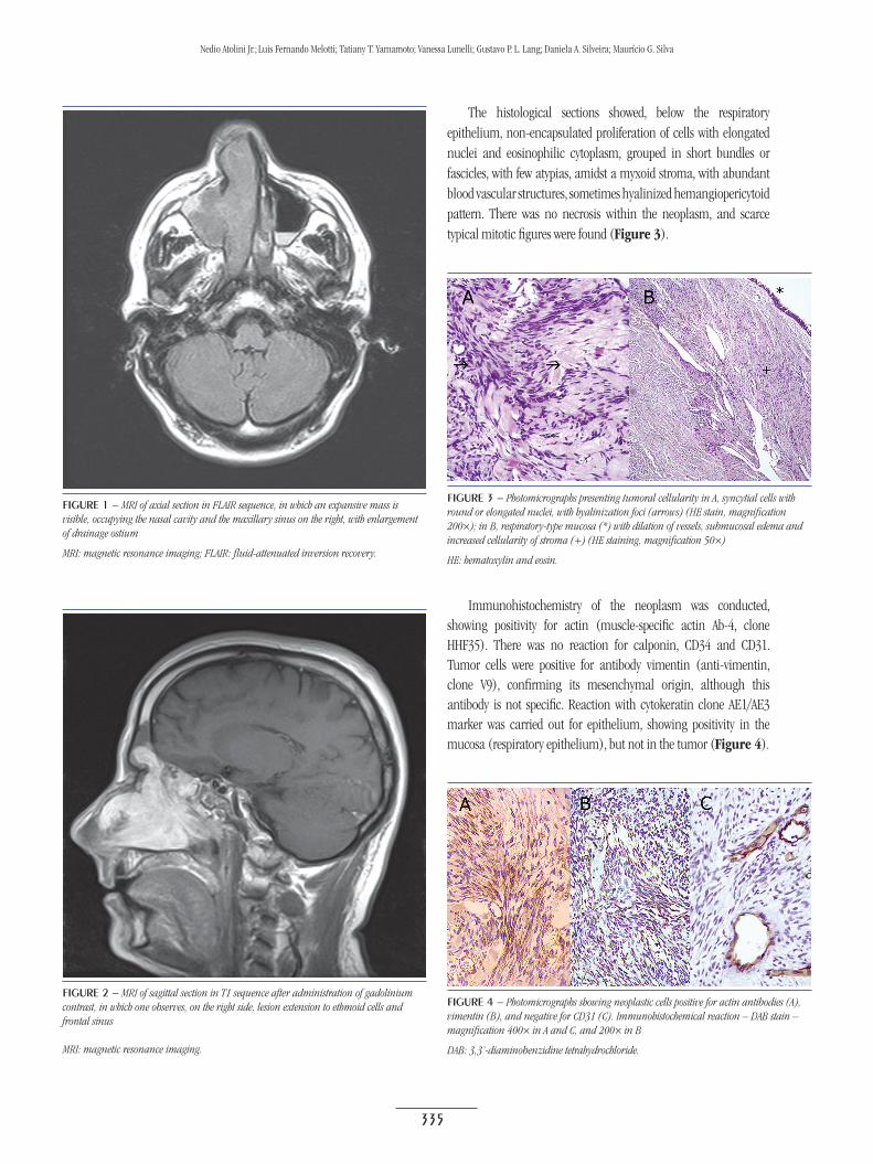
The histological sections showed, below the respiratory epithelium, non-encapsulated proliferation of cells with elongated nuclei and eosinophilic cytoplasm, grouped in short bundles or fascicles, with few atypias, amidst a myxoid stroma, with abundant blood vascular structures, sometimes hyalinized hemangiopericytoid pattern. There was no necrosis within the neoplasm, and scarce typical mitotic figures were found (Figure 3).
figurE 1 − MRI of axial section in FLAIR sequence, in which an expansive mass is visible, occupying the nasal cavity and the maxillary sinus on the right, with enlargement of drainage ostium
MRI: magnetic resonance imaging; FLAIR: fluid-attenuated inversion recovery.
figurE 2 − MRI of sagittal section in T1 sequence after administration of gadolinium contrast, in which one observes, on the right side, lesion extension to ethmoid cells and frontal sinus
MRI: magnetic resonance imaging.
figurE 3 − Photomicrographs presenting tumoral cellularity in A, syncytial cells with round or elongated nuclei, with hyalinization foci (arrows) (HE stain, magnification 200×); in B, respiratory-type mucosa (*) with dilation of vessels, submucosal edema and increased cellularity of stroma (+) (HE staining, magnification 50×)
HE: hematoxylin and eosin.
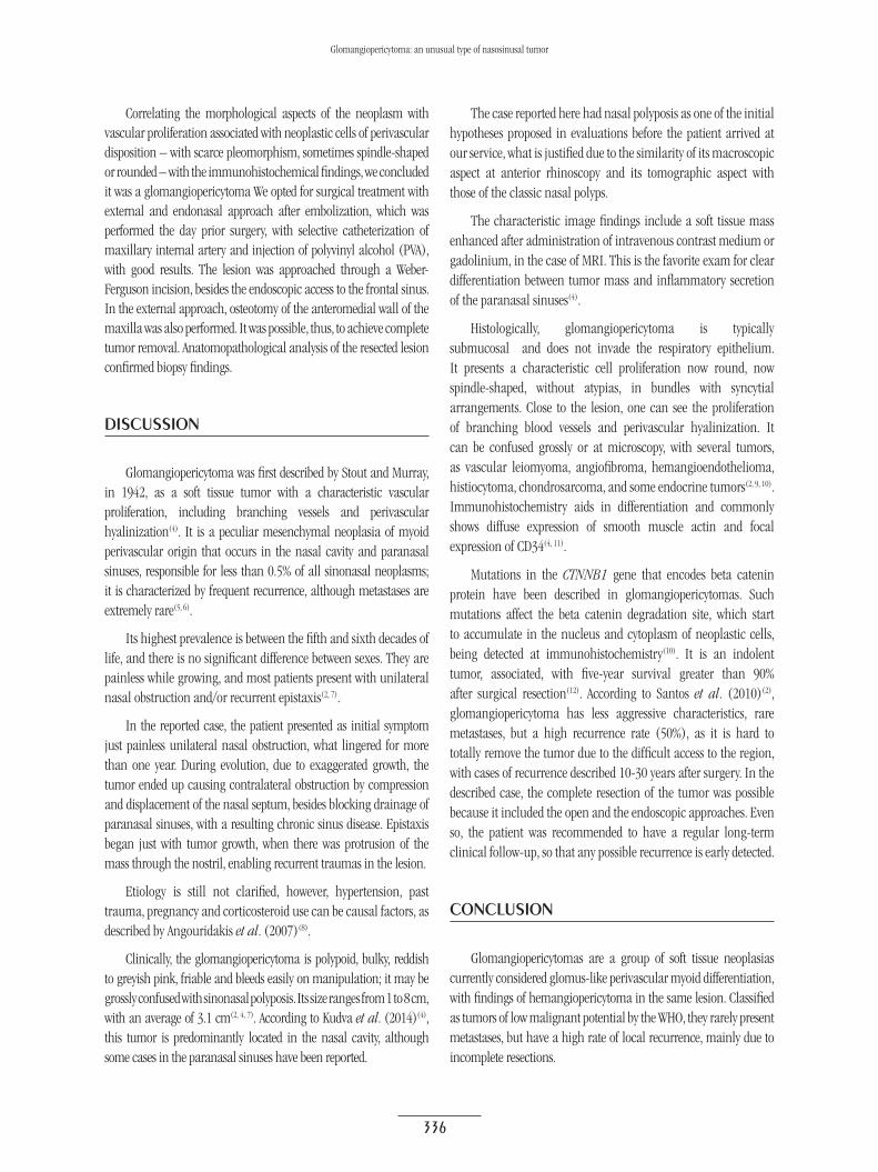
Immunohistochemistry of the neoplasm was conducted, showing positivity for actin (muscle-specific actin Ab-4, clone HHF35). There was no reaction for calponin, CD34 and CD31. Tumor cells were positive for antibody vimentin (anti-vimentin, clone V9), confirming its mesenchymal origin, although this antibody is not specific. Reaction with cytokeratin clone AE1/AE3 marker was carried out for epithelium, showing positivity in the mucosa (respiratory epithelium), but not in the tumor (Figure 4).
figurE 4 − Photomicrographs showing neoplastic cells positive for actin antibodies (A), vimentin (B), and negative for CD31 (C). Immunohistochemical reaction – DAB stain – magnification 400× in A and C, and 200× in B
DAB: 3,3’-diaminobenzidine tetrahydrochloride.
Nedio Atolini Jr.; Luis Fernando Melotti; Tatiany T. Yamamoto; Vanessa Lunelli; Gustavo P. L. Lang; Daniela A. Silveira; Maurício G. Silva
336
Correlating the morphological aspects of the neoplasm with vascular proliferation associated with neoplastic cells of perivascular disposition – with scarce pleomorphism, sometimes spindle-shaped or rounded – with the immunohistochemical findings, we concluded it was a glomangiopericytoma We opted for surgical treatment with external and endonasal approach after embolization, which was performed the day prior surgery, with selective catheterization of maxillary internal artery and injection of polyvinyl alcohol (PVA), with good results. The lesion was approached through a Weber-Ferguson incision, besides the endoscopic access to the frontal sinus. In the external approach, osteotomy of the anteromedial wall of the maxilla was also performed. It was possible, thus, to achieve complete tumor removal. Anatomopathological analysis of the resected lesion confirmed biopsy findings.
DiSCuSSion
Glomangiopericytoma was first described by Stout and Murray, in 1942, as a soft tissue tumor with a characteristic vascular proliferation, including branching vessels and perivascular hyalinization(4). It is a peculiar mesenchymal neoplasia of myoid perivascular origin that occurs in the nasal cavity and paranasal sinuses, responsible for less than 0.5% of all sinonasal neoplasms; it is characterized by frequent recurrence, although metastases are extremely rare(5, 6).
Its highest prevalence is between the fifth and sixth decades of life, and there is no significant difference between sexes. They are painless while growing, and most patients present with unilateral nasal obstruction and/or recurrent epistaxis(2, 7).
In the reported case, the patient presented as initial symptom just painless unilateral nasal obstruction, what lingered for more than one year. During evolution, due to exaggerated growth, the tumor ended up causing contralateral obstruction by compression and displacement of the nasal septum, besides blocking drainage of paranasal sinuses, with a resulting chronic sinus disease. Epistaxis began just with tumor growth, when there was protrusion of the mass through the nostril, enabling recurrent traumas in the lesion.
Etiology is still not clarified, however, hypertension, past trauma, pregnancy and corticosteroid use can be causal factors, as described by Angouridakis et al. (2007)(8).
Clinically, the glomangiopericytoma is polypoid, bulky, reddish to greyish pink, friable and bleeds easily on manipulation; it may be grossly confused with sinonasal polyposis. Its size ranges from 1 to 8 cm, with an average of 3.1 cm(2, 4, 7). According to Kudva et al. (2014)(4), this tumor is predominantly located in the nasal cavity, although some cases in the paranasal sinuses have been reported.
The case reported here had nasal polyposis as one of the initial hypotheses proposed in evaluations before the patient arrived at our service, what is justified due to the similarity of its macroscopic aspect at anterior rhinoscopy and its tomographic aspect with those of the classic nasal polyps.
The characteristic image findings include a soft tissue mass enhanced after administration of intravenous contrast medium or gadolinium, in the case of MRI. This is the favorite exam for clear differentiation between tumor mass and inflammatory secretion of the paranasal sinuses(4).
Histologically, glomangiopericytoma is typically submucosal and does not invade the respiratory epithelium. It presents a characteristic cell proliferation now round, now spindle-shaped, without atypias, in bundles with syncytial arrangements. Close to the lesion, one can see the proliferation of branching blood vessels and perivascular hyalinization. It can be confused grossly or at microscopy, with several tumors, as vascular leiomyoma, angiofibroma, hemangioendothelioma, histiocytoma, chondrosarcoma, and some endocrine tumors(2, 9, 10). Immunohistochemistry aids in differentiation and commonly shows diffuse expression of smooth muscle actin and focal expression of CD34(4, 11).
Mutations in the CTNNB1 gene that encodes beta catenin protein have been described in glomangiopericytomas. Such mutations affect the beta catenin degradation site, which start to accumulate in the nucleus and cytoplasm of neoplastic cells, being detected at immunohistochemistry(10). It is an indolent tumor, associated, with five-year survival greater than 90% after surgical resection(12). According to Santos et al. (2010)(2), glomangiopericytoma has less aggressive characteristics, rare metastases, but a high recurrence rate (50%), as it is hard to totally remove the tumor due to the difficult access to the region, with cases of recurrence described 10-30 years after surgery. In the described case, the complete resection of the tumor was possible because it included the open and the endoscopic approaches. Even so, the patient was recommended to have a regular long-term clinical follow-up, so that any possible recurrence is early detected.
ConCLuSion
Glomangiopericytomas are a group of soft tissue neoplasias currently considered glomus-like perivascular myoid differentiation, with findings of hemangiopericytoma in the same lesion. Classified as tumors of low malignant potential by the WHO, they rarely present metastases, but have a high rate of local recurrence, mainly due to incomplete resections.
Glomangiopericytoma: an unusual type of nasosinusal tumor
337
CorrESPonDing author
Nedio Atolini JuniorRua Uruguai, 1.954, sala 202; Centro; CEP: 99010-111; Passo Fundo-RS, Brasil; Phone: +55 (54) 9685-9398; e-mail: [email protected].
rESuMo
Glomangiopericitomas são tumores de partes moles que apresentam diferenciação mioide perivascular distinta na região sinunasal e correspondem a menos de 0,5% das neoplasias dessa região. Relatamos o caso de um paciente de 39 anos de idade com tumoração intranasal de proliferação celular de padrão hemangiopericitoide e imuno-histoquímico compatível com glomangiopericitoma. Optou-se por tratamento cirúrgico externo e endonasal, com embolização pré-operatória. Os glomangiopericitomas são incomuns e caracterizam-se pela recorrência frequente, sendo raras as metástases. Geralmente indolores, apresentam-se com obstrução nasal unilateral e/ou epistaxe, com massa de aspecto polipoide, avermelhada e friável. O diagnóstico pode ser confirmado pelo exame histopatológico e imuno-histoquímico.
Unitermos: hemangiopericitoma; neoplasias nasais; neoplasias dos seios paranasais; otorrinolaringopatias.
rEfErEnCES
1. El-Naggar AK, Chan JKC, Grandis JR, Takata T, Slootweg PJ, editors. WHO classification of head and neck tumours. 4th ed. Lyon: IARC; 2017. p. 44.
2. Santos Gorjón P, Muñoz Herrera A, Flores Corral T, et al. Hemangiopericitoma naso-sinusal. Revisión de la literatura a propósito de un caso. O.R.L. Aragon. 2010; 13(2): 24-6.
3. Verim A, Ertugay CK, Karaca CT, Gunes P, Sheidaei S, Oysu C. A rare tumor of nasal cavity: glomangiopericytoma. Case Reports in Otolaryngology. 2014; article ID 282958, 3 p.
4. Kudva R, Sharma S, Gurijala R, Nayak DR. Sinonasal-type hemangiopericytoma of nasal cavity: a rare neoplasm – case report with a brief review of literature. RRJMHS. 2014; 3(3): 31-6.
5. Haller F, Bieg M, Moskalev EA, et al. Recurrent mutations within the amino-terminal region of β-catenin are probable key molecular driver events in sinonasal hemangiopericytoma. Am J Pathol. 2015; 185(2): 563-71.
6. Barbara M. Le neoplasie maligne non epiteliali delle V.A.D.S. Galatina Le, Italia: TorGraf; 2014. p. 73-98.
7. Roy NP, Desai DP, Jain SA. Glomangiopericytoma of nasal cavity. Indian J Pathol Microbiol. 2015; 58(4): 554-6.
8. Angouridakis N, Zaraboukas T, Vital J, Vital V. Sinonasal hemangiopericytoma of the middle turbinate: a case report and brief review of the literature. B-ENT. 2007; 3(3): 139-43.
9. Park YK, Park JH, Kim YW, Lee JH, Yang MH. Nasal hemangiopericytoma – a case report. J Korean Med Sci. 1990; 5(3): 173-8.
10. Thompson LDR, Wenig BM. Diagnostic pathology: head & neck. 2nd ed. Salt Lake City, UT: Elsevier, 2016. Glomangiopericytoma. p. 97.
11. Handra-Luca A, Abd Elmageed ZY, Magkou C, Lae M. Immunophenotype heterogeneity in nasal glomangiopericytoma. Case Reports in Otolaryngology. 2015; article ID 308743. 3 p.
12. Dandekar M, McHugh JB. Sinonasal glomangiopericytoma: case report with emphasis on the differential diagnosis. Arch Pathol Lab Med. 2010; vol 134: 1444-9.
Nedio Atolini Jr.; Luis Fernando Melotti; Tatiany T. Yamamoto; Vanessa Lunelli; Gustavo P. L. Lang; Daniela A. Silveira; Maurício G. Silva