Embed Size (px)
Citation preview
GLAUCOMA SOCIETIES
GS1 GLAUCOMA – A DIAGNOSIS BASED ON PROBABILITY M. Atanassov1 1Department of Ophthalmologyq Medical University, Plovdiv - Bulgaria This review discusses the process of making the medical decision for the diagnosis of glaucoma and the determination of therapeutic behavior. We discuss the complex evaluation of the signs of glaucoma – the often vague boundary between norm and pathology in function and structure. We conclude that when building diagnosis, we rely on the likelihood of a structural or functional finding to be the norm or pathology, and diagnosis is often based on the judgment of what is the probability of a patient to develop glaucoma defects after certain time. The same goes for therapy – the art is deciding which treatment regimen most likely to stop or slow the glaucoma progression so that the patient can preserve visual functions.

GS2 OCT EVALUATION OF OPTIC NERVE HEAD IN PATIENTS SUSP ECT FOR GLAUCOMA L. Mishev1 1Eye hospital, Sofia - Bulgaria Aims of the study: Searching for new criterias in interpretation of Optic nerve head (ONH) in patients suspect for glaucoma. Methods: The study was done on 30 patients which were randomly choosed. The focus was on suspect patients with ocular hypertension. The OCT machine that was used is Optovue RTvue-100. The interpretation of optic nerve parameters seems to be very important factor in determining the diagnose for Glaucomatous process.Despite the well known RNFL thickness based protocols and GCC mapping protocols, we feel that there is a need for additional criteiries describing morphological changes in the ONH ,to provide more complete clinical picture and way to exact diagnose. The algorithm for determining the ONH parameters The algorithm is based on «floating canvas» principle.In normal eyes it performs quite well and exact.But there is a problem in determining the borders and size of the ONH and the excavation in myopic disk,tilted disk,hypermetropic disk.The algorithm searches for the pigment epithelium as a most hyperreflective tissue to determine the boundery of the ONH ,but in eyes with parapapilary atrophy the pigment epithelium is way off from the real border of the ONH. Results: We propose the Bruch membrane to be used as a anchor point from which the border to be estimated and we call it «Nerve canal opening» ( NCO) ,position of Lamina cribrosa according to NCO , thicknes of Lamina cribrosa and shape of the optic nerve below the Lamina cribrosa .We belive that this additional signs will be very helpfull for early detection of Glaucomatous process.


GS3 CENTRAL CORNEAL THICKNESS IN PATIENTS WITH ADVANCED PRIMARY OPEN ANGLE GLAUCOMA S. Ivanova1 1Department of Ophthalmology, Alexandrovska Hospital, Sofia - Bulgaria Background:To examine Central Corneal Thickness (CCT) in patients with uncontrolled advanced Primary Open Angle Glaucoma (POAG) and to perform comparative study in relation to CCT in healthy persons of the same age group. To do comparative study of glaucoma changes in patients with advanced POAG, divided in 3 groups according to the corneal thickness Methods: CCT was measured in 40 patients (64 eyes) with advanced POAG - Cup/disc ratio = 0.76 ± 0.09; Intraocular pressure (IOP) = 21.01 ± 3.6 mmHg; MD = -20.1 ± 7.8 dB; PSD = 8.4 ± 2.9 dB. The results were compared to those of the same examinations in 30 healthy persons (60 eyes) of the same age group. The group of patients with advanced POAG was divided in 3 groups according to CCT. The rest of the routine diagnostic methods, used in the ophthalmology practice: establishing visual acuity, tonometry, gonioscopy, ophthalmoscopy, computer perimetry, were done also in all patients. Results: Statistically significant difference between healthy eyes and these with advanced POAG in relation to C/D, IOP, MD, PSD and CCT was found. The cornea was statistically thinner in patients with advanced POAG. When patients with advanced POAG were divided into groups according to corneal thickness, it was found that the worse glaucoma defects were available in the group of patients with thinner corneas. Conclusion: It was found statistically thinner CCT in patients with advanced POAG. There was correlation in changes of CCT and MD. Therefore, the necessity of assessment and eventual rectification of tonometric values according to CCT data during examination and treatment of patients with POAG was accentuated. Presumably, the compensation of IOP in the group with thinner cornea was insufficient or incorrectly interpreted.
GS4 COMBINED SURGERY IN ADVANCED PRIMERY CONGENITAL GLA UCOMA N. Petkova1 1University Medical Centre,Sofia, Sofia - Bulgaria Backgroung: Combined surgery Trabeculotomy /TT/ and Trabeculectomy/TE/is often method of choice in advanced Primary Congenital Glaucoma/PCG/. Purpose: To compare the efficacy and complications of TT and TE with Mitomycin C/MMC/ performed in two steps or combined in one step in advanced PCG. Methods: 35 eyes of 24 children with advanced PCG underwent TT and TE with MMC.The eyes were divided in two groups: group1 - surgery was performed in two steps: TT and after 1- 2 months: TE, because of inadequate efficacy /20/ eyes and group 2: TT and TE with MMC performed in one step /15 eyes/. IOP before surgery was 28 ± 5 mmHg in group 1 and 30 ± 6 mmHg in group 2. IOP before and after surgery,corneal size ,efficacy and complications were compared between the two groups. Follow up period was up to 10 years. Results: Postoperative IOP one month after surgery was 15 ± 4 mmHg in group 1 and 14 ± 4 mmHg in group 2. In the follow up period 30 % in group 1 and 33% in group2 needed additional medical or surgical treatment.Most common complications in the early postoperative period prevailing in group 2 were hypotony , shallow anterior chamber , hyphaema, cataract /rarely-1 eye in group 2/ and in the late postsurgical period - elevation of IOP. Conclusions: In advanced PCG combined one step antiglaucoma surgery /TT+TE with MMC/. Is with a good efficacy similar to that of two step antiglaucoma surgery, but with more complications in the early postsurgical period.
GS5 EFFICACY OF TRABECULECTOMY WITH MITOMYCIN C IN PSEUDOEXFOLIATIVE GLAUCOMA PATIENTS AFTER MINIMUM 2 YEARS FOLLOW -UP M. Konareva-Kostianeva1 1Dept Ophthalmology, Plovdiv - Bulgaria Purpose: To assess the absolute and qualified success of trabeculectomy (TE) with mitomycin C (MMC) in pseudoexfoliative glaucoma patients followed-up more than 2 years. Methods: Forty pseudoexfoliative glaucoma patients undergoing TE with MMC and with minimum of 2-year follow-up are included. We measure the success of TE as absolute success if the intraocular pressure (IOP) is ≤ 18 mmHg and ≥ 6 mmHg without the use of glaucoma medications and as qualified success if the IOP is ≤ 18 mmHg under glaucoma medications. Results: The mean preoperative IOP was 31.1 ± 9.3 mmHg (range 21-57). In mean period of 2.8 ± 1.2 years (range 2-5) after surgery we observe the statistically significant decrease of IOP (mean IOP 14.8 ± 5.4 mmHg, p < 0.001). After more than 2 years 34 eyes are with IOP < 18 mm Hg. An absolute success of operation is observed in 21 (52.5%) patients and a qualified success - in 13 (32.5%) patients. Elevated IOP under glaucoma medications with mean value 24.2 ± 4.6 mmHg, range 19-32 mm Hg (unsuccessful TE) is found in 6 (15%) of the patients. Conclusions: Our results show a successful TE (with absolute and qualified success) in 85% of pseudoexfoliative glaucoma patients after more than 2 years follow-up.
GS6 EXPRESS IMPLANT. TECHNIQUE. FIRST RESULTS P. Guguchkova-Janchuleva1, L. Mishev1, B. Samsonova1, B. Krasimirova1, D. Popov1 1Eye hospital Zrenie, Sofia - Bulgaria Using of ExPress implant in glaucoma treatment changed the standard filtrating glaucoma surgery. The implant allows a control over the subscleral outflow and avoids the postoperative hypotony. The successful outcome of the operation demands knowledge of the technique of insertion consisting of several important steps: Scleral or subscleral application of Mytomicin - Formation of adequate sclera flab - Correctly directed punction of the anterior chamber - Implant insertion maintaining the anterior chamber deep enough, using visco substances or maintainer. Our first results concerning the postoperative IOP are very good without any complications.
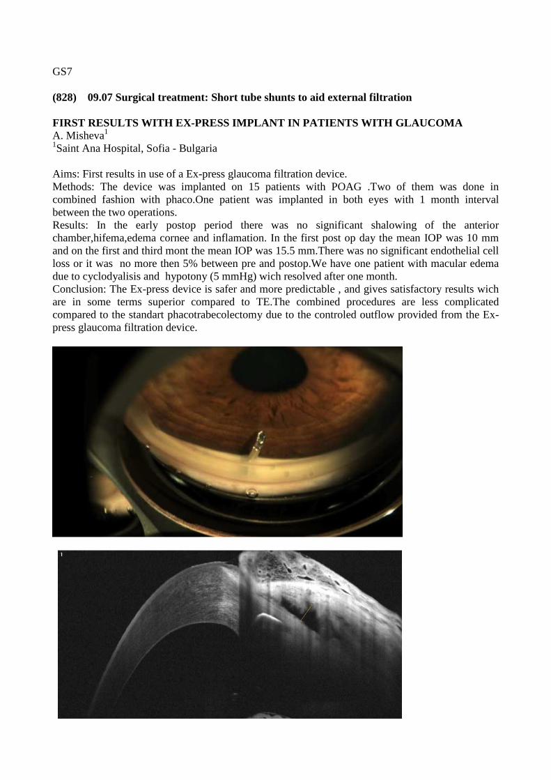
GS7 (828) 09.07 Surgical treatment: Short tube shunts to aid external filtration FIRST RESULTS WITH EX-PRESS IMPLANT IN PATIENTS WIT H GLAUCOMA A. Misheva1 1Saint Ana Hospital, Sofia - Bulgaria Aims: First results in use of a Ex-press glaucoma filtration device. Methods: The device was implanted on 15 patients with POAG .Two of them was done in combined fashion with phaco.One patient was implanted in both eyes with 1 month interval between the two operations. Results: In the early postop period there was no significant shalowing of the anterior chamber,hifema,edema cornee and inflamation. In the first post op day the mean IOP was 10 mm and on the first and third mont the mean IOP was 15.5 mm.There was no significant endothelial cell loss or it was no more then 5% between pre and postop.We have one patient with macular edema due to cyclodyalisis and hypotony (5 mmHg) wich resolved after one month. Conclusion: The Ex-press device is safer and more predictable , and gives satisfactory results wich are in some terms superior compared to TE.The combined procedures are less complicated compared to the standart phacotrabecolectomy due to the controled outflow provided from the Ex-press glaucoma filtration device.


GS8 CONTROL OF INTRAOCULAR PRESSURE IN SECONDARY GLAUCO MA Y. Kirilova 1, T. Hergeldzhieva-Fileva1, P. Vassileva1 1University Eye Hospital, Sofia - Bulgaria Background: To present the results of treatment in patients with secondary glaucoma. Methods: A retrospective study of 24 consecutive patients with secondary non-compensated glaucoma for a period of one year (January 2009- January 2010) was done. Full eye examination, gonioscopy, B-scan, visual field testing and OCT were performed in all patients. The demographic data and the etiology of the secondary glaucoma were analyzed. Treatment was conservative and surgical in all cases. Results: The average age of patients was 42 years (range 24 to 76). The most common cause of secondary glaucoma was anterior synechiae as a result of pseudophakic keratopathy in 8 patients (33%). Other causes included: exfoliative glaucoma - in 5 (21%), pigmented glaucoma – in 4 (17%), post-traumatic aphakia - in 3 (13%), anterior chamber trauma – in 2 (8%), and herpetic keratouveitis - in 2 (8%). The surgical treatment included filtration surgeries: trabeculectomy with Ologen® - in19 patients (79%), filtering procedure with Ex-Press® - in 3 (13%) and filtering procedure with the Ahmed® valve - in 1 (4%). Avastin® of dose of 2.5 mg/0.1 ml was injected in the anterior chamber of 1 patient (4%) with neovascular glaucoma and IOP decreased from 45, 0 mmHg to 28, 0 mmHg in this case. In patients treated with TE and Ologen®, IOP was reduced from 38, 0 mmHg to 17, 3 mmHg on average; in patients with the Ex-Press® drainage device - from 45, 0 mmHg to 12, 0 mmHg; and in the patient with the Ahmed® valve - from 50, 0 mmHg to 17, 3 mmHg. The follow-up period was from 12 to 24 months. Conclusion: A customized approach is necessary in all cases with secondary glaucoma depending on the etiology, disease’s progression, condition of the other eye, and IOP values. Surgery is indicated in most of the patients with secondary glaucoma.
GS9 OCT IN THE DIAGNOSIS AND FOLLOW -UP OF GLAUCOMAS T. Hergeldzhieva-Fileva1, Y. Kirilova1, P. Vassileva1 1University Eye Hospital, Sofia - Bulgaria Background: The irreversible loss of ganglion cells, which is in the basis of glaucoma, leads to attenuation and damage of the retinal nerve fiber layer (RNFL). It is known that 40% of the axons had to be lost for the detection of any changes in visual function (visual field). Structural alterations in the RNFL, detected by OCT, precede those in the optic nerve head and visual field. The purpose of this presentation is to share our experience in using OCT for early diagnosis and follow up of glaucoma patients. Methods: For the period from June 2009 to December 2010, 285 patients with glaucoma or suspected glaucoma were examined with the Stratus OCT ™. The central corneal thickness, chamber angle, peripapillary RNFL, optic nerve head and macular region were scanned and measured. Results: OCT is a suitable diagnostic method for: identifying the integrity of the RNFL in glaucoma suspects with a large excavation, setting up a suspicion for glaucoma before any detectable changes in the visual field, follow up of patients with ocular hypertension, and follow up of glaucoma patients for progressive loss of RNFL. Conclusions: OCT is helpful in the diagnosis and follow up of patients with glaucoma. The interpretation of the results is always in conjunction with clinical data, visual field changes and IOP measurements.


![Principal - Red eyes in the necropsy floor: twenty cases of …ainfo.cnptia.embrapa.br/digital/bitstream/item/139190/1/... · 2016. 2. 17. · casos de hifema em cães e gatos.] Hifema,](https://img.pdfslide.us/doc/110x75/60a808ec21120836b460b9a0/principal-red-eyes-in-the-necropsy-floor-twenty-cases-of-ainfo-2016-2-17.jpg)