Embed Size (px)
Citation preview
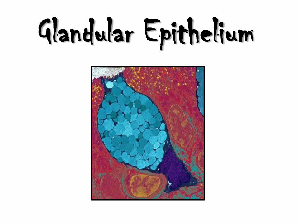
Glandular Epithelium
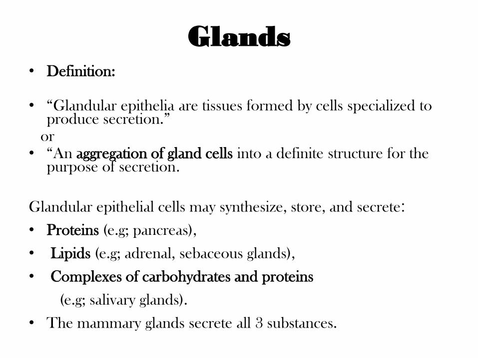
Glands • Definition: • “Glandular epithelia are tissues formed by cells specialized to
produce secretion.” or
• “An aggregation of gland cells into a definite structure for the purpose of secretion.
Glandular epithelial cells may synthesize, store, and secrete:
• Proteins (e.g; pancreas),
• Lipids (e.g; adrenal, sebaceous glands),
• Complexes of carbohydrates and proteins
(e.g; salivary glands).
• The mammary glands secrete all 3 substances.
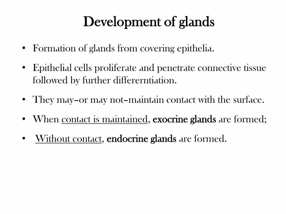
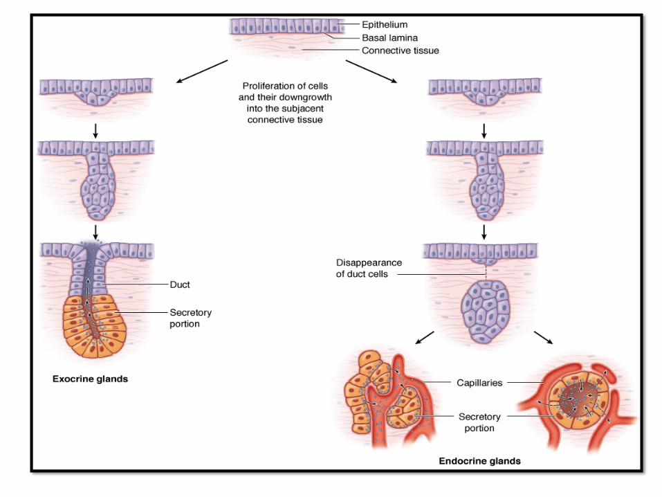
Development of glands
• Formation of glands from covering epithelia.
• Epithelial cells proliferate and penetrate connective tissue
followed by further differerntiation.
• They may–or may not–maintain contact with the surface.
• When contact is maintained, exocrine glands are formed;
• Without contact, endocrine glands are formed.
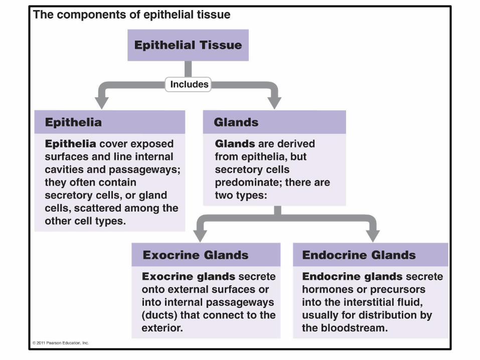
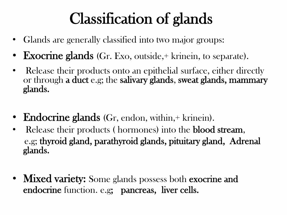
Classification of glands
• Glands are generally classified into two major groups:
• Exocrine glands (Gr. Exo, outside,+ krinein, to separate).
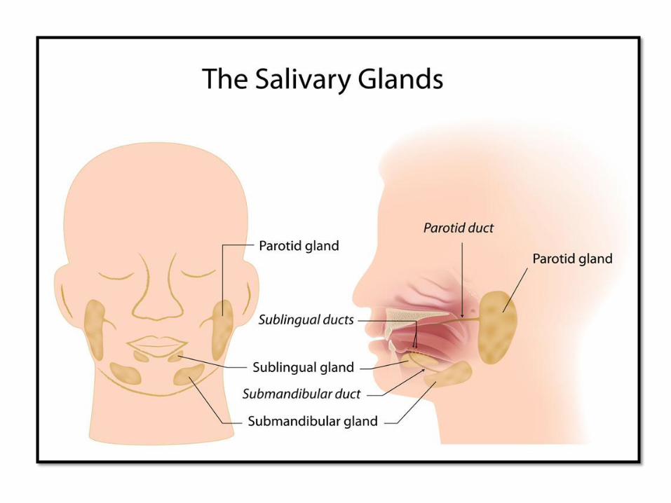
• Release their products onto an epithelial surface, either directly or through a duct e.g; the salivary glands, sweat glands, mammary glands.
• Endocrine glands (Gr, endon, within,+ krinein).
• Release their products ( hormones) into the blood stream,
e.g; thyroid gland, parathyroid glands, pituitary gland, Adrenal glands.
• Mixed variety: Some glands possess both exocrine and
endocrine function. e.g; pancreas, liver cells.
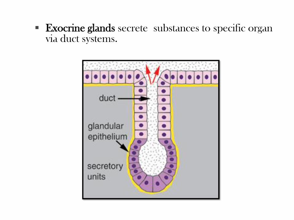
Exocrine glands secrete substances to specific organ via duct systems.
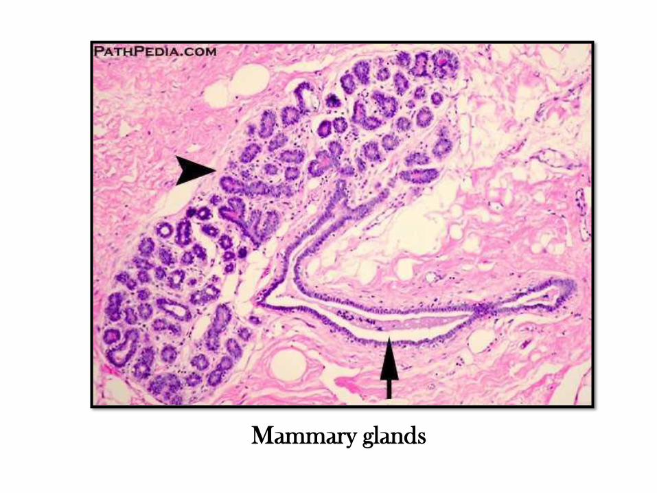
Mammary glands
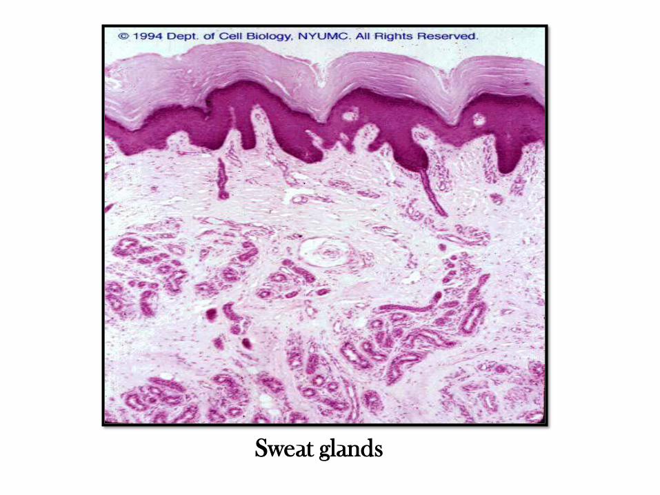
Sweat glands
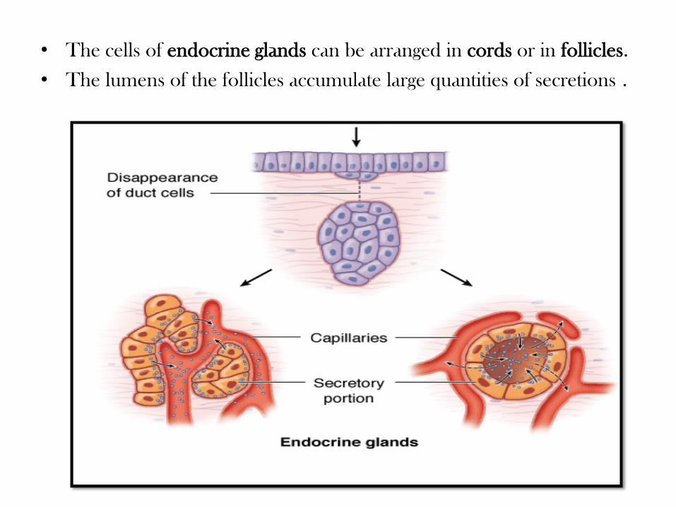
• The cells of endocrine glands can be arranged in cords or in follicles.
• The lumens of the follicles accumulate large quantities of secretions .
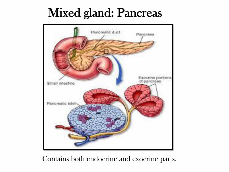
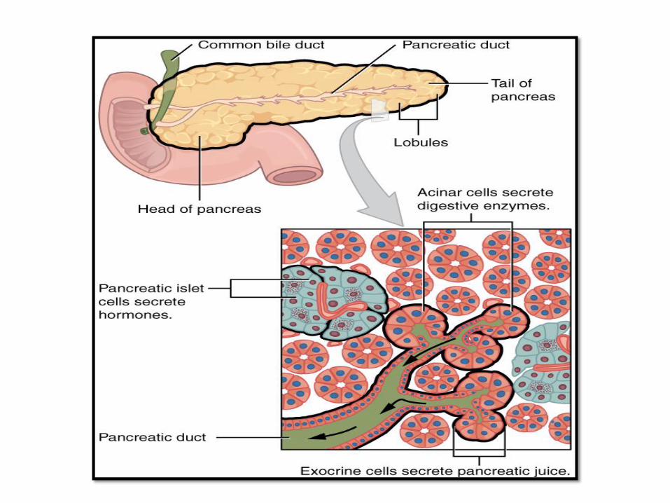
Mixed gland: Pancreas
Contains both endocrine and exocrine parts.
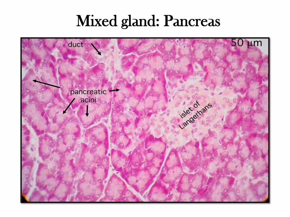
Mixed gland: Pancreas
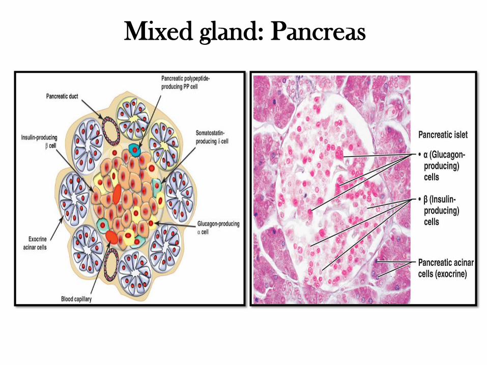
Mixed gland: Pancreas
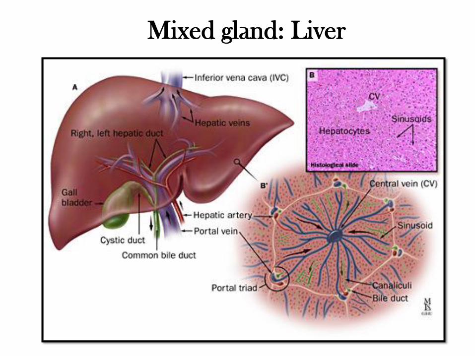
Mixed gland: Liver
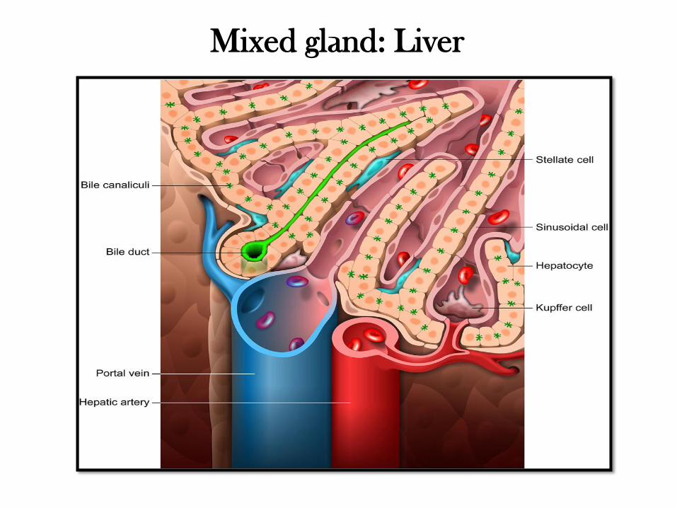
Mixed gland: Liver
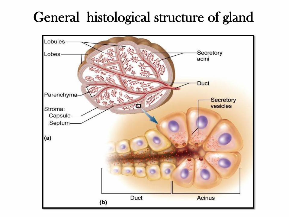
General histological structure of gland
General histological structure of gland
• Externally a gland is surrounded by a dense layer of connective
tissue which forms capsule of the gland.
• From the capsule connective tissue septa extend into the gland,
thereby dividing its substance into a number of lobes.
• Thinner septa subdivide each lobe into smaller lobules.
• Blood vessels and nerves pass along the connective tissue septa to
reach the secretory elements.
• The functional part of a gland, formed by its secretory cells, is
known as parenchyma of the gland.
• The supporting elements of the gland, which consists mainly of
connective tissue, are referred to as stroma of the gland.
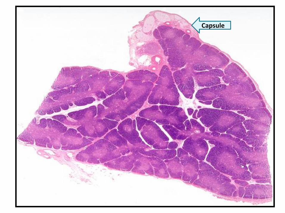
Capsule
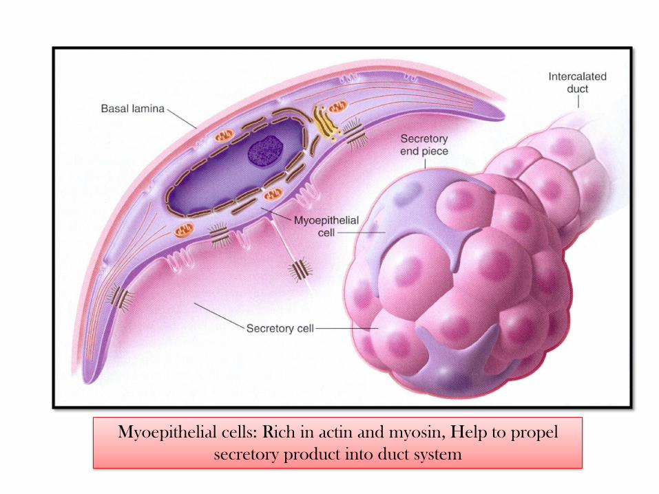
Myoepithelial cells: Rich in actin and myosin, Help to propel
secretory product into duct system
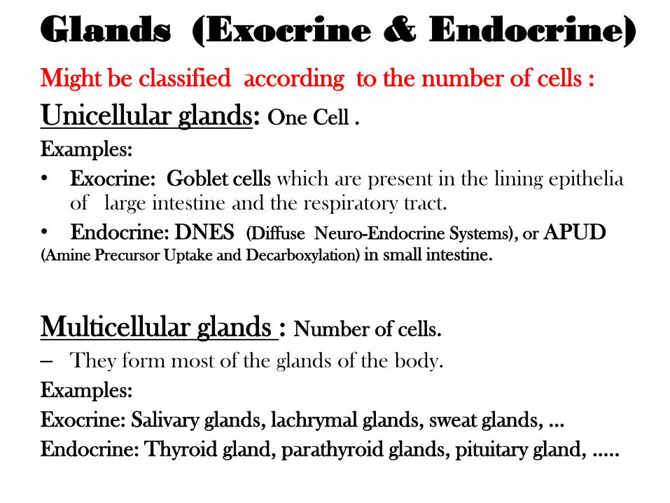
Glands (Exocrine & Endocrine)
Might be classified according to the number of cells :
Unicellular glands: One Cell .
Examples:
• Exocrine: Goblet cells which are present in the lining epithelia
of large intestine and the respiratory tract.
• Endocrine: DNES (Diffuse Neuro-Endocrine Systems), or APUD (Amine Precursor Uptake and Decarboxylation) in small intestine.
Multicellular glands : Number of cells.
– They form most of the glands of the body.
Examples:
Exocrine: Salivary glands, lachrymal glands, sweat glands, …
Endocrine: Thyroid gland, parathyroid glands, pituitary gland, …..
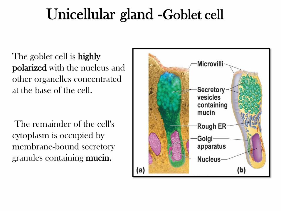
The goblet cell is highly
polarized with the nucleus and
other organelles concentrated
at the base of the cell.
The remainder of the cell's
cytoplasm is occupied by
membrane-bound secretory
granules containing mucin.
Unicellular gland - Goblet cell
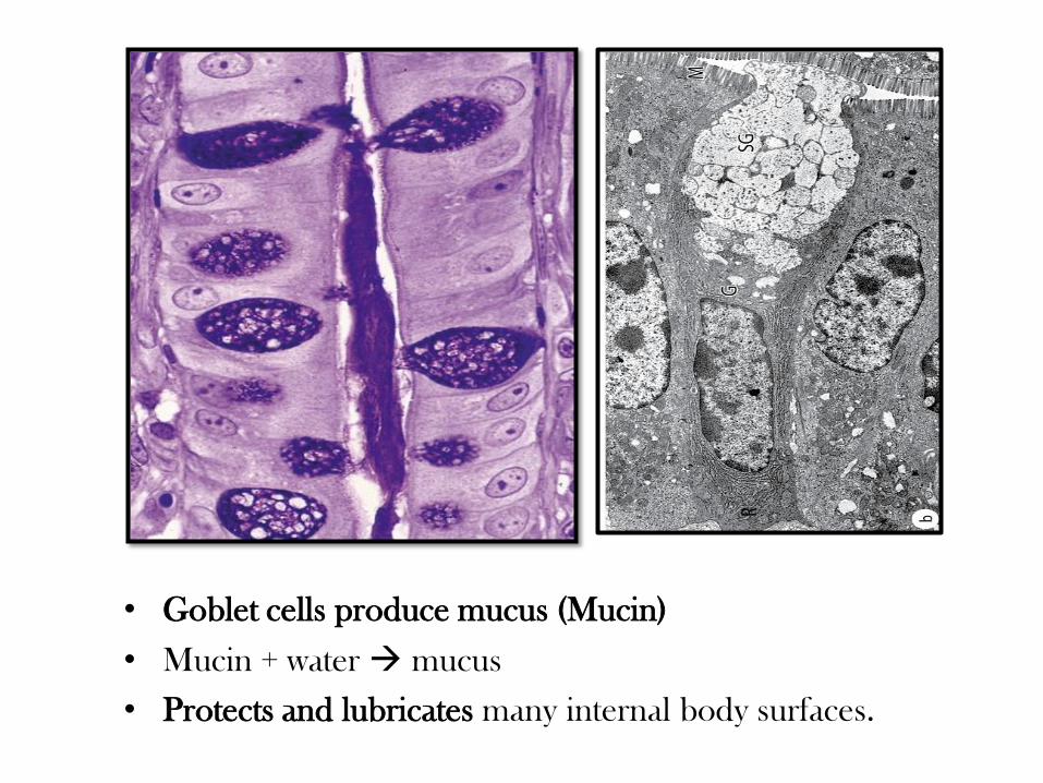
• Goblet cells produce mucus (Mucin)
• Mucin + water mucus
• Protects and lubricates many internal body surfaces.
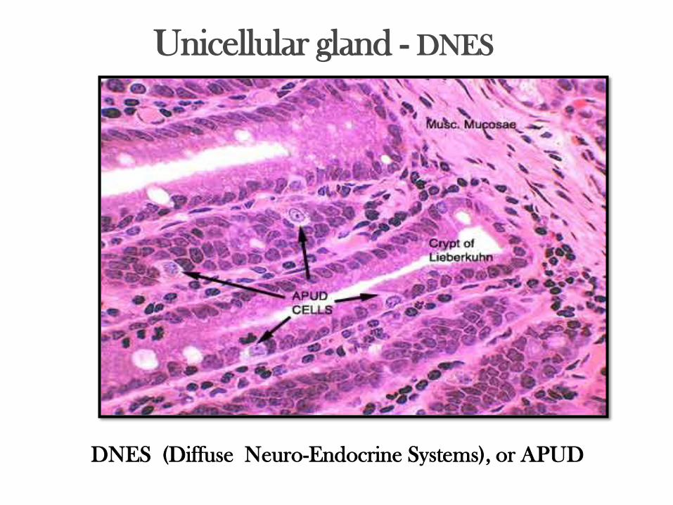
Unicellular gland - DNES
DNES (Diffuse Neuro-Endocrine Systems), or APUD
Exocrine glands
These can also be classified on the basis of:
• Morphology of ducts and secreting portions.
• Nature of secretory product.
• Mode of secretion.
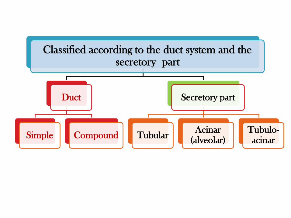
Classified according to the duct system and the secretory part
Duct
Simple Compound
Secretory part
Tubular Acinar
(alveolar) Tubulo-acinar
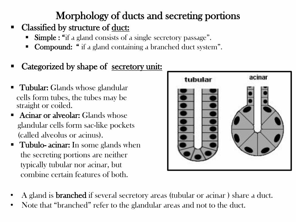
Morphology of ducts and secreting portions Classified by structure of duct:
Simple : “if a gland consists of a single secretory passage”.
Compound: “ if a gland containing a branched duct system”.
Categorized by shape of secretory unit:
Tubular: Glands whose glandular
cells form tubes, the tubes may be straight or coiled.
Acinar or alveolar: Glands whose
glandular cells form sac-like pockets
(called alveolus or acinus).
Tubulo- acinar: In some glands when
the secreting portions are neither
typically tubular nor acinar, but
combine certain features of both.
• A gland is branched if several secretory areas (tubular or acinar ) share a duct.
• Note that “branched” refer to the glandular areas and not to the duct.
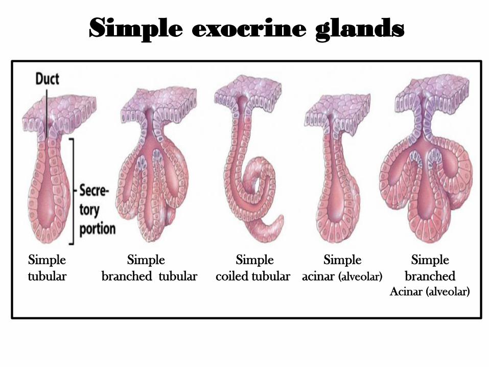
Simple exocrine glands
Simple
tubular
Simple
acinar (alveolar)
Simple
coiled tubular
Simple
branched
Acinar (alveolar)
Simple
branched tubular
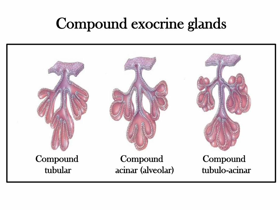
Compound exocrine glands
Compound Compound Compound
tubular acinar (alveolar) tubulo-acinar
Simple
exocrine glands
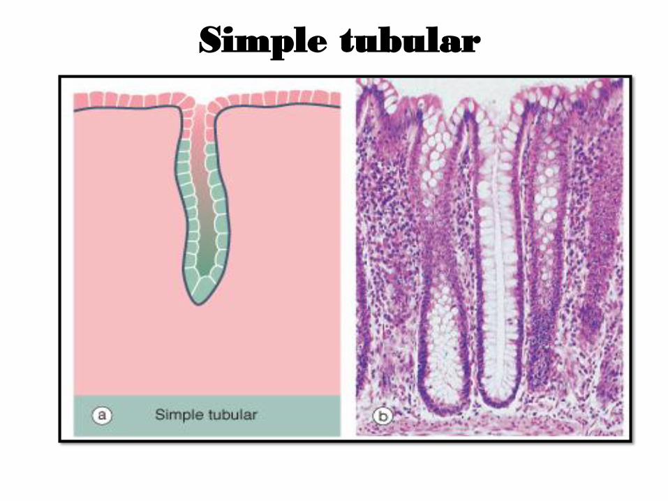
Simple tubular
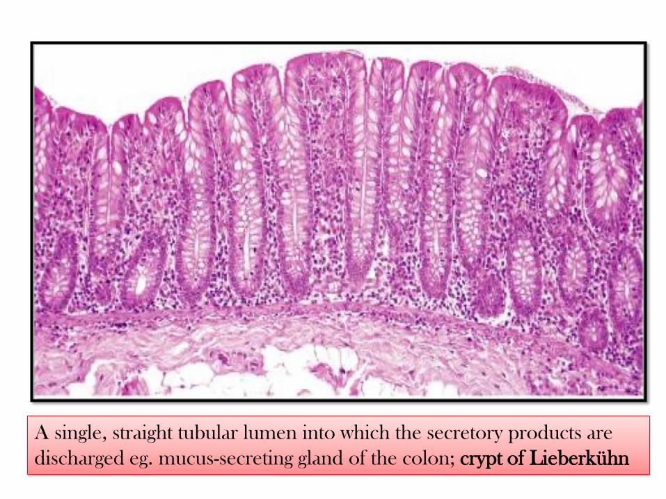
A single, straight tubular lumen into which the secretory products are
discharged eg. mucus-secreting gland of the colon; crypt of Lieberkühn
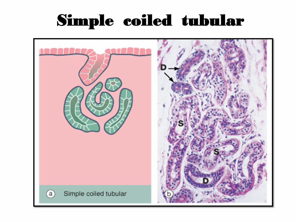
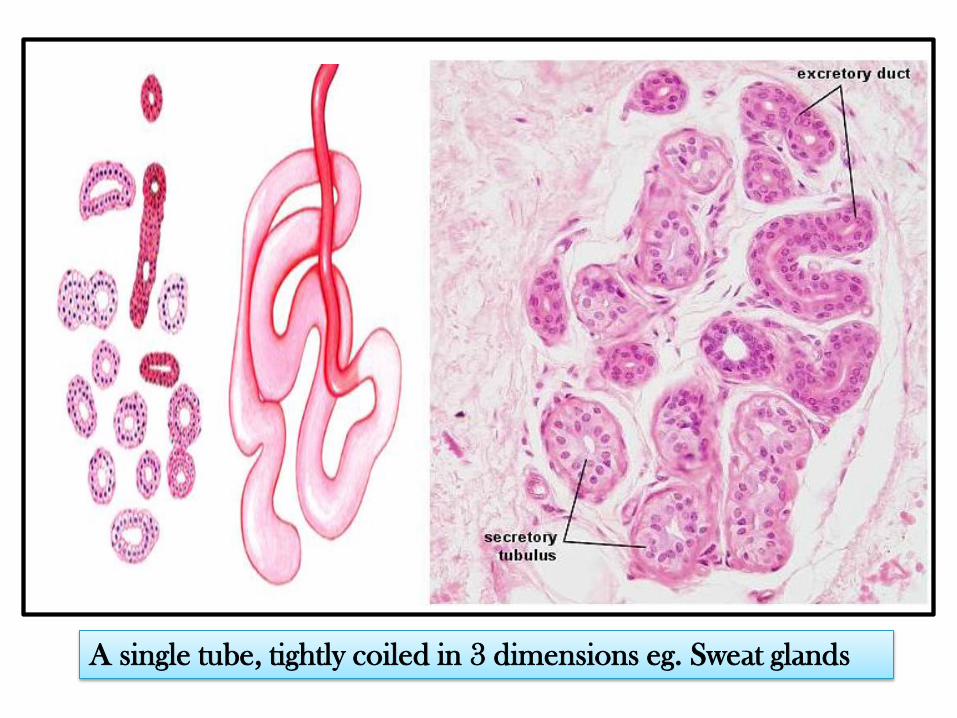
Simple coiled tubular
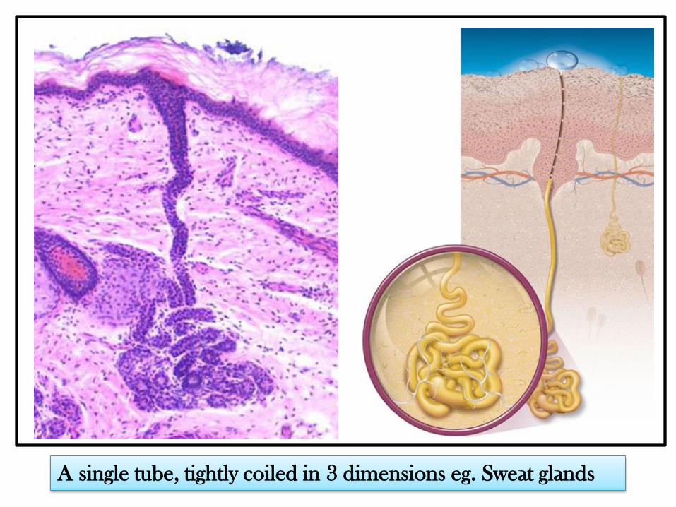
A single tube, tightly coiled in 3 dimensions eg. Sweat glands
A single tube, tightly coiled in 3 dimensions eg. Sweat glands
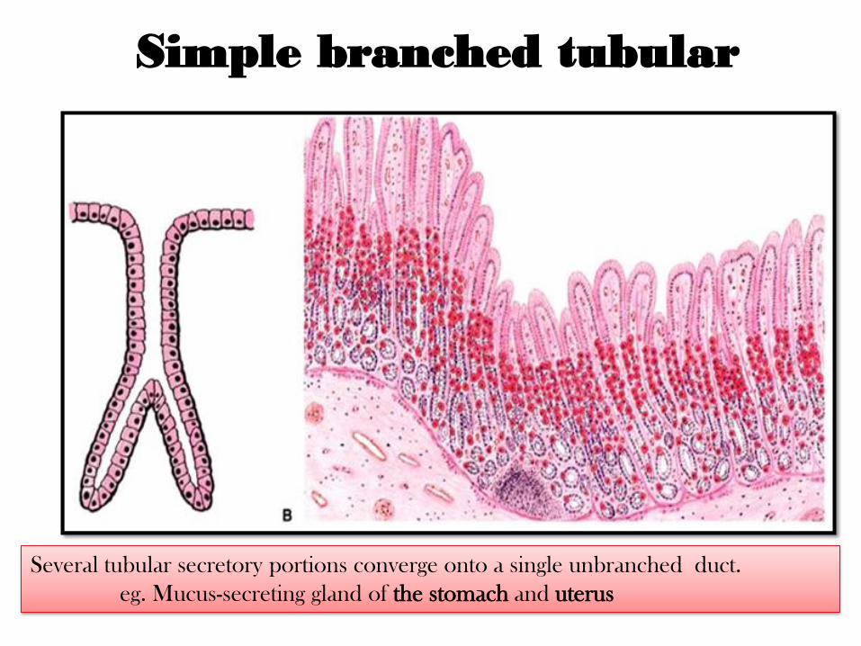
Simple branched tubular
Several tubular secretory portions converge onto a single unbranched duct.
eg. Mucus-secreting gland of the stomach and uterus
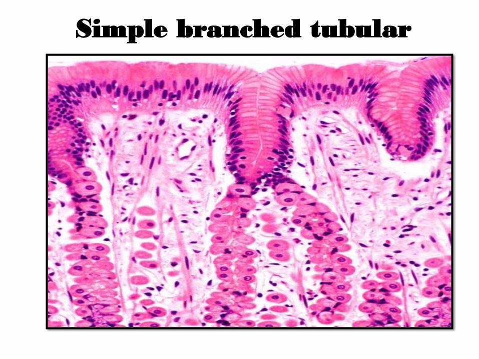
Simple branched tubular
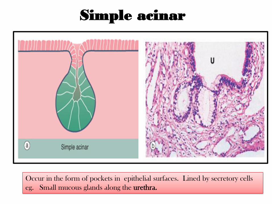
Simple acinar
Occur in the form of pockets in epithelial surfaces. Lined by secretory cells
eg. Small mucous glands along the urethra.
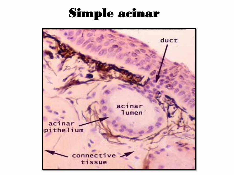
Simple acinar
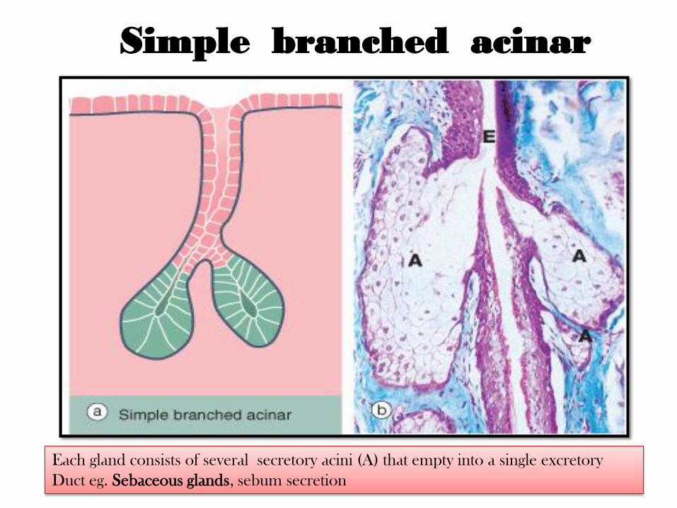
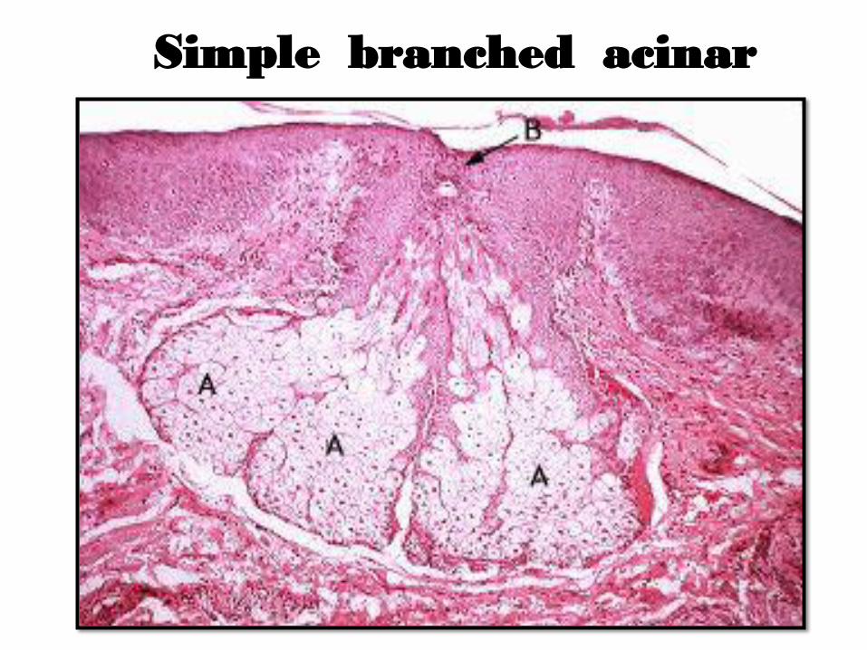
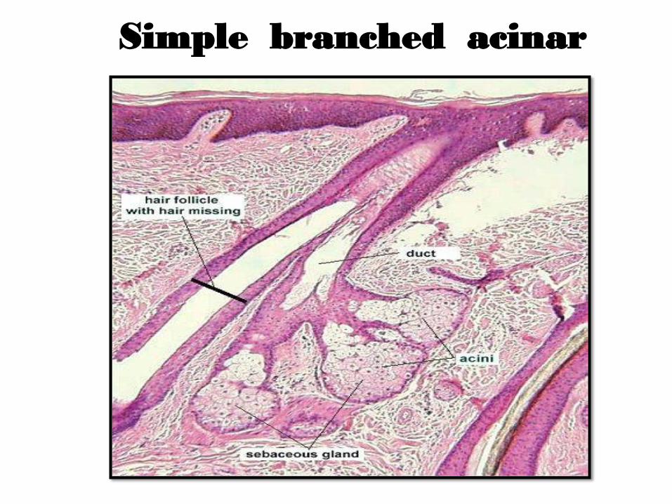
Simple branched acinar
Each gland consists of several secretory acini (A) that empty into a single excretory
Duct eg. Sebaceous glands, sebum secretion
Simple branched acinar
Simple branched acinar
Compound
exocrine glands
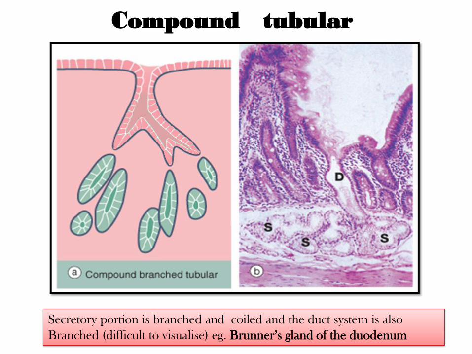
Compound tubular
Secretory portion is branched and coiled and the duct system is also
Branched (difficult to visualise) eg. Brunner’s gland of the duodenum
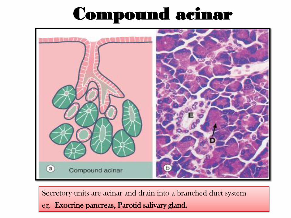
Compound acinar
Secretory units are acinar and drain into a branched duct system
eg. Exocrine pancreas, Parotid salivary gland.
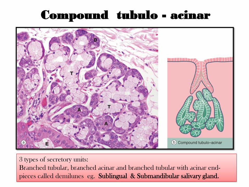
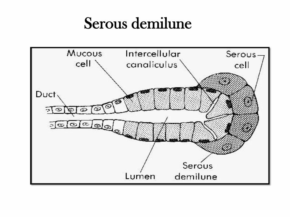
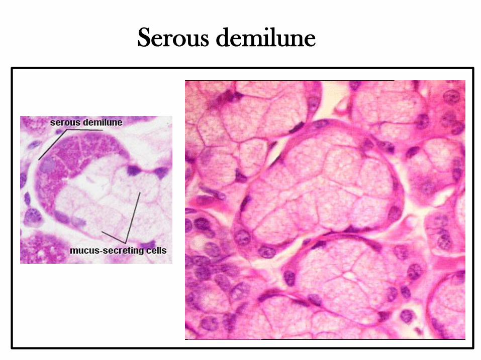
Compound tubulo - acinar
3 types of secretory units:
Branched tubular, branched acinar and branched tubular with acinar end-
pieces called demilunes eg. Sublingual & Submandibular salivary gland.
Classification
on the basis of nature of
secretory product
Classification on the basis of nature of
secretory product
1. Mucous glands: these glands produce a viscid, slimy,
carbohydrate-rich secretion which is called mucus,
e.g; Pyloric glands of stomach
2. Serous glands: these glands produce a thin, watery, protein-
rich secretions, often high in enzymatic activity e.g; Exocrine
pancreas, the parotid salivary gland.
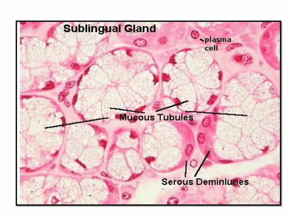
3. Mixed glands: these glands produce both mucous and
serous secretions e.g; the sublingual and submandibular salivary
glands.
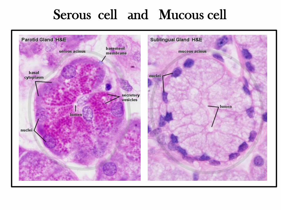
Serous cell and Mucous cell
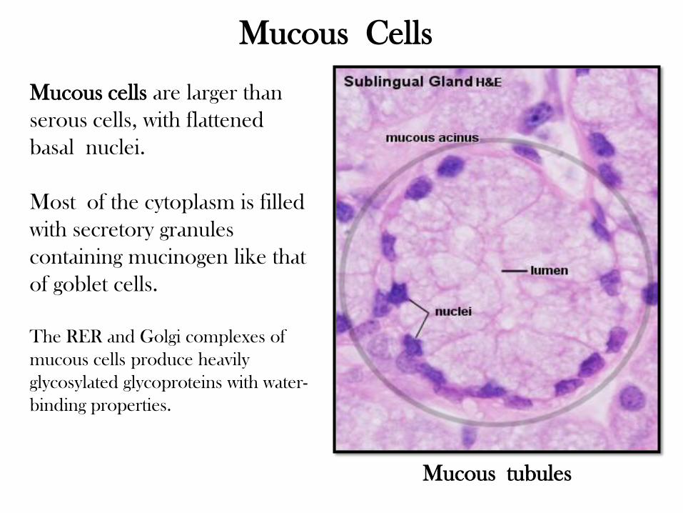
Mucous cells are larger than
serous cells, with flattened
basal nuclei.
Most of the cytoplasm is filled
with secretory granules
containing mucinogen like that
of goblet cells.
The RER and Golgi complexes of
mucous cells produce heavily
glycosylated glycoproteins with water-
binding properties.
Mucous tubules
Mucous Cells
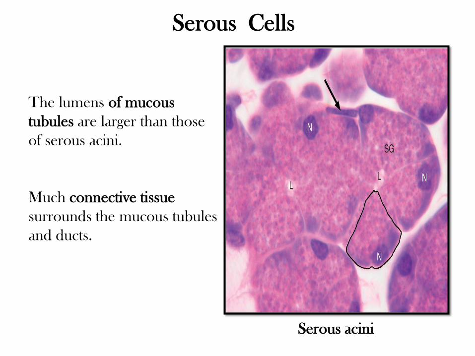
The lumens of mucous
tubules are larger than those
of serous acini.
Much connective tissue
surrounds the mucous tubules
and ducts.
Serous acini
Serous Cells
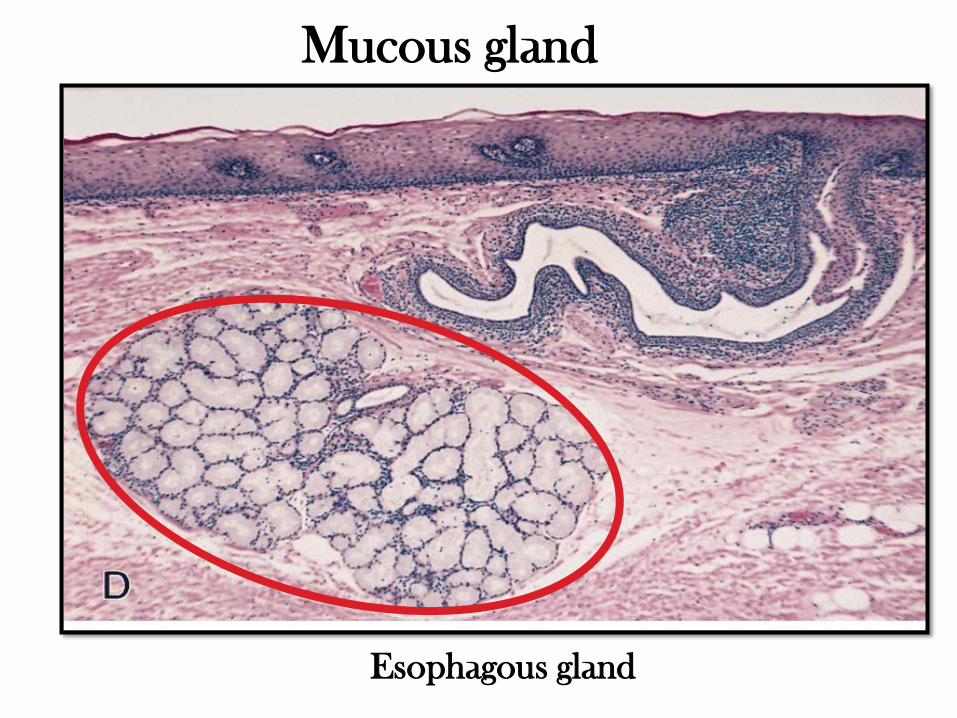
Esophagous gland
Mucous gland
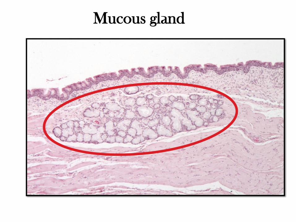
Mucous gland
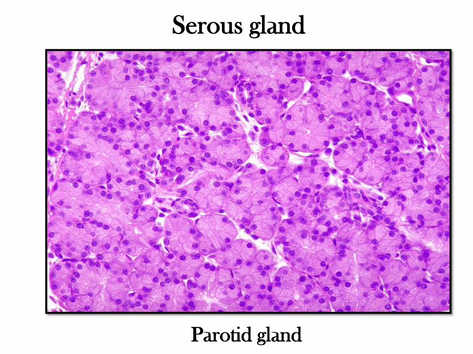
Parotid gland
Serous gland
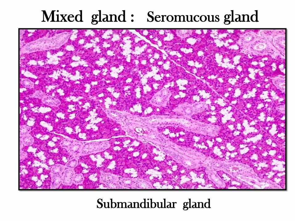
Mixed gland : Seromucous gland
Submandibular gland
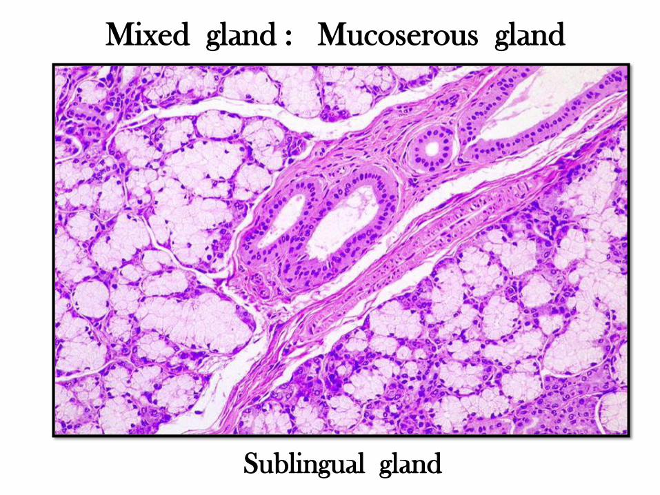
Mixed gland : Mucoserous gland
Sublingual gland
Classification
on the basis of the mode of
secretion
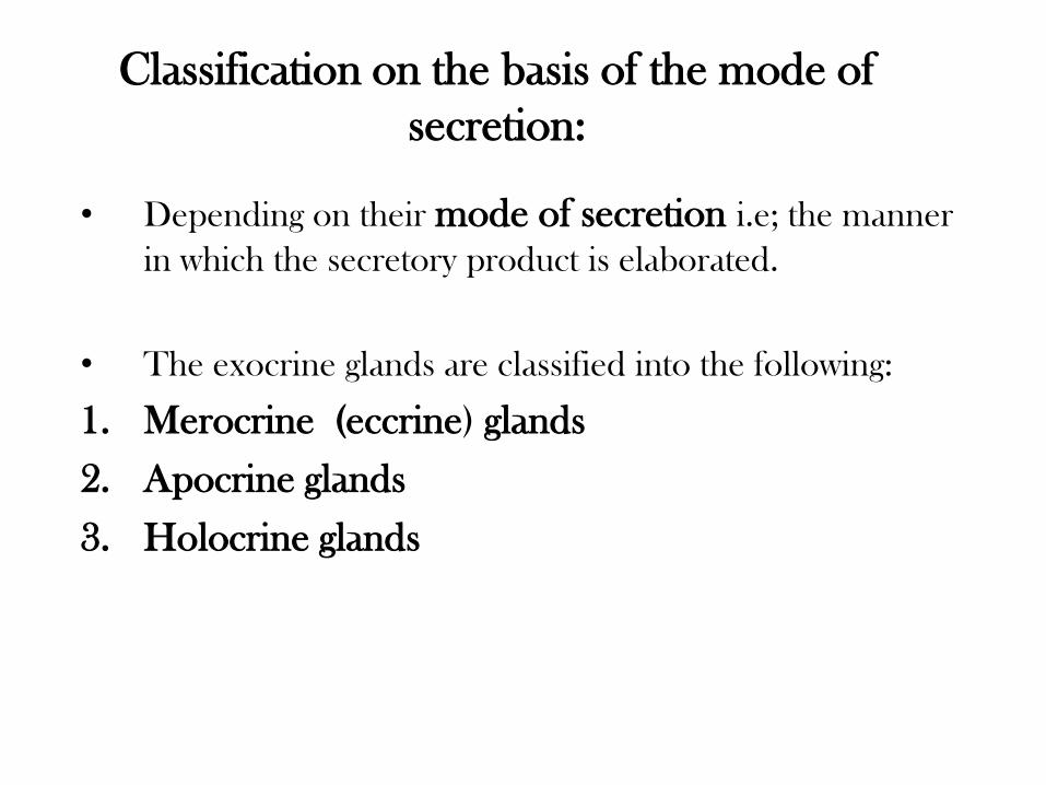
Classification on the basis of the mode of
secretion:
• Depending on their mode of secretion i.e; the manner
in which the secretory product is elaborated.
• The exocrine glands are classified into the following:
1. Merocrine (eccrine) glands
2. Apocrine glands
3. Holocrine glands
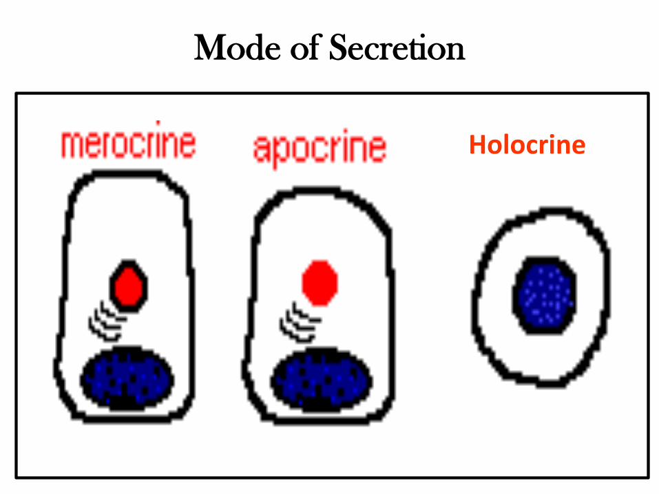
Holocrine
Mode of Secretion
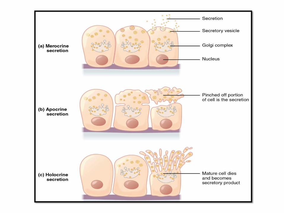
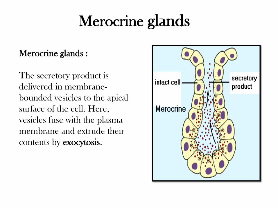
Merocrine glands
Merocrine glands :
The secretory product is
delivered in membrane-
bounded vesicles to the apical
surface of the cell. Here,
vesicles fuse with the plasma
membrane and extrude their
contents by exocytosis.
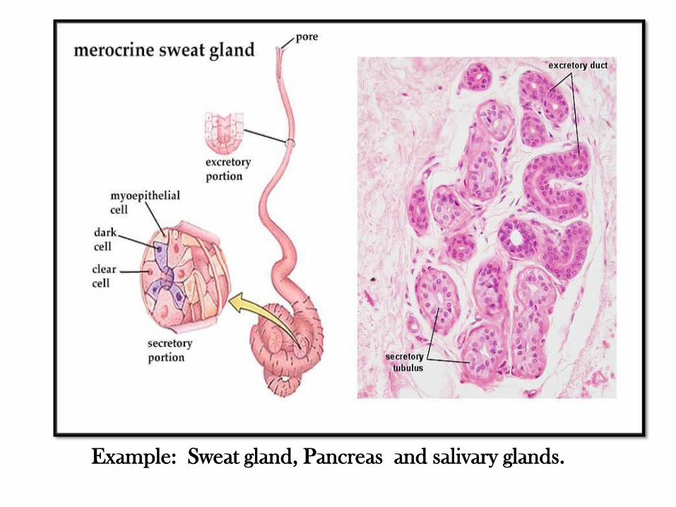
Example: Sweat gland, Pancreas and salivary glands.
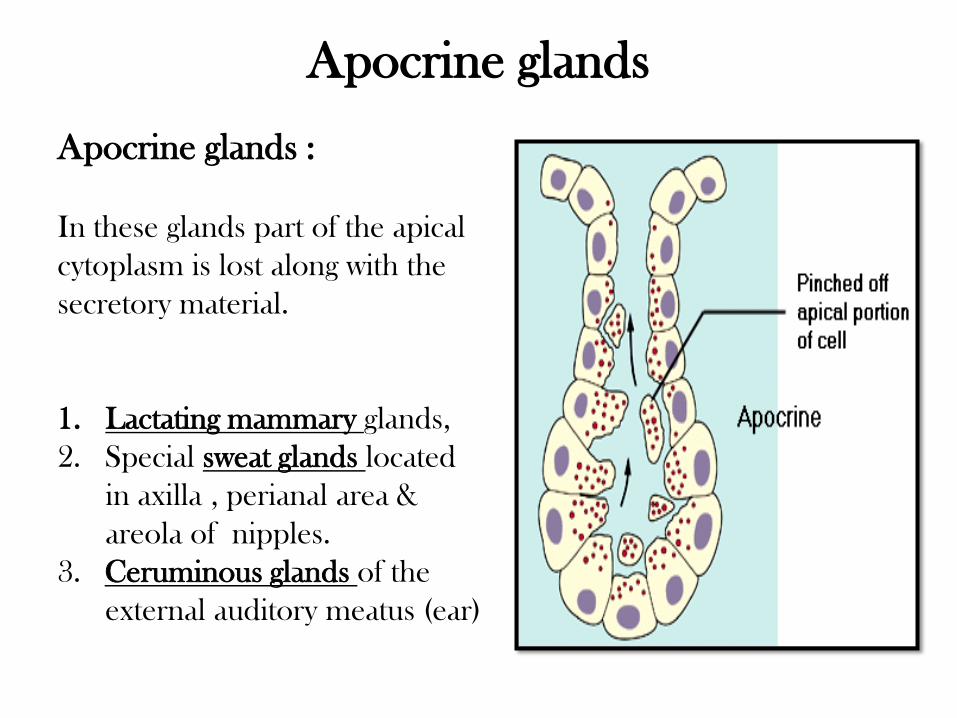
Apocrine glands
Apocrine glands :
In these glands part of the apical
cytoplasm is lost along with the
secretory material.
1. Lactating mammary glands,
2. Special sweat glands located
in axilla , perianal area &
areola of nipples.
3. Ceruminous glands of the
external auditory meatus (ear)
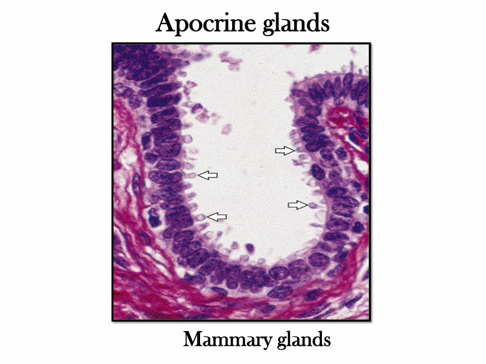
Mammary glands
Apocrine glands
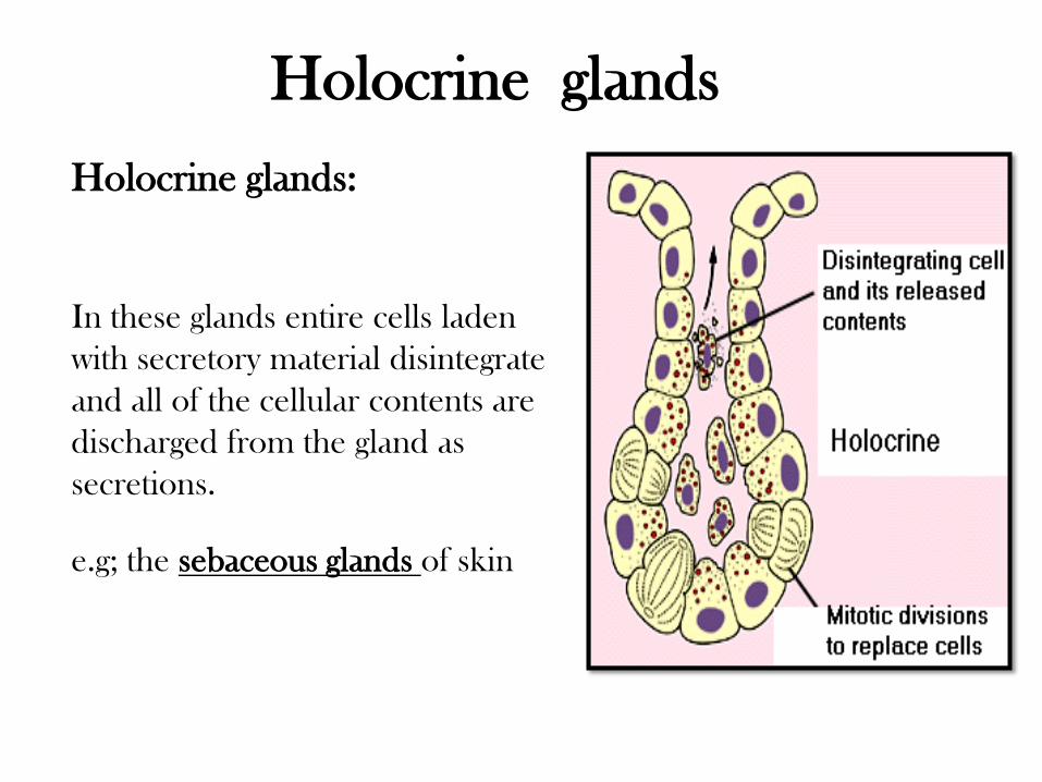
Holocrine glands
Holocrine glands:
In these glands entire cells laden
with secretory material disintegrate
and all of the cellular contents are
discharged from the gland as
secretions.
e.g; the sebaceous glands of skin
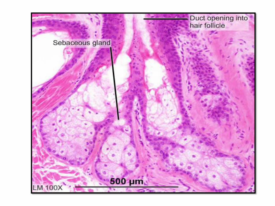
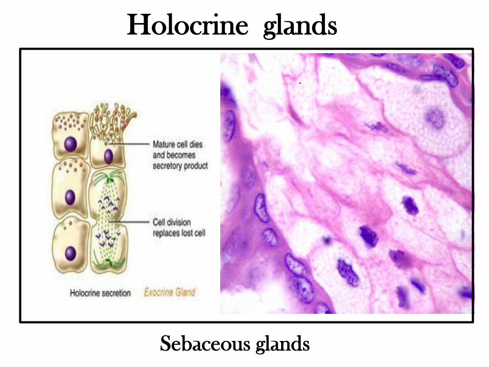
Holocrine glands
Sebaceous glands
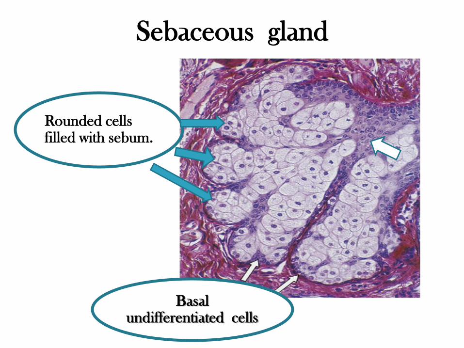
Sebaceous gland
Basal undifferentiated cells
Rounded cells filled with sebum.
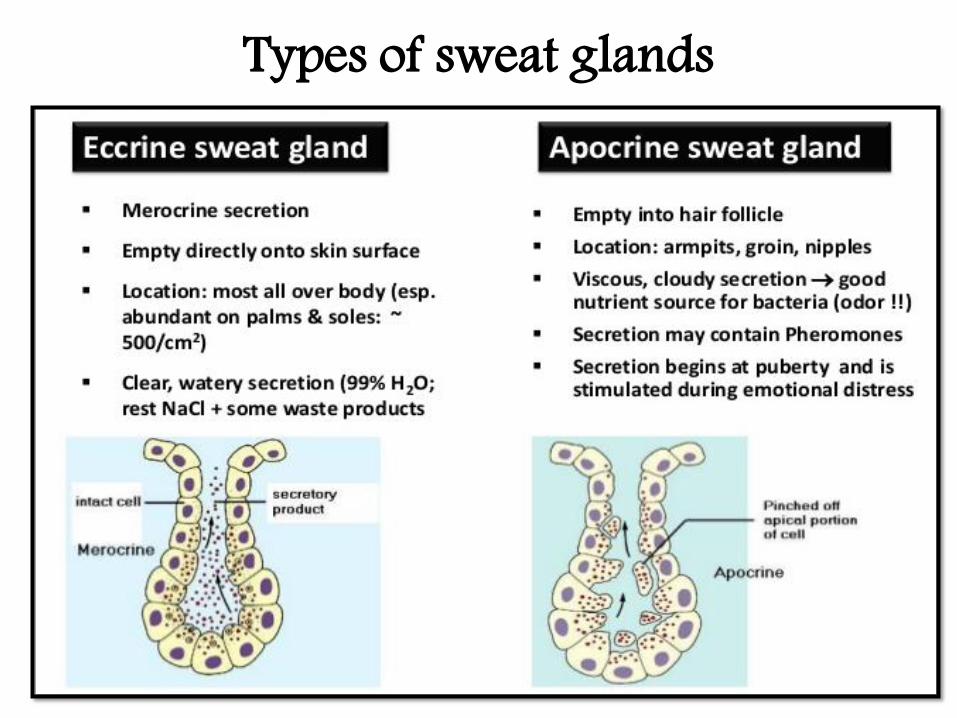
Types of sweat glands
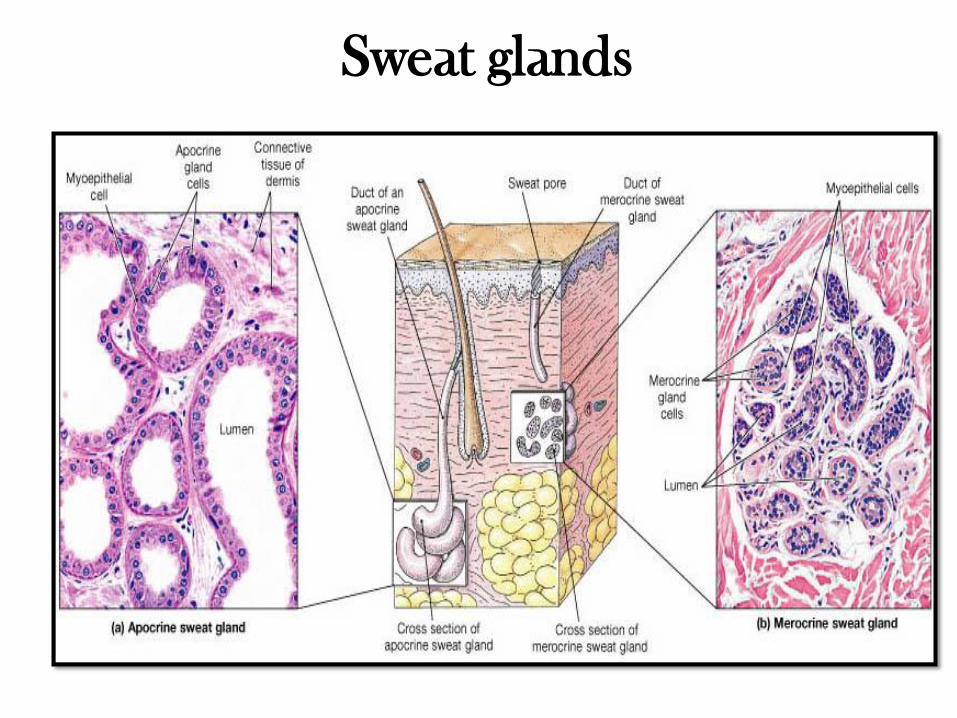
Sweat glands
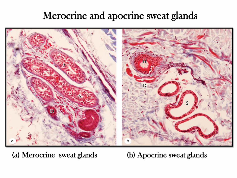
(a) Merocrine sweat glands (b) Apocrine sweat glands
Merocrine and apocrine sweat glands
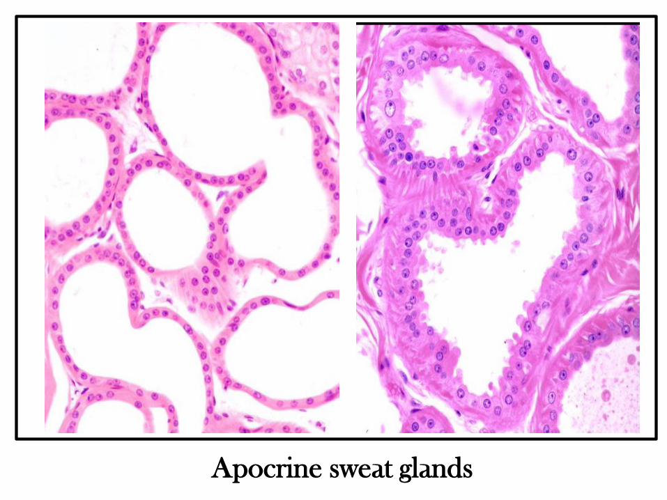
Apocrine sweat glands