Embed Size (px)
Citation preview

UPPER AND LOWER
GASTROINTESTINAL BLEEDING:
clinical and endoscopic approach
Giuliano Lombardi
ENDOSCOPIA GASTROINTESTINALE IN PEDIATRIA E NON SOLO...
PEDIATRIC GASTROINTESTINAL ENDOSCOPY AND BEYOND Roma,12-13 Aprile 2013 PRESIDENTE: PROF. SALVATORE CUCCHIARA
Ospedale Reg. di Pescara U.O.C. di Pediatria Medica, Direttore dott. Giuliano Lombardi
Unità di Gastroenterologia Ped /Servizio Speciale di Endoscopia Digestiva Ped

ANY BLEED THAT OCCURS DISTAL TO THE LIGAMENT OF TREITZ
AND SUPERIOR TO THE ANUS
INCLUDING THE LAST ¼ OF THE DUODENUM, AND THE ENTIRE AREA OF THE JEJUNUM, ILEUM,
COLON, RECTUM
LOWER GASTRO-INTESTINAL BLEEDING (LGIB)

Can present as an ..
ACUTE and life-threatening event
CHRONIC BLEEDING
which might manifest as
iron-deficiency anemia, fecal
occult blood or intermittent scant
hematochetia
Acute bleeding from the colon is usually less dramatic than upper gastrointestinal hemorrhage and is self-limiting in most cases.
Barnert J and Messmann H “Diagnosis and management of lower gastrointestinal bleeding” – Gastroenterol Hepatol 6, 637-646 (2009)
LOWER GASTRO-INTESTINAL BLEEDING (LGIB)

Barnert J and Messmann H “Diagnosis and management of lower gastrointestinal bleeding” – Gastroenterol Hepatol 6, 637-646 (2009)
Results from capsule and double-balloon endoscopy have revolutionized tha management algorithm of small bowel bleeding. Bleeding from the small bowel represents a distinct entity.
LOWER GASTRO-INTESTINAL BLEEDING (LGIB)

Barnert J and Messmann H “Diagnosis and management of lower gastrointestinal bleeding” – Gastroenterol Hepatol 6, 637-646 (2009)
GASTROINTESTINAL BLEEDING
UPPER MIDDLE LOWER

Consensus statement
sul sanguinamento gastrointestinale in età pediatrica (giugno 2005)
Panel
A. Barabino - [SIGENP] Genova
P. Betalli - [SICP] Padova
F. Cosentino - [SIED] Milano
L. Dall’Oglio - [SICP] [SIED] Roma
G. L.’de Angelis - [SIGENP] Parma
C. De Giacomo - [SIGENP] Milano
D. Falchetti - [SICP] Brescia
P. Gandullia - [SIGENP] Genova
G. Guariso - [SIGENP] Padova
G. Lombardi - [SIGENP] Pescara
A. Rossi - [SIED] Milano
V. Tomaselli - [SICP] Milano
Coord: C. Romano (SIGENP) Messina
Società Italiana di Gastroenterologia
Epatologia e Nutrizione Pediatrica
Società Italiana di Endoscopia Digestiva
Società Italiana di Chirurgia Pediatrica
Italian Panel Pediatric Endoscopy

Bleeding definition
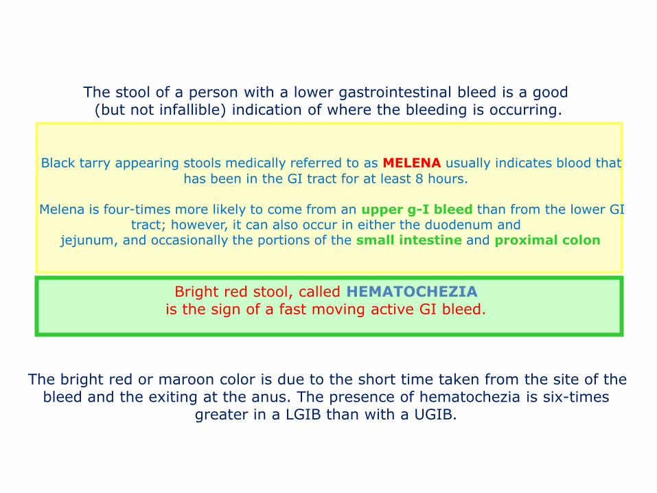
The stool of a person with a lower gastrointestinal bleed is a good (but not infallible) indication of where the bleeding is occurring.
Black tarry appearing stools medically referred to as MELENA usually indicates blood that
has been in the GI tract for at least 8 hours.
Melena is four-times more likely to come from an upper g-I bleed than from the lower GI tract; however, it can also occur in either the duodenum and
jejunum, and occasionally the portions of the small intestine and proximal colon
Bright red stool, called HEMATOCHEZIA is the sign of a fast moving active GI bleed.
The bright red or maroon color is due to the short time taken from the site of the bleed and the exiting at the anus. The presence of hematochezia is six-times
greater in a LGIB than with a UGIB.

CLINICAL DIAGNOSIS
AGE
LOCATION OF THE HEMORRHAGIC SITE
COLOUR AND SEVERITY OF THE BLEEDING
PRESENCE OR ABSENCE OF PAIN AND DIARRHEA

GOOD HISTORY is crucial to determinate the source of bleeding
Acuteness or chronicity of bleeding, color and quantity of the blood in stool or emesis, antecedent symptoms, history of straining, abdominal pain, or trauma.
Anorectal disorders, fissures, and distal polyps produce red blood
Melena rather than bright red blood per rectum is usually a sign of bleeding that comes from a source proximal to the ligament of Treitz
Massive upper GI bleeding can produce bright red blood per rectum if GI transit time is rapid
Blood mixed in stool or dark red blood implies a proximal source with some degree of digestion of the blood
Age- and etiology-specific symptoms to be aware of:
O Hsia, R Halpern and J Mola “Gastrointestinal bleeding” Pediatrics 2008

GOOD HISTORY is crucial to determinate the source of bleeding
A history of vomiting, diarrhea, fever, ill contacts, or travel suggests an infectious etiology
Bloody diarrhea and signs of obstruction suggest volvulus, intussusception, or necrotizing enterocolitis, particularly in the ex-premature infant
Recurrent or forceful vomiting is associated with Mallory-Weiss tears
Familial history of NSAID use may suggest ulcer disease
Age- and etiology-specific symptoms to be aware of:
O Hsia, R Halpern and J Mola “Gastrointestinal bleeding” Pediatrics 2008

GOOD HISTORY is crucial to determinate the source of bleeding
Recent jaundice, easy bruising, and changes in stool color may signal liver disease
Other evidence of coagulation abnormalities elicited from the history may also point to disorders of the kidney or reticuloendothelial system
For complaints of bloody stool, make sure to elicit on history foods or drugs that may give a stool bloody appearance (certain antibiotics, iron supplements, red licorice, chocolate, kool-Aid, flavored gelatin, or bismuth-containing products
Undiagnosed organ dysfunction possibilities to be aware of:
O Hsia, R Halpern and J Mola “Gastrointestinal bleeding” Pediatrics 2008

“ FALSE” GI BLEEDING
DEGLUTITION OF BREAST MILK
EPIXTASIS
HEMOPTYSIS
DRUGS AND FOODS

DIFFERENTIAL DIAGNOSIS
Neonates 1 month – 3 years > 3 years
Swallowed maternal blood Necrotizing enterocolitis Malrotation with midgut volvulus Allergic colitis (cow’s milk protein) Hirschsprung’s disease Anorectal fissures Coagulopathy Drugs
Allergic colitis (cow’s milk protein) Anorectal fissures Intussusception Meckel’s diverticulum Gastrointestinal duplication Polyps Ischemic bowel secondary to volvulus Rectal prolapse Infectious colitis Vascular malformation Drugs
Juvenile polyps IBD Vascular malformation Hemolytic uremic syndrome (HUS) Henoch Schonlein Purpura Iatrogenic Trauma - abuse
MUCOSAL LESION
VASCULAR LESION
DEFECTS IN HEMOSTASIS

OBSCURE GASTROINTESTINAL BLEEDING
unknown origin that persists or recurs after initial negative endoscopy
< 3 yrs > 3 yrs
GASTRIC- DUODENUM
ULCERS POLYPS
INTESTINE MECKEL MECKEL VASCULAR ANOMALIES
COLON ALLERGIC OR ACUTE COLITIS
IBD
5% between Treitz’s ligament and ileo-cecal valve

SIGNS OF IMPAIRED CIRCULATION
INSPECTION OF MOUTH
PURPURIC LESIONS (Shonlein-Henoch)
ORAL or LABIAL SIGNS of Peutz-Jaeghers
HEPATOSPLENOMEGALY
INSPECTION of the ANUS–RECTAL EXAMINATION
ASSESSMENT OF CARDIO-PULMONARY
SKIN COLOR
SENSORY EVALUATION
MUSCLE TONE
PUPIL REACTIVITY
FISICAL EXAM

LABORATORY
Complete blood count
PCR
Hepatic function
Renal function
Stool examination

OBSCURE-OCCULT BLEEDING
UPPER and LOWER ENDOSCOPY -if necessary repeat the exam
PUSH ENTEROSCOPY
-expert hands -also with therapeutic purposes MASSIVE BLEEDING
SCINTIGRAPHY
ANGIOGRAPHY
ENDOSCOPY
SURGERY
BLEEDING RELATIVELY MINOR
ENDOSCOPY

de Franchis R, 1996 - Baveno II
CLINICALLY SIGNIFICANT BLEEDING
TRANSFUSION REQUIREMENT of > 2 Units of blood within 24 h of time zero
PLUS A SYSTOLIC BLOOD PRESSURE OF < 100 MMHG
POSTURALE CHANGE OF > 20 mmHg
PULSE RATE OF > 100 /min AT TIME ZERO
PRIMARY CARE AND “ URGENT MANAGEMENT”

Angiography (positive on 27-77% acute LIB) Endoscopy Enteroscopy (single/double balloon; intraoperative)
Capsule endoscopy Scintigraphy (positive on 45% of LIB)
Surgery
DIAGNOSTIC EVALUATION

Enteroscope, single/double balloon
Antegrade (oral) DBE Retrograde (anal) DBE

Obscure GI bleeding Second-look endoscopy
overt occult
Capsule endoscopy
Angiography
Massive bleeding
positive
Specific management Medical treatment
PE or DBE cauterization Angiography + embolization
Laparoscopy IOE
negative
Further work-up needed?
Observation Medical treatment
Repeat routine endoscopy /CE Meckel’s scan
Laparoscopy/IOE
recurrence
positive negative
Follow up Specific management
No further Work up
NO
NO
YES
YES

Wireless Capsule Endoscopy
• Time efficient, patient friendly, sensitive method to visualize the small bowel
• Disadvantages
– No therapeutics
– Unable to control movement
– Unable to clear bubbles and debris

PillCamTM SB Vascular Lesions

Shonlein Henoch
3-7 aa GI localization 45-75%
~15%: GI bleeding and other GI symptoms preceding skin lesions
DD other vasculitis

Ulcers - Crohn’s disease
- Ulcer isolated (idiopathic, NSAIDs, 6 mercapt, ischemic) - Meckel’s diverticulum, Zollinger Ellison’s syndrome,
Vasculitis - Infections (Clostridium difficile, Salmonella,Tbc, Tifo, Campylobacter
jejuni, Yersinia, Rotavirus)
Small bowel

Crohn's disease
•panenteric inflammation
•characteristically segmental
Lenaerts C and others Pediatrics 83:777-781,1989

Crohn's disease
Example of Crohn's disease involving the small intestine. Here, the mucosal surface demonstrates an irregular nodular appearance with hyperemia and focal superficial ulceration.

MECKEL’S DIVERTICULUM

Treatment only of vascular lesions bleeding Always exclude coagulation disorders Individual therapy; there is no uniformity of treatment
Realistic treatment goals Carefully evaluate the entire small intestine before any surgery Other diseases often coexist
THERAPHY

“expertise”
THERAPHY

VASOACTIVE DRUGS SPLANCHNIC VASOCONSTRICTION,
REDUCING FLOW AND PORTAL PRESSURE
OCTEOTRIDE
4-8 g/kg/die
bolus ev 1-2 µg/Kg (5 min) 1-2 g/kg/h
time: RANGE 28.5-168 h
SOMATOSTATIN 50-100 mcg seguiti da
250 mcg/h ev for 24-120 h
GLYPRESSIN 1-2 mcg IV every
4-6h for 24h
R. De Franchis “ SMT and analogues and other vasoactive drugs in the treatment of bleeding oesophageal varices “
Digestive and Liver Disease 36 ( Suppl.1), 2004, S 93-100
….safe and effective in controlling nonarterial severe GI bleeding in children …..
…an important adjunct in the initial treatment of patients with severe GI bleeding and requiring stabilization before endoscopic or other investigative procedures…

ENDOSCOPIC TREATMENT OF BLEEDING
Therapeutic modalities
Contact thermal devices (eg, heater probe [HP], multipolar electrocautery[MPEC] probes, and hemostatic graspers) Noncontact thermal devices (eg, argon plasma coagulator [APC]) Injection needles Mechanical devices (eg, band ligators, clips, and loops)

CONCLUSIONS
ACCURATE DIAGNOSTIC ITER IS ESSENTIAL
IN MANAGEMENT OF DIGESTIVE BLEEDING
PHISICAL AND PSYCHOLOGICAL DIFFICULTIES
AND THE COSTS OF INAPPROPRIATE INVESTIGATIONS
ARE NOT REFUNDABLE………….!!!!!!!