Embed Size (px)
Citation preview
CASE REPORT
Gitelman syndrome: novel mutation and long-term follow-up
Aditi Sinha • Petr Lnenicka • Biswanath Basu •
Ashima Gulati • Pankaj Hari • Arvind Bagga
Received: 22 May 2011 / Accepted: 13 September 2011 / Published online: 4 October 2011
� Japanese Society of Nephrology 2011
Abstract We report a case of Gitelman syndrome pre-
senting with fatigue, paresthesias, weakness of limbs and
neck muscles since 2.5 years of age. Investigations showed
hypokalemia and hypomagnesemia with urinary magne-
sium wasting. Genetic analysis revealed the presence of a
novel homozygous mutation in the SLC12A3 gene
(c.2879_2883?9ins14bp, p.Val 960 Glu fsx12). Manage-
ment with potassium and magnesium supplements and
spironolactone resulted in a significant improvement in
symptoms. Over a follow-up of 11 years, the patient
showed satisfactory growth and physical development.
Keywords Hypokalemia � Hypomagnesemia �SLC12A3 gene
Introduction
Gitelman syndrome (OMIM 263800) is an autosomal
recessive renal tubular disorder characterized by hypo-
magnesemia, hypokalemic metabolic alkalosis and normal
blood pressure, with modestly increased blood levels of
renin and aldosterone [1, 2]. The condition is secondary to
inactivating mutations of the SLC12A3 gene that encodes
the thiazide-sensitive sodium chloride cotransporter
(NCCT) in the distal convoluted tubules. Most patients
have mild symptoms and the diagnosis is delayed to late
childhood or adulthood [1–3]. The occurrence of symptoms
in early childhood is rare, and information on the long-term
outcome of Gitelman syndrome is limited [4–7]. We report
a 6-year-old girl with Gitelman syndrome, with novel
homozygous mutation of the SLC12A3 gene, who was
followed up for 11 years.
Case report
A 6-year-old girl was referred for evaluation of polyuria,
nocturnal enuresis and intermittent weakness of lower
limbs since 2.5 years of age. The child tired easily and
complained of intermittent limb weakness. While she could
attend school normally, she could not participate in active
sports. She was admitted twice, at 3- and 5-years of age,
with muscle cramps and severe weakness of muscles of the
neck. There was intermittent history of paresthesias of the
neck and limbs. There was no history of seizures, syncope,
vertigo, palpitations, hematuria and use of diuretics or
laxatives. On examination, the blood pressure was
96/56 mmHg, pulse rate 78/min and respiratory rate
20/min. The height was 109.5 cm and weight was 16 kg, at
25th and 3rd percentile, respectively [8]. The muscle tone,
power and deep tendon reflexes were normal.
Blood levels of potassium ranged between 2.4 and
2.7 mEq/L, sodium 133–139 mEq/L, chloride 97–99 mEq/L,
venous pH 7.43–7.58, bicarbonate 20–22 mEq/L, magnesium
1.4–1.5 mg/dL (normal 1.7–2.4 mg/dL), urea 38 mg/dL and
creatinine 0.4 mg/dL. Urinalysis was normal and there was no
glucosuria or abnormal proteinuria. The levels of urinary
potassium were 70–180 mEq/L, sodium 50–117 mEq/L and
chloride 60–109 mEq/L. The urinary calcium excretion
A. Sinha (&) � B. Basu � A. Gulati � P. Hari � A. Bagga
Division of Pediatric Nephrology, Department of Pediatrics,
All India Institute of Medical Sciences,
New Delhi 110029, India
e-mail: [email protected]
P. Lnenicka
Institute of Biology and Medical Genetics of the 1st Faculty of
Medicine of Charles University and General Teaching Hospital,
Prague, Czech Republic
123
Clin Exp Nephrol (2012) 16:306–309
DOI 10.1007/s10157-011-0542-x

ranged between 6 and 30 mg/dL and calcium to creatinine
ratio was normal (0.03–0.18 mg/mg). The fractional excre-
tion of magnesium was 12.6% (normal\5%).
After overnight fasting, a thiazide test was performed by
estimation of serum and urinary electrolytes and creatinine
at baseline and over 4 h, following administration of oral
hydrochlorothiazide at a dose of 1 mg/kg [9]. The maximal
fractional excretion of chloride was 2.42%, similar to the
baseline value of 1.84%, suggesting a diagnosis of Gitel-
man syndrome. Plasma renin activity (1.33 ng/ml/h) and
blood levels of aldosterone (29.3 ng/dL), thyroxine and
thyroid-stimulating hormone were normal. Ultrasonogra-
phy of the abdomen showed normal kidneys with no
evidence of renal stones or nephrocalcinosis.
Family history was remarkable for third-degree parental
consanguinity (Fig. 1). A 9-year-old sister, with similar
symptoms since 4 years of age and with evaluation sug-
gesting hypokalemic metabolic alkalosis, had died recently
in a road traffic accident. Sequencing of the SLC12A3 gene
was carried out in the index case and her parents, and
compared to the reference genomic sequence (Genomic
sequence NC 000016; http://www.ncbi.nlm.nih.gov/gene/
6559). The analysis showed a novel homozygous mutation
of the SLC12A3 gene, in the form of an insertion of 14 base
pairs into the junction of exon 24 and intron 24
(c.2879_2883 ? 9ins14bp, p.Val 960 Glu fsx12) (Fig. 2).
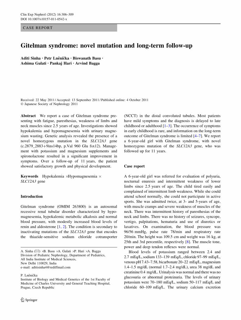
This mutation is expected to lead to the substitution of
valine by glutamic acid at amino acid 960 (p.960Val[Glu)
and a frameshift changing the amino acid sequence
downstream of amino acid 960 and introducing an abnor-
mal stop codon at position 971 (Fig. 3). This change may
also lead to misprocessing of the junction of exon 24 and
intron 24. Either action may result in the production of a
non-functional protein.
The parents showed the above mutation in heterozygous
form. Sequencing also revealed 3 known polymorphisms
(c.122A[G, p.Ala122Ala; c.791C[G, p.Ala264Gly and
c.2952 C[T, p.Ile984Ile) in the patient.
Based on the clinical, biochemical features and muta-
tional analysis, the patient was diagnosed as Gitelman
syndrome. She was treated with supplements of potassium
(2–3 mEq/kg/day), magnesium (as oral magnesium sul-
phate, 0.7–1.2 mEq/kg/day) and indomethacin (1 mg/kg/day).
While there was symptomatic improvement, the patient
complained of tiredness after prolonged activity, and
hypokalemia and hypomagnesemia persisted. Over the next
2 years, the doses of potassium and magnesium supple-
ments were maintained at 3.5 and 1 mEq/kg/day respec-
tively. In view of persistent hypokalemia, the patient also
received treatment with spironolactone without a signifi-
cant increase in the level of serum potassium. She con-
tinued to attend regular school but was unable to participate
in active sports. Menarche was attained at 14 years.
At last follow-up, 11 years after diagnosis, the 17-year-
old remains asymptomatic apart from tiredness after pro-
longed activity. Her present height and weight are 145 cm
(25th percentile) and 53 kg (80th percentile) respectively,
and the sexual maturity rating is stage 5. The blood
level of sodium is 137 mEq/L, potassium 3.4 mEq/L,
pH 7.38, bicarbonate 22 mEq/L, creatinine 0.7 mg/dL,
urea 34 mg/dL and magnesium 1.4 mEq/L. Current medi-
cations include indomethacin and supplements of potas-
sium and magnesium.
Discussion
The diagnosis of Gitelman syndrome in this patient was
based on clinical and biochemical features, and confirmed
by the presence of a homozygous mutation in the SLC12A3
gene [2]. During a follow-up of 11 years, physical growth
and development were normal. The chief concern during
management was an inability to maintain normal levels of
serum magnesium and potassium, resulting in episodic
muscle weakness and feeling of tiredness.
The presentation of Gitelman syndrome is heterogeneous
in terms of age at presentation, clinical features and severity
of biochemical abnormalities. Most patients are diagnosed in
late childhood, beyond 6 years of age, or adulthood during
routine investigation or when evaluated for cramps, fatigue,
paresthesias and tetany [2]. A subgroup of patients have early
onset of symptoms including tetany, paralysis, seizures,
rhabdomyolysis, growth retardation and arrhythmias [7, 9].
The mild nature of clinical and laboratory abnormalities in
our patient likely represent a milder phenotype, which may
Fig. 1 Pedigree chart showing third-degree parental consanguinity.
An elder sibling with similar presentation died in an accident
Clin Exp Nephrol (2012) 16:306–309 307
123

explain absence of overt alkalosis and normal levels of
plasma renin and aldosterone. In comparison with Bartter
syndrome, the degree of volume contraction and the asso-
ciated stimulation of the renin-angiotensin-aldosterone
axis in Gitelman syndrome are mild, and plasma aldosterone
levels might remain within normal range.
Data on long-term follow-up of patients diagnosed in
early childhood is limited. Bettinelli et al. [4] reported a
6-year-old girl with Gitelman syndrome, who was followed
for 14 years. At 20 years of age, the growth and develop-
ment were satisfactory and magnesium supplementation
reduced the risk of symptoms such as tetany. Satisfactory
physical growth has been reported following correction of
electrolyte abnormalities in a case series of children with
Gitelman syndrome [5]. Correction of biochemical abnor-
malities in the present patient also resulted in significant
clinical improvement. At the end of follow-up of 11 years,
she is intermittently symptomatic but has shown satisfac-
tory physical growth.
More than 180 mutations throughout the SLC12A3 gene
have been identified in Gitelman syndrome (Human Gene
Mutation Database, http://www.hgmd.cf.ac.uk). Most are
missense mutations; nonsense, frameshift and splice-site
mutations are less frequent. The condition is genetically
heterogeneous and no hotspots are identified, although
some mutations are more commonly reported in certain
ethnic groups [10]. There is limited evidence of a geno-
type-phenotype correlation, with considerable phenotypic
variability even within families having the same mutations
[10]. A proportion of patients show only a single mutation
in the heterozygous state, instead of being compound het-
erozygous or homozygous for such mutations [11, 12].
Although the mutation in the present case is not previously
reported, its presence in the homozygous state in a patient
with characteristic clinical and biochemical features sup-
ports the diagnosis of Gitelman syndrome. It is likely that
the present mutation resulted in a significant loss of NCCT
channel activity leading to an early onset of symptoms.
Fig. 2 Electrophoregram demonstrating the sequence of base pair at
the junction of the exon 24 and intron 24 of SLC12A3 gene. Arrowson the figure indicate (1) the first base of a 14-base pair length
insertion mutation that introduces a frameshift mutation, (2) the first
base after the frameshift insertion, and (3) the end of the mutated exon
24. The vertical strip denotes the end of the non-mutated exon. The
sequence of the reference normal exon is annotated below the mutated
sequence
308 Clin Exp Nephrol (2012) 16:306–309
123

Confirmation of the pathogenicity of this mutation requires
transcript analysis and functional studies.
The present report adds to the experience of manage-
ment of Gitelman syndrome presenting in early childhood.
The patient showed the presence of a novel mutation,
which was considered pathogenic. Long-term potassium
and magnesium supplementation resulted in improvement
of clinical symptoms and normal physical growth.
References
1. Simon DB, Nelson-Williams C, Bia MJ, Ellison D, Karet FE,
Molina AM, et al. Gitelman’s variant of Bartter’s syndrome,
inherited hypokalemic alkalosis, is caused by mutations in the
thiazide-sensitive Na–Cl cotransporter. Nat Genet. 1996;12:
24–30.
2. Knoers NV. Inherited forms of renal hypomagnesemia: an
update. Pediatr Nephrol. 2009;24:697–705.
3. Peters M, Jeck N, Reinalter S, Leonhardt A, Tonshoff B, Klaus
GG, et al. Clinical presentation of genetically defined patients
with hypokalemic salt-losing tubulopathies. Am J Med.
2002;112:183–90.
4. Bettinelli A, Metta MG, Perini A, Basilico E, Santeramo C.
Long-term follow-up of a patient with Gitelman’s syndrome.
Pediatr Nephrol. 1993;7:67–8.
5. Herrero-Morın JD, Rodrıguez J, Coto E, Gil-Pena H, Alvarez V,
Espinosa L, et al. Gitelman syndrome in Gypsy pediatric patients
carrying the same intron 9 ? 1 G[T mutation. Clinical features
and impact on quality of life. Nephrol Dial Transpl. 2011;26:
151–5.
6. Conti G, Vitale A, Tedeschi S, Syren ML, Pantano R, Chimenz R,
et al. Hypokalaemia and failure to thrive: report of a misleading
onset. J Paediatr Child Health. 2010;46:276–7.
7. Pachulski RT, Lopez F, Sharaf R. Gitelman’s not-so-benign
syndrome. N Engl J Med. 2005;353:850–1.
8. http://www.cdc.gov/growthcharts/percentile_data_files.htm
(accessed 29 April 2011).
9. Colussi G, Bettinelli A, Tedeshi S, de Ferrari ME, Syren ML,
Borsa N, et al. A thiazide test for the diagnosis of renal tubular
hypokalemic disorders. Clin J Am Soc Nephrol. 2007;2:454–60.
10. Riveira-Munoz E, Chang Q, Godefroid N, Hoenderop JG, Bindels
RJ, Dahan K, et al. for the Belgian Network for the Study of
Gitelman syndrome. Transcriptional and functional analyses of
SLC12A3 mutations: new clues for the pathogenesis of Gitelman
syndrome. J Am Soc Nephrol. 2007;18:1271–83.
11. Takeuchi K, Kure S, Kato T, Taniyama Y, Takahashi N, Ikeda Y,
et al. Association of a mutation in thiazide-sensitive Na–Cl
cotransporter with familial Gitelman syndrome. J Clin Endocrinol
Metab. 1996;81:4496–9.
12. Lemmink HH, Knoers NV, Karolyi L, van Dijk H, Niaudet P,
Antignac C, et al. Novel mutations in the thiazide-sensitive NaCl
cotransporter gene in patients with Gitelman syndrome with
predominant localization to the C-terminal domain. Kidney Int.
1998;54:720–30.
Fig. 3 Figure showing expected effect of mutation on protein
synthesis. The upper panel depicts the amino acid sequence generated
by translation of the normal nucleotide sequence at the junction of
exon 24 and intron 24. Normally, the amino acid chain is terminated
at position 1030 due to the presence of a stop codon. Mutation
between nucleotides guanine and thymine (indicated by the arrowpointing upwards in the upper panel) results in the insertion of 14
nucleotides (shown as the underlined sequence in the lower panel).This is expected to result in a frameshift mutation, with the
introduction of a stop codon at position 971
Clin Exp Nephrol (2012) 16:306–309 309
123