Embed Size (px)
Citation preview

GIPC1 Interacts with MyoGEF and Promotes MDA-MB-231Breast Cancer Cell Invasion*
Received for publication, January 25, 2010, and in revised form, July 13, 2010 Published, JBC Papers in Press, July 15, 2010, DOI 10.1074/jbc.M110.107649
Di Wu, Akiko Haruta, and Qize Wei1
From the Department of Biochemistry, Kansas State University, Manhattan, Kansas 66506
GIPC1/synectin, a single PDZ domain-containing protein,binds to numerous proteins and is involved in multiple biologi-cal processes, including cell migration. We reported previouslythatMyoGEF, a guanine nucleotide exchange factor, plays a rolein regulating breast cancer cell polarization and invasion. Here,we identify GIPC1 as an interacting partner of MyoGEF. Bothin vitro and in vivo binding assays show that the GIPC1 PDZdomain binds to the PDZ-binding motif at the C terminus ofMyoGEF. Immunofluorescence analysis shows that GIPC1 andMyoGEF colocalize to the cell leading edge. Depletion of GIPC1byRNAi inMDA-MB-231 cells causes cells to shift fromapolar-ized to a rounded morphology. Matrigel invasion assays showthat RNAi-mediated depletion of GIPC1 dramatically decreasesMDA-MB-231 cell invasion. Notably, an anti-MyoGEF peptideantibody, whose epitope is located at the C terminus of MyoGEF,interferes with GIPC1-MyoGEF complex formation. Treatmentof MDA-MB-231 cells with the anti-MyoGEF peptide antibodydisrupts cell polarization and invasion. Thus, our results suggestthat GIPC1-MyoGEF complex formation plays an importantrole in regulating MDA-MB-231 breast cancer cell polarizationand invasion.
Rho GTPase signaling plays a central role in regulating cellmigration (1). Evidence has accumulated indicating thatsmall GTPase proteins, including Rac1, Cdc42, and RhoA,can be activated at the cell leading edge, where they regulateactin polymerization and membrane protrusion, thus con-tributing to the regulation of cell migration (2–5). Guaninenucleotide exchange factors (GEFs)2 activate the smallGTPase proteins by catalyzing the exchange of bound GDPfor GTP, whereas GTPase-activating proteins inactivate thesmall GTPase proteins by increasing their low intrinsicGTPase activity (6, 7). Therefore, localization of GEFs tospecific subcellular locations is critically important for spa-tiotemporal activation of small GTPase proteins (3, 8). Atleast two different mechanisms have been implicated in reg-ulating the localization of GEFs. First, protein-protein inter-actions involving the pleckstrin homology domain of GEFs
can target GEFs such as Dbl and Trio to their destinationssuch as the actin cytoskeleton (9–11). Second, the PDZ-binding motif is found in �40% of GEFs, and binding of PDZdomain-containing proteins to the PDZ-binding motif ofGEFs such as Syx1, kalirin-7, and bPIX can target GEFs tospecific locations such as the plasma membrane (12–14).GIPC1/synectin, a single PDZ domain-containing protein,
acts as a scaffolding protein to function inmultiple biologicalprocesses such as protein trafficking, endocytosis, andreceptor clustering (15, 16). Accumulating evidence furtherindicates that GIPC1 plays a role in regulating cell polarityand motility. Murine primary arterial endothelial cells de-rived from GIPC1-ablated mice show decreased migrationand impaired polarization (17). Syndecan-4, Syx1, andendoglin regulate cell migration through interactions withGIPC1/synectin (18–20). GIPC1 interacts with 5T4, a trans-membrane glycoprotein that is involved in tumor metastasis(21–23). Moreover, a recent study shows that cancerous breasttissues express an increased level of GIPC1 comparedwith nor-mal breast tissues (24). However, it is not clear whether GIPC1plays a role in regulating breast cancer cell migration and/orinvasion.We reported previously that MyoGEF, a guanine nucleo-
tide exchange factor, can activate RhoA/RhoC and is impli-cated in regulating breast cancer cell invasion (25). Usingyeast two-hybrid screening, we identified GIPC1 as one ofthe MyoGEF-interacting proteins. In this study, we demon-strate that the GIPC1 PDZ domain can bind to the PDZ-binding motif at the C terminus of MyoGEF. Depletion ofGIPC1 by RNAi inhibits the invasion activity of MDA-MB-231 breast cancer cells. Moreover, we show that an anti-MyoGEF peptide antibody can bind to the C terminus ofMyoGEF and interfere with the in vitro interaction betweenGIPC1 and MyoGEF. Treatment of MDA-MB-231 cells withthe anti-MyoGEF peptide antibody disrupts cell polarizationand invasion. Thus, our results indicate that complex forma-tion between GIPC1 and MyoGEF plays a role in regulatingMDA-MB-231 cell polarization and invasion.
EXPERIMENTAL PROCEDURES
Yeast Two-hybrid Screening—Yeast two-hybrid screeningwas carried out as described previously (26). Briefly, full-lengthhuman MyoGEF was used as bait to screen a mouse 11-dayembryo Matchmaker cDNA library (Clontech). Synthetic de-fined medium lacking leucine, tryptophan, and histidine wasused to identify the positive yeast colonies. The filter lift assayfor �-galactosidase activity was then carried out to confirm thepositive colonies. The cDNA fragments encoding the potential
* This work was supported, in whole or in part, by National Institutes of HealthGrant P20 RR015563 from the National Center for Research Resources. Thiswork was also supported by the Terry C. Johnson Center for Basic CancerResearch. This is Contribution 10-211-J from the Kansas Agricultural Exper-iment Station (Manhattan, KS).
1 To whom correspondence should be addressed: Dept. of Biochemistry, Kan-sas State University, 141 Chalmers Hall, Manhattan, KS 66506. Tel.: 785-532-6736; Fax: 785-532-7278; E-mail: [email protected].
2 The abbreviation used is: GEF, guanine nucleotide exchange factor.
THE JOURNAL OF BIOLOGICAL CHEMISTRY VOL. 285, NO. 37, pp. 28643–28650, September 10, 2010© 2010 by The American Society for Biochemistry and Molecular Biology, Inc. Printed in the U.S.A.
SEPTEMBER 10, 2010 • VOLUME 285 • NUMBER 37 JOURNAL OF BIOLOGICAL CHEMISTRY 28643
by guest on July 9, 2018http://w
ww
.jbc.org/D
ownloaded from

MyoGEF-interacting partners were recovered from the posi-tive yeast colonies and subjected to DNA sequencing. Themouse gipc1 cDNA was amplified using the following primerpair: 5�-GAATTCAATGCCACTGGGACTGGGG-3� (for-ward primer; the underlined nucleotide sequence is the recog-nition site for EcoRI) and 5�-CTCGAGGTAGCGGCCAAC-CTTGGC-3� (reverse primer; the underlined nucleotidesequence is the recognition site for XhoI).Plasmids and Cell Culture—pEGFP-MyoGEF and pCS3-
MyoGEF were described previously (27). GIPC1 and MyoGEFcDNA fragments were subcloned into pEGFP-C3 andpCS3�MT vectors to generate plasmids encoding GFP- orMyc-tagged polypeptides. All plasmids encoding GST-taggedMyoGEF or GIPC1 fragments were generated by subcloningthe cDNA fragments into the pGEX-6p-1 vector. MDA-MB-231 breast cancer cells were purchased from American TypeCulture Collection (Manassas, VA). MDA-MB-231 cellswere grown in Leibovitz’s L-15 medium supplemented with10% fetal bovine serum. HeLa cells were purchased fromClontech and were grown in DMEM supplemented with 10%fetal bovine serum. Transfection was done with Lipo-fectamine 2000 (Invitrogen) according to the manufacturer’sinstructions. siRNAs specific for human GIPC1 were pur-chased from Invitrogen (siRNA1, GCU ACG CCU UCAUCA AGC GCA UCA A; siRNA2, CCA ACG UCA AGGAGCUGUAUGGCAA; and siRNA3, UGUGGAGCCUGUUAC CUC CGC AUU U).Protein Expression and in Vitro Translation—GST-fused
polypeptides were expressed in a bacterial expression system.BL21 bacterial cells expressing GST-fused polypeptides werehomogenized by sonication and lysed in PBS containing 1%Triton X-100 for 1 h at 4 °C. The GST fusion proteins werepurified using glutathione-conjugated agarose beads, elutedwith 100 mM Tris-HCl (pH 7.5) and 5 mM glutathione, anddialyzed against 50 mM Tris-HCl (pH 7.5) and 50 mM NaCl.In vitro translatedMyc-tagged proteins were synthesized usingthe TNT SP6 quick coupled transcription/translation system(Promega) according to the manufacturer’s instructions.Immunoprecipitation and GST Pulldown Assays—Immuno-
precipitation and GST pulldown assays were carried out asdescribed previously (27, 28). Briefly, transfected cells werelysed in radioimmune precipitation assay lysis buffer (50 mM
Tris-HCl (pH 7.5), 150 mM NaCl, 0.25% deoxycholate, 1%Nonidet P-40, 1 mM EDTA, 1 mM PMSF, 1 mM Na3VO4, and1 mMNaF with protease inhibitor mixture) for 10 min on ice.Cell extracts were collected and precleared with proteinA/G-agarose beads. The precleared lysate was incubatedwith agarose-conjugated anti-Myc antibody overnight at4 °C. After washing four times with radioimmune precipita-tion assay lysis buffer, the bound proteins were eluted withSDS loading buffer. For GST pulldown experiments, theimmobilized GST-fused polypeptides were incubated with invitro translated Myc-tagged proteins or with cell lysates fromtransfected cells overnight at 4 °C. After washing four timeswith binding buffer (50 mM Tris-HCl (pH 7.4), 100 mM NaCl,0.05% Triton X-100, 10% glycerol, 0.2 mM EDTA, and 1 mM
DTT), the beads were resuspended in SDS loading buffer toelute the bound proteins.
Immunoblotting—Cell lysates and immunoprecipitated andGST pulldown proteins were separated on 7 or 4–12% SDS-polyacrylamide gel, transferred to an Immobilon-P transfermembrane (Millipore), blocked in 5% nonfat milk, and incu-bated with primary antibodies as indicated. The following pri-mary antibodies were used: mouse anti-Myc (9E10, 1:1000),rabbit anti-GFP (1:1000), and rabbit anti-�-tubulin (1:2000)(Santa Cruz Biotechnology); goat anti-GIPC1 (1:250; NovusBiologicals, Littleton, CO); and rabbit anti-MyoGEF (1:100)(25, 27). The blots werewashed and incubatedwith horseradishperoxide-conjugated secondary antibodies (1:5000; Santa CruzBiotechnology) for 1 h at 23 °C. The blots were visualized bySuperSignal West Pico luminol/enhancer solution (Pierce).Immunofluorescence—Immunofluorescence was carried
out as described previously (25, 26). MDA-MB-231 cellstransfected with plasmids or siRNA were trypsinized; cul-tured on fibronectin-coated coverslips for an additional 1, 3,or 5 h; and then fixed with 4% paraformaldehyde. For RhoAstaining, cells were fixed with 10% TCA for 10 min on ice.The primary antibodies used for immunofluorescence weremouse monoclonal anti-Myc (9E10, 1:1000), rabbit poly-clonal anti-MyoGEF (1:50), and goat polyclonal anti-GIPC1(1:100; Abcam). The following secondary antibodies werepurchased from Invitrogen: Alexa Fluor 594-labeled donkeyanti-mouse IgG (1:500), Alexa Fluor 350-labeled donkey anti-mouse IgG (1:500), Alexa Fluor 594-labeled donkey anti-goat IgG (1:500), Alexa Fluor 488-labeled donkey anti-goatIgG (1:500), Alexa Fluor 594-labeled donkey anti-rabbit IgG(1:500), and Alexa Fluor 488-labeled donkey anti-rabbit IgG(1:500). Actin filaments were stained with rhodamine- or FITC-phalloidin (Invitrogen). Images were taken using a Leica DMI6000 B microscope and processed by blind deconvolution. Todetermine the cell polarity, long (L) and short (S) axes of indi-vidual cells were measured using the NIH ImageJ program.Cells were counted as polarized (L/S ratio � 2.0) or nonpolar-ized (L/S ratio � 2.0).Matrigel Invasion Assays—Transfected or antibody-treated
MDA-MB-231 cells were trypsinized, and �1 � 105 cells (inLeibovitz’s L-15 medium containing 3% BSA) were seededon the upper wells of BioCoat Matrigel chambers (BD Bio-sciences). The lower wells were filled with Leibovitz’s L-15medium containing 10% FBS. The transfected cells thenunderwent chemoattraction across the Matrigel and filter(pore size of 8 �m) to the lower surface of the transwells for22 h. The nonmigrating cells on the upper chambers wereremoved with a cotton swab. The migrated cells on the lowersurface of the membrane were fixed in 4% paraformalde-hyde, stained with 1% crystal violet, and then photographedat five different and random fields with a 20� objective. Datawere collected from three independent experiments, eachdone in triplicate. Migrated cells were counted, and meandifferences (�S.E.) between groups were analyzed using Stu-dent’s t test.In Vitro Antibody Delivery—MDA-MB-231 cells were
treated with normal IgG or anti-MyoGEF antibody using invitro PULSin protein/antibody and peptide delivery reagent(Genesee Scientific, San Diego, CA) according to the manu-facturer’s instructions. Cells were grown in a 24-well tissue
GIPC1-MyoGEF Interaction
28644 JOURNAL OF BIOLOGICAL CHEMISTRY VOLUME 285 • NUMBER 37 • SEPTEMBER 10, 2010
by guest on July 9, 2018http://w
ww
.jbc.org/D
ownloaded from
culture plate. 1 �g of normal rabbit IgG (Sigma) or anti-MyoGEF antibody was used for each well of a 24-well tissueculture plate. 4 h after antibody treatment, the treated cellswere subjected to Matrigel invasion assays as describedabove or processed for immunofluorescence staining withphalloidin and Alexa Fluor 594-labeled goat anti-rabbit IgG(Invitrogen).
RhoA/RhoC Activation Assays—RhoA/RhoC activationassays were preformed as described previously (25, 29).
RESULTS
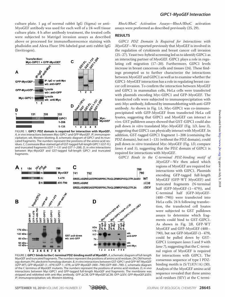
GIPC1 PDZ Domain Is Required for Interactions withMyoGEF—We reported previously that MyoGEF is involved inthe regulation of cytokinesis and breast cancer cell invasion(25–27). Yeast two-hybrid screening led us to identify GIPC1 asan interacting partner of MyoGEF. GIPC1 plays a role in regu-lating cell migration (17–20). Furthermore, GIPC1 levelsincrease in breast cancerous cells and tissues (24). These find-ings prompted us to further characterize the interactionsbetweenMyoGEF andGIPC1 aswell as to examinewhether theGIPC1-MyoGEF interaction has a role in regulating breast can-cer cell invasion. To confirm the interaction betweenMyoGEFand GIPC1 in mammalian cells, HeLa cells were transfectedwith plasmids encoding Myc-GIPC1 and GFP-MyoGEF. Thetransfected cells were subjected to immunoprecipitation withanti-Myc antibody, followed by immunoblotting with anti-GFPantibody. As shown in Fig. 1A, Myc-GIPC1 was co-immuno-precipitated with GFP-MyoGEF from transfected HeLa celllysates, suggesting that GIPC1 and MyoGEF can interact invivo. GST pulldown assays showed that GST-GIPC1 could alsopull down in vitro translated Myc-MyoGEF (Fig. 1D, lane 3),suggesting that GIPC1 can physically interact withMyoGEF. Inaddition, GST-tagged GIPC1 fragment 1–208 (containing thePDZ domain), but not 1–131 (without the PDZ domain), couldpull down in vitro translated Myc-MyoGEF (Fig. 1D, comparelanes 4 and 5), suggesting that the PDZ domain of GIPC1 isrequired for interactions with MyoGEF.GIPC1 Binds to the C-terminal PDZ-binding motif of
MyoGEF—We then asked whichregions of MyoGEF are required forinteractions with GIPC1. Plasmidsencoding GFP-tagged full-lengthMyoGEF (GFP-WT MyoGEF) andtruncated fragments (N-terminalhalf (GFP-MyoGEF-(1–479)), andC-terminal half (GFP-MyoGEF-(400–790)) were transfected intoHeLa cells. 24 h following transfec-tion, the transfected cell lysateswere subjected to GST pulldownassays to determine which frag-ments could bind to GST-GIPC1.As shown in Fig. 2B, GFP-WTMyoGEF and GFP-MyoGEF-(400–790), but not GFP-MyoGEF-(1–479),could be pulled down by GST-GIPC1 (compare lanes 5 and 9 withlane 7), suggesting that theC-termi-nal region of MyoGEF is requiredfor interactions with GIPC1. Theconsensus sequence of type I PDZ-binding motifs is (S/T)X(V/A) (30).Analysis of theMyoGEF amino acidsequence revealed that three aminoacid residues (SEV) at the C termi-
FIGURE 1. GIPC1 PDZ domain is required for interaction with MyoGEF.A, in vivo interactions between Myc-GIPC1 and GFP-MyoGEF. IP, immunopre-cipitation; wb, Western blotting. B, schematic diagram of GIPC1 and its trun-cated fragments. The numbers represent the positions of the amino acid res-idues. C, Coomassie Blue-stained gel of GST-tagged full-length GIPC1 (GST-FL)and truncated fragments (GST-1–131 and GST-1–208). D, in vitro interactionsbetween Myc-MyoGEF and GST-tagged full-length GIPC1 and truncatedfragments.
FIGURE 2. GIPC1 binds to the C-terminal PDZ-binding motif of MyoGEF. A, schematic diagram of full-lengthMyoGEF and truncated fragments. The numbers represent the positions of amino acid residues. DH, Dbl homol-ogy domain; PH, pleckstrin homology domain. B, in vitro interactions between GST-GIPC1 and GFP-WT MyoGEF(GFP-WT), GFP-MyoGEF-(1– 479) (GFP-1– 479), or GFP-MyoGEF-(400 –790) (GFP-400 –790). C, schematic diagramof the C terminus of MyoGEF mutants. The numbers represent the positions of amino acid residues. D, in vivointeractions between Myc-GIPC1 and GFP-tagged full-length MyoGEF and fragments. The membrane wasstripped and reblotted with anti-Myc antibody. GFP-�C38, GFP-MyoGEF�C38; GFP-�SEV, GFP-MyoGEF�SEV;IP, immunoprecipitation; wb, Western blotting.
GIPC1-MyoGEF Interaction
SEPTEMBER 10, 2010 • VOLUME 285 • NUMBER 37 JOURNAL OF BIOLOGICAL CHEMISTRY 28645
by guest on July 9, 2018http://w
ww
.jbc.org/D
ownloaded from

nus of MyoGEF appear to be a type I PDZ-binding motif.Therefore, we generated several truncated or mutated versionsof MyoGEF (Fig. 2C): �C38 (lacking the C-terminal 38 aminoacid residues) and�SEV (lacking the putative type I PDZ-bind-ing motif). Plasmids encoding GFP-MyoGEF�C38, GFP-MyoGEF�SEV, or GFP-WTMyoGEF were cotransfected witha plasmid encoding Myc-GIPC1 into HeLa cells. The trans-fected cell lysates were then subjected to immunoprecipitationwith anti-Myc antibody, followed by immunoblotting withanti-GFP antibody. As shown in Fig. 2D, Myc-GIPC1 could beco-immunoprecipitated with GFP-WT MyoGEF, but not withGFP-MyoGEF�C38 and GFP-MyoGEF�SEV (compare lane 3with lanes 6 and 9), suggesting that three amino acid residues(SEV) at the C terminus ofMyoGEF are critical for interactionswith GIPC1.Colocalization of MyoGEF and GIPC1 at the Cell Periphery
and Cell Leading Edge—To further confirm the interactionbetween MyoGEF and GIPC1, we transfected plasmids encod-ing GFP-MyoGEF andMyc-GIPC1 into MDA-MB-231 cells toexamine whether both proteins colocalize in transfected cells.24 h after transfection, the transfected cells were trypsinizedand replated on fibronectin-coated coverslips. After incubationfor an additional 60 or 180 min, the transfected cells were fixedand processed for immunofluorescence analysis. Before cellsbecame polarized (after a 60-min incubation), GFP-MyoGEFand Myc-GIPC1 colocalized to the cell periphery (Fig. 3A,arrowheads in panels a–c). After cells became polarized (after a180-min incubation), GFP-MyoGEF and Myc-GIPC1 wereconcentrated to the cell leading edge (arrowheads in panelsd–f). Because three amino acid residues (SEV) at theC terminusof MyoGEF were required for interactions with GIPC1 (Fig.2D), we asked whether a MyoGEF mutant lacking these threeC-terminal amino acids (�SEV) could still colocalize withGIPC1 in transfected cells. MDA-MB-231 cells were trans-fected with plasmids encoding Myc-GIPC1 and GFP-MyoGEF�SEV. GFP-MyoGEF�SEV showed diffuse distribu-tions in the cytoplasm and did not colocalize with Myc-GIPC1to the cell periphery or to the cell leading edge (Fig. 3A, panelsg–l). These results further confirm that GIPC1 interacts withMyoGEF through binding to the PDZ-binding motif at the Cterminus of MyoGEF.To determine whether endogenous MyoGEF colocalizes
with endogenous GIPC1, untransfected MDA-MB-231 cellswere grown on fibronectin-coated coverslips for 60 or 180min and then subjected to immunofluorescence staining forMyoGEF andGIPC1. As shown in Fig. 3B, MyoGEF andGIPC1were colocalized to the cell periphery (arrowheads in panelsa–c) or to the cell leading edge (arrowheads in panels d–f).Depletion of GIPC1Disrupts Cell Polarity and Localization of
MyoGEF—We reported previously that depletion of MyoGEFby RNAi disruptsMDA-MB-231 cell polarity (25). The interac-tion between GIPC1 andMyoGEF led us to ask whether deple-tion of GIPC1 also affects MDA-MB-231 cell polarization.MDA-MB-231 cells were transfected with control or GIPC1siRNAs. 48 h after transfection, MDA-MB-231 cells treatedwith GIPC1 siRNA exhibited a rounded morphology (Fig. 4C).We then examined whether depletion of GIPC1 has an impacton the localization of MyoGEF. A plasmid encoding Myc-
MyoGEF was cotransfected with control or GIPC1 siRNAs intoMDA-MB-231 cells. 48 h after transfection, the transfectedcells were trypsinized, replated on fibronectin-coated coverslips,and incubated for an additional 60 min or 180 min. The trans-fected cells were fixed and stained with anti-Myc antibody andphalloidin. InMDA-MB-231 cells transfectedwith control siRNAand the Myc-MyoGEF-expressing plasmid, MyoGEF localized tothe cell periphery before cells became polarized (Fig. 4E, panelsa–c) or to the cell leading edge after cells becamepolarized (panelsd–f). Cells transfectedwithGIPC1 siRNA and theMyc-MyoGEF-expressing plasmid did not become polarized (Fig. 4E, comparepanelsd–fwithpanels j–l). Inaddition,MyoGEFdidnot localize tothe cell periphery (Fig. 4E, panels g–l). Our results suggest thatGIPC1 is required for the localization of MyoGEF during cellspreading and cell polarization.
FIGURE 3. Colocalization of MyoGEF and GIPC1 in MDA-MB-231 cells.A, colocalization of Myc-GIPC1 and GFP-WT MyoGEF (GFP-WT) or GFP-MyoGEF�SEV (GFP-�SEV). Scale bar 25 �m. B, colocalization of endogenousMyoGEF and GIPC1. Scale bar 20 �m.
GIPC1-MyoGEF Interaction
28646 JOURNAL OF BIOLOGICAL CHEMISTRY VOLUME 285 • NUMBER 37 • SEPTEMBER 10, 2010
by guest on July 9, 2018http://w
ww
.jbc.org/D
ownloaded from

Binding of an Anti-MyoGEF Peptide Antibody to the C Ter-minus of MyoGEF Interferes with the GIPC1-MyoGEFInteraction—Because GIPC1 could bind to the C-terminalPDZ-binding motif of MyoGEF (Fig. 2D), we reasoned that a
peptide antibody against the C ter-minus ofMyoGEFmight bind to theC terminus of MyoGEF, thus inter-fering with the binding of GIPC1 tothe PDZ-binding motif at the C ter-minus of MyoGEF. As describedpreviously (27), a peptide corre-sponding to the C-terminal 18amino acid residues (773MRGPHI-IQLDTPLSASEV790) was used toraise an anti-MyoGEF antibody.This peptide contains the PDZ-bindingmotif (SEV). To identify theepitope of the anti-MyoGEFpeptideantibody, HeLa cells exogenously ex-pressing different truncated versionsof GFP- or Myc-tagged MyoGEFwere subjected to immunoblotanalysis with the anti-MyoGEF pep-tide antibody as well as with theanti-GFP or anti-Myc antibody. Asexpected, the anti-MyoGEF peptideantibody recognized the C-terminalhalf of MyoGEF (amino acids 501–
790), but not the N-terminal half (amino acids 1–500) or aMyoGEF mutant lacking the C-terminal 38 amino acids (datanot shown). Furthermore, the anti-MyoGEF peptide antibodydid not recognize a MyoGEFmutant lacking the C-terminal 10amino acids (Fig. 5A). Deletion of the PDZ-bindingmotif (SEV)from the C terminus of MyoGEF also decreased the ability tobind the anti-MyoGEF peptide antibody (Fig. 5B), suggestingthat the PDZ-binding motif (SEV) contributes directly to theepitope.We then asked whether binding of the anti-MyoGEF peptide
antibody to the C terminus of MyoGEF interferes with the inter-action between GIPC1 andMyoGEF. GST-taggedMyoGEF frag-ment 501–790 (GST-MyoGEF-(501–790)) (Fig. 5C) was incu-bated with the anti-MyoGEF peptide antibody. Antibody-treatedGST-MyoGEF-(501–790) was then used in GST pulldown assaysto examine its interaction with in vitro translatedMyc-GIPC1. Asshown in Fig. 5D, pretreatment with the anti-MyoGEF peptideantibody decreased the binding of GST-MyoGEF-(501–790) toMyc-GIPC1 (compare lanes 2 and 4). These results suggest thatthe anti-MyoGEF peptide antibody can compete with GIPC1 forbinding to the C terminus ofMyoGEF.Treatment of MDA-MB-231 Cells with the Anti-MyoGEF
Peptide Antibody Interferes with Cell Polarization and Invasion—We reported previously that depletion of MyoGEF decreasesthe invasion activity of MDA-MB-231 cells (25). In vitro bind-ing assays showed that the anti-MyoGEF peptide antibodycould compete with GIPC1 for binding to the C terminus ofMyoGEF (Fig. 5D). Thus, we reasoned that treatment of MDA-MB-231 cells with the anti-MyoGEF peptide antibody mightinterfere with the GIPC1-MyoGEF interaction and disrupt cellpolarization and invasion. To test this possibility, we examinedthe effect of antibody treatment on MDA-MB-231 cell polar-ization and invasion. 4 h after treatmentwith normal rabbit IgGor anti-MyoGEF antibody, the treated cells were subjected to
FIGURE 4. Depletion of GIPC1 disrupts cell polarity and the localization of MyoGEF. A, immunoblot analysisof MDA-MB-231 cells transfected with siRNA against GIPC1 (siGIPC1). B, the image in A was quantitated usingthe NIH ImageJ program. C, phase-contrast images of MDA-MB-231 cells transfected with control (siCont) orGIPC1 siRNAs. D, quantitation of nonpolarized MDA-MB-231 cells treated with control or GIPC1 siRNAs. E, local-ization of Myc-MyoGEF in MDA-MB-231 cells transfected with control or GIPC1 siRNAs. Scale bar 20 �m.
FIGURE 5. Binding of the anti-MyoGEF peptide antibody to the C terminusof MyoGEF interferes with the GIPC1-MyoGEF interaction. A, immunoblotanalysis of MDA-MB-231 cells transfected with plasmids encoding Myc-tagged wild-type MyoGEF (Myc-WT) or mutant MyoGEF lacking the C-termi-nal 10 amino acid residues (Myc-�C10). WB, Western blot. B, immunoblotanalysis of MDA-MB-231 cells transfected with plasmids encoding GFP-WTMyoGEF (GFP-WT) or GFP-MyoGEF�SEV (GFP-�SEV). C, 10 �g of purified GST-MyoGEF-(501–790) was subjected to SDS-PAGE, followed by Coomassie Bluestaining. D, in vitro interactions between GST-MyoGEF-(501–790) (GST-501–790) and Myc-GIPC1 in the presence or absence of the anti-MyoGEF peptideantibody (Ab).
GIPC1-MyoGEF Interaction
SEPTEMBER 10, 2010 • VOLUME 285 • NUMBER 37 JOURNAL OF BIOLOGICAL CHEMISTRY 28647
by guest on July 9, 2018http://w
ww
.jbc.org/D
ownloaded from

immunofluorescence staining with phalloidin and Alexa Fluor594-labeled goat anti-rabbit IgG. MDA-MB-231 cells treatedwith the anti-MyoGEF peptide antibody did not polarize (Fig.6A, compare panels a–c and d–f). In addition, MDA-MB-231cells treated with normal rabbit IgG or anti-MyoGEF antibodywere subjected to Matrigel invasion assays. Fig. 6B shows thattreatment with anti-MyoGEF antibody decreased the invasionactivity of MDA-MB-231 cells. Consistent with these findings,depletion of GIPC1 by RNAi in MDA-MB-231 cells alsodecreased cell invasion (Fig. 6B). These results indicate thatGIPC1-MyoGEF complex formationmay play a role in regulat-ing the polarization and invasion activity of MDA-MB-231cells.Depletion of GIPC1 Interferes with RhoALocalization butNot
with Activation of RhoA and RhoC—Disruption of GIPC1 func-tion interfered with the localization of MyoGEF (Fig. 4). Wehave also shown previously that depletion of MyoGEF inter-feres with the activation of RhoA/RhoC and decreases theinvasion activity of MDA-MB-231 cells (25). Thus, we asked
whether depletion of GIPC1 has animpact on the activation and/orlocalization of RhoA/RhoC. Immu-nofluorescence staining of TCA-fixed cells with anti-RhoA antibodyhas been used to monitor the local-ization of active RhoA (31, 32). Toexamine the effect of GIPC1 deple-tion on the localization of activeRhoA, MDA-MB-231 cells weretransfected with control or GIPC1siRNAs. 48 h after transfection, thetransfected cells were trypsinizedand replated on fibronectin-coatedcoverslips. After incubation for anadditional 1, 3, or 5 h, the trans-fected cells were fixed with TCAand then subjected to immunofluo-rescence staining with antibodiesspecific for MyoGEF and RhoA.In cells transfected with controlsiRNA, both MyoGEF and RhoAlocalized to the cell periphery (Fig.7A, arrowheads in panels a–c) or tothe cell leading edge (arrowheads inpanels d–i). Conversely, cells trans-fected with GIPC1 siRNA did notpolarize, and both MyoGEF andRhoC did not localize to the cellperiphery (Fig. 7A, panels j–s).These findings suggest that deple-tion of GIPC1 interferes with thelocalization of MyoGEF and activeRhoA. We then asked whetherdepletion of GIPC1 has an impacton the activation of RhoA/RhoC.MDA-MB-231 cells transfectedwith control or GIPC1 siRNAs for72 h were subjected to rhotekin
FIGURE 6. Treatment with the anti-MyoGEF peptide antibody interfereswith MDA-MB-231 cell polarization and invasion. A, shown is the immu-nofluorescence staining of MDA-MB-231 cells treated with normal IgG or anti-MyoGEF antibody. Scale bar 20 �m. B, MDA-MB-231 cells treated with anti-MyoGEF antibody or GIPC1 siRNA (siGIPC1) were subjected to Matrigelinvasion assays. The results were quantitated as described under “Experimen-tal Procedures.” *, p � 0.05; **, p � 0.01.
FIGURE 7. Effect of GIPC1 depletion on the activation and localization of RhoA/RhoC. A, shown is theimmunofluorescence staining of MyoGEF and RhoA in MDA-MB-231 cells transfected with control (siCont) orGIPC1 (siGIPC1) siRNAs. B, depletion of GIPC1 did not decrease the amount of active RhoA and RhoC in MDA-MB-231 cells. C and D, the image in B was quantitated using the NIH ImageJ program.
GIPC1-MyoGEF Interaction
28648 JOURNAL OF BIOLOGICAL CHEMISTRY VOLUME 285 • NUMBER 37 • SEPTEMBER 10, 2010
by guest on July 9, 2018http://w
ww
.jbc.org/D
ownloaded from

pulldown assays for RhoA activation (25, 27). As shown in Fig. 7(B–D), depletion of GIPC1 did not interfere with the activationof RhoA and RhoC.Expression of GIPC1 in Breast Cancer Cell Lines—We
showed previously thatMyoGEF is expressed in invasive breastcancer cells (MDA-MB-231 and MDA-MB-435S) but is notdetectable in noninvasive (MDA-MB-361 and MCF-7) orpoorly invasive (MDA-MB-468) breast cancer cells (25). Asshown in Fig. 8, however, GIPC1 is expressed in invasive, non-invasive, and poorly invasive breast cancer cells, suggesting thatthe interplay between GIPC1 and other specific factors such asMyoGEF may be critical for breast cancer cell invasion.
DISCUSSION
In this study, we have demonstrated that the GIPC1 PDZdomain can bind to the PDZ-bindingmotif at the C terminus ofMyoGEF. Depletion of GIPC1 in MDA-MB-231 breast cancercells by RNAi disrupts cell polarization and decreases cell inva-sion. Treatment of MDA-MB-231 breast cancer cells with ananti-MyoGEF peptide antibody that can interfere with the invitro interaction between GIPC1 and MyoGEF also leads toimpaired cell polarity and decreased cell invasion. Our resultssuggest that GIPC1-MyoGEF complex formation plays animportant role in regulating the polarization and invasionactivity of MDA-MB-231 breast cancer cells.We have shown previously that MyoGEF can activate RhoA
and RhoC in MDA-MB-231 cells (25). However, we found thatdepletion of GIPC1 by RNAi did not affect RhoA and RhoCactivation in MDA-MB-231 cells (Fig. 7). Thus, binding ofGIPC1 toMyoGEFmay not have a role in controllingMyoGEFactivity toward RhoA and RhoC. Instead, our results suggestthat the GIPC1-MyoGEF interaction may be important for therecruitment of MyoGEF to the cell leading edge (Figs. 3 and 4).Consistently, depletion of GIPC1 interferes with the localiza-tion of active RhoA (Fig. 7). It has also been shown that GIPC1/synectin can bind to a RhoGEF called Syx1 and that theGIPC1-Syx1 interaction plays a role in regulating endothelial cellmigration and tube formation (19). In addition, binding ofGIPC1 to Syx1 is responsible for targeting Syx1 to the cellmem-brane (19). Therefore, GIPC1 likely acts as a scaffolding proteinto recruit Rho GEFs such as MyoGEF and Syx1 to specific sub-cellular locations, thus leading to localized activation of RhoGTPase proteins. Activation and/or localization of RhoGTPaseproteins has been implicated in the regulation of cell migrationand/or invasion (2–5).
Membrane vesicle trafficking is also implicated in regulatingcell polarization and migration (16, 33–37). A line of evidenceindicates that GIPC1 is implicated in endocytosis (15, 16, 38,39). Furthermore, depletion of PLEKHG6/MyoGEF repressesdextran uptake in EGF-stimulated A431 cells (40), suggestingthat MyoGEF may also play a role in regulating endocytosis.However, it is not clear at present whether theGIPC1-MyoGEFinteraction is implicated in regulating membrane vesicletrafficking.GIPC1/synectin can bind to a number of proteins, including
syndecan-4 (41), the GTPase-activating protein RGS-GAIP(42), the transmembrane protein M-SemF (43), receptor tyro-sine kinases TrkA and TrkB (44, 45), integrins �5 and �6 (46),neuropilin-1 (47), the insulin-like growth factor type 1 receptor(48), themyeloid cell-surfacemarker CD93 (49), themelanoso-malmembrane protein gp75 (50), the humanT-cell lymphotro-phic virus type 1 Tax oncoprotein (51), megalin (LDL receptor)(52, 53), 5T4 (21), the TGF�III receptor (54), the �-adrenergicreceptor (55), the human lutropin receptor (56), dopamine D2and D3 receptors (57), and GLUT1 and myosin VI (38, 58). It islikely that the list of GIPC1-interacting partners will continueto increase. Thus, it appears to be important to dissect out theroles of specific binding partners and the causes of the variouscell phenotypes resulting from depletion of GIPC1. Our resultsshow that the anti-MyoGEFpeptide antibody can interferewithGIPC1-MyoGEF complex formation and that treatment withthe anti-MyoGEF peptide antibody impairs cell polarity anddecreases cell invasion (Fig. 6). These findings suggest thatbinding of GIPC1 to MyoGEF plays an important role in regu-lating MDA-MB-231 breast cancer cell polarization and inva-sion. However, it remains to be determined whether binding ofGIPC1 to other interacting partners also contributes to the reg-ulation of breast cancer cell polarization and invasion.
Acknowledgments—We thank Drs. Robert S. Adelstein and MaryAnne Conti for critical reading and comments on the manuscript.
REFERENCES1. Raftopoulou, M., and Hall, A. (2004) Dev. Biol. 265, 23–322. Burridge, K., and Wennerberg, K. (2004) Cell 116, 167–1793. Jaffe, A. B., and Hall, A. (2005) Annu. Rev. Cell Dev. Biol. 21, 247–2694. Pertz, O., Hodgson, L., Klemke, R. L., and Hahn, K. M. (2006)Nature 440,
1069–10725. Kurokawa, K., and Matsuda, M. (2005)Mol. Biol. Cell 16, 4294–43036. Mackay, D. J., and Hall, A. (1998) J. Biol. Chem. 273, 20685–206887. Zheng, Y. (2001) Trends Biochem. Sci. 26, 724–7328. Rossman, K. L., Der, C. J., and Sondek, J. (2005)Nat. Rev. Mol. Cell Biol. 6,
167–1809. Bellanger, J. M., Astier, C., Sardet, C., Ohta, Y., Stossel, T. P., and Debant,
A. (2000) Nat. Cell Biol. 2, 888–89210. Seipel, K., O’Brien, S. P., Iannotti, E., Medley, Q. G., and Streuli, M. (2001)
J. Cell Sci. 114, 389–39911. Vanni, C., Parodi, A., Mancini, P., Visco, V., Ottaviano, C., Torrisi, M. R.,
and Eva, A. (2004) Oncogene 23, 4098–410612. Garcia-Mata, R., and Burridge, K. (2007) Trends Cell Biol. 17, 36–4313. Penzes, P., Johnson, R. C., Sattler, R., Zhang, X., Huganir, R. L., Kambam-
pati, V., Mains, R. E., and Eipper, B. A. (2001) Neuron 29, 229–24214. Park, E., Na, M., Choi, J., Kim, S., Lee, J. R., Yoon, J., Park, D., Sheng, M.,
and Kim, E. (2003) J. Biol. Chem. 278, 19220–1922915. Hasson, T. (2003) J. Cell Sci. 116, 3453–3461
FIGURE 8. Expression of GIPC1 in breast cancer cell lines. Breast cancer celllysates were subjected to immunoblot analysis with antibodies specific forGIPC1 (upper panel) or �-tubulin (lower panel).
GIPC1-MyoGEF Interaction
SEPTEMBER 10, 2010 • VOLUME 285 • NUMBER 37 JOURNAL OF BIOLOGICAL CHEMISTRY 28649
by guest on July 9, 2018http://w
ww
.jbc.org/D
ownloaded from

16. Buss, F., Luzio, J. P., and Kendrick-Jones, J. (2002) Traffic 3, 851–85817. Chittenden, T. W., Claes, F., Lanahan, A. A., Autiero, M., Palac, R. T.,
Tkachenko, E. V., Elfenbein, A., Ruiz de Almodovar, C., Dedkov, E., To-manek, R., Li, W., Westmore, M., Singh, J. P., Horowitz, A., Mulligan-Kehoe, M. J., Moodie, K. L., Zhuang, Z. W., Carmeliet, P., and Simons, M.(2006) Dev. Cell 10, 783–795
18. Tkachenko, E., Elfenbein, A., Tirziu, D., and Simons, M. (2006) Circ. Res.98, 1398–1404
19. Liu, M., and Horowitz, A. (2006)Mol. Biol. Cell 17, 1880–188720. Lee, N. Y., Ray, B., How, T., and Blobe, G. C. (2008) J. Biol. Chem. 283,
32527–3253321. Awan, A., Lucic, M. R., Shaw, D. M., Sheppard, F., Westwater, C., Lyons,
S. A., and Stern, P. L. (2002) Biochem. Biophys. Res. Commun. 290,1030–1036
22. Myers, K. A., Rahi-Saund, V., Davison, M. D., Young, J. A., Cheater, A. J.,and Stern, P. L. (1994) J. Biol. Chem. 269, 9319–9324
23. Carsberg, C. J., Myers, K. A., Evans, G. S., Allen, T. D., and Stern, P. L.(1995) J. Cell Sci. 108 (Pt 8), 2905–2916
24. Rudchenko, S., Scanlan, M., Kalantarov, G., Yavelsky, V., Levy, C., Es-tabrook, A., Old, L., Chan, G. L., Lobel, L., and Trakht, I. (2008) BMCCancer 8, 248
25. Wu, D., Asiedu, M., and Wei, Q. (2009) Oncogene 28, 2219–223026. Asiedu, M., Wu, D., Matsumura, F., andWei, Q. (2009)Mol. Biol. Cell 20,
1428–144027. Wu, D., Asiedu, M., Adelstein, R. S., and Wei, Q. (2006) Cell Cycle 5,
1234–123928. Wei, Q. (2005) J. Biol. Chem. 280, 37790–3779729. Liu, B. P., and Burridge, K. (2000)Mol. Cell. Biol. 20, 7160–716930. Songyang, Z., Fanning, A. S., Fu, C., Xu, J., Marfatia, S. M., Chishti, A. H.,
Crompton, A., Chan, A. C., Anderson, J. M., and Cantley, L. C. (1997)Science 275, 73–77
31. Charras, G. T., Hu, C. K., Coughlin, M., and Mitchison, T. J. (2006) J. CellBiol. 175, 477–490
32. Yonemura, S., Hirao-Minakuchi, K., and Nishimura, Y. (2004) Exp. CellRes. 295, 300–314
33. Mellor, H. (2004) Curr. Biol. 14, R434–43534. Ulrich, F., and Heisenberg, C. P. (2009) Traffic 10, 811–81835. Caswell, P., and Norman, J. (2008) Trends Cell Biol. 18, 257–26336. Jones,M. C., Caswell, P. T., andNorman, J. C. (2006)Curr. Opin. Cell Biol.
18, 549–55737. Le Roy, C., and Wrana, J. L. (2005) Dev. Cell 9, 167–16838. Reed, B. C., Cefalu, C., Bellaire, B. H., Cardelli, J. A., Louis, T., Salamon, J.,
Bloecher, M. A., and Bunn, R. C. (2005)Mol. Biol. Cell 16, 4183–420139. Naccache, S. N., Hasson, T., andHorowitz, A. (2006) Proc. Natl. Acad. Sci.
U.S.A. 103, 12735–1274040. D’Angelo, R., Aresta, S., Blangy, A., Del Maestro, L., Louvard, D., and
Arpin, M. (2007)Mol. Biol. Cell 18, 4780–479341. Gao, Y., Li, M., Chen, W., and Simons, M. (2000) J. Cell. Physiol. 184,
373–37942. De Vries, L., Lou, X., Zhao, G., Zheng, B., and Farquhar,M. G. (1998) Proc.
Natl. Acad. Sci. U.S.A. 95, 12340–1234543. Wang, L. H., Kalb, R. G., and Strittmatter, S. M. (1999) J. Biol. Chem. 274,
14137–1414644. Lou, X., Yano, H., Lee, F., Chao, M. V., and Farquhar, M. G. (2001) Mol.
Biol. Cell 12, 615–62745. Kato, H., Ohno, K., Hashimoto, K., and Sato, K. (2004) FEBS Lett. 572,
123–12846. El Mourabit, H., Poinat, P., Koster, J., Sondermann, H., Wixler, V., Wege-
ner, E., Laplantine, E., Geerts, D., Georges-Labouesse, E., Sonnenberg, A.,and Aumailley, M. (2002)Matrix Biol 21, 207–214
47. Cai, H., and Reed, R. R. (1999) J. Neurosci. 19, 6519–652748. Wu, J., O’Donnell, M., Gitler, A. D., and Klein, P. S. (2006) Development
133, 3651–366049. Bohlson, S. S., Zhang, M., Ortiz, C. E., and Tenner, A. J. (2005) J. Leukoc.
Biol. 77, 80–8950. Liu, T. F., Kandala, G., and Setaluri, V. (2001) J. Biol. Chem. 276,
35768–3577751. Rousset, R., Fabre, S., Desbois, C., Bantignies, F., and Jalinot, P. (1998)
Oncogene 16, 643–65452. Gotthardt, M., Trommsdorff, M., Nevitt, M. F., Shelton, J., Richardson,
J. A., Stockinger, W., Nimpf, J., and Herz, J. (2000) J. Biol. Chem. 275,25616–25624
53. Lou, X., McQuistan, T., Orlando, R. A., and Farquhar, M. G. (2002) J. Am.Soc. Nephrol. 13, 918–927
54. Blobe, G. C., Liu, X., Fang, S. J., How, T., and Lodish, H. F. (2001) J. Biol.Chem. 276, 39608–39617
55. Hu, L. A., Chen, W., Martin, N. P., Whalen, E. J., Premont, R. T., andLefkowitz, R. J. (2003) J. Biol. Chem. 278, 26295–26301
56. Hirakawa, T., Galet, C., Kishi,M., andAscoli,M. (2003) J. Biol. Chem. 278,49348–49357
57. Jeanneteau, F., Diaz, J., Sokoloff, P., and Griffon, N. (2004)Mol. Biol. Cell15, 696–705
58. Bunn, R. C., Jensen, M. A., and Reed, B. C. (1999) Mol. Biol. Cell 10,819–832
GIPC1-MyoGEF Interaction
28650 JOURNAL OF BIOLOGICAL CHEMISTRY VOLUME 285 • NUMBER 37 • SEPTEMBER 10, 2010
by guest on July 9, 2018http://w
ww
.jbc.org/D
ownloaded from

Di Wu, Akiko Haruta and Qize WeiInvasion
GIPC1 Interacts with MyoGEF and Promotes MDA-MB-231 Breast Cancer Cell
doi: 10.1074/jbc.M110.107649 originally published online July 15, 20102010, 285:28643-28650.J. Biol. Chem.
10.1074/jbc.M110.107649Access the most updated version of this article at doi:
Alerts:
When a correction for this article is posted•
When this article is cited•
to choose from all of JBC's e-mail alertsClick here
http://www.jbc.org/content/285/37/28643.full.html#ref-list-1
This article cites 58 references, 32 of which can be accessed free at
by guest on July 9, 2018http://w
ww
.jbc.org/D
ownloaded from