Embed Size (px)
Citation preview
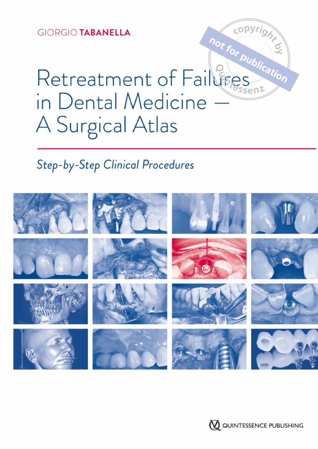

Giorgio Tabanella
Retreatment of Failures in Dental Medicine — A Surgical Atlas
Step-by-Step Clinical Procedures
Retreatment of Failures in Dental Medicine — A Surgical AtlasStep-by-Step Clinical Procedures
GIORGIO TABANELLA
Berlin, Barcelona, Chicago, Istanbul, London, Milan, Mexico City, Moscow, Paris, Prague, Seoul, Tokyo, Warsaw
A CIP record for this book is available from the British Library.ISBN: 978-1-78698-094-6
Quintessenz Verlags-GmbHIfenpfad 2–412107 BerlinGermanywww.quintessenz.de
© 2019 Quintessenz Verlags-GmbH, BerlinAll rights reserved. This book or any part thereof may not be reproduced, stored in a retrieval system, or transmitted in any form or by any means, whether electronic, mechanical, photocop-ying, or otherwise, without prior written permission of the publisher.
Editing: Natalie Ward, Quintessence Publishing Co Ltd, London, UKProject Management: Änne B. Kappeler, Quintessenz Verlags-GmbH, Berlin, GermanyLayout and Production: René Kirchner, Quintessenz Verlags-GmbH, Berlin, Germany
Printed and bound in Croatia by Graficki Zavod Hrvatske d.o.o., Zagreb
For Lorena, Edoardo, and Alice
Quintessence Publishing Co Ltd, Grafton Road, New Malden, Surrey KT3 3AB, United Kingdom www.quintpub.co.uk
V
FOREWORD
Altered macro and micro designs of oral im-plants have resulted in increased primary stability (tapered implants) and faster jaw-bone response (moderately rough implant surfaces), which have facilitated and hastened the process of osseointegration. In turn, im-plant surgery procedures have been adjusted to take advantage of these improvements in terms of immediate implant placement after extraction, one-stage surgery with early or immediate non-functional/functional loading and implants concurrently inserted with bone augmentation procedures. Reported out-comes have shown similar implant survival figures in selected patients compared with the more time-consuming traditional techniques.
The elegant images and videos in the pres-ent publication illustrate these procedures and are accompanied by well-presented an-amnestic data, radiographic examinations and proper treatment plans. The publication shows how compromised teeth that may be
due to a patient’s mishaps, poor compliance or less-successful previous dentistry are profes-sionally handled through careful extractions, immediate implant placement and augmen-tation with mainly bovine bone material. By reducing the number of surgical procedures, the total treatment time is significantly short-ened and patients are left with healthy tissue and esthetic implant/tooth-supported restor-ations. The positive psychological impact of this for many of them cannot be emphasised enough.
In conclusion, this case series may serve as a guideline for many dental surgeons involved in implant dentistry.
Göteborg, 16 June 2019The Brånemark Clinic
Bertil Friberg, DDS, MDS, PhD

VI
ACKNOWLEDGMENTS
The author would like to express his deepest gratitude to Lorena Bordi for offering her as-sistance during this challenging project and Dr Emanuele Nicolini for his technical support during the shooting of the surgeries.
VII
INTRODUCTION
Complications or failures are unfavourable consequences of a disease, a health condi-tion or a therapy. They can also be iatrogenic and might affect the prognosis of a disease. Complications or failures can occur at the lev-el of the organization, objective, team or work environment and can have a strong negative emotional impact on the practitioners who experience them. However, patient harm can occur even when a physician is knowledgea-ble and skilled and follows the best practice with excellent execution. Furthermore, fail-ures may be related to patient behaviour, as not only doctors and medical staff but also patients play a major role in the success of a therapy. In fact, poor patient compliance such as smoking habits, poor oral hygiene or non-compliance with postoperative instructions can lead to failure.
There is a general idea in the community that any medical issue can be solved with medica-tion or doctors’ interventions. This is not always valid: failure is part of the medical profession.
On the opposite, a medical error is a prevent-able adverse effect that harms patients. It can occur at different stages of the treatment process: diagnosis, prognosis, treatment or technique. In the medical profession, these types of errors are generally considered to be human errors. Medical errors are associated with various factors including inexperienced clinicians, new procedures, patient age, com-plex or urgent treatments, poor communica-tion, improper documentation, inadequate assistance, poorly designed treatment and errors of judgement. Practitioner risk factors include prejudiced thinking, sleep deprivation, fatigue, depression and burnout.
Cost-cutting measures may also compromise treatment outcomes. Many medical achieve-
ments have been obtained after significant investment and at a very high cost. As a re-sult, some therapies may not be affordable for some patients, who could find themselves ex-cluded from the benefits of the most advanced treatments.
Some failures may be related to the limita-tions of the medical profession; others may occur when doctors are mainly focused on their own ego rather than on defining the best treatment for their patients. This is a risk that may also cause a loss of trust in the medical profession.
Few doctors can claim that they have gone through their entire career without failing at something. However, the best doctors are those who have experienced fewer failures and who have been also able to manage their failures. In fact, medicine may have some lim-itations as regards to the treatment of certain pathologies.
Positive thinking after a complication or fail-ure is the key. It is tremendously useful to admit when things go wrong; it is especially important to think about what could have been done to avoid the failure. Furthermore, com-plications and failures are an intrinsic part of many competitive professional environments. The best approach is to try to learn from an adverse event so that one can be more suc-cessful in the future.
In general, physicians are embarrassed to talk about their mistakes. However, sharing fail-ures with colleagues can make the discussion about them feel normal. Medicine revolves around a culture of perfectionism and this is certainly also what medical professionals want. Being able to cope with failures is not a problem unique to the medical field. However, compared with other professions, medicine blends work life with real life. In fact, most
VIII
doctors do not describe what they do as a job because being a doctor is what they are and not only what they do. A doctor’s need to be successful in medicine is almost like a reflec-tion of his or her personal success in life.
Considering failures as part of medicine in general would help doctors to share their negative experiences and turn them into pos-itive ones. This idea should be incorporated into medical education as a first step towards normalizing the culture of failure in medicine. Doctors need to be less hard on themselves; in fact, most adverse events are caused not by negligence or carelessness but by inev-itable human error. Doctors ought to strive to do things better, yet they also need to ac-knowledge their fallibility and implement ap-proaches to reduce the aspect of human error. The acceptance of this concept would repre-sent a change of attitude and would create a more productive and positive atmosphere. Adverse events should be analyzed and meas-ured so that changes can be introduced to prevent their recurrence. Writing and talking as well as lecturing about failures would help to reduce medical errors in the long term and increase patient safety. The goal of this project is to share critical information for the retreat-ment of failures in dental medicine. Human performance, optimization of the diagnostic and therapeutic process, access to technology and state-of-the-art treatment are all benefi-cial in the drive to expand medical knowledge.
The perception of failure is quite common in trainees and trainers. We consciously judge ourselves against internal criteria. The only people who can remedy a failure are those re-sponsible for it. The point is to recognize the nature of the failure and research the problem. Many of most successful doctors have recov-ered from a failure by focusing on the medical work. To remedy a failure, a doctor essentially needs to prepare for the next treatment as he
should have prepared for the last. We all expe-rience good and bad days, but luck should have nothing to do with a successful treatment. The preparation for treating a patient should include skills, attitude, knowledge, training, decision making and common sense. Trainees should be seen to demonstrate a progressive acquisition of knowledge, skills and experi-ence. Also, senior doctors should periodically be checked by their peers to evaluate whether their knowledge and practice is up to date.
Medicine is linked to other sciences, but at the same time is independent and isolated in the sense that it deals with diseases and patients and could therefore be considered unique. First, it is important to define the disease and ask specific questions: What is a medical doc-tor supposed to do? Treat the symptoms? The pathology? Or both? If we have the tendency to treat what we hear, see and feel rather than the disease itself, we are not doing the right thing. The patient and the disease should be treated simultaneously. Apart from this, the patient’s health ought to be restored by avoid-ing ‘false’ methods of treatment. Doctors need to be aware that, as medical profession-als, they could be influenced in their clinical decision making by their morals, values and judgements as well as by their mental or emo-tional status. This reality may have an influ-ence on the analytical evaluation of patients.
An accurate system of diagnosis must be considered the main pillar of all treatments. Nevertheless, failure is part of medicine and must be considered as such in order to prop-erly treat and hopefully cure patients. Fur-thermore, ‘fashions’ in medicine such as new drugs or technologies need to be considered carefully. A calm and judicious summing up must be based on evidence.
In order to better guide us in our clinical de-cision making, every decision about the care
IX
of any individual should be based on explic-it, conscientious and judicious use of cur-rent best evidence. This approach, known as evidence-based medicine, leads to improve-ments in medical knowledge, provides a valid framework for teaching, contributes to decision making, allows for better commu-nication with patients and enables doctors to treat patients more successfully. However, evidence-based medicine does not necessar-ily ensure that better decisions are made. It is not entirely clear how doctors incorporate evidence into their final decisions, since it seems that the practice of medicine is never entirely free of personal judgement. This dif-ficulty is mainly related to the fact that many treatment outcomes cannot be properly iden-tified or measured. It is also unclear which parameters should be considered when de-termining the outcomes of a specific treat-ment, and some conclusions could also be considered unethical from a different point of view, for example, the treatment of a failing dentition in a young versus an elderly patient. Defining an esthetic outcome or a patient’s quality of life during and after treatment may be very difficult, especially if these variables are not quantifiable. Evidence-based medi-cine often rejects the opinion of experts who are mainly doctors who have interpreted re-search findings and objectives. Meta-analy-ses are generally considered the most precise statistical methods to evaluate the efficacy of a treatment. However, they might not include some studies that could be of clinical rele-vance but have low statistical power. There-fore, evidence-based medicine may be inter-preted as biased, since it is based on those treatments for which there is evidence, yet without considering the efficacy of individual treatments. It is obvious that complex issues cannot be solved with simplistic solutions and decision making seems, then, to be a reason-ing process that is partially based on personal values.
The nature of error in medicine is mainly re-lated to the lack of expertise and training. It is also evident that accidents and mistakes generally do not have a single cause but result from a string of latent flaws. It is also true that there is a trend to underestimate the role of social, relational and organizational factors as well as patient compliance. There is a strong link between poor communication, errors and adverse events.
There is an urgent need in medicine to in-crease attention to errors, adverse outcomes, quality of treatment and results. The more treatments performed, the higher the risk of failure. However, few studies focus on the cause of adverse effects, and those that do are mainly in a hospital setting. The cause may lie in the combination of several human factors such as a lack of communication, an exces-sive workload or inadequate training. How-ever, doctors’ personalities also play a role, including overconfidence. Human decisions are responsible for almost all failures, includ-ing those related to patient compliance. Some failures also occur as a result of motivational problems or inadequate management. Lack of experience, inadequate knowledge, a stress-ful environment and inadequate equipment are other important considerations when as-sessing complications and failures, as are the economic context and personal characteris-tics of the patient, such as the language spo-ken, the patient’s personality and the level of compliance.
A patient’s disease is the most powerful pre-dictor of the clinical outcome. As there is very little if any research on the aspects discussed here, it is of extreme value to share the re-treatment of complications and failures with colleagues. The ultimate goal is to acknowl-edge the potential for error and to finally help doctors improve the safety and overall quality of care of their patients.
XI
TABLE OF CONTENT
Foreword VAcknowledgments VIIntroduction VII
Chapter 1 Endo failure – Tooth loss and perforation of the buccal plate | 1Introduction 4Anamnesis/medical history 4Dental history 4Chief complaint 5Radiographic findings 5Clinical findings 7Diagnosis 7Prognosis 8Indications and objectives 8 Criteria for decision making 8Treatment planning 9 Instruments and techniques 9Treatment results 26Take-home message 37References 40
Chapter 2 Diagnostic failure – Mistreated trauma associated with periapical chronic lesions and anatomical bone deformities | 43Introduction 46Anamnesis/medical history 46Dental history 46Chief complaint 46Radiographic findings 49Clinical findings 52Diagnosis 53Prognosis 53Indications and objectives 53 Criteria for decision making 53Treatment planning 54 Instruments and techniques 54Treatment results 92Take-home message 96References 98
XII
Chapter 3 Perio failure – Recurrent and excessive scaling and root planing | 101Introduction 104Anamnesis/medical history 105Dental history 105Chief complaint 105Radiographic findings 105Clinical findings 108Diagnosis 109Prognosis 109Indications and objectives 109 Criteria for decision making 109Treatment planning 110 Instruments and techniques 110Treatment results 126Take-home message 133References 136
Chapter 4 Implant failure – Iatrogenic bone grafting and implant placement: Ailing dental implants associated with oroantral communication | 139Introduction 142Anamnesis/medical history 142Dental history 142Chief complaint 143Radiographic findings 143Clinical findings 144Diagnosis 144Prognosis 144Indications and objectives 145 Criteria for decision making 145Treatment planning 147 Instruments and techniques 147Treatment results 181Take-home message 183References 184

Tooth loss and perfora tion of the buccal plate
CH
APT
ER 1 ENDO FAILURE
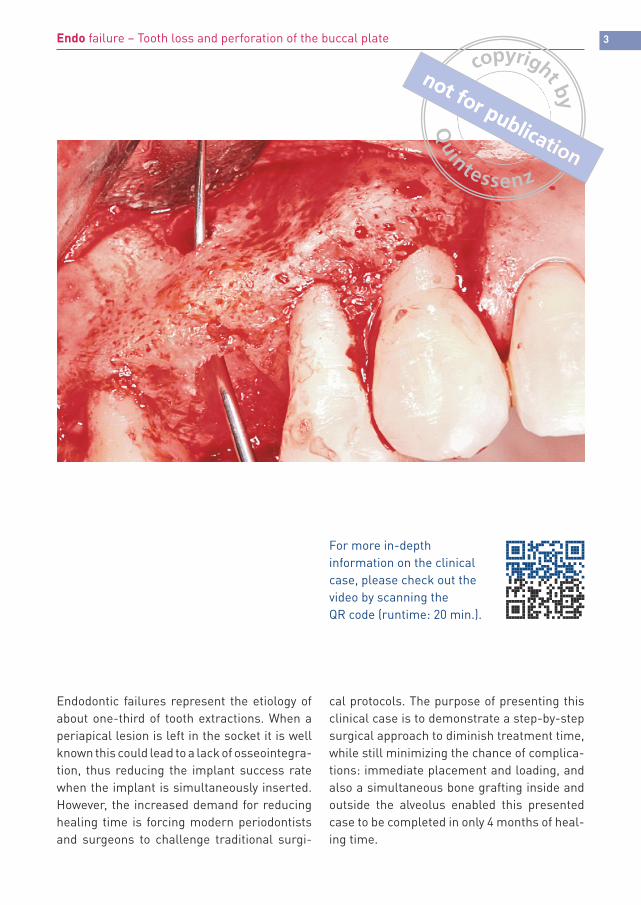
3Endo failure – Tooth loss and perforation of the buccal plate
Endodontic failures represent the etiology of about one-third of tooth extractions. When a periapical lesion is left in the socket it is well known this could lead to a lack of osseointegra-tion, thus reducing the implant success rate when the implant is simultaneously inserted. However, the increased demand for reducing healing time is forcing modern periodontists and surgeons to challenge traditional surgi-
cal protocols. The purpose of presenting this clinical case is to demonstrate a step-by-step surgical approach to diminish treatment time, while still minimizing the chance of complica-tions: immediate placement and loading, and also a simultaneous bone grafting inside and outside the alveolus enabled this presented case to be completed in only 4 months of heal-ing time.
For more in-depth information on the clinical case, please check out the video by scanning the QR code (runtime: 20 min.).
4 CHAPTER 1
1.1 INTRODUCTION
The risk of treating patients who have been through iatrogenic treatment must be properly calculated, since a minor complication during treatment may cause a tremendous amount of bone loss and soft tissue deformities. These complications can also be higher in numbers and more severe when a medical mistake has previously occurred. Furthermore, the psychological aspect of these patients who, in case of complications would need multi-ple surgeries and treatments, may effect the treatment itself. In any case, the treatment fo-cus is to achieve a peri-implant positive bony architecture that will support and stabilize the surrounding mucosa. Soft and hard tis-sue manipulation will help to create a stable peri-implant sealing that will maintain the esthetic treatment results over time. The pre-sented philosophy of work can lead implantol-ogy to a different level, since patients’ desires and quality of life during treatment could be significantly improved. Thus, factors crucial to achieve a biomimetic and long-lasting result will also be emphasized.
Patients who have been unlucky enough to ex-perience complications resulting from treat-ment have a unique approach towards the new specialist who is going to cure them: they are doubtful, as well as reluctant, but still willing to be treated. Their emotions are predominant and they have lost respect and confidence for medical discipline and doctors. In general, this type of patient does not have a positive or cooperative approach towards the new team of specialists. It is therefore imperative to first define a correct diagnosis and then visualize the final desirable results. Modern techno-logy can help to envisage what the patient and surgeon would like to achieve, however digital tools need to be supported by biological prin-ciples. Only biology, anatomy, vascularization, type of defect, potential for tissue regenera-
tion, surgical skills, and experience will con-firm if what was visualized during the diag-nostic phase is achievable or not.
Another important point is to approach these retreatment cases with advanced techniques and modified protocols in order to reduce the overall healing time, since these patients are drained of all energy after previous treatment.
Therefore, a reduced timing is an important parameter to avoid patients opting for the easiest and fastest treatment, such as a fixed or removable partial denture: this particular treatment will then force patients to face fur-ther retreatments, especially those who were young at the time of the first treatment.
Once the diagnosis has been defined and a patient’s chief complaint registered, the final treatment planning can be executed.
1.2 ANAMNESIS/MEDICAL HISTORY
A 45-year-old female patient presented with pain and no suppuration on the inferior border of the right zygomatic bone (Figs 1 to 8). Her medical history reported no significant sys-temic diseases except for a mild hypotension. The patient, at the time of treatment, was not under any medication.
1.3 DENTAL HISTORY
The patient reported to have received dental treatment for almost all her life. In her child-hood she received orthodontic treatment and minor caries were treated mainly with amal-gam and only a few with composite restor-ations on teeth #17, 16, 26, 27, 37, 45 and 46. After this initial phase of dental treatment she had no further decay. At about the age of 25 she lost the first mandibular left molar (#36) and after a few years, tooth #47. Both mandib-ular molars were mobile at the time of extrac-
5Endo failure – Tooth loss and perforation of the buccal plate
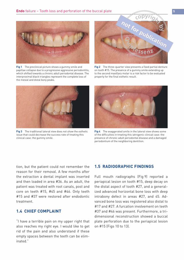
Fig 1 The preclinical picture shows a gummy smile and papillae collapse due to a progressive aggressive periodontitis, which shifted towards a chronic adult periodontal disease. The interproximal black triangles represent the complete loss of the mesial and distal bony peaks.
Fig 3 The traditional lateral view does not show the esthetic issue that could decrease the success rate of treating this clinical case: the gummy smile.
Fig 2 The three-quarter view presents a fixed partial denture on tooth #15. The presence of a gummy smile extending up to the second maxillary molar is a risk factor to be evaluated properly for the final esthetic result.
Fig 4 The exaggerated smile in the lateral view shows some of the difficulties in treating this iatrogenic clinical case: the presence of chronic adult periodontal disease and a damaged periodontium of the neighboring dentition.
tion, but the patient could not remember the reason for their removal. A few months after the extraction a dental implant was inserted and then loaded in area #36. As an adult, the patient was treated with root canals, post and core on teeth #15, #45 and #46. Only teeth #15 and #37 were restored after endodontic treatment.
1.4 CHIEF COMPLAINT
“I have a terrible pain on my upper right that also reaches my right eye. I would like to get rid of the pain and also understand if these empty spaces between the teeth can be elim-inated.”
1.5 RADIOGRAPHIC FINDINGS
Full mouth radiographs (Fig 9) reported a periapical lesion on tooth #15, deep decay on the distal aspect of tooth #27, and a general-ized advanced horizontal bone loss with deep intrabony defect in areas #27, and 45. Ad-vanced bone loss was registered also distal to #17 and #27. A furcation involvement on teeth #37 and #46 was present. Furthermore, a tri-dimensional reconstruction showed a buccal plate perforation due to the periapical lesion on #15 (Figs 10 to 13).
6 CHAPTER 1
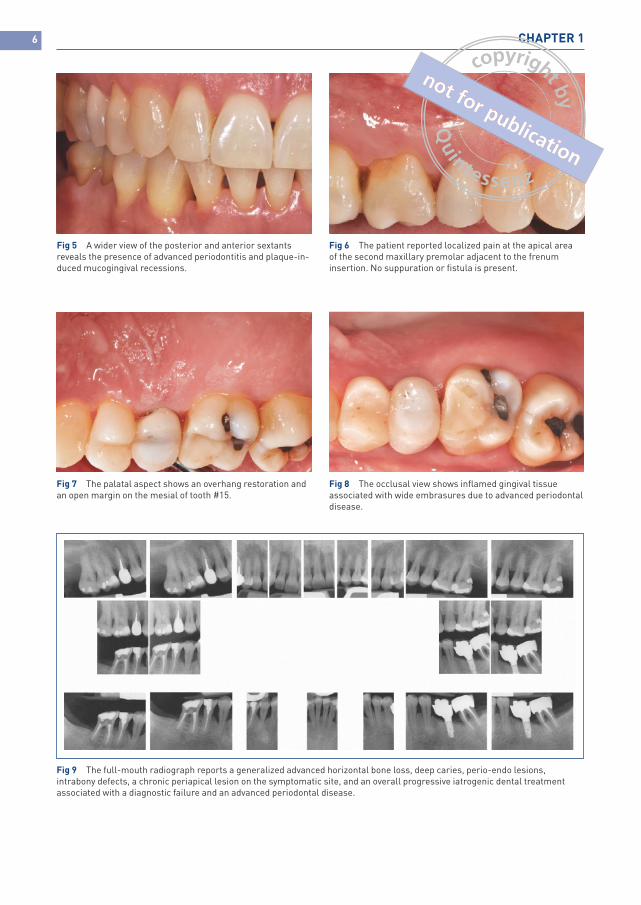
Fig 5 A wider view of the posterior and anterior sextants reveals the presence of advanced periodontitis and plaque-in-duced mucogingival recessions.
Fig 6 The patient reported localized pain at the apical area of the second maxillary premolar adjacent to the frenum insertion. No suppuration or fistula is present.
Fig 7 The palatal aspect shows an overhang restoration and an open margin on the mesial of tooth #15.
Fig 8 The occlusal view shows inflamed gingival tissue associated with wide embrasures due to advanced periodontal disease.
Fig 9 The full-mouth radiograph reports a generalized advanced horizontal bone loss, deep caries, perio-endo lesions, intrabony defects, a chronic periapical lesion on the symptomatic site, and an overall progressive iatrogenic dental treatment associated with a diagnostic failure and an advanced periodontal disease.
7Endo failure – Tooth loss and perforation of the buccal plate
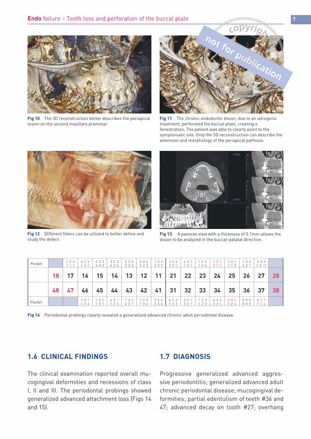
Fig 10 The 3D reconstruction better describes the periapical lesion on the second maxillary premolar.
Fig 12 Different filters can be utilized to better define and study the defect.
Fig 11 The chronic endodontic lesion, due to an iatrogenic treatment, perforated the buccal plate, creating a fenestration. The patient was able to clearly point to the symptomatic site. Only the 3D reconstruction can describe the extension and morphology of the periapical pathosis.
Fig 13 A panorex view with a thickness of 0.1mm allows the lesion to be analyzed in the buccal-palatal direction.
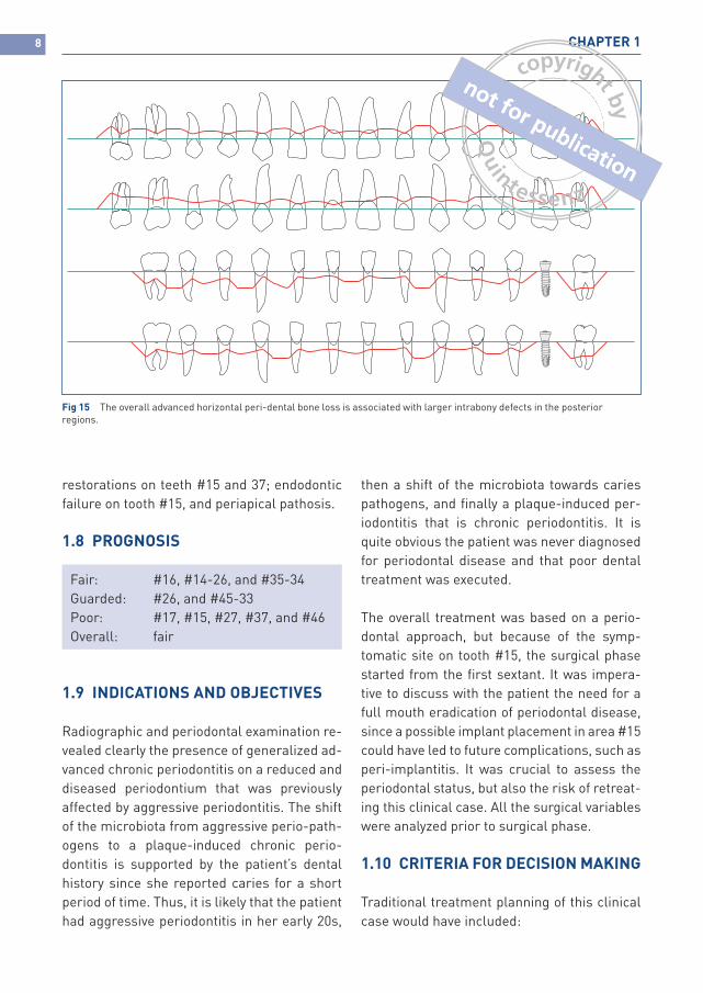
Fig 14 Periodontal probings clearly revealed a generalized advanced chronic adult periodontal disease.
Pocket5 2 47 5 5
4 2 45 4 5
4 2 34 3 3
3 2 34 2 4
3 2 43 2 4
4 2 44 2 3
5 2 33 2 3
3 2 43 3 5
4 2 55 3 5
5 2 45 3 4
5 2 53 5 5
5 2 55 5 3
5 3 44 2 7
4 3 85 3 10
18 17 16 15 14 13 12 11 21 22 23 24 25 26 27 28
48 47 46 45 44 43 42 41 31 32 33 34 35 36 37 38
Pocket5 3 76 3 4
7 3 55 3 5
4 3 75 3 5
7 2 54 3 7
7 2 37 3 4
2 2 43 3 4
4 3 25 3 3
3 2 73 2 7
7 2 36 2 3
4 3 43 2 5
5 3 55 2 4
0 0 00 0 0
8 7 77 6 6
1.6 CLINICAL FINDINGS
The clinical examination reported overall mu-cogingival deformities and recessions of class I, II and III. The periodontal probings showed generalized advanced attachment loss (Figs 14 and 15).
1.7 DIAGNOSIS
Progressive generalized advanced aggres-sive periodontitis; generalized advanced adult chronic periodontal disease; mucogingival de-formities; partial edentulism of teeth #36 and 47; advanced decay on tooth #27; overhang
8 CHAPTER 1
Fig 15 The overall advanced horizontal peri-dental bone loss is associated with larger intrabony defects in the posterior regions.
restorations on teeth #15 and 37; endodontic failure on tooth #15, and periapical pathosis.
1.8 PROGNOSIS
Fair: #16, #14-26, and #35-34Guarded: #26, and #45-33Poor: #17, #15, #27, #37, and #46Overall: fair
1.9 INDICATIONS AND OBJECTIVES
Radiographic and periodontal examination re-vealed clearly the presence of generalized ad-vanced chronic periodontitis on a reduced and diseased periodontium that was previously affected by aggressive periodontitis. The shift of the microbiota from aggressive perio-path-ogens to a plaque-induced chronic perio-dontitis is supported by the patient’s dental history since she reported caries for a short period of time. Thus, it is likely that the patient had aggressive periodontitis in her early 20s,
then a shift of the microbiota towards caries pathogens, and finally a plaque-induced per-iodontitis that is chronic periodontitis. It is quite obvious the patient was never diagnosed for periodontal disease and that poor dental treatment was executed.
The overall treatment was based on a perio-dontal approach, but because of the symp-tomatic site on tooth #15, the surgical phase started from the first sextant. It was impera-tive to discuss with the patient the need for a full mouth eradication of periodontal disease, since a possible implant placement in area #15 could have led to future complications, such as peri-implantitis. It was crucial to assess the periodontal status, but also the risk of retreat-ing this clinical case. All the surgical variables were analyzed prior to surgical phase.
1.10 CRITERIA FOR DECISION MAKING
Traditional treatment planning of this clinical case would have included:
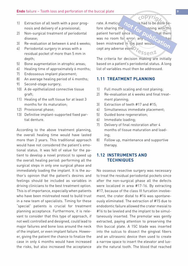
9Endo failure – Tooth loss and perforation of the buccal plate
1) Extraction of all teeth with a poor prog-nosis and delivery of a provisional;
2) Non-surgical treatment of periodontaldisease;
3) Re-evaluation at between 4 and 6 weeks;4) Periodontal surgery in areas with a
residual pocket of more than 5 mm indepth;
5) Bone augmentation in atrophic areas;6) Healing time of approximately 6 months;7) Endosseous implant placement;8) An average healing period of 4 months;9) Second-stage surgery;10) A de-epithelialized connective tissue
graft;11) Healing of the soft tissue for at least 3
months for its maturation;12) Provisional phase;13) Definitive implant-supported fixed par-
tial denture.
According to the above treatment planning, the overall healing time would have lasted more than 2 years. This traditional approach would have not considered the patient’s emo-tional status. It was felt of value for the pa-tient to develop a novel protocol to speed up the overall healing period: performing all the surgical steps in only one surgical phase and immediately loading the implant. It is the au-thor’s opinion that the patient’s desires and feelings should be included as variables in driving clinicians to the best treatment option. This is of importance, especially when patients who have been mistreated need to build trust in a new team of specialists. Timing for these “special” patients is crucial for treatment planning acceptance. Furthermore, it is rele-vant to consider that this type of approach, if not well controlled and designed, could lead to major failures and bone loss around the neck of the implant, or even implant failure. Howev-er, giving the patient the chance to finalize the case in only 4 months would have increased the risks, but also increased the acceptance
rate. A meticulous analysis had to be done be-fore sharing the treatment planning with the patient herself since it was evident that there was no room for error: any patient that has been mistreated in the past would never ac-cept any adverse event.
The criteria for decision making are initially based on a patient’s periodontal status. A long list of variables must then be addressed.
1.11 TREATMENT PLANNING
1) Full mouth scaling and root planing;2) Re-evaluation at 4 weeks and final treat-
ment planning;3) Extraction of teeth #17 and #15;4) Simultaneous immediate placement;5) Guided bone regeneration;6) Immediate loading;7) Delivery of final restoration after 4
months of tissue maturation and load-ing;
8) Follow up, maintenance and supportivetherapy.
1.12 INSTRUMENTS AND TECHNIQUES
No osseous resective surgery was necessary to treat the residual periodontal pockets since after the non-surgical phase all the defects were localized in area #17-16. By extracting #17, because of the class III furcation involve-ment, the crater distal to #16 was spontane-ously eliminated. The extraction of #15 due to endodontic failure allowed the crater mesial to #16 to be leveled and the implant to be simul-taneously inserted. The premolar was gently extracted, paying attention to preserving the thin buccal plate. A 15C blade was inserted into the sulcus to dissect the gingival fibers and an ultrasonic device then used to create a narrow space to insert the elevator and lux-ate the natural tooth. The blood that reached
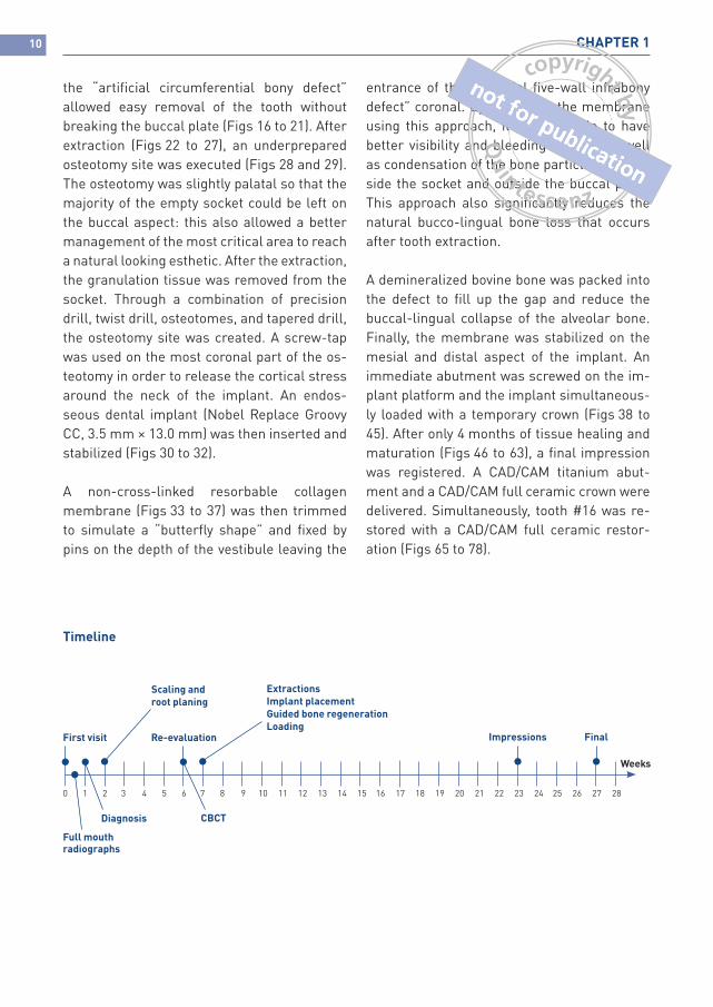
10 CHAPTER 1
the “artificial circumferential bony defect” allowed easy removal of the tooth without breaking the buccal plate (Figs 16 to 21). After extraction (Figs 22 to 27), an underprepared osteotomy site was executed (Figs 28 and 29). The osteotomy was slightly palatal so that the majority of the empty socket could be left on the buccal aspect: this also allowed a better management of the most critical area to reach a natural looking esthetic. After the extraction, the granulation tissue was removed from the socket. Through a combination of precision drill, twist drill, osteotomes, and tapered drill, the osteotomy site was created. A screw-tap was used on the most coronal part of the os-teotomy in order to release the cortical stress around the neck of the implant. An endos-seous dental implant (Nobel Replace Groovy CC, 3.5 mm × 13.0 mm) was then inserted and stabilized (Figs 30 to 32).
A non-cross-linked resorbable collagen membrane (Figs 33 to 37) was then trimmed to simulate a “butterfly shape” and fixed by pins on the depth of the vestibule leaving the
entrance of the “artificial five-wall infrabony defect” coronal. By stabilizing the membrane using this approach, it was possible to have better visibility and bleeding control, as well as condensation of the bone particles both in-side the socket and outside the buccal plate. This approach also significantly reduces the natural bucco-lingual bone loss that occurs after tooth extraction.
A demineralized bovine bone was packed into the defect to fill up the gap and reduce the buccal-lingual collapse of the alveolar bone. Finally, the membrane was stabilized on the mesial and distal aspect of the implant. An immediate abutment was screwed on the im-plant platform and the implant simultaneous-ly loaded with a temporary crown (Figs 38 to 45). After only 4 months of tissue healing and maturation (Figs 46 to 63), a final impression was registered. A CAD/CAM titanium abut-ment and a CAD/CAM full ceramic crown were delivered. Simultaneously, tooth #16 was re-stored with a CAD/CAM full ceramic restor-ation (Figs 65 to 78).
Timeline
Weeks
FinalImpressions
Full mouth radiographs
CBCT
First visit
Diagnosis
Scaling and root planing
Extractions Implant placementGuided bone regenerationLoading
Re-evaluation
0 1 2 3 4 5 6 7 8 9 10 11 12 13 14 15 16 17 18 19 20 21 22 23 24 25 26 27 28
11Endo failure – Tooth loss and perforation of the buccal plate
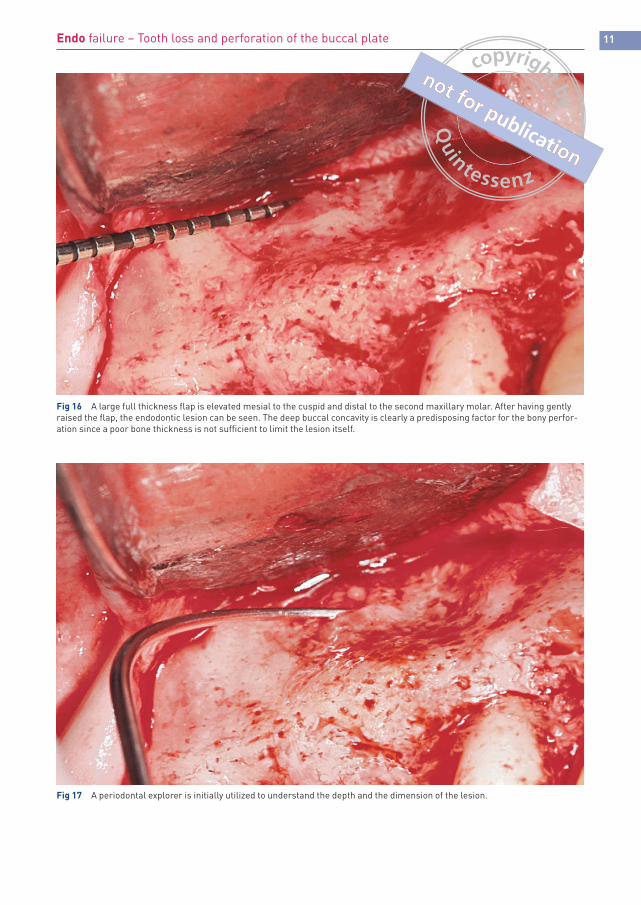
Fig 16 A large full thickness flap is elevated mesial to the cuspid and distal to the second maxillary molar. After having gently raised the flap, the endodontic lesion can be seen. The deep buccal concavity is clearly a predisposing factor for the bony perfor-ation since a poor bone thickness is not sufficient to limit the lesion itself.
Fig 17 A periodontal explorer is initially utilized to understand the depth and the dimension of the lesion.
12 CHAPTER 1
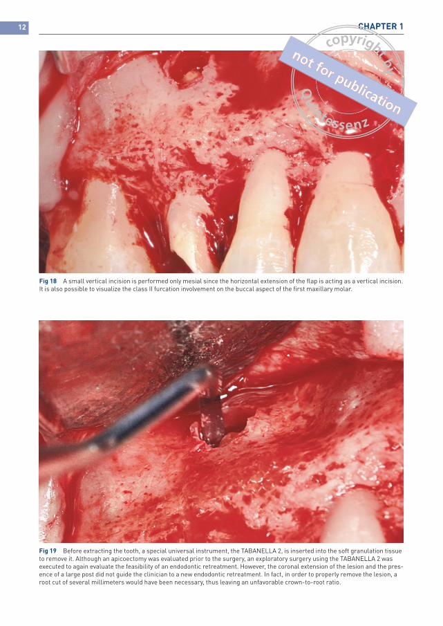
Fig 18 A small vertical incision is performed only mesial since the horizontal extension of the flap is acting as a vertical incision. It is also possible to visualize the class II furcation involvement on the buccal aspect of the first maxillary molar.
Fig 19 Before extracting the tooth, a special universal instrument, the TABANELLA 2, is inserted into the soft granulation tissue to remove it. Although an apicoectomy was evaluated prior to the surgery, an exploratory surgery using the TABANELLA 2 was executed to again evaluate the feasibility of an endodontic retreatment. However, the coronal extension of the lesion and the pres-ence of a large post did not guide the clinician to a new endodontic retreatment. In fact, in order to properly remove the lesion, a root cut of several millimeters would have been necessary, thus leaving an unfavorable crown-to-root ratio.
13Endo failure – Tooth loss and perforation of the buccal plate
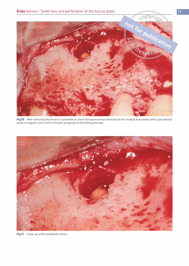
Fig 20 After removing the lesion it is possibile to check the apicocoronal extension of the residual bony defect with a periodontal probe or explorer and confirm the poor prognosis of the failing premolar.
Fig 21 Close-up of the endodontic lesion.
14 CHAPTER 1
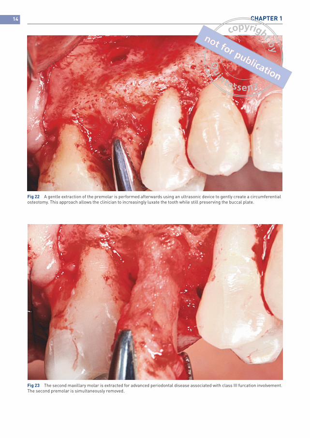
Fig 22 A gentle extraction of the premolar is performed afterwards using an ultrasonic device to gently create a circumferential osteotomy. This approach allows the clinician to increasingly luxate the tooth while still preserving the buccal plate.
Fig 23 The second maxillary molar is extracted for advanced periodontal disease associated with class III furcation involvement. The second premolar is simultaneously removed.
15Endo failure – Tooth loss and perforation of the buccal plate
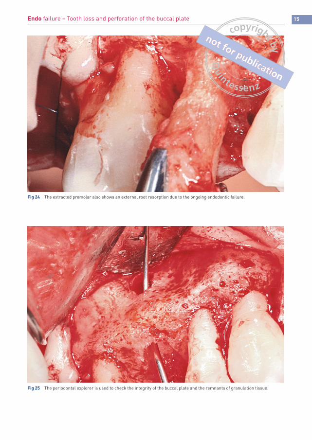
Fig 24 The extracted premolar also shows an external root resorption due to the ongoing endodontic failure.
Fig 25 The periodontal explorer is used to check the integrity of the buccal plate and the remnants of granulation tissue.
16 CHAPTER 1
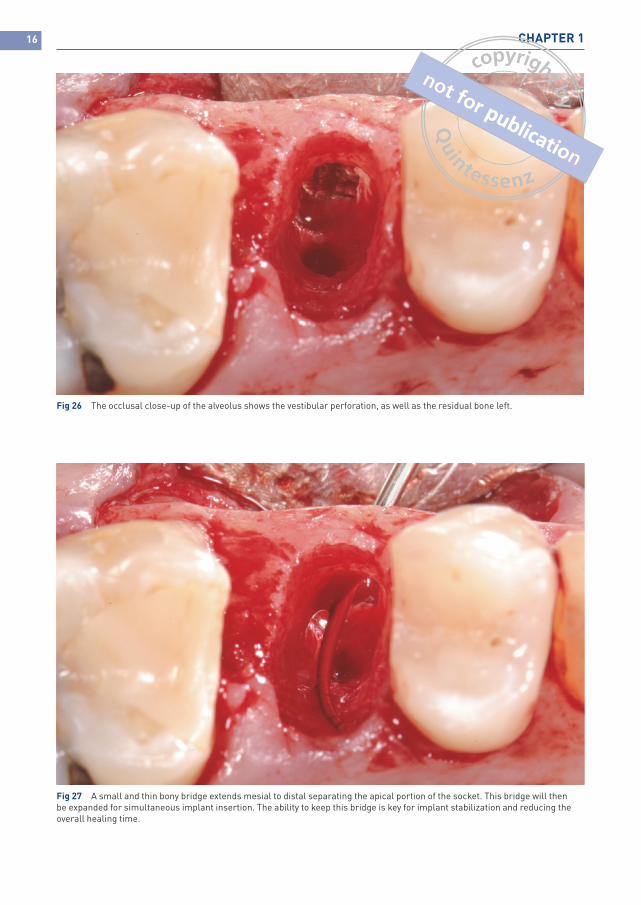
Fig 26 The occlusal close-up of the alveolus shows the vestibular perforation, as well as the residual bone left.
Fig 27 A small and thin bony bridge extends mesial to distal separating the apical portion of the socket. This bridge will then be expanded for simultaneous implant insertion. The ability to keep this bridge is key for implant stabilization and reducing the overall healing time.
17Endo failure – Tooth loss and perforation of the buccal plate
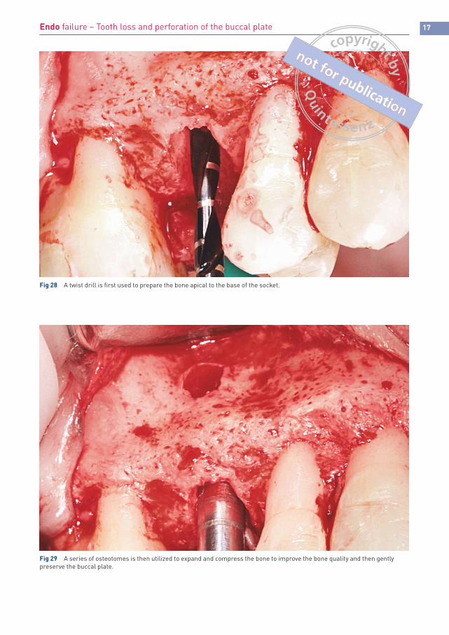
Fig 28 A twist drill is first used to prepare the bone apical to the base of the socket.
Fig 29 A series of osteotomes is then utilized to expand and compress the bone to improve the bone quality and then gently preserve the buccal plate.
18 CHAPTER 1
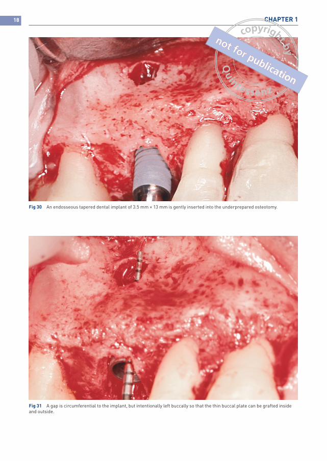
Fig 30 An endosseous tapered dental implant of 3.5 mm × 13 mm is gently inserted into the underprepared osteotomy.
Fig 31 A gap is circumferential to the implant, but intentionally left buccally so that the thin buccal plate can be grafted inside and outside.
19Endo failure – Tooth loss and perforation of the buccal plate
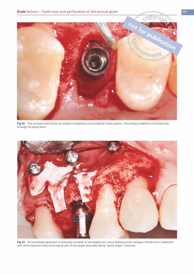
Fig 32 The occlusal view shows an implant completely surrounded by empty spaces. The primary stability is achieved only through the basal bone.
Fig 33 An immediate abutment is manually screwed. A resorbable non-cross-linked porcine collagen membrane is stabilized with three titanium tacks at the apical part of the target area after being “pants shape” trimmed.

20 CHAPTER 1
Fig 34 A demineralized bovine bone is packed and compressed with the TABANELLA 1. Two pins then fix the membrane on its narrowest mesial and distal coronal extensions.
Fig 35 The guided bone regeneration allows for overbuilding of the site and tridimensionally reconstructing the missing bone.
21Endo failure – Tooth loss and perforation of the buccal plate
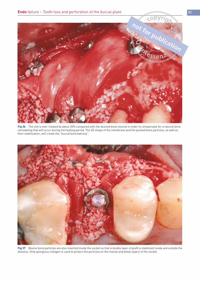
Fig 36 The site is over-treated by about 20% compared with the desired bone volume in order to compensate for a natural bone remodeling that will occur during the healing period. The 3D shape of the membrane and the packed bone particles, as well as their stabilization, will create the “buccal bone balcony”.
Fig 37 Bovine bone particles are also inserted inside the socket so that a double layer of graft is stabilized inside and outside the alveolus. Only spongious collagen is used to protect the particles on the mesial and distal aspect of the socket.
22 CHAPTER 1
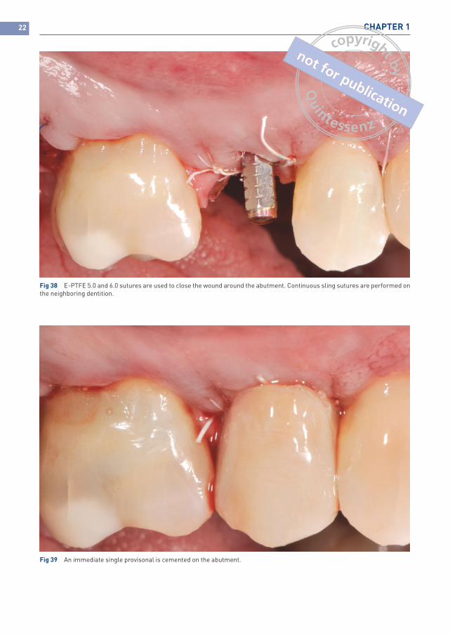
Fig 38 E-PTFE 5.0 and 6.0 sutures are used to close the wound around the abutment. Continuous sling sutures are performed on the neighboring dentition.
Fig 39 An immediate single provisonal is cemented on the abutment.

23Endo failure – Tooth loss and perforation of the buccal plate
Fig 40 Occlusal view of the sutured sextant
Fig 41 Lateral view of the treated sextant.
24 CHAPTER 1
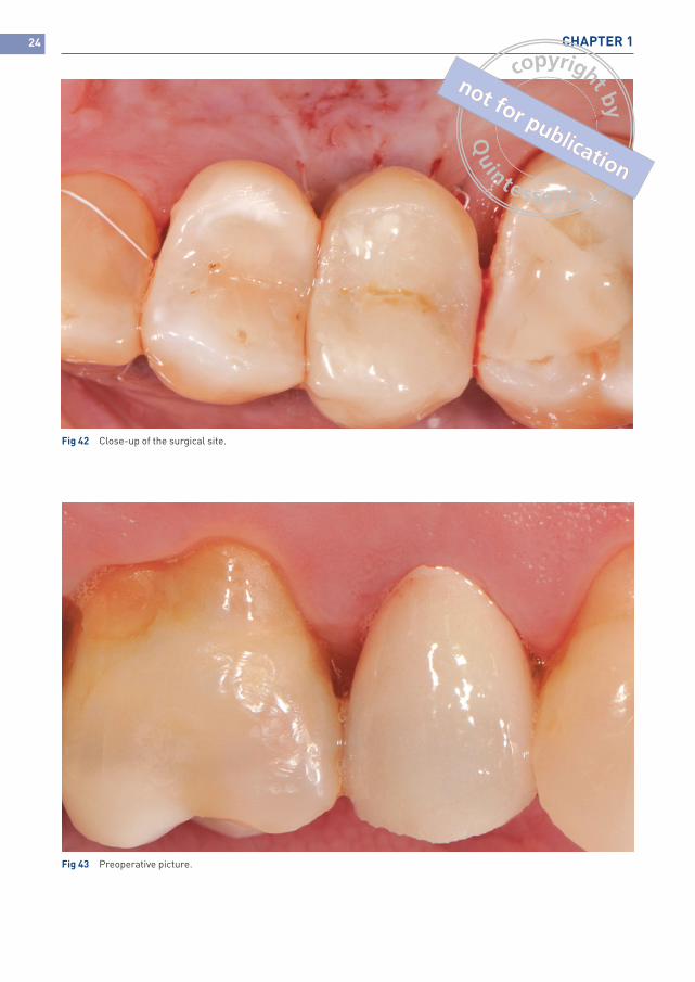
Fig 42 Close-up of the surgical site.
Fig 43 Preoperative picture.
25Endo failure – Tooth loss and perforation of the buccal plate
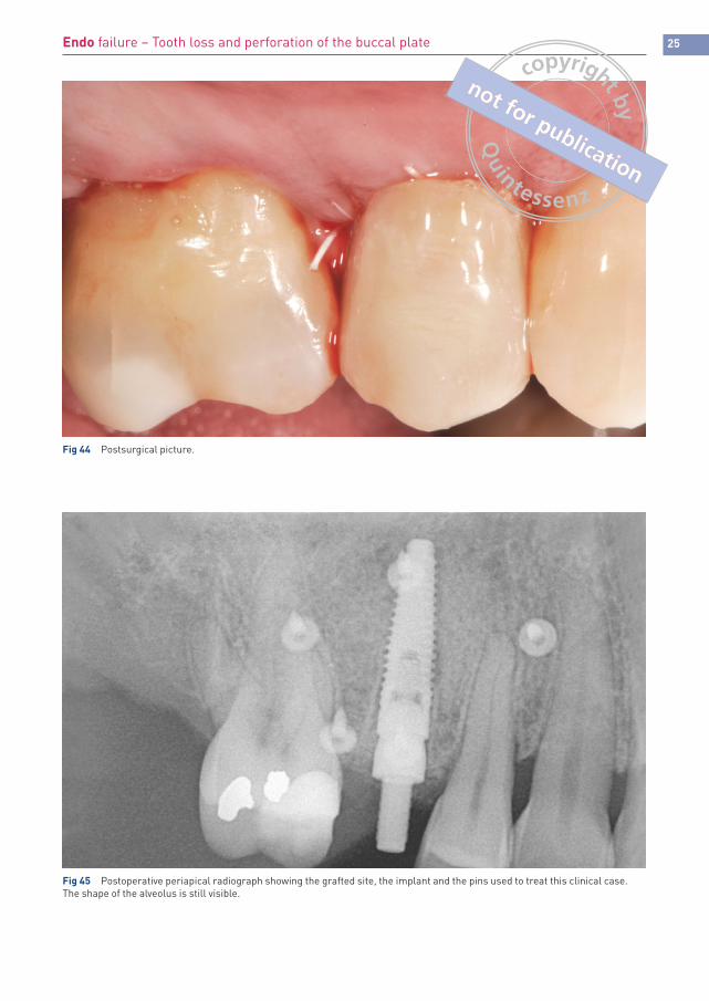
Fig 44 Postsurgical picture.
Fig 45 Postoperative periapical radiograph showing the grafted site, the implant and the pins used to treat this clinical case. The shape of the alveolus is still visible.
26 CHAPTER 1
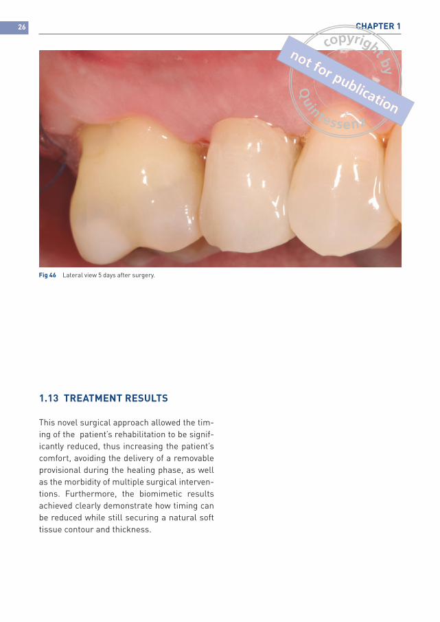
Fig 46 Lateral view 5 days after surgery.
1.13 TREATMENT RESULTS
This novel surgical approach allowed the tim-ing of the patient’s rehabilitation to be signif-icantly reduced, thus increasing the patient’s comfort, avoiding the delivery of a removable provisional during the healing phase, as well as the morbidity of multiple surgical interven-tions. Furthermore, the biomimetic results achieved clearly demonstrate how timing can be reduced while still securing a natural soft tissue contour and thickness.