Embed Size (px)
Citation preview
8/4/2019 giesel (1)
http://slidepdf.com/reader/full/giesel-1 1/6
a report by
Fabian Rengier ,1, 2 Hendrik von Tengg-Kobligk ,1 Christ ian Zechmann ,1 Hans-Ulr ich Kauczor 3 and Frederik L Giesel 1, 3
1. Department of Radiology, German Cancer Research Centre (DKFZ); 2. Research Training Group 1126: Intelligent Surgery,
University Hospital Heidelberg; 3. Department of Diagnostic and Interventional Radiology, University Hospital Heidelberg
In the last few years, medical imaging and image post-processing
techniques have rapidly advanced. Today’s multislice computed
tomography (MSCT) and high-performance magnetic resonance imaging
(MRI) can acquire thousands of images within a breath-hold, recording
volumes at incredibly high spatial resolution.1
MRI has experienced improvements in a variety of aspects such as
increasing image quality and reducing acquisition times, thusstrengthening its usefulness in daily clinical routine.2 Both modalities have
their advantages and disadvantages, but thanks to rapid evolution during
the last decades both have grown beyond two dimensions, giving rise to
new opportunities for the medical and bioengineering community.3
Rapid prototyping is one of the most recently evolving techniques in
this field4 and is expected to lead great progress in different industrial
fields, including healthcare. This article will illustrate the pathway from
medical imaging via 3D virtual visualisation to 3D solid objects using
the rapid prototyping technique and discuss the medical applications
and implications.
From Medical Imaging to 3D Solid Objects
The process chain from medical imaging to 3D solid objects can be
divided into three major parts: image acquisition, image post-
processing and rapid prototyping. Images are acquired using CT or MRI,
stored at a picture archiving and communication system (PACS) and
transferred to a dedicated image post-processing workstation (see
Figure 1). On the workstation, 3D segmentation and visualisation are
performed and the segmented structures are exported as machine-
readable data with the possibility of further geometric modelling using
computer-aided design (CAD) software. Such data can then be used by
rapid prototyping machines to generate a 3D solid object.
Image Acquisition
3D data volumes of adequate image quality are of vital importance forthe basis of the process chain. To begin with, high spatial resolution
with a reconstructed slice thickness not exceeding 1mm and nearly
isotropic voxel size is essential to minimise partial volume effects and
step artefacts during image reformation,5 as well as to obtain highly
detailed imaging information. In cases of vascular applications, optimal
timing of contrast material injection is required to achieve sufficient
and homogenous enhancement of the arterial vasculature and to
avoid streak artefacts from adjacent veins.6,7 Today, 3D data can be
easily acquired using both CT and MRI. However, CT is still the
preferred imaging modality compared with MRI because isotropy is
easier to achieve and less time-consuming and, fundamentally,
because segmentation algorithms work better with CT data.Nevertheless, MRI offers the possibility of acquiring 3D data of
any structure within the body without radiation exposure.8
Furthermore, 3D data can also be acquired using positron emission
tomography (PET), single photon emission computed tomography
(SPECT) or ultrasound.
Image Post-processing
The 3D data are stored using the common digital imaging and
communications in medicine (DICOM) format and transferred to a
dedicated image post-processing workstation for image analysis and
reconstruction. By processing and recording extremely large streams of
data, high-performance computers can conduct state-of-the-art imagepost-processing that transforms radiological individual images into 3D
and even 4D worlds (adding synchronised motion). It is a technology
that radiologists and hospital employees have used for some time. In
this context, volume rendering (VR), maximum intensity projection
(MIP) and other techniques are highly appreciated by the clinicians. For
example, minimally invasive vascular surgery is planned and performed
almost exclusively using 3D image post-processing to pinpoint the
extent of the disease and treat it accordingly.9 Furthermore, 3D
reconstruction of complex multifragment fractures is helpful for the
orthopaedic surgeon to plan the operation and choose the correct
osteosynthesis material.
This next step in visualisation is achieved by using intricate mathematical
algorithms to derive individual structures from radiological 3D volumes,
transforming those structures and altering them as appropriate and
Beyond the Eye – Medical Applications of 3D Rapid Prototyping Objects
© T O U C H B R I E F I N G S 2 0 0 8
Frederik L Giesel is a Physician and Senior Researcher in the
Department of Radiology at the National German Cancer
Research Centre in Heidelberg. He is also an Honorary
Visiting Lecturer at the University of Sheffield. His research
focuses on image analysis, 3D visualisation and image post-
processing in neuro-imaging. He holds several patents for
contrast media, undertakes various clinical trials and has
broad expertise in industrial co-operation. Recently,
Dr Giesel gained an international MBA to extend his
expertise from medicine to economics and is a lecturer at
the Frankfurt School of Finance and Management.
Fabian Rengier is a Research Fellow in the Department of
Radiology at the National German Cancer Research Centre
in Heidelberg. He is a Junior Lecturer in the Institute of
Anatomy and Cell Biology at the University of Heidelberg,
Head of the Concise Anatomy academic project and a
founding member of the Virtual Anatomy working group. Hisresearch focuses on new cardiovascular imaging and image
post-processing techniques. He has received grants from the
German Research Foundation (DFG), the German National
Academic Foundation and the University of Heidelberg.
Dr Rengier attended medical school in Heidelberg.
Digital Radiography
76
8/4/2019 giesel (1)
http://slidepdf.com/reader/full/giesel-1 2/6
77E U R O P E A N M E D I C A L I M A G I N G R E V I E W
Beyond the Eye – Medical Applications of 3D Rapid Prototyping Objects
necessary. Such segmentation ultimately renders radiological imaging
data into a virtual 3D reconstruction of the segmented structures in the
form of slice contours or 3D triangle mesh models.10 This virtual model is
then exported as machine-readable data, the kind of data that is needed
to create models – a procedure the automotive industry calls rapid
prototyping. Outputs are normally saved in initial graphics exchange
specification (IGES), surface tessellation language (STL) or virtual reality
modelling language (VRML) format. These output files can be either
directly transferred to a rapid prototyping machine or further processed
using CAD software. CAD offers powerful geometrical modelling tools
that can be used, for example, to prepare surgical implants or medical
phantoms depending on their purpose.
Rapid Prototyping
In general, rapid prototyping can be defined as an approach or
methodology used to quickly manufacture physical models using 3D
CAD data. Rapid prototyping has also been referred to as solid free-
form, computer-automated or layered manufacturing. Rapid
prototyping has its obvious use as a truly 3D method for visualisationand better haptic impression.
Currently, rapid prototyping is mainly devoted to producing 3D
prototypes and models. The word ‘rapid’ should be interpreted rather
figuratively – producing complex, individual models can take any time
between hours and days. However, complex models would take weeks or
months to produce using traditional approaches. In this way, rapid
prototyping has revolutionised product development in the non-medical
world and opens tremendous opportunities in the medical arena. The
principle of rapid prototyping is to use 3D computer models for the
construction of 3D solid physical models by the addition of layers of
material.11
By building the solid object layer by layer, even complex-shaped structures can be produced that would be difficult or impossible
using conventional methods of material removal.12
Rapid prototyping refers to a number of established manufacturing
techniques and a multitude of experimental technologies either in
development or used by small groups of individuals. Each technique is
based on different materials and offers different possibilities for all kinds
of purposes. Established rapid prototyping techniques include
sterolithography (SLA) based on photopolymers, selective laser sintering
(SLS) based on plastic, metal or ceramic powders, laminated object
manufacturing (LOM) based on paper or plastic films, fused deposition
modelling (FDM) based on thermoplastics or eutectic metals, solid ground
curing (SGC) based on photopolymers, electron beam melting (EBM)
based on metal powders and inkjet printing techniques using different
kinds of fine powders.
Medical Applications and Implications
Medical applications are some of the most compelling applications of
rapid prototyping. In the last decade, rapid prototyping has been used for
a broad variety of medical purposes, including individual patient care,research, education and training. It is useful and beneficial for patients as
it produces even complex solid models of anatomical structures.
Individual Patient Care
Anatomical Information for Surgery and Radiation Therapy
Rapid prototyping objects can improve and facilitate diagnosis, pre-
operative planning of surgical procedures and intra-operative
orientation. This is especially helpful in craniofacial and maxillofacial
surgery,13–18 but is also beneficial in many other applications ranging
from pelvic surgery,19,20 neurosurgery (see Figure 2)21 including spine
surgery,22 cardiovascular surgery23,24 and visceral surgery.25 Studies
dealing with these applications have demonstrated significant
improvements in diagnosis and pre-operative planning due to better
3D appreciation of the pathology and increased accuracy of
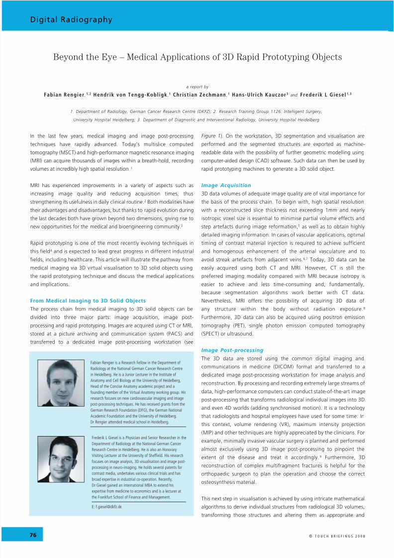
Figure 1: The Process Chain from Medical Imaging to3D Solid Objects
The process chain from medical imaging to 3D solid objects can be divided into three major
parts. Images are acquired using computed tomography or magnetic resonance imaging,
stored in a picture archiving and communication system (PACS) and transferred to a
dedicated image post-processing workstation. On the workstation, 3D segmentation and
visualisation are performed and a computer-aided design (CAD) model of the segmented
structures can be generated. Such data can then be used by rapid prototyping machines tocreate the 3D solid object.
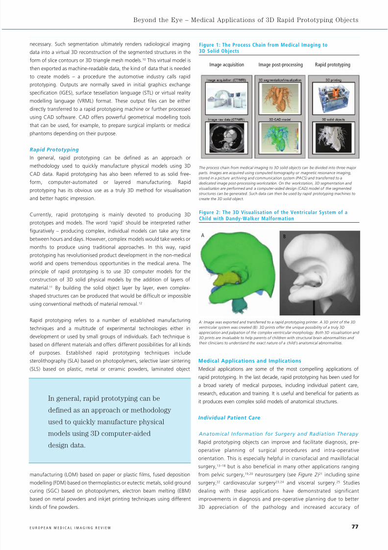
Figure 2: The 3D Visualisation of the Ventricular System of aChild with Dandy-Walker Malformation
A: Image was exported and transferred to a rapid prototyping printer. A 3D print of the 3D
ventricular system was created (B). 3D prints offer the unique possibility of a truly 3D
appreciation and palpation of the complex ventricular morphology. Both 3D visualisation and
3D prints are invaluable to help parents of children with structural brain abnormalities and
their clinicians to understand the exact nature of a child’s anatomical abnormalities.
A B
Image acquisition Image post-processing Rapid prototyping
In general, rapid prototyping can be
defined as an approach or methodology
used to quickly manufacture physical
models using 3D computer-aided
design data.
8/4/2019 giesel (1)
http://slidepdf.com/reader/full/giesel-1 3/6
78 E U R O P E A N M E D I C A L I M A G I N G R E V I E W
Digital Radiography
measurements and the possibility of planning, preparing and
simulating the surgical procedure in advance.15 Furthermore, 3D
replicas of the surgically treated structures can be intra-operatively
viewed side by side to the patient and thus facilitate orientation and
navigation particularly in complex cases. These advantages are
associated with reduced operating times, allowing for cost-effective
use of operating rooms.26 In this way, the advantages exceed the
limitations of the technique, namely the time and costs for creating
rapid prototyping objects. Moreover, rapid prototyping is a helpful
tool for radiation treatment planning and simulation27,28 and can be
used to create individual radiation shields.29
Prostheses and Implants
In addition to being useful for surgical planning and navigation, the rapid
prototyping technique can serve for producing medical prostheses and
implants, in particular for bone reconstructions. The great potential of
the rapid prototyping technique lies within the possibility of customised
prostheses and implants. Commercially available standard-sized bone
replacement parts may be sufficient for most surgical procedures and
cases, but not for all cases of any given procedure.
There are three reasons emphasising the need for individually producedprostheses. First, there are patients outside the standard range with
respect to size or other special requirements caused by disease or genetics.
Second, surgical outcome may be improved using customised devices
because standard prostheses or implants do not always adequately match
the individual anatomy. Third, customised prostheses and implants allow
for minimisation of the amount of resected patient tissue. Hence, the time
and costs for the production of customised rapid prototyping prostheses
and implants seem to be reasonable in selected patients.
The rapid prototyping technique has been applied to the reconstruction
of a variety of anatomical structures, showing the potential of this
technique in a time where individual patient care is becoming more andmore important. Customised prostheses and implants using rapid
prototyping have been successfully used for skull reconstructions,13,30 hip
replacements,31 femoral reconstructions,32 hemi-knee joints33,34 and
dental restorations.35
The rapid prototyping technique is beneficial not only for bone
reconstructions but also for replacing soft tissues, as rapid prototyping can
be applied to a variety of materials. Individual auricular prostheses36,37
probably provide the most vivid impression on the possible usefulness of the
technique. In patients with a missing ear, a mirrored scan of the remaining
ear is used for manufacturing a flesh-like rapid prototyping ear model.
Visualisation and Perception
Medical images most often clearly depict the pathology and its
patient-specific characteristics. However, medical images may be
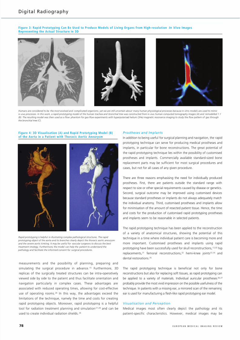
Figure 3: Rapid Prototyping Can Be Used to Produce Models of Living Organs from High-resolution In Vivo ImagesRepresenting the Actual Structure in 3D
Humans are considered to be the most evolved and complicated organisms, yet we are still uncertain about many human physiological processes because in vitro models are used to mimic
in vivo processes. In this work, a rapid prototyping model of the human trachea and bronchial tree was constructed from in vivo human computed tomography images (A) and remodelled 1:1
(B). The resulting model was then used as a flow phantom for gas-flow experiments with hyperpolarised helium (3He) magnetic resonance imaging to study the flow pattern of gas through
the bronchial tree (C).
A B C
Figure 4: 3D Visualisation (A) and Rapid Prototyping Model (B)of the Aorta in a Patient with Thoracic Aortic Aneurysm
A
B
Rapid prototyping is helpful in illustrating complex pathological structures. The rapid prototyping object of the aorta and its branches clearly depict the thoracic aortic aneurysm
and the severe aortic kinking. It may be useful for vascular surgeons to discuss the best
treatment strategy. Furthermore, the model can help the patient to understand the
pathology and facilitate the informed consent for surgical procedures.
8/4/2019 giesel (1)
http://slidepdf.com/reader/full/giesel-1 4/6
79E U R O P E A N M E D I C A L I M A G I N G R E V I E W
Beyond the Eye – Medical Applications of 3D Rapid Prototyping Objects
difficult to grasp for the patients themselves because they most often
do not have any previous knowledge for interpreting medical images,
and radiologists or surgeons may have difficulties in explaining the
patient his or her disease. The expression ‘grasp’ has a double
meaning: rapid prototyping may help the patient to understand the
pathology by providing a real, touchable model.19 Consequently,
patients will feel more comfortable giving informed consent for
surgical procedures,26 especially parents having to decide for their
children (see Figure 2).
Research
Rapid prototyping offers new opportunities for scientific research. On the
one hand, research with phantoms produced by rapid prototyping can help
to elucidate physiological processes that are not yet fully understood (see
Figure 3).38–41 On the other hand, rapid prototyping objects may contribute
to a better understanding of complex pathologies.24,38,42 Complex
pathologies are characterised by either complex morphology or functional
consequences. Complex morphologies may be better to depict 3D solid
objects than 2D medical images or 3D visualisations.24
Functionalconsequences can be assessed with patient-based phantoms simulating in
vivo conditions38 and can provide new insights into haemodynamic or
aerodynamic aspects of cardiovascular or airway diseases. By using the
established CAD techniques from the automotive industry, medical data can
be post-processed in terms of deformation or pressure processes as well.
Moreover, rapid prototyping offers the possibility to evaluate medical
imaging and post-processing techniques43 and to develop artificial organs.44
Current research also focuses on tissue11 and neural engineering.42
Education
Both surgical and minimally invasive procedures require a thorough
knowledge of anatomical structures and their topographical relations.This comprehensive knowledge is traditionally learned through the
preparation of human cadavers during pre-clinical studies at medical
school, and then put into practice and consolidated during actual
surgeries. However, gaining greater experience in the special area of
interest before operating on a patient is desirable. 2D medical images or
3D visualisations on a 2D screen are insufficient for obtaining an intuitive
understanding of complex anatomical details.24,45 Rapid prototyping
objects enhance 3D learning especially in challenging anatomical and
pathological conditions (see Figure 4).
Training
Furthermore, the possibility of surgical training procedures in general
and patient-specific procedures in complex cases improves the abilities
and results of surgeons.46 Rapid prototyping objects allow for intensive
training of young surgeons mimicking in vivo situations and real tissues
without the risk of damage to the patient.47,48 Trainees do not hesitate
to perform difficult procedures on rapid prototyping objects as they
may do in patients. After being trained, they will feel more
self-confident when going to the operating room. Furthermore, the
pre-operative simulation of a specific, complex and sensitive surgery
provides the unique opportunity to employ surgical instruments
identical to those used in the actual procedure, in order to determine
the best operating strategy.15 Hence, it increases the surgeon’s
confidence in the operation.
Discussion
Rapid prototyping has grown beyond its initial use in industrial sectors,
such as the automobile industry, and today can be regarded as one of
the most promising techniques to be associated with medical imaging.
A variety of rapid prototyping applications have recently emerged and
will probably find their way into the clinical arena, in medical
education, training and medical research. Although the medical
applications are relatively young, their enormous potential has already
been demonstrated in several studies.16,19,26
The application of rapid prototyping techniques in surgery is beneficial for
diagnosis, treatment planning and intra-operative navigation, especially in
complex cases where 2D source images or 3D virtual visualisations are
insufficient to give a complete understanding of the pathology. 13–25
Furthermore, rapid prototyping objects are useful for training surgeons
because they allow surgical procedures to be simulated in a realistic
manner.15 Additionally, customised prostheses and implants can be
manufactured using the presented process chain.30–37 Finally, 3D solid
objects are highly beneficial for communication between doctors, patients
and family members. These applications will probably gain further
importance when more attention is paid to individual patient care.
Medical research has already profited by rapid prototyping giving
new insights into physiological and pathological processes.38,39 Efforts
have been made on the development of artificial organs and tissues
using rapid prototyping.11,42,44 Much of the medical research using
rapid prototyping directly focuses on patients due to the individuality
of the technique itself; therefore, we predict that the knowledge
gained will eventually be transferred to the clinics and subsequently
the benefit to the patients themselves.
The traditional approach of teaching anatomy mainly focuses on normal
anatomy without considering its variation and pathological changes.
Medical students will only experience a greater variety when dealing with
patients. Rapid prototyping could serve as the medium to bring
anatomical variations from clinics to pre-clinical studies in order to
improve the understanding of anatomy. By adapting the transparency or
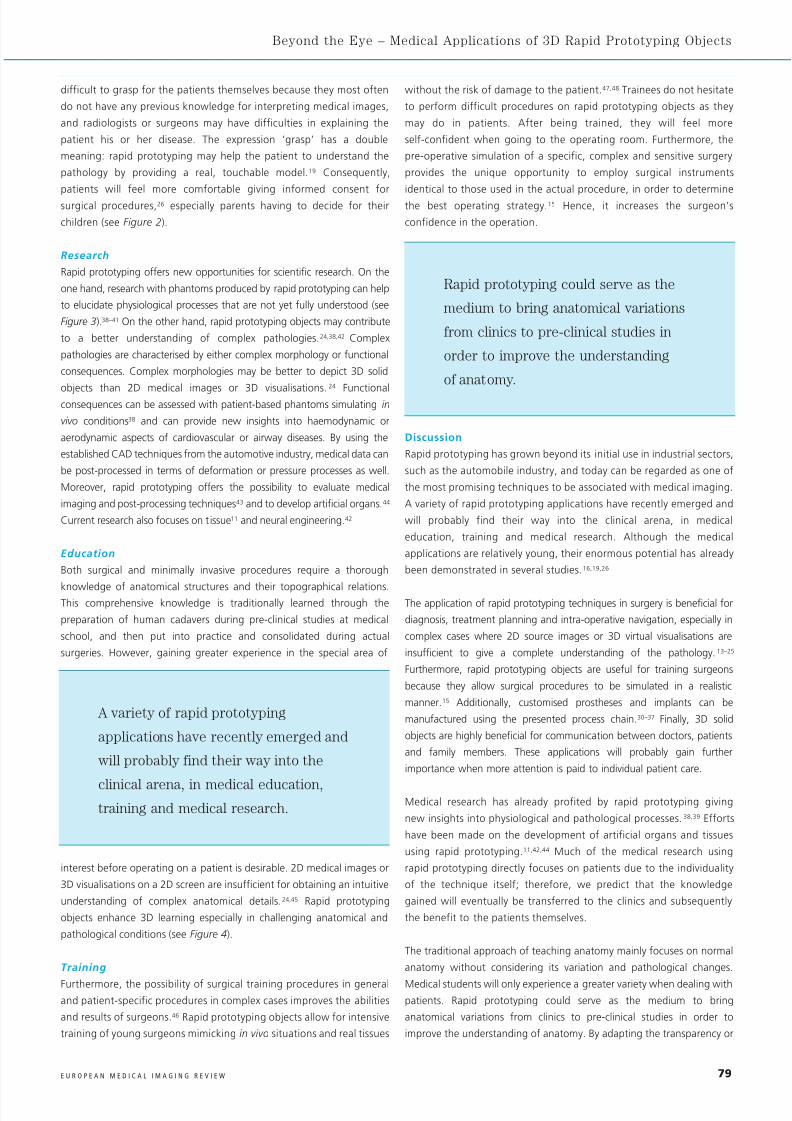
A variety of rapid prototyping
applications have recently emerged and
will probably find their way into the
clinical arena, in medical education,
training and medical research.
Rapid prototyping could serve as the
medium to bring anatomical variations
from clinics to pre-clinical studies in
order to improve the understanding
of anatomy.
8/4/2019 giesel (1)
http://slidepdf.com/reader/full/giesel-1 5/6
80 E U R O P E A N M E D I C A L I M A G I N G R E V I E W
Digital Radiography
rigidity of the used material, certain aspects can be emphasised for the
trainee or medical student as well.
The process chain from medical imaging to 3D solid objects involves
knowledge from a variety of fields, ranging from the acquisition of raw
data to image post-processing and the manufacturing of the
final models. Radiologists are the most important players in this
process chain as they combine expert know-how in both image
acquisition and post-processing. Nevertheless, the process chain only
runs smoothly if radiologists, computer scientists and material
scientists work closely together.
The greatest limitation to rapid prototyping is that it can only be applied
to objects not exceeding a certain dimension because the printers are not
yet able to handle extremely large objects. Future developments may
overcome this limitation. The costs and time needed for rapid
prototyping should not be regarded as limitations because they are due
to the individuality of each rapid prototyping object.
Conclusions
Rapid prototyping has established a variety of medical applications
such as surgical and interventional planning and training, bone
reconstructions or medical education. A tremendous growth in
utilisation as well as application development can be anticipated in the
field of individual patient care, medical education and training, as well
as medical research. ■
Acknowledgements
We greatly appreciate the support by VitalRecon Ltd, Frankfurt,
Germany, in providing image analysis, segmentation and
manufacturing of rapid prototyping models. Fabian Rengier received a
grant from the German Research Foundation (DFG) under the auspices
of the ‘Research training group 1126: Intelligent Surgery –
Development of new computer-based methods for the future
workplace in surgery’. We further acknowledge the support by the
Klaus Tschira Foundation and by 4D concepts, Gross Gerau, Germany,
in particular Rainer Neumann.
1. Kido T, Kurata A, Higashino H, et al., Cardiac imaging using
256-detector row four-dimensional CT: preliminary clinical
report, Radiat Med , 2007;25:38–44.
2. Meaney JF, Goyen M. Recent advances in contrast-enhanced
magnetic resonance angiography, Eur Radio l , 2007;17
(Suppl. 2):B2–B6.
3. Doi K. Diagnostic imaging over the last 50 years: research
and development in medical imaging science and technology,
Phys Med B io l , 2006;51:R5–27.
4. McGurk M, Amis AA, Potamianos P, Goodger NM, Rapid
prototyping techniques for anatomical modelling in medicine,
Ann R Col l Surg Engl , 1997;79:169–74.
5. Mahesh M, Search for isotropic resolution in CT from
conventional through multiple-row detector, Radiographi cs,
2002;22:949–62.
6. Johnson TR, Nikolaou K, Wintersperger BJ, et al.,Optimization of contrast material administration for
electrocardiogram-gated computed tomographic angiography
of the chest, J Comput Ass ist Tomogr , 2007;31:265–71.
7. Suzuki S, Furui S, Kaminaga T, Yamauchi T, Measurement of
vascular diameter in vi t ro by automated software for CT
angiography: effects of inner diameter, density of contrast
medium, and convolution kernel, AJR Am J Roentgenol , 2004;
182:1313–17.
8. von Tengg-Kobligk H, Weber TF, Rengier F, et al., Imaging
modalities for the thoracic aorta, J Cardiovasc Surg (Tor ino),
2008;49:429–47.
9. von Tengg-Kobligk H, Weber TF, Rengier F, et al., Image
postprocessing of aortic CTA and MRA, Radio loge, 2007;
47:1003–11.
10. Hahn HK, Millar WS, Klinghammer O, et al., A reliable and
efficient method for cerebral ventricular volumetry in pediatricneuroimaging, Methods Inf Med , 2004;43:376–82.
11. Peltola SM, Melchels FP, Grijpma DW, Kellomaki M, A review
of rapid prototyping techniques for tissue engineering
purposes, Ann Med , 2008;40:268–80.
12. Webb PA, A review of rapid prototyping (RP) techniques in
the medical and biomedical sector, J Med Eng Technol ,
2000;24: 149–53.
13. D'Urso PS, Earwaker WJ, Barker TM, et al., Custom
cranioplasty using stereolithography and acrylic, Br J Plast
Surg , 2000;53: 200–204.
14. Faber J, Berto PM, Quaresma M, Rapid prototyping as a tool
for diagnosis and treatment planning for maxillary canine
impaction, Am J Orthod Dentofacial Orthop , 2006;129:583–9.
15. Mavili ME, Canter HI, Saglam-Aydinatay B, et al., Use of
three-dimensional medical modeling methods for precise
planning of orthognathic surgery, J Craniofac Surg,
2007;18:740–47.
16. Muller A, Krishnan KG, Uhl E, Mast G, The application of
rapid prototyping techniques in cranial reconstruction and
preoperative planning in neurosurgery, J Craniofac Surg,
2003; 14:899–914.
17. Poukens J, Haex J, Riediger D, The use of rapid prototyping
in the preoperative planning of distraction osteogenesis of
the cranio-maxillofacial skeleton, Comput Aided Surg , 2003;8:
146–54.
18. Wagner JD, Baack B, Brown GA, Kelly J, Rapid 3-dimensional
prototyping for surgical repair of maxillofacial fractures: a
technical note, J Oral Maxi l lofac Surg, 2004;62:898–901.
19. Guarino J, Tennyson S, McCain G, et al., Rapid prototyping
technology for surgeries of the pediatric spine and pelvis:
benefits analysis, J Pediatr Orthop , 2007;27:955–60.
20. Hurson C, Tansey A, O'Donnchadha B, et al., Rapid
prototyping in the assessment, classification and preoperative
planning of acetabular fractures, Injury , 2007;38:1158–62.
21. Wurm G, Tomancok B, Pogady P, et al., Cerebrovascular
stereolithographic biomodeling for aneurysm surgery.
Technical note, J Neurosurg, 2004;100:139–45.22. Paiva WS, Amorim R, Bezerra DA, Masini M, Application of
the stereolithography technique in complex spine surgery, Arq
Neuropsiquiatr , 2007;65:443–5.
23. Armillotta A, Bonhoeffer P, Dubini G, et al., Use of rapid
prototyping models in the planning of percutaneous
pulmonary valved stent implantation, Proc Ins t Mech Eng [H] ,
2007;221: 407–16.
24. Kim MS, Hansgen AR, Wink O, et al., Rapid prototyping: a
new tool in understanding and treating structural heart
disease, Circulat ion , 2008;117:2388–94.
25. Hiramatsu H, Yamaguchi H, Nimi S, Ono H, [Rapid
prototyping of the larynx for laryngeal frame work surgery],
Nippon J ib i i nkoka Gakkai Kaiho , 2004;107:949–55.
26. D'Urso PS, Barker TM, Earwaker WJ, et al., Stereolithographic
biomodelling in cranio-maxillofacial surgery: a prospective
trial, J Craniomaxi l lofac Surg, 1999;27:30–37.27. Kalet IJ, Wu J, Lease M, et al., Anatomical information in
radiation treatment planning, Proc AMIA Symp , 1999;291–5.
28. Sun SP, Wu CJ, Using the full scale 3D solid anthropometric
model in radiation oncology positioning and verification, Conf
Proc I EEE Eng Med B io l Soc , 2004;5:3432–5.
29. Zemnick C, Woodhouse SA, Gewanter RM, et al., Rapid
prototyping technique for creating a radiation shield,
J Prosthet Dent , 2007;97:236–41.
30. Singare S, Liu Y, Li D, et al., Individually Prefabricated
Prosthesis for Maxilla Reconstuction, J Prosthodont , 2008;
17:135–40.
31. Dai KR, Yan MN, Zhu ZA, Sun YH, Computer-aided custom-
made hemipelvic prosthesis used in extensive pelvic lesions,
J Arthroplasty , 2007;22:981–6.
32. Harrysson OL, Hosni YA, Nayfeh JF, Custom-designed
orthopedic implants evaluated using finite element analysis
of patient-specific computed tomography data: femoral-
component case study, BMC Musculoskelet D i sord , 2007;8:91.
33. He J, Li D, Lu B, et al., Custom fabrication of composite tibial
hemi-knee joint combining CAD/CAE/CAM techniques, Proc
Ins t Mech Eng [H] , 2006;220:823–30.
34. Wang Z, Teng Y, Li D, [Fabrication of custom-made artificial
semi-knee joint based on rapid prototyping technique:
computer-assisted design and manufacturing], Zhongguo Xiu
Fu Chong J i an Wai Ke Za Zhi , 2004;18:347–51.
35. Lee MY, Chang CC, Ku YC, New layer-based imaging and
rapid prototyping techniques for computer-aided design and
manufacture of custom dental restoration, J Med Eng Technol ,
2008;32:83–90.
36. Subburaj K, Nair C, Rajesh S, et al., Rapid development of
auricular prosthesis using CAD and rapid prototyping
technologies, Int J Oral Maxi l lofac Surg, 2007;36:938–43.
37. Ciocca L, Mingucci R, Gassino G, Scotti R, CAD/CAM ear
model and virtual construction of the mold, J Prosthet Dent ,
2007; 98:339–43.
38. Canstein C, Cachot P, Faust A, et al., 3D MR flow analysis in
realistic rapid-prototyping model systems of the thoracicaorta: comparison with in vivo data and computational fluid
dynamics in identical vessel geometries, Magn Reson Med ,
2008;59: 535–46.
39. Chung SK, Son YR, Shin SJ, Kim SK, Nasal airflow during
respiratory cycle, Am J Rhinol , 2006;20:379–84.
40. Giesel FL, Mehndiratta A, Tengg-Kobligk H, et al., Rapid
prototyping raw models on the basis of high resolution
computed tomography lung data for respiratory flow
dynamics, Acad Radiol , in press.
41. Giesel FL, Hard A, Hahn HK, et al., 3D-Reconstructions of the
cerebral ventricles and volume quantification in children with
brain malformations, Acad Radiol , in press.
42. Tek P, Chiganos TC, Mohammed JS, et al., Rapid prototyping
for neuroscience and neural engineering, J Neurosci Methods,
2008;172:263–9.
43. Winder RJ, Sun Z, Kelly B, et al., Abdominal aortic aneurysmand stent graft phantom manufactured by medical rapid
prototyping, J Med Eng Technol , 2002;26:75–8.
44. Taga I, Funakubo A, Fukui Y, Design and development of an
artificial implantable lung using multiobjective genetic
algorithm: evaluation of gas exchange performance, ASAIO J ,
2005;51:92–102.
45. Suzuki M, Ogawa Y, Kawano A, et al., Rapid prototyping of
temporal bone for surgical training and medical education,
Acta Otolaryngol , 2004;124:400–402.
46. Knox K, Kerber CW, Singel SA, et al., Rapid prototyping to
create vascular replicas from CT scan data: making tools to
teach, rehearse, and choose treatment strategies, Catheter
Cardiovasc Interv , 2005;65:47–53.
47. Bruyere F, Leroux C, Brunereau L, Lermusiaux P, Rapid
prototyping model for percutaneous nephrolithotomy
training, J Endourol , 2008;22:91–6.
48. Sulaiman A, Boussel L, Taconnet F, et al., In vi t ro non-rigid
life-size model of aortic arch aneurysm for endovascular
prosthesis assessment, Eur J Cardiothorac Surg ,
2008;33:53–7.
8/4/2019 giesel (1)
http://slidepdf.com/reader/full/giesel-1 6/6
20TH
ANNUAL MEETING AND POSTGRADUATE COURSEJUNE 23 – 26
VALENCIA / ESESGAR 2009
EUROPEAN SOCIETY OF GASTROINTESTINAL AND ABDOMINAL RADIOLOGY
OFFICERS OF THE
ESGAR EXECUTIVE COMMITTEE
PRESIDENT
B. Marincek (Zurich/CH)
PRESIDENT-ELECT
Y. Menu (Le Kremlin-Bicêtre/FR)
VICE PRESIDENT
F. Caseiro-Alves (Coimbra/PT)
SECRETARY
S. Jackson (Plymouth/UK)
TREASURER
A. Palkó (Szeged/HU)
PAST PRESIDENT
C. Bartolozzi (Pisa/IT)
BY-LAWS COMMITTEE
C. Matos (Brussels/BE)
EDUCATION COMMITTEE
A. Laghi (Latina/IT)
MEMBERSHIP COMMITTEE
J.S. Laméris (Amsterdam/NL)
MEETING PRESIDENT
L. Marti-Bonmati (Valencia/ES)
PRE-MEETING PRESIDENT
M. Laniado (Dresden/DE)
PRE-PRE-MEETING PRESIDENT
G. Morana (Treviso/IT)
FELLOWS REPRESENTATIVES
L.H. Ros Mendoza (Zaragoza/ES)
W. Schima (Vienna/AT)
CENTRAL ESGAR OFFICENeutorgasse 9/2a
AT – 1010 Vienna, Austria
Phone: +43 1 535 89 27
Fax: +43 1 535 70 37
E-Mail: [email protected]
MEETING PRESIDENTDr. Luis Martí-Bonmatí
Dr Peset University Hospital
Resonancia Magnetica. Servicio de Radiologia
Gaspar Aguilar, 90
ES – 46017 Valencia, Spain
Abstract submission and registration open on October 31, 2008.
www.esgar.org
![1 1 1 1 1 1 1 ¢ 1 1 1 - pdfs.semanticscholar.org€¦ · 1 1 1 [ v . ] v 1 1 ¢ 1 1 1 1 ý y þ ï 1 1 1 ð 1 1 1 1 1 x](https://img.pdfslide.us/doc/110x75/5f7bc722cb31ab243d422a20/1-1-1-1-1-1-1-1-1-1-pdfs-1-1-1-v-v-1-1-1-1-1-1-y-1-1-1-.jpg)


![$1RYHO2SWLRQ &KDSWHU $ORN6KDUPD +HPDQJL6DQH … · 1 1 1 1 1 1 1 ¢1 1 1 1 1 ¢ 1 1 1 1 1 1 1w1¼1wv]1 1 1 1 1 1 1 1 1 1 1 1 1 ï1 ð1 1 1 1 1 3](https://img.pdfslide.us/doc/110x75/5f3ff1245bf7aa711f5af641/1ryho2swlrq-kdswhu-orn6kdupd-hpdqjl6dqh-1-1-1-1-1-1-1-1-1-1-1-1-1-1.jpg)


![1 1 1 1 1 1 1 ¢ 1 , ¢ 1 1 1 , 1 1 1 1 ¡ 1 1 1 1 · 1 1 1 1 1 ] ð 1 1 w ï 1 x v w ^ 1 1 x w [ ^ \ w _ [ 1. 1 1 1 1 1 1 1 1 1 1 1 1 1 1 1 1 1 1 1 1 1 1 1 1 1 1 1 ð 1 ] û w ü](https://img.pdfslide.us/doc/110x75/5f40ff1754b8c6159c151d05/1-1-1-1-1-1-1-1-1-1-1-1-1-1-1-1-1-1-1-1-1-1-1-1-1-1-w-1-x-v.jpg)

![[XLS] · Web view1 1 1 2 3 1 1 2 2 1 1 1 1 1 1 2 1 1 1 1 1 1 2 1 1 1 1 2 2 3 5 1 1 1 1 34 1 1 1 1 1 1 1 1 1 1 240 2 1 1 1 1 1 2 1 3 1 1 2 1 2 5 1 1 1 1 8 1 1 2 1 1 1 1 2 2 1 1 1 1](https://img.pdfslide.us/doc/110x75/5ad1d2817f8b9a05208bfb6d/xls-view1-1-1-2-3-1-1-2-2-1-1-1-1-1-1-2-1-1-1-1-1-1-2-1-1-1-1-2-2-3-5-1-1-1-1.jpg)