Embed Size (px)
Citation preview
Teaching Day March 2013
Gibran F Bu7 MSc

• 0) Introduc,on • 1) History • 2) Ocular cranial nerves • 3) Fundoscopy

Intro… WHAT DO THEY WANT!!!!!!
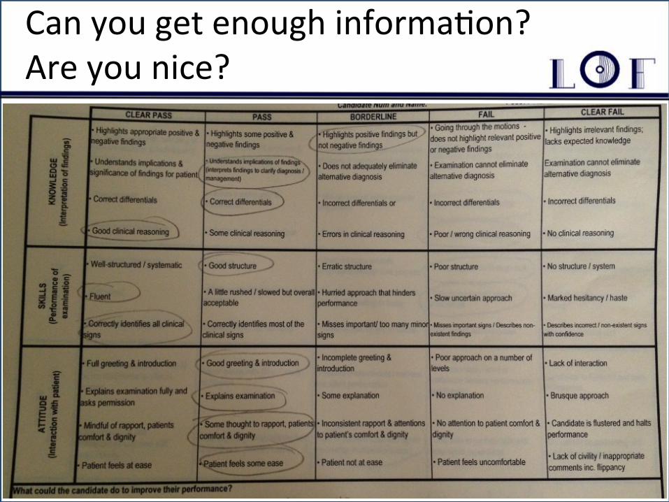
Can you get enough informa,on? Are you nice?
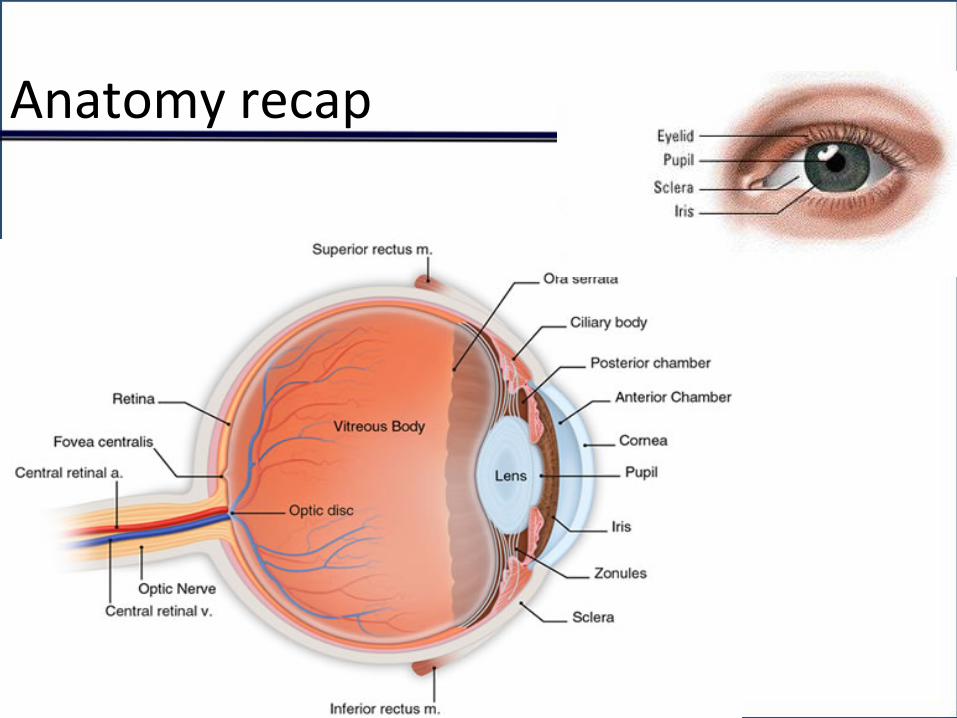
Anatomy recap
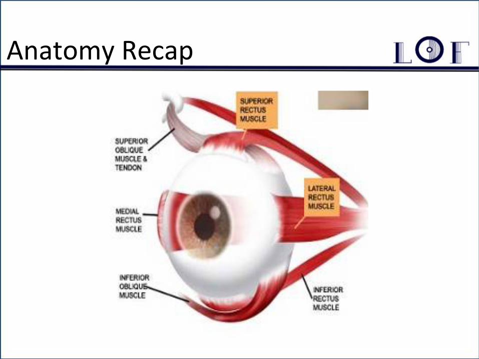
Anatomy Recap
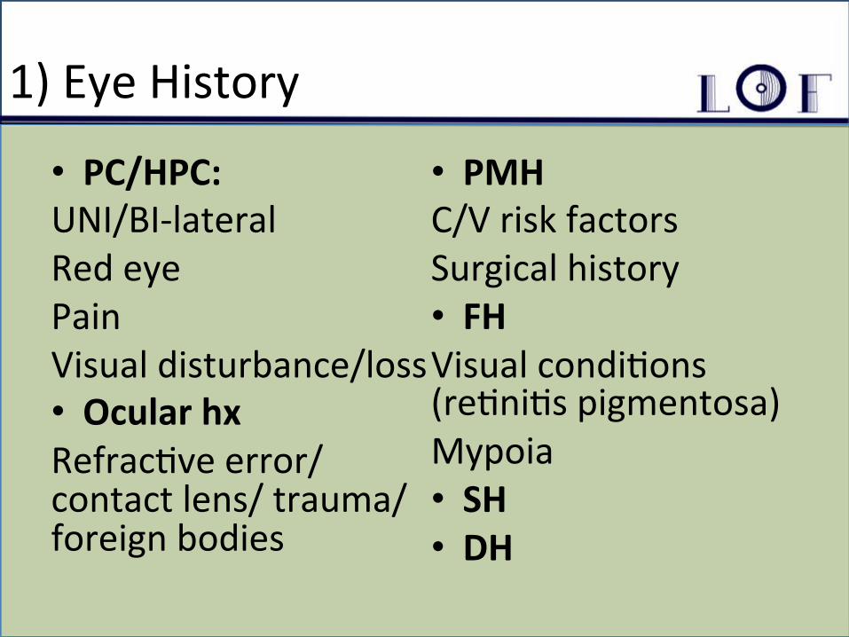
1) Eye History
• PC/HPC: UNI/BI-‐lateral Red eye Pain Visual disturbance/loss • Ocular hx Refrac,ve error/ contact lens/ trauma/ foreign bodies
• PMH C/V risk factors Surgical history • FH Visual condi,ons (re,ni,s pigmentosa) Mypoia • SH • DH
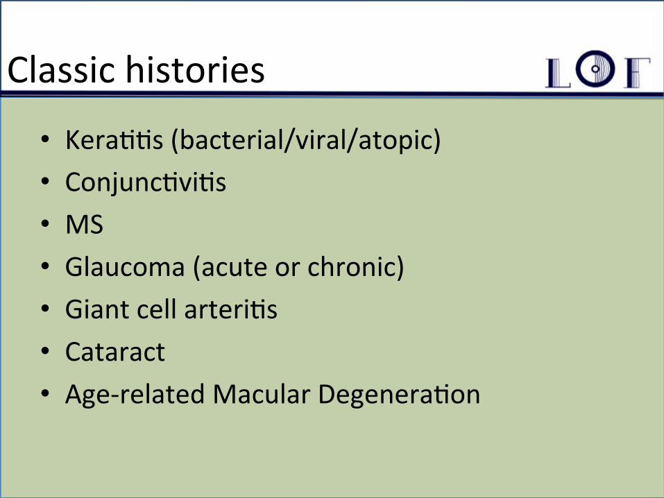
Classic histories
• Kera,,s (bacterial/viral/atopic) • Conjunc,vi,s • MS • Glaucoma (acute or chronic) • Giant cell arteri,s • Cataract • Age-‐related Macular Degenera,on

Vision
No Loss
Pain, Red eye, Purulent discharge
Pain/itchy/scratchy/ red eyes, watery
discharge
Loss/disturbance
Gradual
Peripheral field constricMon
Central vision disturbance and metamorphosia
Color vision disturbance, temporal
improvement in shortsightedness
Sudden
No pain
Flashing lights, floaters
‘Filling up’
Pain
Eye
Photophobia, red eye, (irregular
pupil)
Non-‐red eye, retro bulbar pain
Temple/forehead/jaw
Head, assoc. photophobia
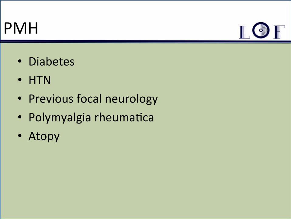
PMH
• Diabetes • HTN • Previous focal neurology • Polymyalgia rheuma,ca • Atopy
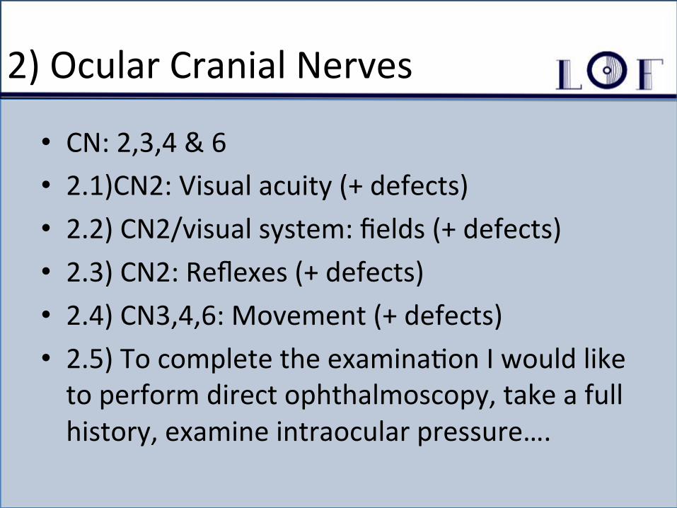
2) Ocular Cranial Nerves
• CN: 2,3,4 & 6 • 2.1)CN2: Visual acuity (+ defects) • 2.2) CN2/visual system: fields (+ defects) • 2.3) CN2: Reflexes (+ defects) • 2.4) CN3,4,6: Movement (+ defects) • 2.5) To complete the examina,on I would like to perform direct ophthalmoscopy, take a full history, examine intraocular pressure….
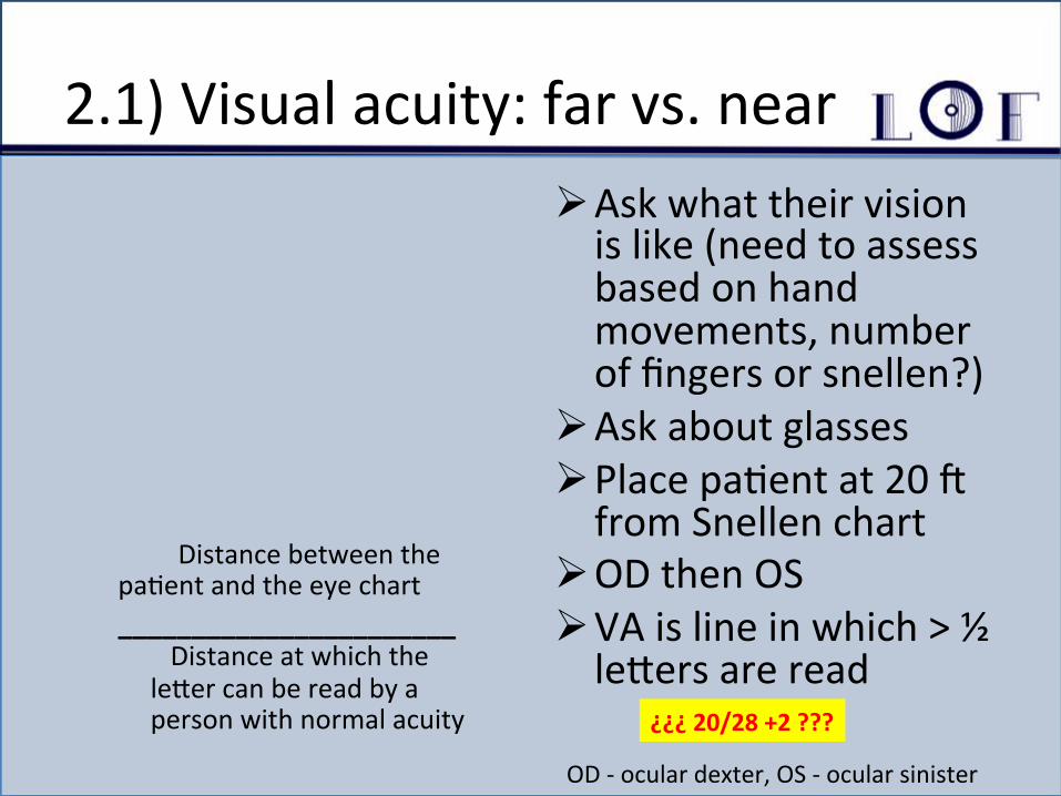
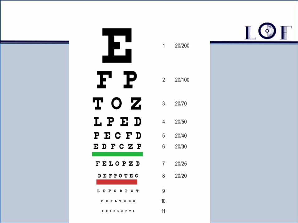
2.1) Visual acuity: far vs. near
Distance between the pa,ent and the eye chart _______________________
Distance at which the leeer can be read by a person with normal acuity
Ø Ask what their vision is like (need to assess based on hand movements, number of fingers or snellen?)
Ø Ask about glasses Ø Place pa,ent at 20 f from Snellen chart
Ø OD then OS Ø VA is line in which > ½ leeers are read
¿¿¿ 20/28 +2 ???
OD -‐ ocular dexter, OS -‐ ocular sinister

2.1) Visual acuity: near • Test of funcMon • (may get away with men,oning it) • Use a card if available (at ~40 cm) • Record (or at least remember!)

2.1) Ishihara plates • Men,on it – may be
prompted to use the book and stopped and asked to explain use.
So: • Check pt can read • Open on control page • Proceed with examina,on
making sure to note which pages pt has difficulty with

2.2) Visual fields
Confronta,on • Same level, same eye. • Red Pin to measure color satura,on
Visual inaeen,on Blind spot defect

2.3) Reflexes: Pupillary Examina,on • Inspect • Direct – Constric,on of ipsilateral eye
• Consensual – Constric,on of contralateral eye
• Swinging light test -‐ RAPD • AccommodaMon • Defects: – Fixed dilated – Tonic Holmes Aides pupil – Pinpoint – Horner’s syndrome – RAPD – op,c neuri,s – AR Pupil
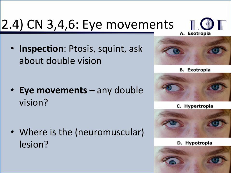
2.4) CN 3,4,6: Eye movements
• InspecMon: Ptosis, squint, ask about double vision
• Eye movements – any double vision?
• Where is the (neuromuscular) lesion?

How to work out which muscle is affected
1. Which eye is affected 2. Which movement is affected 3. Which muscle(s) is responsible 4. Which nerve is responsible
Important defect: Internuclear ophthalmoplegia:-‐ Nystagmus and impaired adbuc,on on lateral gaze. Lesion is in MEDIAL LONGITUDINAL FASICULUS on side of nystagmus *May be feature of Mul,ple Sclerosis*

3) Fundoscopy
• Turn on the scope • Set to 0 • Red reflex (leukocoria/darkened) • Look in the eye • Locate a blood vessel and follow back to the disc • Inspect quadrants • Inspect macula by gerng them to look directly into the light
Fundus signs
• Gross observa,ons • Specific Structures: – Vessels: arteries and veins – ONH – Re,na – Macula
Likely 3rd year Diagnoses • Diabe,c Re,nopathy • HTN re,nopathy • Macular Degenera,on • Re,ni,s pigmentosa
Others to consider? • Macular hole • Epire,nal membrane

HTN
IV) PAPILLODEMA III) Coeon wool spots and flame hemorrhages
I) SILVER/COPPER WIRING
II) AV NIPPING
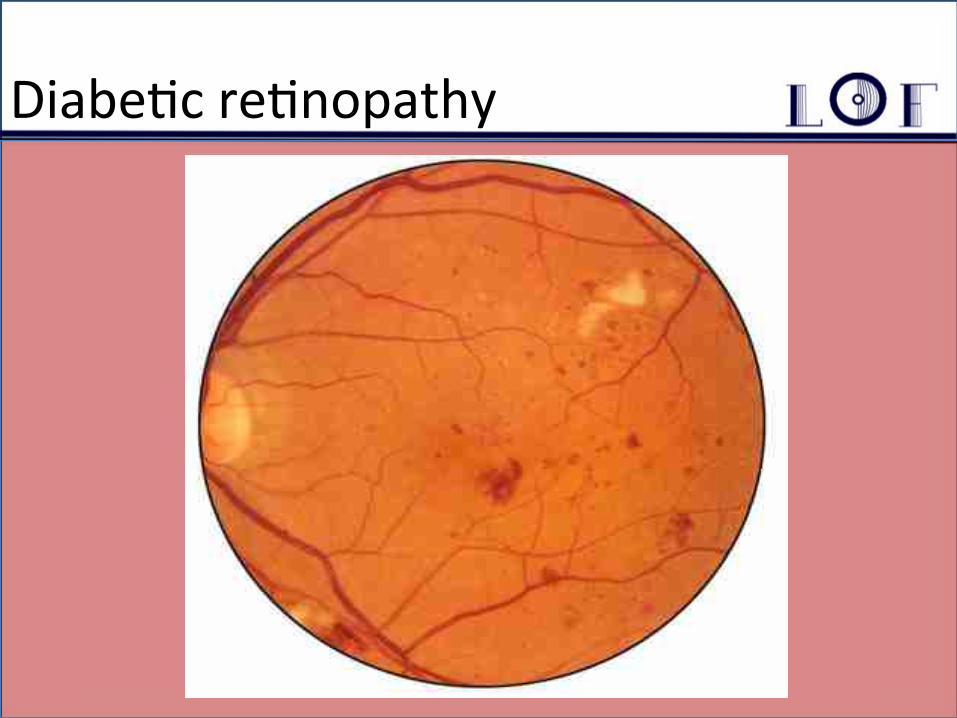
Diabe,c re,nopathy
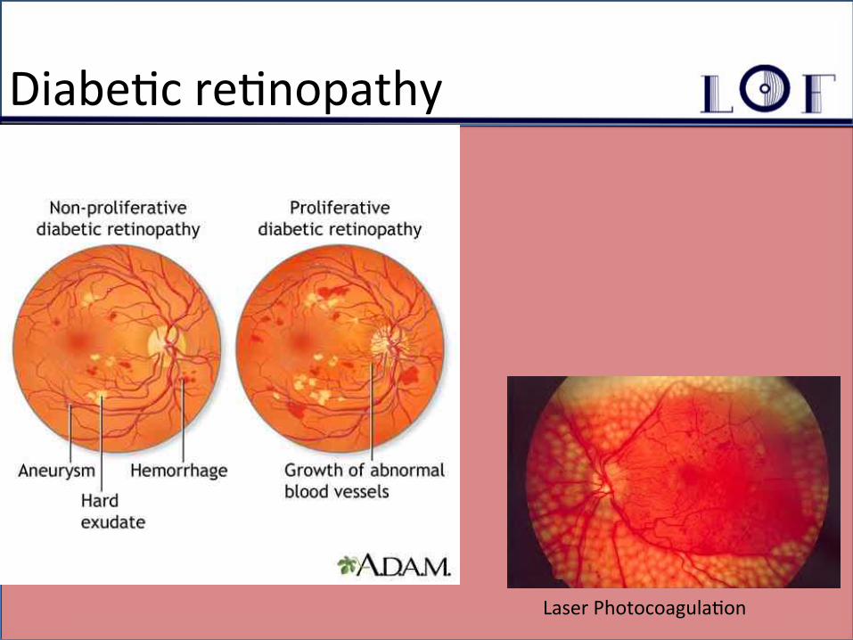
Diabe,c re,nopathy
Laser Photocoagula,on
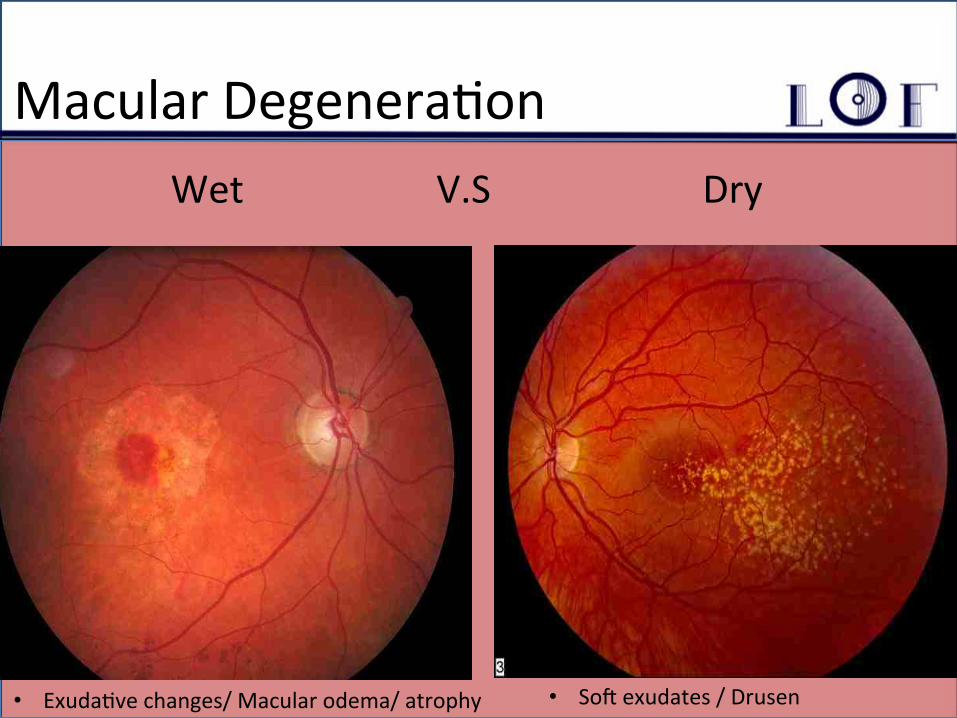
Macular Degenera,on Wet V.S Dry
• Sof exudates / Drusen • Exuda,ve changes/ Macular odema/ atrophy

Macular Degenera,on
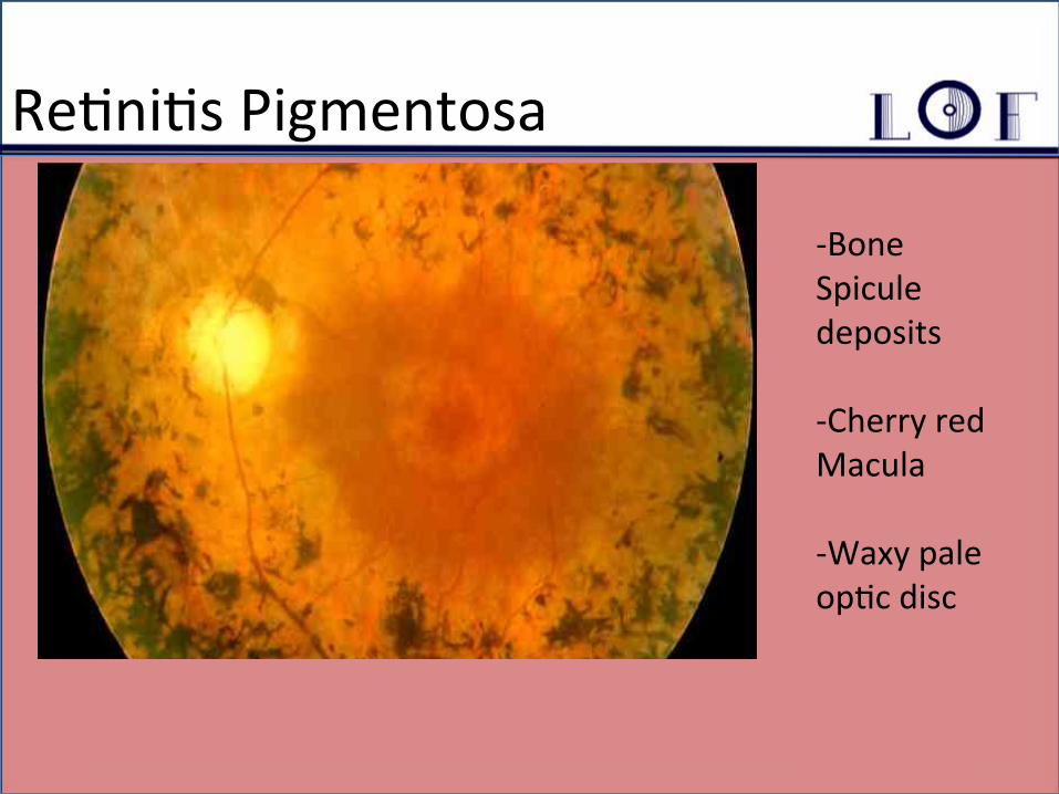
Re,ni,s Pigmentosa
-‐Bone Spicule deposits -‐Cherry red Macula -‐Waxy pale op,c disc
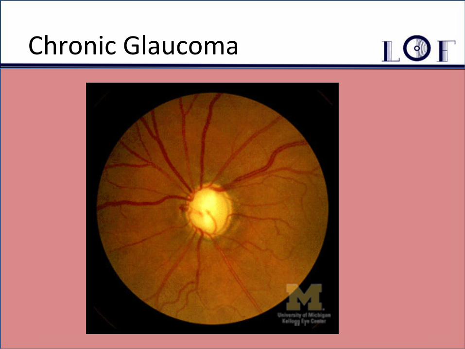
Chronic Glaucoma
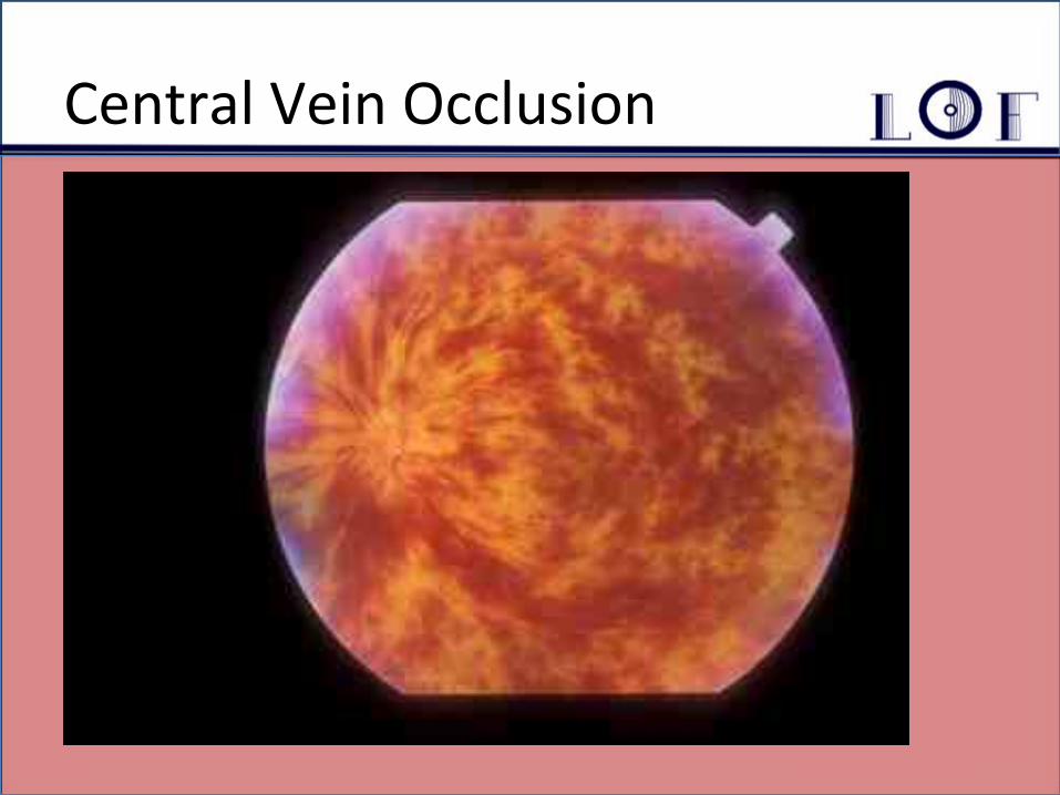
Central Vein Occlusion
!
Good Luck!!!!
• Sources: Picture credits to google etc… www.uninteforsight.com www.labordeeyegroup.com hep://www.health.state.mn.us/divs/s/mch/webcourse/vision/mod4a.cfm hep://web1.ncoptometry.org/nonpro.aspx