Embed Size (px)
Citation preview
Giants in Obstetrics and Gynecology Series:A profile of John C. Hobbins, MDRoberto Romero, MD, DMedSci, Editor-in-Chief for Obstetrics
J ohn C. Hobbins, MD, DistinguishedProfessor at the University of Colo-
rado, and former Professor and Chief ofObstetrics at Yale University and at theUniversity of Colorado, is an undisputedgiant in our specialty. John has been apioneer of ultrasound imaging in obstet-rics and gynecology; evenmore so, it is theinspirational vitality of such an extraordi-nary man whose influence on the evolu-tion of modern obstetrical practice isperhaps best reflected in the legions ofresidents and fellows he trained. They, inturn, have taught others, building onwhatthey learned from John to become aca-demic leaders in our specialty and chang-ing the practice of obstetrics through theirwork.
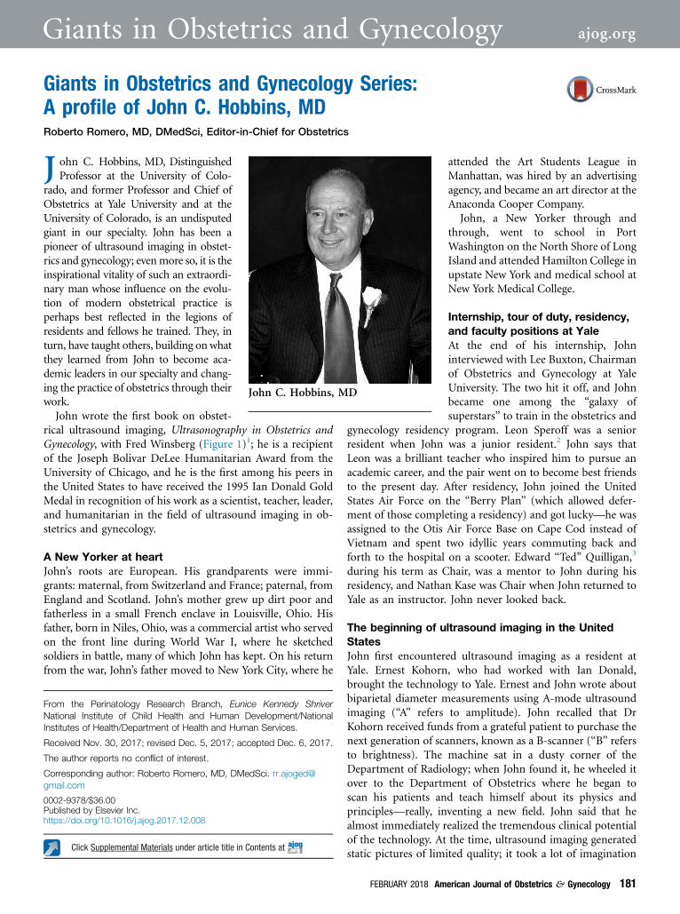
John wrote the first book on obstet-rical ultrasound imaging, Ultrasonography in Obstetrics andGynecology, with Fred Winsberg (Figure 1)1; he is a recipientof the Joseph Bolivar DeLee Humanitarian Award from theUniversity of Chicago, and he is the first among his peers inthe United States to have received the 1995 Ian Donald GoldMedal in recognition of his work as a scientist, teacher, leader,and humanitarian in the field of ultrasound imaging in ob-stetrics and gynecology.
A New Yorker at heartJohn’s roots are European. His grandparents were immi-grants: maternal, from Switzerland and France; paternal, fromEngland and Scotland. John’s mother grew up dirt poor andfatherless in a small French enclave in Louisville, Ohio. Hisfather, born in Niles, Ohio, was a commercial artist who servedon the front line during World War I, where he sketchedsoldiers in battle, many of which John has kept. On his returnfrom the war, John’s father moved to New York City, where he
attended the Art Students League inManhattan, was hired by an advertisingagency, and became an art director at theAnaconda Cooper Company.
John, a New Yorker through andthrough, went to school in PortWashington on the North Shore of LongIsland and attended Hamilton College inupstate New York and medical school atNew York Medical College.
Internship, tour of duty, residency,and faculty positions at YaleAt the end of his internship, Johninterviewed with Lee Buxton, Chairmanof Obstetrics and Gynecology at YaleUniversity. The two hit it off, and Johnbecame one among the “galaxy ofsuperstars” to train in the obstetrics and
gynecology residency program. Leon Speroff was a seniorresident when John was a junior resident.2 John says thatLeon was a brilliant teacher who inspired him to pursue anacademic career, and the pair went on to become best friendsto the present day. After residency, John joined the UnitedStates Air Force on the “Berry Plan” (which allowed defer-ment of those completing a residency) and got lucky—he wasassigned to the Otis Air Force Base on Cape Cod instead ofVietnam and spent two idyllic years commuting back andforth to the hospital on a scooter. Edward “Ted” Quilligan,3
during his term as Chair, was a mentor to John during hisresidency, and Nathan Kase was Chair when John returned toYale as an instructor. John never looked back.
The beginning of ultrasound imaging in the UnitedStatesJohn first encountered ultrasound imaging as a resident atYale. Ernest Kohorn, who had worked with Ian Donald,brought the technology to Yale. Ernest and John wrote aboutbiparietal diameter measurements using A-mode ultrasoundimaging (“A” refers to amplitude). John recalled that DrKohorn received funds from a grateful patient to purchase thenext generation of scanners, known as a B-scanner (“B” refersto brightness). The machine sat in a dusty corner of theDepartment of Radiology; when John found it, he wheeled itover to the Department of Obstetrics where he began toscan his patients and teach himself about its physics andprinciples—really, inventing a new field. John said that healmost immediately realized the tremendous clinical potentialof the technology. At the time, ultrasound imaging generatedstatic pictures of limited quality; it took a lot of imagination
Click Supplemental Materials under article title in Contents at
John C. Hobbins, MD
From the Perinatology Research Branch, Eunice Kennedy ShriverNational Institute of Child Health and Human Development/NationalInstitutes of Health/Department of Health and Human Services.
Received Nov. 30, 2017; revised Dec. 5, 2017; accepted Dec. 6, 2017.
The author reports no conflict of interest.
Corresponding author: Roberto Romero, MD, DMedSci. [email protected]
0002-9378/$36.00Published by Elsevier Inc.https://doi.org/10.1016/j.ajog.2017.12.008
FEBRUARY 2018 American Journal of Obstetrics & Gynecology 181
Giants in Obstetrics and Gynecology ajog.org
and knowledge of anatomy to figure out what the black andwhite lines represented.
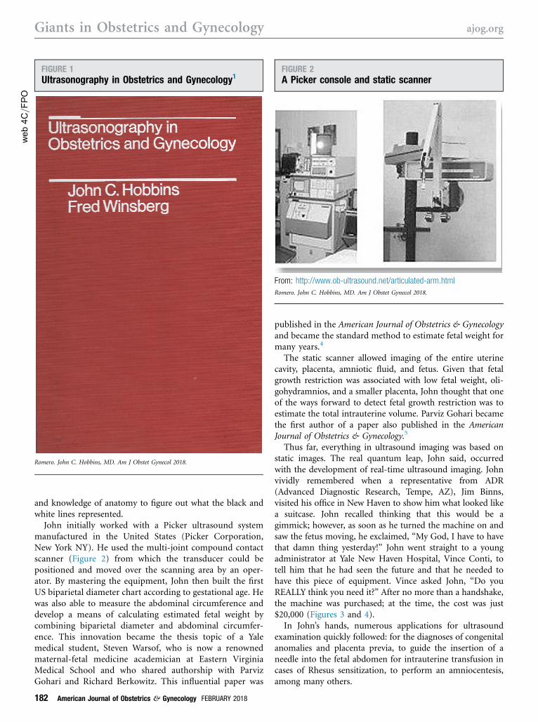
John initially worked with a Picker ultrasound systemmanufactured in the United States (Picker Corporation,New York NY). He used the multi-joint compound contactscanner (Figure 2) from which the transducer could bepositioned and moved over the scanning area by an oper-ator. By mastering the equipment, John then built the firstUS biparietal diameter chart according to gestational age. Hewas also able to measure the abdominal circumference anddevelop a means of calculating estimated fetal weight bycombining biparietal diameter and abdominal circumfer-ence. This innovation became the thesis topic of a Yalemedical student, Steven Warsof, who is now a renownedmaternal-fetal medicine academician at Eastern VirginiaMedical School and who shared authorship with ParvizGohari and Richard Berkowitz. This influential paper was
published in the American Journal of Obstetrics & Gynecologyand became the standard method to estimate fetal weight formany years.4
The static scanner allowed imaging of the entire uterinecavity, placenta, amniotic fluid, and fetus. Given that fetalgrowth restriction was associated with low fetal weight, oli-gohydramnios, and a smaller placenta, John thought that oneof the ways forward to detect fetal growth restriction was toestimate the total intrauterine volume. Parviz Gohari becamethe first author of a paper also published in the AmericanJournal of Obstetrics & Gynecology.5
Thus far, everything in ultrasound imaging was based onstatic images. The real quantum leap, John said, occurredwith the development of real-time ultrasound imaging. Johnvividly remembered when a representative from ADR(Advanced Diagnostic Research, Tempe, AZ), Jim Binns,visited his office in New Haven to show him what looked likea suitcase. John recalled thinking that this would be agimmick; however, as soon as he turned the machine on andsaw the fetus moving, he exclaimed, “My God, I have to havethat damn thing yesterday!” John went straight to a youngadministrator at Yale New Haven Hospital, Vince Conti, totell him that he had seen the future and that he needed tohave this piece of equipment. Vince asked John, “Do youREALLY think you need it?” After no more than a handshake,the machine was purchased; at the time, the cost was just$20,000 (Figures 3 and 4).
In John’s hands, numerous applications for ultrasoundexamination quickly followed: for the diagnoses of congenitalanomalies and placenta previa, to guide the insertion of aneedle into the fetal abdomen for intrauterine transfusion incases of Rhesus sensitization, to perform an amniocentesis,among many others.
web4C=F
PO
FIGURE 1Ultrasonography in Obstetrics and Gynecology1
Romero. John C. Hobbins, MD. Am J Obstet Gynecol 2018.
FIGURE 2A Picker console and static scanner
From: http://www.ob-ultrasound.net/articulated-arm.htmlRomero. John C. Hobbins, MD. Am J Obstet Gynecol 2018.
Giants in Obstetrics and Gynecology ajog.org
182 American Journal of Obstetrics & Gynecology FEBRUARY 2018

Perhaps one of his less well-known contributions is that,before he went home every night, he rolled his precious ADRultrasound system to Labor and Delivery just in case someonehad a question. John’s openness is how residents, privateattendings, and faculty members learned to use ultrasoundimaging. Rather than locking the ultrasound unit in his lab-oratory, John encouraged its usage and assumed the risk forthe transducer’s breakdown from time to time. This singledecision, reflective of John’s generosity, is perhaps responsiblefor the rapid adoption of ultrasound imaging as a clinical toolby the obstetrics faculty and trainees at Yale. We all learned bydoing; there were no books or courses at the time. However,John recognized the importance of teaching ultrasound im-aging and organized the first courses of obstetric and gyne-cologic ultrasound imaging at Yale for both academicphysicians and private practitioners. He arranged the coursesto have a theoretic component in the morning and a practicalhands-on session in the afternoon.
Ultrasound imaging for the diagnosis of fetalcongenital anomaliesJohn saw that ultrasound imaging offered a detailed exami-nation of fetal anatomy, thus the visualization and measure-ment of congenital anomalies. Previously, the inability toobtain a biparietal diameter resulted in only a hint to thepresence of anencephaly, reported by Campbell et al6 in theUnited Kingdom. John and his coauthors reported the pro-ficiency of ultrasound imaging for the diagnosis of severalcongenital anomalies. Over the course of a little more thantwo years, 2548 ultrasound examinations of high-risk patientswere performed in John’s unit. Of those patients at riskbecause of a previous anomaly, 10 of 122 had a recurrentanomaly. His team concluded that ultrasound-derived infor-mation could be used to assess the risk of a recurrent anomaly
and optimize the care of pregnant women who are diagnosedwith congenital anomaly.7 This concept is the basis for thecurrent practice of offering a midtrimester ultrasound scan toscreen for congenital anomalies. Figure 5 shows a congenitaldiaphragmatic hernia that was diagnosed with the contactscanner and published in that seminal paper.
A flurry of activity followed, because ultrasound imagingwas used to identify anomalies in virtually every organ sys-tem. For example, patients at risk for fetal skeletal dysplasiaswere referred to the unit for prenatal imaging. Supportingthis preliminary work conducted by John and his key coin-vestigator M. Jeremiah Mahoney was Edmund Crelin, Pro-fessor and Chair of Anatomy at Yale. “Ed, who was interestedin short-limb dysplasias, gave Jeremiah and me permission togo off hours into his scary, poorly lit, morgue-ish lab deep inthe bowels of the medical school with a fish tank and aportable (the ADR) ultrasound unit,” John said. “I rememberscanning his fetal cadaver long bones under very creepy cir-cumstances. I asked an engineer from the company whatwould happen if I, inadvertently, let the transducer fall intothe water in the tank. He used one word e electrocution. Ididn’t know whether he was joking..”
web4C=F
PO
FIGURE 3The ADR Model 2130, a linear-array scanner
Advanced Diagnostic Research, Tempe AZ. From: http://www.ob-ultrasound.net/adr.htmlRomero. John C. Hobbins, MD. Am J Obstet Gynecol 2018.
FIGURE 4An ADR image of a 12-week-old fetus
Advanced Diagnostic Research, Tempe AZ. From: http://www.ob-ultrasound.net/adrearly.htmlRomero. John C. Hobbins, MD. Am J Obstet Gynecol 2018.
ajog.org Giants in Obstetrics and Gynecology
FEBRUARY 2018 American Journal of Obstetrics & Gynecology 183

John further explained: “We went to the AnatomyDepartment to be sure that the hyperechogenic lines actuallycorresponded to fetal bones. Those who use ultrasound im-aging today and are able to recognize the complexity of thefetal heart, thyroid, or the intraocular structures may notrealize that there was a time when we were not sure if we wereactually seeing fetal long bones reliably or knew how tostandardize the measurement.” Soon, the diagnosis of osteo-genesis imperfecta,8 thanatophoric dysplasia,9 and chon-droectodermal dysplasia10 were reported from John’s unit.Some diagnoses (ie, Ellis van Creveld syndrome) rested onboth sonographic and fetoscopic findings: fetoscopy identi-fied the ectodermal findings and polydactyly, because thelatter could not be diagnosed with ultrasound imaging at thattime.
Fetoscopy, fetal blood sampling, and prenataldiagnosis with fetal blood, a partnership withM. Jeremiah MahoneyFurther development of fetoscopy by John and Jeremiah, apediatrician and Vice Chairman of the Department ofHuman Genetics at Yale, led to fetal blood sampling in uterofor the prenatal diagnosis of hemoglobinopathies and otherconditions, which included congenital infections, hemophilia,and other disorders.11-13 New Haven had become a destina-tion for people from all over the world to observe Johnperform fetoscopy and fetal blood sampling. Among thosewho benefited from John’s generosity was Aris Antsaklis,who subsequently addressed the problem of thalassemia inGreece, as well as many others who are known to havefounded centers of their own. The famous “Fetoscopy Group”continues to meet yearly to discuss issues in prenataldiagnosis.
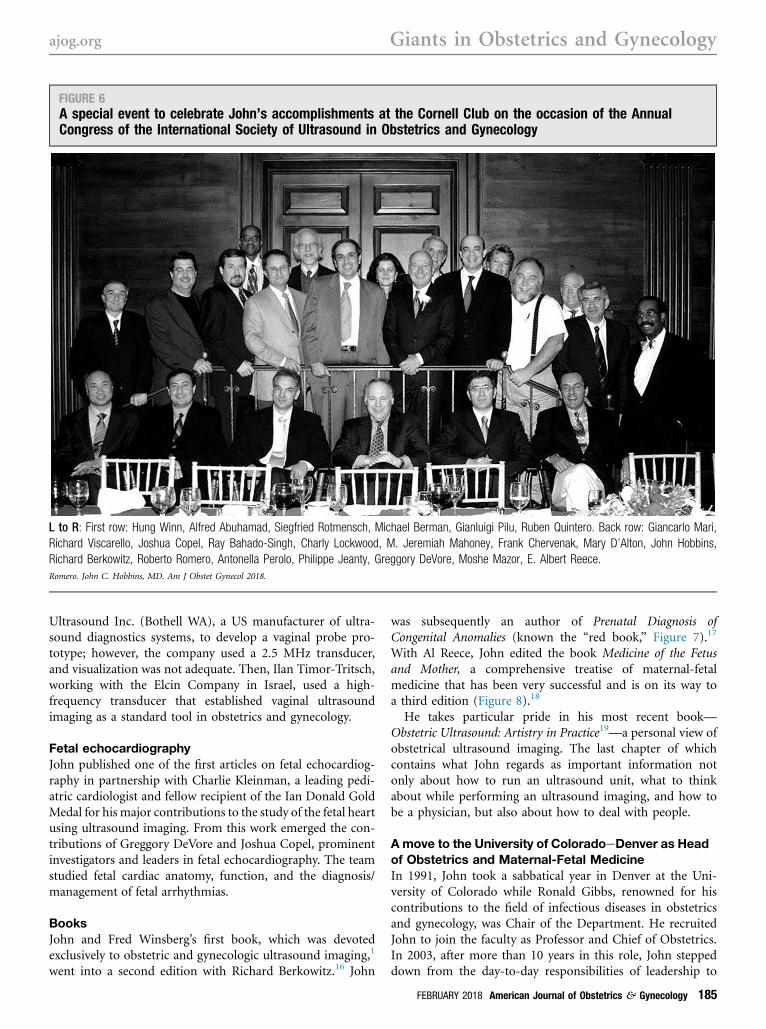
John’s traineesJohn held the position as the division director ofMaternal-FetalMedicine at Yale from 1976e1991. A prominent group ofclinical fellows graduated from the program. Frank Chervenak,Professor and Chair at Cornell, hosted a special event at theCornell Club to celebrate John’s accomplishments on theoccasion of the Annual Congress of the International Society ofUltrasound in Obstetrics and Gynecology (Figure 6).
Gianluigi Pilu was a visitor/trainee who has become one ofthe most productive people in the field of ultrasound imag-ing. “When our family traveled to Bologna in 1982, he wasthe very young guy assigned to make our family comfortableon our sabbatical,” John said. “We could tell immediately thathe was something special.”
Beyond the formal maternal-fetal medicine fellowshiptraining, there was an extraordinary group of individuals whotrained in ultrasound imaging in John’s unit: Philippe Jeanty,Gianluigi Pilu, Moshe Mazor, Albert Reece, Frank Chervenak,Charly Lockwood, Hung Winn, Khalil Tabsh, Sigi Rotmensch,Enrique Oyarzun, Mohammed Emamian, Jorge Andres Robert,Angela Scioscia, Alfred Abuhamad, Ray Bahado-Singh, ZionHagay, Mary D’Alton, Antonella Perolo, Macor Wan, and BoHyun Yoon. John’s trainees have gone on to become leaders inobstetrics and gynecology, and in medicine, occupying suchpositions as Deans of Schools of Medicine, Associate Deans,Chairs of Departments in the United States and abroad, leadersat the National Institutes of Health, and Division Heads ofMaternal-Fetal Medicine, among others, and have beenrecognized as outstanding leaders in their own right.
“Larry Platt and I have happily worked together on variousprojects over the years,” John also recalled. “Larry, Jim Binns,and I established the Gottesfeld-Hohler Memorial Founda-tion (GoHo) many years ago to provide fora for ultrasoundimaging education. One of GoHo’s (Larry, John, and JoshCopel) biggest triumphs was to recently organize an inter-national think tank in June for discussion about how to dealwith Zika and its effect on the fetal central nervous system.We’ve just submitted the summary for publication.”
Importance of ultrasound imaging in laborAn early pioneer of ultrasound imaging in labor, John’stutelage of one of his fellows, Phillipe Jeanty, led to thedevelopment of perineal ultrasound imaging to excludeplacenta previa;14 its use progressed from there to measuringthe station of the head in labor, detecting molding, andassessing the angle of progression as a prognostic factor forvaginal delivery.15 The latter work was done with AntonioBarbera and was first published as an American College ofObstetricians and Gynecologists video. Intrapartum sonog-raphy is an emerging field, because it is becoming increasinglyclear that ultrasound imaging can help identify the patientwho would have a difficult operative vaginal delivery.
Transvaginal ultrasound imagingE. Albert Reece had the original idea to develop transvaginalultrasound imaging. Al and John worked with ATL
FIGURE 5Diaphragmatic hernia
The A indicates a transverse scan at the level of the diaphragm thatshows the defect. From Touloukian RJ. Intestinal atresia. Clin Perinatol1978;5:3. With permission, John C. Hobbins MD.Romero. John C. Hobbins, MD. Am J Obstet Gynecol 2018.
Giants in Obstetrics and Gynecology ajog.org
184 American Journal of Obstetrics & Gynecology FEBRUARY 2018
Ultrasound Inc. (Bothell WA), a US manufacturer of ultra-sound diagnostics systems, to develop a vaginal probe pro-totype; however, the company used a 2.5 MHz transducer,and visualization was not adequate. Then, Ilan Timor-Tritsch,working with the Elcin Company in Israel, used a high-frequency transducer that established vaginal ultrasoundimaging as a standard tool in obstetrics and gynecology.
Fetal echocardiographyJohn published one of the first articles on fetal echocardiog-raphy in partnership with Charlie Kleinman, a leading pedi-atric cardiologist and fellow recipient of the Ian Donald GoldMedal for his major contributions to the study of the fetal heartusing ultrasound imaging. From this work emerged the con-tributions of Greggory DeVore and Joshua Copel, prominentinvestigators and leaders in fetal echocardiography. The teamstudied fetal cardiac anatomy, function, and the diagnosis/management of fetal arrhythmias.
BooksJohn and Fred Winsberg’s first book, which was devotedexclusively to obstetric and gynecologic ultrasound imaging,1
went into a second edition with Richard Berkowitz.16 John
was subsequently an author of Prenatal Diagnosis ofCongenital Anomalies (known the “red book,” Figure 7).17
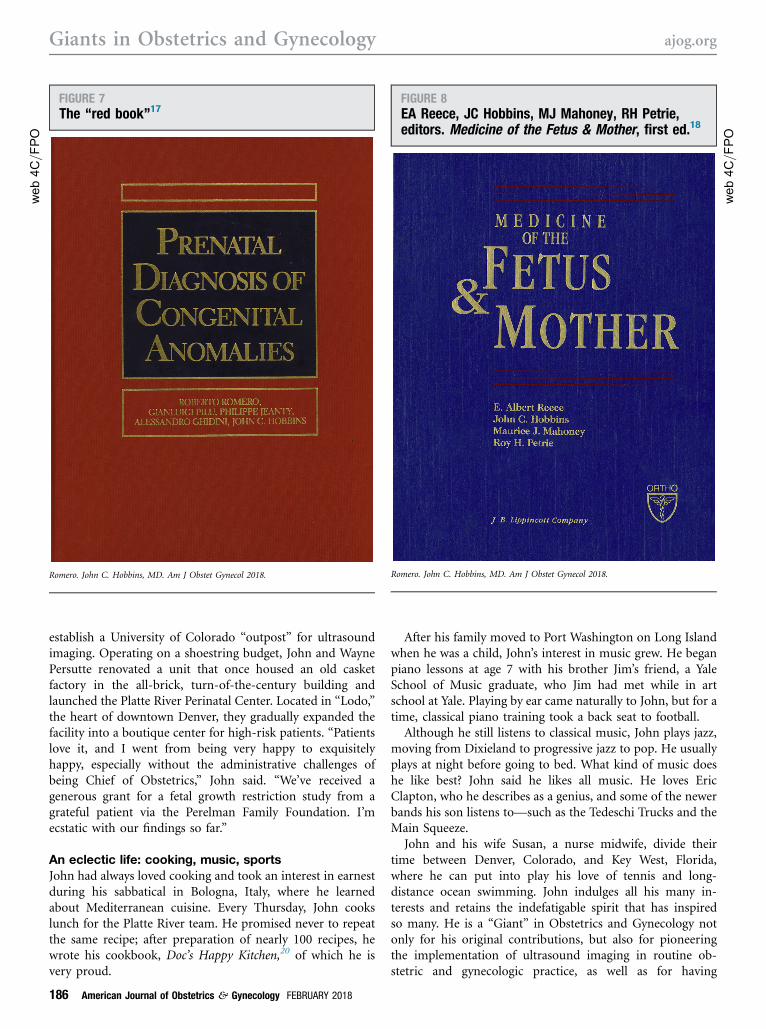
With Al Reece, John edited the book Medicine of the Fetusand Mother, a comprehensive treatise of maternal-fetalmedicine that has been very successful and is on its way toa third edition (Figure 8).18
He takes particular pride in his most recent book—Obstetric Ultrasound: Artistry in Practice19—a personal view ofobstetrical ultrasound imaging. The last chapter of whichcontains what John regards as important information notonly about how to run an ultrasound unit, what to thinkabout while performing an ultrasound imaging, and how tobe a physician, but also about how to deal with people.
Amove to the University of ColoradoeDenver as Headof Obstetrics and Maternal-Fetal MedicineIn 1991, John took a sabbatical year in Denver at the Uni-versity of Colorado while Ronald Gibbs, renowned for hiscontributions to the field of infectious diseases in obstetricsand gynecology, was Chair of the Department. He recruitedJohn to join the faculty as Professor and Chief of Obstetrics.In 2003, after more than 10 years in this role, John steppeddown from the day-to-day responsibilities of leadership to
FIGURE 6A special event to celebrate John’s accomplishments at the Cornell Club on the occasion of the AnnualCongress of the International Society of Ultrasound in Obstetrics and Gynecology
L to R: First row: Hung Winn, Alfred Abuhamad, Siegfried Rotmensch, Michael Berman, Gianluigi Pilu, Ruben Quintero. Back row: Giancarlo Mari,Richard Viscarello, Joshua Copel, Ray Bahado-Singh, Charly Lockwood, M. Jeremiah Mahoney, Frank Chervenak, Mary D’Alton, John Hobbins,Richard Berkowitz, Roberto Romero, Antonella Perolo, Philippe Jeanty, Greggory DeVore, Moshe Mazor, E. Albert Reece.Romero. John C. Hobbins, MD. Am J Obstet Gynecol 2018.
ajog.org Giants in Obstetrics and Gynecology
FEBRUARY 2018 American Journal of Obstetrics & Gynecology 185
establish a University of Colorado “outpost” for ultrasoundimaging. Operating on a shoestring budget, John and WaynePersutte renovated a unit that once housed an old casketfactory in the all-brick, turn-of-the-century building andlaunched the Platte River Perinatal Center. Located in “Lodo,”the heart of downtown Denver, they gradually expanded thefacility into a boutique center for high-risk patients. “Patientslove it, and I went from being very happy to exquisitelyhappy, especially without the administrative challenges ofbeing Chief of Obstetrics,” John said. “We’ve received agenerous grant for a fetal growth restriction study from agrateful patient via the Perelman Family Foundation. I’mecstatic with our findings so far.”
An eclectic life: cooking, music, sportsJohn had always loved cooking and took an interest in earnestduring his sabbatical in Bologna, Italy, where he learnedabout Mediterranean cuisine. Every Thursday, John cookslunch for the Platte River team. He promised never to repeatthe same recipe; after preparation of nearly 100 recipes, hewrote his cookbook, Doc’s Happy Kitchen,20 of which he isvery proud.
After his family moved to Port Washington on Long Islandwhen he was a child, John’s interest in music grew. He beganpiano lessons at age 7 with his brother Jim’s friend, a YaleSchool of Music graduate, who Jim had met while in artschool at Yale. Playing by ear came naturally to John, but for atime, classical piano training took a back seat to football.
Although he still listens to classical music, John plays jazz,moving from Dixieland to progressive jazz to pop. He usuallyplays at night before going to bed. What kind of music doeshe like best? John said he likes all music. He loves EricClapton, who he describes as a genius, and some of the newerbands his son listens to—such as the Tedeschi Trucks and theMain Squeeze.
John and his wife Susan, a nurse midwife, divide theirtime between Denver, Colorado, and Key West, Florida,where he can put into play his love of tennis and long-distance ocean swimming. John indulges all his many in-terests and retains the indefatigable spirit that has inspiredso many. He is a “Giant” in Obstetrics and Gynecology notonly for his original contributions, but also for pioneeringthe implementation of ultrasound imaging in routine ob-stetric and gynecologic practice, as well as for having
web4C=F
PO
FIGURE 7The “red book”17
Romero. John C. Hobbins, MD. Am J Obstet Gynecol 2018.
web4C=F
PO
FIGURE 8EA Reece, JC Hobbins, MJ Mahoney, RH Petrie,editors. Medicine of the Fetus & Mother, first ed.18
Romero. John C. Hobbins, MD. Am J Obstet Gynecol 2018.
Giants in Obstetrics and Gynecology ajog.org
186 American Journal of Obstetrics & Gynecology FEBRUARY 2018
inspired and trained generations of leaders in our disci-pline. His charisma and contagious enthusiasm for all thatultrasound imaging can do have made him belovedthroughout the world. -
REFERENCES
1. Hobbins JC, Winsberg F. Ultrasonography in obstetrics and gyne-cology. Easton (MD): Williams & Wilkins Co; 1983.2. Romero R. Giants in obstetrics and gynecology series: a profile ofLeon Speroff. MD. Am J Obstet Gynecol 2017;217:263.e1-8.3. Romero R. A profile of Dr Edward J. Quilligan. Am J Obstet Gynecol2017;216:547-51.e3.4. Warsof SL, Gohari P, Berkowitz RL, Hobbins JC. The estimation offetal weight by computer-assisted analysis. Am J Obstet Gynecol1977;128:881-92.5. Gohari P, Berkowitz RL, Hobbins JC. Prediction of intrauterine growthretardation by determination of total intrauterine volume. Am J ObstetGynecol 1977;127:255-60.6. Campbell S, Johnstone FD, Holt EM, May P. Anencephaly: earlyultrasonic diagnosis and active management. Lancet 1972;2:1226-7.7. Hobbins JC, Grannum PA, Berkowitz RL, Silverman R, Mahoney MJ.Ultrasound in the diagnosis of congenital anomalies. Am J ObstetGynecol 1979;134:331-45.8. Chervenak FA, Romero R, Berkowitz RL, et al. Antenatal sonographicfindings of osteogenesis imperfecta. Am J Obstet Gynecol 1982;143:228-30.
9. Chervenak FA, Blakemore KJ, Isaacson G, Mayden K, Hobbins JC.Antenatal sonographic findings of thanatophoric dysplasia with cloverleafskull. Am J Obstet Gynecol 1983;146:984-5.10. Mahoney MJ, Hobbins JC. Prenatal diagnosis of chondroectodermaldysplasia (Ellis-van Creveld syndrome) with fetoscopy and ultrasound.N Engl J Med 1977;297:258-60.11. Hobbins JC, Mahoney MJ. In utero diagnosis of hemoglobinopathies:technic for obtaining fetal blood. N Engl J Med 1974;290:1065-7.12. Chang H, Hobbins JC, Cividalli G, et al. In utero diagnosis of: he-moglobinopathies: hemoglobin synthesis in fetal red cells. N Engl J Med1974;290:1067-8.13. Alter BP, Friedman S, Hobbins JC, et al. Prenatal diagnosis of sickle-cell anemia and alpha G Philadelphia: study of a Fetus also at risk forH b S/betaþ-thalassemia. N Engl J Med 1976;294:1040-1.14. Jeanty P, D’alton M, Romero R, Hobbins JC. Perineal scanning. AmJ Perinatol 1986;3:289-95.15. Barbera AF, Pombar X, Perugino G, Lezotte DC, Hobbins JC. A newmethod to assess fetal head descent in labor with transperineal ultra-sound. Ultrasound Obstet Gynecol 2009;33:313-9.16. Hobbins JC, Winsberg F, Berkowitz RL. Ultrasonography inobstetrics and gynecology. 2nd ed. Easton (MD): Williams & Wilkins Co;1983.17. Romero R, Pilu G, Jeanty P, Ghidini A, Hobbins JC. Prenatal diag-nosis of congenital anomalies. New York: Appleton & Lange; 1988.18. Reece EA, Hobbins JC, Mahoney MJ, Petrie RH, editors. Medicine ofthe Fetus & Mother. Philadelphia PA: JB Lippincott Company; 1993.19. Hobbins JC. Obstetric ultrasound: artistry in practice. Malden (MA):Blackwell Publishing; 2008.20. Hobbins J.Doc’s happy kitchen. San Francisco: BlurbBooks Inc; 2016.
ajog.org Giants in Obstetrics and Gynecology
FEBRUARY 2018 American Journal of Obstetrics & Gynecology 187
web4C=F
PO
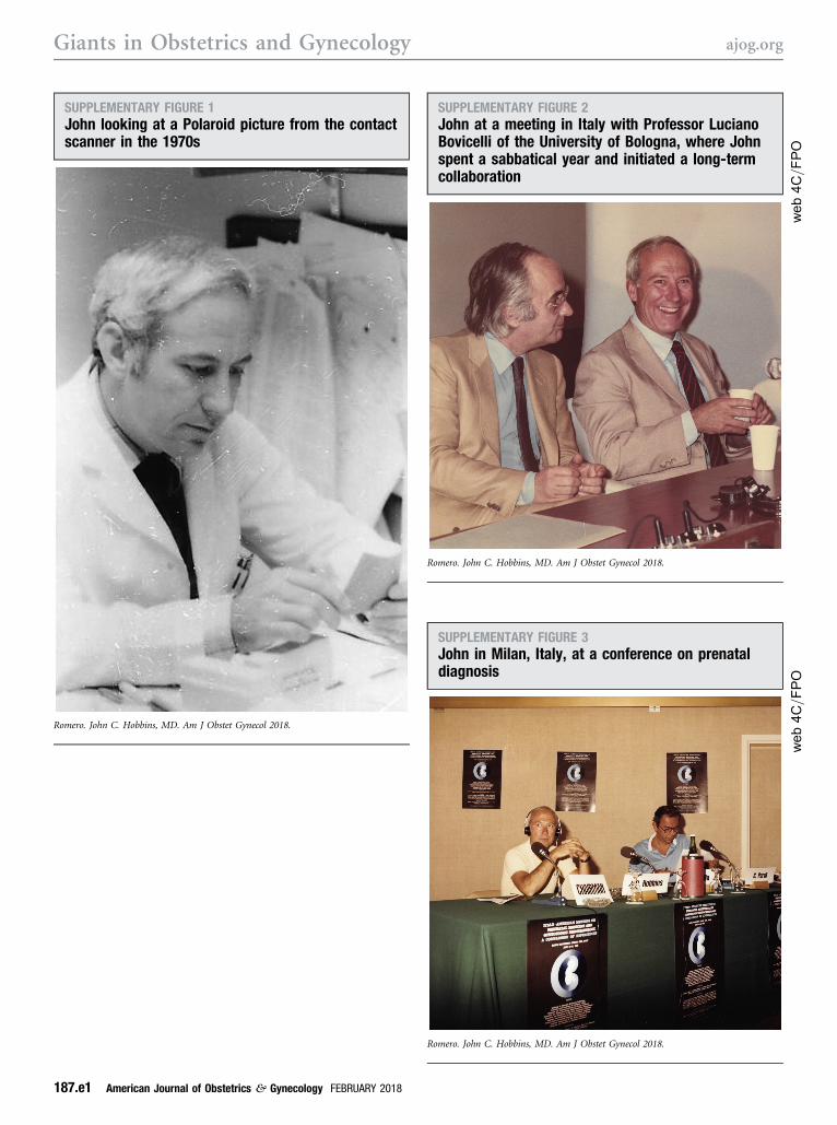
SUPPLEMENTARY FIGURE 2John at a meeting in Italy with Professor LucianoBovicelli of the University of Bologna, where Johnspent a sabbatical year and initiated a long-termcollaboration
Romero. John C. Hobbins, MD. Am J Obstet Gynecol 2018.
SUPPLEMENTARY FIGURE 1John looking at a Polaroid picture from the contactscanner in the 1970s
Romero. John C. Hobbins, MD. Am J Obstet Gynecol 2018.
web4C=F
PO
SUPPLEMENTARY FIGURE 3John in Milan, Italy, at a conference on prenataldiagnosis
Romero. John C. Hobbins, MD. Am J Obstet Gynecol 2018.
Giants in Obstetrics and Gynecology ajog.org
187.e1 American Journal of Obstetrics & Gynecology FEBRUARY 2018

SUPPLEMENTARY FIGURE 4A picnic at Yale with members of the Perinatal Unit;rain interrupted the event, but it was all taken instride
Romero. John C. Hobbins, MD. Am J Obstet Gynecol 2018.
SUPPLEMENTARY FIGURE 5John using the contact scanner in the 1970s
Romero. John C. Hobbins, MD. Am J Obstet Gynecol 2018.
web4C=F
PO
SUPPLEMENTARY FIGURE 6John in his office, relaxing between procedures
Romero. John C. Hobbins, MD. Am J Obstet Gynecol 2018.
SUPPLEMENTARY FIGURE 7Medical intelligence article in the New EnglandJournal of Medicine that described a method forobtaining fetal blood using the fetoscope
The hand with the syringe is that of M. Jeremiah Mahoney (Departmentof Human Genetics, Pediatrics, Obstetrics and Gynecology at Yale).Romero. John C. Hobbins, MD. Am J Obstet Gynecol 2018.
ajog.org Giants in Obstetrics and Gynecology
FEBRUARY 2018 American Journal of Obstetrics & Gynecology 187.e2

web4C=F
PO
SUPPLEMENTARY FIGURE 8Event at the Cornell Club in 2002
L to R: Roberto Romero, Frank Chervenak (Professor and Chair at Cornell), John Hobbins, E. Albert Reece (Dean of the School of Medicine at theUniversity of Maryland), and M. Jeremiah Mahoney (Department of Human Genetics, Pediatrics, Obstetrics and Gynecology at Yale).Romero. John C. Hobbins, MD. Am J Obstet Gynecol 2018.
web4C=F
PO
SUPPLEMENTARY FIGURE 9Playing the piano, International Society ofUltrasound in Obstetrics and Gynecology 2002
Picture by Ilan Timor-Tritsch: “To my all-time role model, John the Great,Ilan 2002 ISUOG.”Romero. John C. Hobbins, MD. Am J Obstet Gynecol 2018.
Giants in Obstetrics and Gynecology ajog.org
187.e3 American Journal of Obstetrics & Gynecology FEBRUARY 2018

web4C=F
PO
SUPPLEMENTARY FIGURE 10John in action on the tennis court
Romero. John C. Hobbins, MD. Am J Obstet Gynecol 2018.
web4C=F
PO
SUPPLEMENTARY FIGURE 11John’s latest book, Doc’s Happy Kitchen20
The book emerged from John’s long-standing interest in culinary pur-suits. The book promises “really good meals, STAT.” Photographs byRachel Rourk.Romero. John C. Hobbins, MD. Am J Obstet Gynecol 2018.
ajog.org Giants in Obstetrics and Gynecology
FEBRUARY 2018 American Journal of Obstetrics & Gynecology 187.e4